Characterization of
Maltose and Trehalose Transport in Corynebacterium glutamicum
Inaugural-Dissertation zur
Erlangung des Doktorgrades
der Mathematisch-Naturwissenschaftlichen Fakultät der Universität zu Köln
vorgelegt von
Alexander Wolfgang Henrich aus Damscheid
Köln, Februar 2011
Berichterstatter:
Herr Prof. Dr. Reinhard Krämer Frau Prof. Dr. Karin Schnetz
Tag der Disputation: 14. April 2011
“Observation is a passive science,
experimentation an active one.”
Claude Bernard
Kurzzusammenfassung
Zur Produktion von Aminosäuren mit dem kommerziell bedeutsamen Bakterium Corynebacterium glutamicum kann aus Stärkehydrolysat gewonnene Maltose als gute und günstige Kohlenstoffquelle dienen. In der vorliegenden Arbeit wurde das Maltose/Maltodextrin Aufnahme-System MusIFGK2-E von C. glutamicum identifiziert und charakterisiert; ein ABC Transporter mit einem Km von 1,2 ± 0,2 µM und Vmax von 26,2 ± 1,0 nmol/(min*mg TG). Es konnte gezeigt werden, dass das System kein Operon bildet und dass es von dem transkriptionellen Regulator RamA reguliert wird. Zusätzlich konnte eine regulatorische Verbindung zur Phosphotransferase System vermittelten Glukose Verstoffwechslung aufgezeigt werden. Weitere Analysen identifizierten das Protein MusI, welches zwar für die Aufnahme von Maltose essentiell, dessen Funktion aber noch unbekannt ist.
Neben der Bedeutung als Aminosäureproduzent ist C. glutamicum ein wichtiger Modellorganismus für die Zellwand und deren Synthese innerhalb der Corynebacterineae.
In diesem Zusammenhang spielt Trehalose als Komponente von Mycolaten, wichtigen Bausteinen der Zellwand von Corynebakterien and Mycobakterien, eine bedeutende Rolle. In dieser Arbeit konnte zum ersten Mal gezeigt werden, dass C. glutamicum, im Gegensatz zu bereits publizierten Daten, mit einem Trehalose Aufnahme-System ausgestattet ist. Die Aufnahme wird über den ABC Transporter TusFGK2-E mit einem Km
von 0,16 ± 0,02 µM und Vmax von 2,5 ± 0,1 nmol/(min*mg TG) vermittelt. Trehalose kann anschließend durch TreS in Maltose umgewandelt werden und dann über den Maltose Abbauweg verstoffwechselt werden. Beachtenswerterweise konnte in dieser Arbeit ein zweiter Abbauweg für Trehalose / Maltose identifiziert werden. Dieser Weg beinhaltet die TreS vermittelte Umwandlung von Trehalose in Maltose, TreX vermittelte Maltose-1- Phosphate und anschließende α-Glucan Synthese durch GlgE, vergleichbar zu einem Abbauweg in M. tuberculosis (Kalscheuer et al., 2010a).
Diese Ergebnisse stellten die Hypothese von Tropis et al., 2005 in Frage, dass man zur Synthese von Trehalose-Mycolaten Biosynthese und Export von Trehalose benötigt. Mit Hilfe einer vierfach-Deletionsmutante, die weder fähig ist Trehalose zu synthetisieren noch aufzunehmen, konnte gezeigt werden, dass die Synthese von Trehalose-Mycolaten durch externe Zugabe des Zuckers, auch ohne die vorherige Aufnahme von Trehalose möglich ist. Dies zeigte dass die Trehalose Export Hypothese weiterhin Gültigkeit besitzt und dass das Trehalose Export-System von C. glutamicum noch zu identifizieren ist.
Abstract Abstract
For the production of amino acids with the commercially important Gram-positive bacterium Corynebacterium glutamicum maltose derived from starch hydrolysate can serve as a good and cheap carbon source. The present study identified and characterized the maltose/maltodextrin uptake system MusIFGK2-E of C. glutamicum being an ABC transport system with a Km of 1.2 ± 0.2 µM and a Vmax of 26.2 ± 1.0 nmol/(min*mg cdm).
Analyses showed that the system is not transcribed in an operon and that it is regulated by the transcriptional regulator RamA. In addition a regulatory connection to phosphotransferase system mediated glucose utilization was found. Further it was shown that the maltose uptake system possesses an additional protein MusI with unknown function that is important for the uptake of maltose.
Besides being an important amino acid producer, C. glutamicum is a major model organism for the cell envelope and its synthesis of the Corynebacterineae. In this context trehalose plays a crucial role as component of mycolates, important building blocks of the cell wall of corynebacteria and mycobacteria. This study showed for the first time that C. glutamicum, in contradiction to previously published data, is equipped with an uptake system for trehalose and is further capable to use trehalose for growth. The uptake of trehalose is mediated by the ABC transport system TusFGK2-E with a Km of 0.16 ± 0.02 µM and a Vmax of 2.5 ± 0.1 nmol/(min*mg cdm). Trehalose can be utilized for growth via TreS mediated conversion into maltose and further degradation by the known maltose pathway. Notably, this study revealed a second pathway for the utilization of trehalose / maltose involving TreS mediated conversion of trehalose to maltose, TreX mediated maltose-1-phosphat formation and α-glucan formation by GlgE a pathway similar to the one found in M. tuberculosis (Kalscheuer et al., 2010a).
These results challenged the hypothesis of Tropis et al., 2005 that for the synthesis of trehalose mycolates biosynthesis and export of trehalose is needed. But with the help of a quadruple-mutant, that is neither able to synthesize nor to import trehalose, it was shown that the formation of trehalose mycolates is possible without the uptake of externally present trehalose leaving the trehalose export hypothesis valid and the trehalose export system of C. glutamicum to be identified.
1 INTRODUCTION... 1
1.1 Corynebacterium glutamicum... 1
1.2 Cell wall composition of corynebacteria... 1
1.3 Trehalose: An important component of the cell wall of C. glutamicum ... 3
1.4 Trehalose synthesis in C. glutamicum ... 4
1.5 Trehalose transport ... 5
1.6 Maltose metabolism in C. glutamicum ... 7
1.7 General transport mechanisms... 9
1.8 The maltose uptake system MalFGK2-E of E. coli ...10
1.9 Thesis objectives...12
2 MATERIALS AND METHODS... 13
2.1 Bacterial strains, plasmids and oligonucleotides...13
2.2 Media and culture conditions...18
2.3 Molecular biology methods...19
2.3.1 DNA digestion, ligation and purification ...19
2.3.2 Competent cells and transformation ...19
2.3.3 Polymerase chain reaction ...19
2.3.4 Agarose gel electrophoresis ...19
2.3.5 Construction of C. glutamicum mutant strains...19
2.3.6 RNA hybridization experiments ...20
2.3.7 5’-RACE ...21
2.3.8 RT-PCR...21
2.4 Biochemical methods ...21
2.4.1 HPLC analyses...21
2.4.2 Extraction of glycolipids ...22
2.4.3 TLC for maltose and trehalose separation ...22
2.4.4 Screening assay...22
Contents
2.4.5 Enzyme assay ...23
2.4.6 Glutamate production ...23
2.4.7 Radioactive transport assays...23
3 RESULTS ... 24
3.1 Maltose uptake in Corynebacterium glutamicum ...24
3.1.1 Maltose positively influences glucose utilization ...24
3.1.2 Biochemical characterization of glucose uptake in C. glutamicum ...27
3.1.3 Biochemical characterization and first regulatory insight of the maltose uptake system of C. glutamicum...29
3.1.4 Identification of the maltose uptake system of C. glutamicum...34
3.1.5 Transcriptional organization of the maltose uptake system MusFGK2-E ...37
3.1.6 MusI: A novel component of an ACB transporter for maltose uptake ...40
3.1.7 Expression analyses of the maltose uptake system...42
3.1.8 Substrate specificity of MusIFGK2-E ...43
3.1.9 Connections between maltose and glucose metabolism...45
3.2 Trehalose metabolism of C. glutamicum...47
3.2.1 C. glutamicum utilizes trehalose for growth ...47
3.2.2 Biochemical characterization of trehalose uptake in C. glutamicum...49
3.2.3 Identification of the trehalose uptake system of C. glutamicum...51
3.2.4 Transcriptional organization of tusFGK-E ...53
3.2.5 Direct evolution of C. glutamicum for the utilization of trehalose as sole carbon source ...54
3.3 The trehalose export hypothesis...55
3.3.1 Screening system for mutants defective in trehalose export ...58
3.3.2 Approach for the biochemical characterization of trehalose export ...60
3.3.3 TreX: A new player in trehalose/maltose metabolism ...61
3.3.4 A new approach to characterize the trehalose export biochemically ...63
4. DISCUSSION... 67
4.1 The maltose uptake system MusIFGK2-E of C. glutamicum...67
4.2 Regulation of MusIFGK2-E ...69
4.3 Nonclassical regulatory phenomena...71
4.4 The trehalose uptake system TusFGK2-E of C. glutamicum ...73
4.5 TreS: An important enzyme in trehalose metabolism...77
4.6 Trehalose export in C. glutamicum ...77
4.7 A second pathway for trehalose and maltose degradation...80
4.8 Model of maltose and trehalose metabolism in C. glutamicum ...83
5 SUMMARY... 85
6 REFERENCES... 87
Abbreviations
Abbreviations
ABC ATP-binding cassette ADP Adenosine 5’-diphosphate AGM Arabinogalactan mycolate ATP Adenosine 5’-triphosphate
ATCC American Type Culture Collection
b Bases
cAMP Cyclic Adenosin Monophosphate cdm Cell dry matter
GMM Glucose monomycolate
h Hours
HPLC High performance liquid chromotography IPTG Isopropyl β-D-1-thiogalactopyranoside
Kb Kilo basepares
Km Michaelis constant
KPi Potassium phosphate buffer
LB Luria-Bertani
M Molar
mM Millimolar
OD Optical density
OPA Ortho-phthaldialdehyde ORF Open reading frame PEP Phosphoenolpyruvate PCR Polymerase chain reaction PP-pathway Pentose phosphate pathway PTS Phosphotransferase System TDM Trehalose dimycolate TLC Thin-layer chromatography
TM C. glutamicum spontaneous-mutant capable to grow on trehalose TMM Trehalose monomycolate
TY Tryptone Yeast (complex medium)
U Units
Vmax Maximum velocity (v/v) Volume per volume (w/v) Weight per volume
WT Wild type (C. glutamicum ATCC 13032)
1 Introduction
1.1 Corynebacterium glutamicum
The Gram-positive GC-rich bacterium Corynebacterium glutamicum was isolated in 1957 by Udaka und Kinoshita (Kinoshita et al., 1957). C. glutamicum is a non-pathogenic member of the Corynebacteriaceae, who form together with the families Mycobacteriaceae, Nocardiaceae, Gordoniaceae, Dietziaceae und Tsukamurellaceae a suborder of the Actinomycetales, named Corynebacterianeae (Stackebrandt et al., 1997).
Today C. glutamicum is commercially used for the industrial production of amino acids reaching an annual production of more than two million tons in 2005, mainly L-glutamic acid and L-lysine (Leuchtenberger et al., 2005).
Besides being an important amino acid producer, C. glutamicum is a major model organism for the cell envelope and its synthesis of the suborder Corynebacterianeae, including pathogenic members such as Mycobacterium tuberculosis, Mycobacterium leprae and Corynebacterium diphtheriae. Moreover, the 3.3 Mb genome of C. glutamicum was completely sequenced by independent research groups in Europe and Japan (Kalinowski et al., 2003; Ikeda and Nakagawa, 2003), making the organism a good target for genome wide genetic research.
1.2 Cell wall composition of corynebacteria
Members of the suborder of the Corynebacterianeae such as corynebacteria and mycobacteria show a unique cell envelope design which shows major differences compared to other Gram-positive bacteria. Their cell envelope comprises not only a peptidoglycan layer, with some additional polysaccharides or polyol phosphate polymers like in other gram-positive bacteria, but also an outer membrane which is typical for Gram- negative bacteria (Fig. 1). This additional membrane contains extractable lipids and long- chain mycolic acids that are covalently linked to peptidoglycan via a unique polysaccharide arabinogalactan network (Brennan & Nikaido, 1995). The innermost layer is the plasma membrane. In C. glutamicum it consists almost exclusively of phospholipids with negatively charged head-groups, namely phosphatidylglycerol, di- phosphatidylglycerol (cardiolipin), phosphatidylinositol and phosphatidylinositol mannosides. These lipids posses to over 90% palmitic (C16:0) and oleic (C18:1) acids as fatty acid chains (Hoischen & Krämer, 1990; Özcan et al., 2007). Adjacent to the plasma membrane a peptidoglycan layer, a common feature of Gram-positive bacteria, can be found forming the first layer of the cell wall skeleton. In Corynebacterianeae this layer is composed of β-1,4-linked N-acetylglucosamine and N-acetyl muramic acid residues linked
Introduction 2 to tri- or tetrapeptides such as L-Ala-D-Glu-meso-diaminopimelic acid or L-Ala-D-Glu- meso-diaminopimelic acid-D-Ala (Schleifer & Kandler, 1972).
Figure 1: Cell wall structure of Corynebacterianeae according to Puech et al., 2001.
The heteropolysaccharide arabinogalactan forms the second layer, which is composed of a linear alternating β-D-galactofuranosyl backbone connected to a 3,5-branched α-D- arabinofuranosyl structure. The galactan domain of this layer is attached to peptidoglycan via a special “linker unit” and its arabinan domain is linked to mycolic acids, forming the mycolyl-arabinogalactan-peptidoglycan (mAGP) complex (Puech et al., 2001; Seidel et al., 2007).
Mycolic acids are 2-alkyl-branched, 3-hydroxylated long chain fatty acids which represent major and specific constituents of the third layer of the cell wall skeleton, the mycolate layer. The number of carbon atoms and the degree of desaturation of main and lateral chains vary according to the genus. In mycobacteria the fatty acid chains are very long (C60-90), whereas the alkyl-chain in norcadia and corynebacteria is shorter (norcadomycolic acids C44-60; corynomycolic acids C22-36) (De Briel et al., 1992; Barry et al., 1998;
Rafidinarivo et al., 2009).
While this additional barrier in Gram-negative bacteria is a typical bilayer of phospholipid and lipopolysaccharide, in mycobacteria and corynebacteria the cell-wall-linked mycolates and corynomycolates certainly participate in this barrier since the disruption of genes that code for mycoloyltransferases causes a decrease in the amount of cell-wall-bound
Outer layer
Mycolate layer
Arabinogalactan
Peptidoglycan
Cytoplasmic membrane
Porin
Trehalose dimycolate
Trehalose monomycolate
Phospholipid
Protein
mycolates and affects the permeability of the envelope of the mutants (Puech et al., 2000). Electron microscopy of freeze-fractured preparations and chemical analyses indicated that the inner part of the mycolate layer consists mainly of arabinogalactan mycolate and lesser amounts of extractable mycolate (Puech et al., 2001). Parts of the extractable mycolates are trehalose monomycolate (TMM) and trehalose dimycolate (TDM), which derive from the esterification of one or two mycolic acids with a molecule trehalose (Shimakata & Minatogawa, 2000; Tropis et al., 2005). The outer layer consists almost exclusively of these mycolates (TMM; TDM) (Daffé & Draper, 1998; Puech et al., 2001). Recently, Hoffmann and co-workers found with the help of cryo-electron tomography and the investigation of frozen-hydrated cryosections of Mycobacterium smegmatis, Myobacterium bovis and C. glutamicum that the outermost layer, the mycolate layer, is a morphologically symmetrical lipid bilayer (Hoffmann et al., 2008). This stands in contrast to the, so far preferred, model that the mycolate layer is asymmetrical.
Analogue to the outer membrane in Gram-negative bacteria, the mycolate layer of mycobacteria is thought to be responsible for the low permeability of the cell envelope (Brennan & Nikaido, 1995). To facilitate exchange of substances across this barrier, channel-forming proteins, so-called porins, such as PorA are necessary to allow the passage of hydrophilic substances (Costa-Riu et al., 2003; Hünten et al., 2005 a/b; Ziegler et al., 2008).
On the surface of this membrane-like structure corynebacteria posses an outer layer which is formed of up to 90% of polysaccharides, composed of glucose, arabinose and various lipids, mostly TDM and TMM, but also phospholipids such as phosphatidylinositol mannosides and phosphatidylglycerol (Puech et al., 2001).
1.3 Trehalose: An important component of the cell wall of C. glutamicum It has been shown that mycolates are a crucial part of the cell envelope of Gram-positive bacteria belonging to the suborder of the Corynebacterianeae. These mycolates are formed from trehalose esterified by one or two mycolic acids and mycolic acids covalently linked to arabinogalactan (Brennan & Nikaido, 1995). As trehalose is involved not only in the synthesis of TMM and TDM, but also in the formation of arabinogalactan mycolate (AGM), trehalose was thought to be essential for these three components of the mycolate layer (Shimakata & Minatogawa, 2000).
In mycobacteria, the mycolate layer is the part of the cell envelope that causes the high resistance of these bacteria to most common antibiotics e.g. in M. tuberculosis, the causal agent of tuberculosis. Furthermore, trehalose dimycolate, also known as cord factor, plays a role in the persistence of this pathogenic bacterium in the host cell, presumably by
Introduction 4 inhibiting the fusion between lysosomes and phagosomes containing the bacteria (Crowe et al., 1994). This makes it important to study the role of trehalose thoroughly in its close relative C. glutamicum, possibly opening new opportunities for drug development.
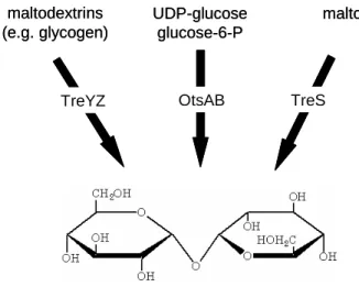
1.4 Trehalose synthesis in C. glutamicum
In C. glutamicum three different trehalose synthesis pathways have been identified TreYZ, OtsAB and TreS (Fig. 2). As only one of these pathways is present in most bacteria this fact underlines the importance of trehalose in the general physiology of mycobacteria and corynebacteria, e.g. as a crucial part of the cell envelope and during stress adaptation under certain conditions (Gebhardt, 2005; Tropis et al., 2005).
Figure 2: Trehalose synthesis pathways in C. glutamicum, according to Wolf et al., 2003.
In C. glutamicum the TreYZ-pathway is the main trehalose synthesis pathway (Tzvetkov et al., 2003; Wolf et al., 2003). It is responsible for the synthesis and the accumulation of trehalose as a response to hyperosmotic stress. In a two step reaction the terminal maltosyl-residue of maltodextrin is converted via the maltooligosyltrehalose synthase (TreY) into a trehalosyl unit which is then cut off by a maltooligosyltrehalose hydrolase (TreZ) leading to trehalose (Maruta et al., 1996 a/b/c; Kim et al., 2000). The second pathway starts with the condensation of UDP-glucose and glucose-6-phosphate by the osmotically regulated trehalose synthesis (Ots) A enzyme yielding trehalose-6-phosphate which is then dephosphorylated to trehalose by OtsB (Argüelles, 2000). For the OtsAB- pathway in Corynebacterium matruchotii it was discussed by Shimakata and Minatogawa (2000) that the production of trehalose-6-phosphate by OtsA is important for the synthesis of trehalose monomycolate, an important component of the cell envelope. This conclusion seems only to be true for C. matruchotii, since it has been shown for C. glutamicum that a
maltodextrins (e.g. glycogen)
UDP-glucose glucose-6-P
maltose
TreYZ OtsAB TreS
maltodextrins (e.g. glycogen)
UDP-glucose glucose-6-P
maltose
TreYZ OtsAB TreS
strain deleted in the otsA gene was able to synthesize trehalose mycolate (Wolf et al., 2003). The OtsAB-pathway is more important for the synthesis of trehalose as compatible solute after a hyperosmotic shock, when the cells face carbon- and nitrogen-limitation at the same time (Gebhardt, 2005). This is underlined by the fact that the otsA gene is upregulated five-fold after an osmotic up-shift (Wolf et al., 2003).
The third way to synthesize trehalose is the TreS-pathway that is a one step reaction in which the trehalose synthase (TreS) catalyzes the conversion of maltose into trehalose. In C. glutamicum TreS is thought to be a possible substitute for a missing trehalase, since it has been shown in in vitro assays that it catalyzes maltose synthesis from trehalose and therefore, it can be important for the recycling of trehalose after an osmotic up-shift (Wolf et al., 2003).
1.5 Trehalose transport
For C. glutamicum it has been published that there is no uptake system present for trehalose (Gebhardt, 2005). This has been concluded by two observations / experiments.
At first growth of C. glutamicum monitored for 10 h with trehalose as sole carbon source revealed that it is not able to utilize the disaccharide for growth (Wolf, 2002). A second approach measuring the uptake of [14C]-trehalose strengthened the hypothesis that trehalose can not be taken up by C. glutamicum. A mutant deficient in trehalose synthesis (C. glutamicum ∆otsA ∆treS ∆treY) was able to incorporate external [14C]-trehalose in TMMs, so that radioactivity was only detected in the cell wall , whereas there was no uptake into the cytoplasm detected, showing that the synthesis of TMM takes place in the cell envelope (Gebhardt, 2005). When the respective triple-mutant was supplemented with external trehalose, the cells were able to synthesize trehalose monomycolate, showing that external trehalose is important for mycolate synthesis in C. glutamicum. Further experiments revealed that the triple-mutant is also capable of using other sugars (glucose, maltose or maltotriose) for the synthesis of mycolated glycolipids. Thus it was stated that in principle mycolate synthesis is generally possible if an α-glucosyl-containing sugar is present in the medium (Tropis et al., 2005). From these experiments it was concluded that C. glutamicum must be equipped with a trehalose export system that facilitates the transport of trehalose across the plasma membrane, as trehalose is produced in the cytoplasm and the esterification with mycolic acids seems to take place in the cell envelope (Tropis et al., 2005).
Escherichia coli synthesizes internal trehalose as an osmoprotectant under high osmolarity, independent of the carbon source, and is also able to utilize trehalose as carbon source both under low and high osmolality (Boos et al., 1990). In E. coli the uptake of trehalose is facilitated by a phosphotransferase system (PTS) leading to trehalose-6-
Introduction 6 phosphate (Boos et al., 1990). It continues with hydrolysis to trehalose and proceeds by splitting trehalose by an amylotrehalase, releasing one glucose residue with the simultaneous transfer of the other to a polysaccharide acceptor (Boos et al., 1990). In this organism the trehalose-specific permease (EIITre) is a permease of the EIIBC domain type that lacks a covalently bound EIIA domain. Instead, enzyme IITre-mediated phosphorylation of trehalose requires the activity of enzyme IIAGlc, a component of the major glucose transport system (Klein et al., 1995). In addition to the uptake and the subsequent degradation of trehalose in the cytoplasm, E. coli also harbors a periplasmic trehalase TreA that is induced under high osmolarity to ensure the utilization of trehalose by hydrolysis to glucose and subsequent uptake via PTS. Under these conditions both uptake and internal hydrolysis of trehalose are turned off (Boos et al., 1987; Boos et al., 1990).
Phosphotransferase systems that facilitate the transport of trehalose have been found in several organisms including Bacillus subtilis, Pseudomonas fluorescens, Salmonella typhimurium, Vibrio parahaemolyticus and Spiroplasma citri (Kubota et al., 1979; Postma et al., 1986; Schöck & Dahl, 1996; Matthijs et al., 2000; André et al., 2003).
In all those organisms the uptake of trehalose is tightly connected to other sugar import systems through common domains. The Gram-negative soil bacterium Sinorhizobium meliloti takes up trehalose via an ABC transport system with a trehalose/maltose-binding protein (Jensen et al., 2002). Such systems can also be found in Streptomyces reticuli and hyperthermophilic archaea such as Thermococcus litoralis, Thermus thermophilus, and Sulfolobus solfataricus (Xavier et al., 1996; Elferink et al., 2001; Schlösser 2000; Silva et al., 2005; Herman et al., 2006).
During the present work it was published that M. tuberculosis and M. smegmatis also possess an ABC transport system for the uptake of trehalose (Kalscheuer et al., 2010).
Both systems are specific for trehalose and are thought to be important for the recycling of external trehalose derived as a byproduct during the biosynthesis of the mycolic acid cell envelope (Kalscheuer et al., 2010).
Saccharomyces cerevisiae possesses at least two different uptake mechanisms for trehalose; a high-affinity trehalose-H+ symporter (AGT1) as well as a constitutive low- affinity transport system which is thought to be a facilitated diffusion process (Stambuk et al., 1998; Plourde-Owobi et al., 1999). Both transporters could also mediate trehalose efflux under stress conditions (Stambuk et al., 1998).
With regard to trehalose export systems, hardly anything is known. For M. tuberculosis it was suggested that TMMs are synthesized in the cytoplasm and afterwards transported via an ABC transporter outside the cell (Takayama et al., 2005). Such a mechanism would
avoid the transport of free trehalose but does not seem to be realized by C. glutamicum as mentioned previously.
1.6 Maltose metabolism in C. glutamicum
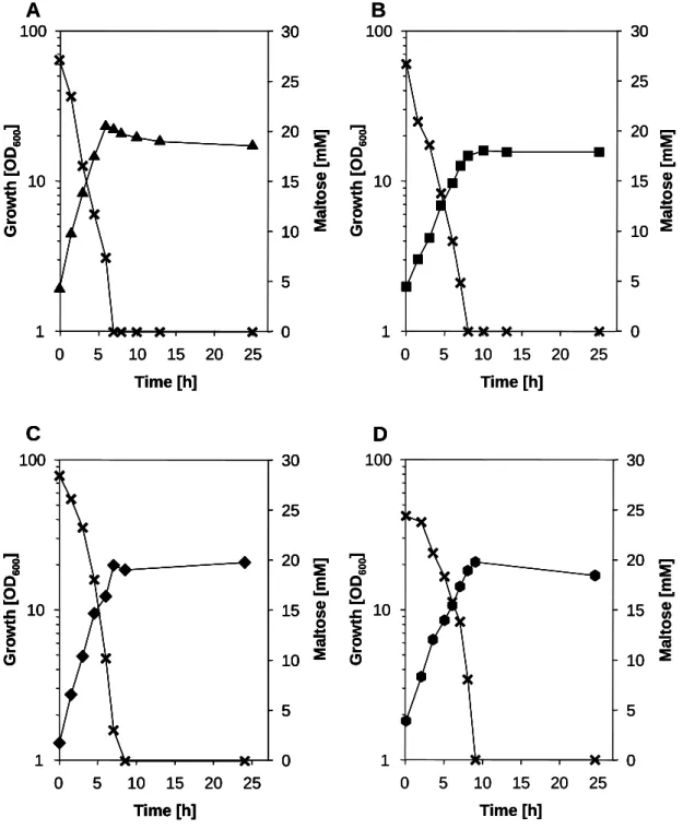
Maltose is a disaccharide formed by two α-1,4-glycosidic linked glucose molecules. High amounts of maltose arise from the enzymatic degradation of starch which can be an alternative as a cheap carbon source for the cultivation of various bacteria. Growth experiments with C. glutamicum in minimal medium containing maltose revealed that C. glutamicum can efficiently exploit maltose as carbon source (Seibold, 2007). Further it has been shown that maltose positively influences glucose utilization increasing the productivity of an amino acid producing C. glutamicum strain (Krause et al., 2010), making it even more interesting to study maltose metabolism.
In contrast to other Gram-positive bacteria the maltose/maltodextrin metabolism in C. glutamicum has not been fully investigated. The maltose metabolic pathway starts with the uptake of maltose by a so far unknown and not characterized transport system. The incoming maltose is then channeled into metabolism by the 4-α-glucanotransferase (MalQ), which catalyzes the transfer from maltosyl and longer dextrinyl residues onto maltose, producing glucose and maltodextrins (Kempkes, 2009).
Figure 3: Maltose metabolism in C. glutamicum. MalQ - 4-α-glucanotransferase; MalP - maltodextrin phosphorylase; Glk - glucokinase; Pgm - phosphoglucomutase, after Seibold et al., 2009.
The released glucose is then phosphorylated by the glucokinase (Glk) yielding glucose-6- phoshate which is then further metabolized (Fig.3). The maltodextrins are degraded by the
membrane transporter
maltodextrins maltose
glucose maltose
glucose-1-P Pi
glucose-6-P
Pgm
ATP ADP
glycolysis
PP-pathway MalQ
MalP
Glk
inside outside
membrane transporter
maltodextrins maltose
glucose maltose
glucose-1-P Pi
glucose-6-P
Pgm
ATP ADP
glycolysis
PP-pathway MalQ
MalP
Glk
inside outside
Introduction 8 maltodextrin phosphorylase (MalP), which forms glucose-1-phoshate by sequential phosphorolysis of the non-reducing end of larger dextrins (Seibold et al., 2009).
The so far revealed maltose metabolism in C. glutamicum is very similar to the well studied maltose and maltodextrin metabolism of the Gram-negative bacterium E. coli and the archeaon T. litoralis (Boos & Shuman, 1998; Xavier et al., 1999), but it is rather different to the pathways found in other Gram-positive bacteria such as Lactobacillus sanfrancisco (Neubauer et al., 1994) and B. subtilis (Schönert et al., 2006).
In B. subtilis maltose is taken up by a maltose specific PTS and becomes phosphorylated.
The cytoplasmic maltose-phosphate is then hydrolyzed by an α-glucosidase (MalA), resulting in glucose and glucose-6-phosphate which is then channeled into metabolism (Schönert et al., 2006). In contrast, L. sanfrancisco takes up maltose by an H+-symporter and cleaves it afterwards via a maltose phosphorylase into glucose and β-glucose-1- phosphate.
Figure 4: Interconnections between maltose and trehalose metabolic pathways. For the maltose metabolism an unknown import system, MalQ – 4-α-glucanotransferase, and Glk – glucokinase are needed. Trehalose synthesis starting from maltodextrins involves the maltooligosyltrehalose synthase (TreY) and the maltooligosyltrehalose trehalohydrolase (TreZ).
The second pathway is catalysed by the trehalose-6-phosphat synthase (OtsA) and the trehalose- 6-phosphate phosphatase (OtsB), respectively, starting with UDP-glucose and glucose-6- phosphate. The third pathway is catalyzed by TreS, with maltose-trehalose interconverting activity.
The export of trehalose for mycolate synthesis is facilitated by an unknown transport system.
membrane exporter
glucose-6-P
maltose maltose
glucose maltodextrin/α-glucan
trehalose trehalose mycolate synthesis
inside outside
importer
TreS TreYZ
OtsAB
UDP-glucose
glycolysis PP-pathway ATP ADP
Glk
MalQ membrane
exporter
glucose-6-P
maltose maltose
glucose maltodextrin/α-glucan
trehalose trehalose mycolate synthesis
inside outside
importer
TreS TreYZ
OtsAB
UDP-glucose
glycolysis PP-pathway ATP ADP
Glk
MalQ
Interestingly, the metabolic pathways for maltose and trehalose are closely interconnected in C. glutamicum (Fig. 4). Incoming maltose can be converted to trehalose by TreS and it has also been proposed that trehalose derived after an osmotic up-shift might be recycled by TreS via maltose (Wolf et al., 2003). Furthermore, MalQ derived maltodextrins are connected to trehalose synthesis via the TreYZ-pathway and the OtsAB-pathway uses glucose-6-phosphate derived during maltose metabolism (Fig. 4). The synthesized trehalose again is important for the cell envelope structure, as building block of mycolates (Tropis et al., 2005).
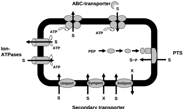
1.7 General transport mechanisms
Protein facilitated transport processes can proceed via two principal mechanisms, a passive and an active one. The pore- or channel-facilitated diffusion enables the thermodynamically favored transport of substances along an existing concentration gradient (from high to low concentrations). These proteins can be addressed as water filled pores that enable the passage of unspecific substances or they function as specific channels over biological membranes, presenting passive transport processes. A good example is LamB, the specific diffusion pore for maltose and maltodextrins, in the outer membrane of E. coli (Boos & Shuman, 1998).
Figure 5: Important classes of bacterial transport systems. S - substrate; X - ion (e.g. H+, Na+), which gradient drives substrate transport; ATP - adenosine 5’-triphosphate; PEP – phospoenolpyruvate, according to Schneider, 2000.
S
S
ATP
ATP
ATP
S X S S
X ATP
S
S
PEP
S S~P
Ion- ATPases
ABC-transporter
PTS
Secondary transporter
Antiport Symport
Uniport S
S
ATP
ATP
ATP
S X S S
X ATP
S
S
PEP
S S~P
Ion- ATPases
ABC-transporter
PTS
Secondary transporter
Antiport Symport
Uniport
Introduction 10 When substances are accumulated in the cytoplasm against an existing concentration gradient (from low to high concentrations) the uptake has to be coupled to an energy dependent step. These active transport processes can be grouped in different classes (Fig. 5). Primary active transport systems utilize the free energy of ATP hydrolysis for the transport of substances. Ion-ATPases, such as detoxication-systems in bacteria for heavy metals (e.g. Cd2+ or AsO43-; Schneider, 2000) and ABC transporter (see 1.8) belong to this class of transport systems. Secondary active transport systems couple the transport of substances to an existing electrochemical gradient (mostly ions such as H+ and Na+). In principle three different secondary active transport mechanisms exist.
During a uniport, also known as facilitated diffusion, which is in fact a passive transport process, transport is driven by the concentration gradient of the transported substance in only one direction, e.g. found in S. cerevisiae for the uptake of xylose (Saloheimo et al., 2007). During a symport two substances are transported in the same direction over biological membranes, using the energy derived from the transport of the co-substance down its concentration gradient. A well studied example is the lactose permease LacY of E. coli that transports lactose in symport with H+ ions (Kaback et al., 2010). On the contrary one substrate is transported in one direction at the same time as co-transporting another substance in the other direction during an antiport (Fig. 5).
Exclusive to bacteria are the phosphotransferase systems (PTS) for sugar uptake, like the main uptake system for glucose in C. glutamicum (Moon et al., 2005), where the source of energy is phospoenolpyruvate (PEP). The phosphoryl group of PEP is transferred to the imported sugar via several proteins; this process is called group translocation. As the transported sugar is chemically modified during the transport process a concentration gradient is provided preventing return transport of the substrate (Fig. 5).
1.8 The maltose uptake system MalFGK2-E of E. coli
The so far best studied of all maltose/maltodextrin transport systems is the ABC transporter MalFGK2-E of E. coli which serves as a model for ATP-binding cassette importers in general (Bordignon et al., 2010). ATP-binding cassette (ABC) transporters are found in all three domains of life and form one of the largest protein superfamilies of paralogous sequences. They are common membrane proteins that facilitate the uptake and export of a large variety of substrates ranging from sugars, amino acids, peptides, vitamins, ions, and even polypeptides across biological membranes.
ABC transport systems share a common structural organization comprising two transmembrane domains (TMDs) and two nucleotide-binding domains (NBDs). The TMDs form the translocation pore and determine the substrate specificity. The NBDs bind and hydrolyze ATP, thereby generating conformational changes that are coupled to the TMDs
MalE
MalF MalG
MalK MalK
ATP
ADP + Pi
and ultimately lead to substrate translocation. Transmembrane domains and nucleotide- binding domains can be arranged as separate proteins, like in prokaryotes, as half- transporters or as a single polypeptide chain. Prokaryotic ABC transport systems that facilitate the uptake of substances have been shown to require a third component that is important for substrate capture and initiation of the transport cycle addressed to as solute- binding protein (SBP) (Eitinger et al., 2011).
The maltose/maltodextrin uptake system MalFGK2-E of E. coli comprises at least five genes which encode the proteins building the system. LamB is a specific outer membrane porin, which is important for the uptake of maltose into the periplasm under low external maltose concentrations. The maltose binding protein MalE, which is located in the periplasm, forms the substrate recognition site and binds maltose and maltodextrins with high affinity (Boos & Shuman, 1998). The membrane spanning part consists of the two permeases MalF and MalG. On the cytoplasmic side they are joined by two MalK proteins, the ATP-hydrolyzing subunits of the transporter, which provide the necessary energy for the active transport (Fig. 6) (Dippel & Boos, 2005).
Figure 6: The maltose uptake system MalFGK2-E of E. coli.
Introduction 12 1.9 Thesis objectives
The first part of this work aimed to characterize and identify the maltose uptake system of C. glutamicum with the help of biochemical methods and to identify possible regulatory interconnections. So far it has been published that the inactivation of the four genes encoding enzyme II permeases of the phosphotransferase systems in C. glutamicum had no effect on growth of the respective mutants in minimal medium containing maltose (Moon et al., 2005). Further it was proposed that intracellular maltose and not maltose- phosphates functions as substrate for the key enzyme 4-α-glucanotransferase of the proposed maltose metabolisation pathway (Seibold et. al., 2009), excluding that maltose is taken up via PTS. This stands in contrast to the observations that PTS-enzymes EI and Hpr, two proteins of the PTS phosphorylation cascade, are necessary for growth on maltose as sole carbon source (Parche et al., 2001). Although maltose uptake seemed likely to be facilitated by an H+-symporter or ABC-transporter the involvement of a phosphotransferase system had to be considered as well.
The second part of this project aimed the identification and characterization of the trehalose export system of C. glutamicum. The disaccharide trehalose has been shown to be important for C. glutamicum as a component of mycolates which make up a crucial part of the outer membrane of the Corynebacterianeae. Already published data indicated that mycolate biosynthesis is dependent on external trehalose, which is most probably exported by C. glutamicum via an unknown and not characterized transport system (Tropis et al., 2005). But the export mechanism of the internal produced trehalose has not been elucidated yet. As trehalose is of such major importance in terms of cell wall synthesis for C. glutamicum, there is increasing interest identifying the responsible export system. After the identification of this system its impact on cell envelope synthesis should be revealed.
2 Materials and Methods
2.1 Bacterial strains, plasmids and oligonucleotides
All strains used in this work are listed in table 1.
Table 1: Bacterial strains used in this study.
Strain Genotype Reference
E. coli
DH5αmcr endA1 supE44 thi-1 λ-recA1 gyrA96 relA1 deoR ∆(lacZYA-argF) U196
∈80DlacZ ∆M15mcrA ∆(mmr hsdRMS mcrBC)
Grant et al., 1990
C. glutamicum
ATCC 13032 Wild type Abe et al., 1967
∆malQ Wild type with a chromosomal deletion in the malQ gene
this study
∆malQ ∆treS Wild type with chromosomal deletions in the malQ and treS genes
this study
∆ptsG Wild type with a chromosomal deletion in the ptsG gene
this study
∆mus Wild type with a chromosomal deletion from the cg2703 to cg2708 gene (including cg2704, cg2705 and cg2707)
this study
∆tus Wild type with a chromosomal deletion from the cg0831 to cg0835 gene (including cg0832, cg0833 and cg0834)
this study
IMmusK Wild type with a chromosomal insertion in the cg2708 gene
this study
IMmusI Wild type with a chromosomal insertion in the cg2701 gene
this study
IMmusG Wild type with a chromosomal insertion in the cg2703 gene
this study
IMmusE Wild type with a chromosomal insertion in the cg2705 gene
this study
IMcg2707 Wild type with a chromosomal insertion in the cg2707 gene
this study
∆otsA ∆treS ∆treY Wild type with chromosomal deletions in the otsA, treS and treY genes
Wolf et al., 2003
Materials and Methods 14
Table 1 (continued)
Strain Genotype Reference
C. glutamicum
∆otsA ∆treS ∆treY
∆tus
Wild type with chromosomal deletions in the otsA, treS and treY and from the cg0831 to cg0835 gene (including cg0832, cg0833 and cg0834)
this study
∆hpr Wild type with a chromosomal deletion in the hpr gene
Lindner et al., 2011
∆treS Wild type with chromosomal deletion in the treS gene
Wolf et al., 2003
IMtreX Wild type with a chromosomal insertion in the treX gene
Ute Meyer, this study
∆malQ IMtreX Wild type with a chromosomal deletion in the malQ gene and a chromosomal insertion in the treX gene
Ute Meyer, this study
trehalose mutant Wild type spontaneous mutant growing on trehalose, unknown genotype
this study
Res 167 Wild type with chromosomal deletions in the cglIM, cglIR, cglllR, and NxR genes
Tauch et al., 2002
∆sugR Wild type with chromosomal deletion in the sugR gene
Engels & Wendisch, 2007
RG1 Wild type with chromosomal deletion in the ramB gene
Gerstmeier et al., 2004
RG2 Wild type with chromosomal deletion in the ramA gene
Cramer et al., 2006
All plasmids used in this study are listed in table 2.
Table 2: Plasmids used in this study
Plasmid Description Reference
pK19mobsacB deletion vector, KanR, oriVE.c., oriT, mob, sacB Schäfer et al.,1994 pK19mobsacB_
MalQDel
pK19mobsacB derivative, containing up- and downstream regions of the malQ gene
Seibold, 2008
pK19mobsacB_
ptsGDel
pK19mobsacB derivative, containing up- and downstream regions of the ptsG gene
this study
Table 2 (continued)
Plasmid Description Reference
pK19mobsacB_
musDel
pK19mobsacB derivative, containing up- and downstream regions of the cg2703 and the cg2708 gene, respectively
this study
pK19mobsacB_
tusDel
pK19mobsacB derivative, containing up- and downstream regions of the cg0831 and the cg0835 gene, respectively
this study
pDrive cloning vector; KanR, lacZ Qiagen, Hilden pDrive_IMmusI pDrive derivative, containing an internal region
of the cg2701 gene for inactivation
this study
pDrive_IMmusG pDrive derivative, containing an internal region of the cg2703 gene for inactivation
this study
pDrive_IMmusE pDrive derivative, containing an internal region of the cg2705 gene for inactivation
this study
pDrive_IMmusK pDrive derivative, containing an internal region of the cg2708 gene for inactivation
this study
pDrive_IMcg2707 pDrive derivative, containing an internal region of the cg2707 gene for inactivation
this study
pDrive_IMtreX pDrive derivative, containing an internal region of the treX gene for inactivation
Ute Meyer, this study
pXMJ19 shuttle vector, Ptac, lacIq, CmR Jakoby et al., 1999 pXMJ19_musFGK-E pXMJ19 derivative, carrying the genes from
cg2703 to cg2708 (including cg2704, cg2705 and cg2707) for expression
this study
pXMJ19_tusFGK-E pXMJ19 derivative, carrying the genes from cg0831 to cg0835 (including cg0832, cg0833 and cg0834) for expression
this study
pXMJ19_musI pXMJ19 derivative, carrying the gene cg2701 for expression
this study
pXMJ19_treS pXMJ19 derivative, carrying the gene treS for expression
this study
pEKEx2 shuttle vector, KanR, oriVE. c., oriVC. g.,Ptac , laqIq Eikmanns et al., 1991
Materials and Methods 16 All oligonucleotides used in this study are listed in table 3.
Table 3: Oligonucletides used in this study. Restriction sites are underlined; linker regions are highlighted in bold, T7 promoter sequences are shown in italics.
Oligonucleotide Sequence (5’-3’) Function
MalQRegioFor GATAGGCACGCTCACTG verification of malQ
deletion
MalQRegioRev GGTCTCTGCGACGAATG verification of malQ
deletion PtsG_DEL1 GTCGACGGGCATAATCTGACAGTGTG ptsG deletion PtsG_DEL2 CTTATAAATTTGGAGTGTGAAGGTTATTGCGTG
GGACGCCAAGAACTGATGGG
ptsG deletion
PtsG_DEL3 CACGCAATAACCTTCACACTCCAAATTTATAAG CCGCTGACTTCATTCGATCC
ptsG deletion
PtsG_DEL4 GGATCCTAAGGACGCCATGTCAAACC ptsG deletion
cg2703_Del1BamHI GGGGATCCAACCGCGAACTGCT mus deletion, BamHI cg2703_Del2 CTTATAAATTTGGAGTGTGAAGGTTATTGCGTG
CGAACGATCCGTAGTGTGAG
mus deletion
cg2703_Del3 CACGCAATAACCTTCACACTCCAAATTTATAAG ACGACTACCTGCTGCCATAC
mus deletion
cg2703_Del4BamHI GGGGATCCTGCCTTGGCAATGA mus deletion, BamHI
cg0831_Del1BamHI GGGGATCCGGCGATGCCATTGT tus deletion, BamHI cg0831_Del2 CTTATAAATTTGGAGTGTGAAGGTTATTGCGTG
GGCAGCTTAGCCAACTTC
tus deletion
cg0831_Del3 CACGCAATAACCTTCACACTCCAAATTTATAAG CGCCAACTATCCAACACCTC
tus deletion
cg0831_Del4BamHI GGGGATCCTGCAACCGCTCCAT tus deletion, BamHI dptsG_test for TCGTACGGTGTGGTTAAG verification of ptsG
deletion
dptsG_test rev AGTATGCACCGCGTAATC verification of ptsG deletion
d2703_test for TAGATGGCGCACAGTGACTC verification of mus deletion
d2703_test rev TAACTACCGCAACACCGATG verification of mus deletion
cg2703for TCTAGATGGCGCACAGTGACTCACTT expression of mus, XbaI
Table 3 (continued)
Oligonucleotide Sequence (5’-3’) Function
cg2703rev ACCGGTCGAGTATGCGATTCATGGTT expression of mus, AgeI
cg0831for GCTCTAGATGCGTTCTGCTCCTGACCTT expression of tus, XbaI
cg0831rev CGGGATCCTTTGCGTTGCGATTCGGATT expression of tus, BamHI
Cg2701_sonde1 TTCGCTGACCTAGTCATCGT inactivation + RNA
probe + RT-PCR of cg2701
Cg2701_sonde2 GGGCCCTAATACGACTCACTATAGGG ACTGCGAGGAAGAACAGGTA
inactivation + RNA probe + RT-PCR of cg2701
Cg2703_sonde1 GCTTCTTGGAGGCCACATTG inactivation + RNA
probe + RT-PCR of cg2703
Cg2703_sonde2 GGGCCCTAATACGACTCACTATAGGG TATCGCGGTTACCGTTGGAG
inactivation + RNA probe + RT-PCR of cg2703
Cg2705_sonde1 TCATCACTTGGCAGGACTAC inactivation + RNA
probe + RT-PCR of cg2705
Cg2705_sonde2 GGGCCCTAATACGACTCACTATAGGG ACTCAGCCATGGAATCAGAC
inactivation + RNA probe + RT-PCR of cg2705
Cg2707_sonde1 CCTATTCCGCCTATCTCGTC inactivation + RT-
PCR of cg2707 Cg2707_sonde2 GGGCCCTAATACGACTCACTATAGGG
GGCGATAGTCGGTTCGTATT
inactivation + RT- PCR of cg2707
Cg2708_sonde1 ACTGAAGATCGCCGGCAAGT inactivation + RNA
probe + RT-PCR of cg2708
Cg2708_sonde2 GGGCCCTAATACGACTCACTATAGGG ATTATCCTCCGGCGTCATGG
inactivation + RNA probe + RT-PCR of cg2708
cg2701_for GGGGATCCTTCTCCACGCAGAGGCACAT expression of cg2701, BamHI cg2701_rev GGGGATCCATGACGTGGATACCACTACC expression of
cg2701, BamHI
Materials and Methods 18
Table 3 (continued)
Oligonucleotide Sequence (5’-3’) Function
cg0832_IN 1 GGATCCTCAGACGGCACCAT RNA probe of
cg0832 cg0832_sonde2 GGGCCCTAATACGACTCACTATAGGG
GTGCGGAAGAGTA
inactivation + RNA probe of cg0832 cgtreS XbaI GCTCTAGAAGGCGAAAGTGGTGAAAGAC expression of treS,
XbaI
cgtreSrev KpnI GCGGTACCTTGTTGATGCGCACTACAGC expression of treS, KpnI
ptsG-NB-T7_for CAAACTGACGACGACATC RNA probe of ptsG
ptsG-NB-T7_rev GGGCCCTAATACGACTCACTATAGGG TGGCAGGAAGTAGAAGAC
RNA probe of ptsG
treX-IM-for CACCGCCGACAATAAAGC inactivation of treX
treX-IM-rev GTGGATGCGTTGGATGTG inactivation of treX
2704_SP1 AAGTTGGTACCGCGGAGT 5’-RACE
2704_SP2 AGGCGAAGATGTTGACTG 5’-RACE
2704_SP3 CACGATGAACGGCACCAA 5’-RACE
2705_SP1 CCTGCCAAGTGATGAAGC 5’-RACE
2705_SP2 TTCGTCCTTGCCAATCTC 5’-RACE
2705_SP3 AACATCGCCTTCCTCGTC 5’-RACE
2708_SP1 ATGGCGATGTCACGGTCA 5’-RACE
2708_SP2 ACGTCCTTGTCTCCGATGAA 5’-RACE
2708_SP3 AACTCGCCATCGGCGATT 5’-RACE
2.2 Media and culture conditions
E. coli as well as pre-cultures of C. glutamicum WT and its derivatives were cultivated aerobically in LB or TY complex medium (Sambrook & Russel, 2001) at 37°C and 30°C, respectively. For the main-culture the cells of an overnight pre-culture were washed twice with 0.9% (w/v) NaCl before inoculating. C. glutamicum main-cultures were grown under aerobic conditions at 30°C as 50 ml cultures in 500 ml baffled shaking flasks at 125 rpm in CGC minimal medium (Eikmanns et al., 1991) with the addition of 1% - 2% (w/v) carbon source as indicated in the Results and Discussion section.
For the selection of integration mutants and expression plasmids 25 µg ml-1 chloramphenicol and/or 50 µg ml-1 kanamycin was added. For induction of the expression plasmid pXMJ19 and its derivatives 250 mM IPTG was added to the medium. Growth of bacterial cultures was measured photometrically at 600 nm (OD600).
2.3 Molecular biology methods
2.3.1 DNA digestion, ligation and purification
Standard techniques such as DNA digestion (enzymes purchased from New England Biolabs; Schwalbach), ligation using T4 ligase (New England Biolabs, Schwalbach) and plasmid DNA purification (NucleoSpin® Plasmid DNA Purification kit; Macherey-Nagel, Düren) were performed according to the manufacturer’s manuals.
2.3.2 Competent cells and transformation
Competent E. coli cells were prepared and transformed according to Inoue et al., 1990.
Competent C. glutamicum cells were prepared as described by Liebl et al., 1989, and transformed by electroporation (2.5 kV, 600 Ω, 2.5 µF) with a MICRO PULSERTM (Bio- Rad, München).
2.3.3 Polymerase chain reaction
The amplification of specific DNA fragments was performed by PCR (polymerase chain reaction, Mullis et al., 1986) using the PRECISOR High-Fidelity DNA Polymerase, the EconoTaq® PLUS GREEN 2X Master Mix (BioCat GmbH, Heidelberg) and the Taq polymerase from NEB (New England Biolabs, Schwalbach) according to the manufacturer’s manuals. Oligonucleotides were obtained from Eurofins MWG Operon (Ebersberg, Germany).
2.3.4 Agarose gel electrophoresis
Gel electrophoresis of DNA was performed using 0.9% agarose gels in 1 x TAE buffer (1 x TAE: 0.04 M Tris, 0.5 mM EDTA, pH 7.5 adjusted with acetic acid) as described by Sambrook et al. ,1989. After electrophoresis, DNA was stained with ethidium bromide. For detection of stained DNA, the Image Master VDS system (Amersham Biosciences, Freiburg) was used. DNA was isolated from agarose gels using the NucleoSpin® Extract kit (Macherey-Nagel, Düren) according to the manufacturer’s manual.
2.3.5 Construction of C. glutamicum mutant strains
For the construction of C. glutamicum deletions the method of allelic replacement described by Schäfer et al., 1994, was used. Derivatives of the vector pK19mobsacB were constructed using the method of cross over PCR after Link et al., 1997. For the allelic replacement, competent C. glutamicum cells were transformed with pK19mobsacB derivatives by means of electroporation (van der Rest et al., 1999). Plasmid integration into the genome was verified by selecting kanamycin-resistant and sucrose-sensitive colonies (the expression of the sacB gene encoding the levan sucrase is toxic in sucrose-
Materials and Methods 20 containing media). To promote re-excision of the plasmid DNA, positive clones were grown overnight in LB broth containing 25 µg ml-1 kanamycin, washed and cultivated in CgXII without phosphate and ammonium source containing 0.5% glucose for 6 h. The starved cells were plated on LB agar supplemented with 10% (w/v) sucrose in different dilutions, usually between 10 -3 and 10 -5. Positive colonies obtained from the subsequent selection (KmS, SucR) were tested for allelic replacement by PCR. Single or double deletion strains were used instead of C. glutamicum WT to generate strains carrying multiple gene deletions. For the construction of strains deleted in trehalose uptake 1.5%
trehalose was added to the selection agar.
For the construction of inactivation mutants pDrive derivatives were constructed with internal parts of the target genes. The gene was inactivated by inserting the vector via homologous recombination into the gene in the genomes of C. glutamicum WT (Schäfer et al. 1994). In short, competent WT cells were transformed with pDrive derivatives by means of electroporation (van der Rest et al., 1999). Plasmid integration into the genome was verified by selecting kanamycin-resistant colonies and PCR.
The trehalose spontaneous-mutant was derived by cultivating C. glutamicum WT in minimal medium with 4% (w/v) trehalose as sole carbon source at 30°C for several days until the culture started to grow. Cells from this culture were repeatedly cultivated with trehalose to gain a C. glutamicum strain that grows fast with trehalose.
2.3.6 RNA hybridization experiments
RNA hybridization experiments were performed as described in Möker et al., 2004. In short, for purification of RNA from C. glutamicum cells were harvested in the exponential growth phase, broken with glass beads in a Fast-Prep® 120 instrument (Q-Biogene, Heidelberg), and the RNA purified from the disrupted cells with the NucleoSpin® RNA II kit (Macherey-Nagel, Düren) according to the manufacturer’s manual. For the construction of RNA anti-sense probes of the desired gene intragenic fragments of a size of ~ 500 bp were amplified with primers carrying the T7 RNA polymerase promoter sequence (Tab. 3).
The resulting DNA fragments were used for DIG-11-dUTP-labeled anti-sense RNA generation by in vitro transcription using T7 RNA polymerase.
For Northern Blot analyses total RNA from C. glutamicum WT was separated on an agarose gel containing 17% (v/v) formaldehyde and transferred onto a nylon membrane.
RNA was cross-linked to the membrane by means of ultraviolet irradiation. Hybridization with the constructed probes and detection steps were carried out according to the DIG application manual (Roche Applied Science, Mannheim). Chemiluminescence was detected with the CCD camera of the LAS 1000 CH system (Fuji, Sraubenhardt).
Changes in gene transcription were monitored by RNA hybridization experiments using total RNA transferred to a nylon membrane using a Minifold Slot-Blot system (Schleicher
and Schuell, Dassel). Hybridization and detection steps were carried out according to the Northern Blot analyses.
2.3.7 5’-RACE
The transcriptional start points were determined by means of the 5'-RACE technique (rapid amplification of 5’-cDNA ends). The 5’,3’-RACE Kit (Roche Applied Science, Mannheim) was used according to the manufacturer’s manual. First strand cDNA was synthesized from total RNA (up to 2 mg) using a gene-specific primer (annealing ~ 250 bp downstream of the translational start point), reverse transcriptase and dNTPs. The mixture was incubated for 1 h at 55°C and then the single-s tranded cDNA was purified from unincorporated primers and nucleotides. Subsequently, terminale transferase was used to add a homopolymeric A-tail to the 3’-end of the single-stranded cDNA, thereby tagging the 5’-end or transcriptional start point of the respective mRNA. Tailed DNA was then amplified by PCR using an oligo-dT-anchor primer (supplied by the manufacturer) and a gene-specific primer. The obtained cDNA was further re-amplified by a second PCR with a nested gene-specific primer and the oligo-dT-anchor primer. The resulting PCR product was used as template in a sequencing reaction to determine the last nucleotide upstream of the poly A-tail, which represents the 5’-end of the respective mRNA (Möker et al., 2004).
2.3.8 RT-PCR
For the determination of whether genes are transcribed monocistronically or in an operon RT-PCRs (reverse transcription polymerase chain reaction) was used. Total cDNA was generated as described earlier and used for PCR with specific primer combinations (Tab. 3), in additional reactions DNA and RNA were used as control.
2.4 Biochemical methods 2.4.1 HPLC analyses
For the quantitative analysis of carbohydrates, amino acids and organic acids HPLC (high performance liquid chromatography) analyses on VWR/Hitachi EliteLaChrom systems were used. Cells were separated from the growth medium by centrifugation and analysis was performed from the supernatant.
Carbohydrates and organic acids were separated via a pre-column (ChromCart 30x4 mm CC30/4 Nucleogel Sugar 810-H, Macherey-Nagel, Düren) and an ion-exchange main- column (300x7.8 mm Nucleogel Sugar 810H, Macherey-Nagel, Düren), at 40 °C with a