LMX1B target genes
DISSERTATION ZUR ERLANGUNG DES DOKTORGRADES DER NATURWISSENSCHAFTEN (DR. RER. NAT.)
DER FAKULT ¨ AT F ¨ UR BIOLOGIE UND VORKLINISCHE MEDIZIN DER UNIVERSIT ¨ AT REGENSBURG
vorgelegt von
Natalya Stepanova
aus
Astana, Kasachstan
im Jahr 2015
Die Arbeit wurde angeleitet von:
Prof. Dr. Ralph Witzgall Unterschrift:
1 Introduction 5
1.1 Inside the nephron . . . 5
1.2 Nail-Patella Syndrome . . . 6
1.3 LMX1B and its target genes . . . 6
1.3.1 ABRA – Actin Binding Rho Activating . . . 7
1.3.2 ARL4C – ADP-Ribosylation Factor-Like 4C . . . 8
1.4 The podocyte actin cytoskeleton . . . 10
1.4.1 (Re)organization of the actin cytoskeleton after podocyte injury . 11 1.5 Actin and actin-associated proteins . . . 12
1.5.1 Actin . . . 13
1.5.2 α-Actinins . . . 14
1.5.3 Focal Adhesion Kinase . . . 14
1.5.4 Non-muscle myosin heavy chain IIA . . . 14
1.5.5 Paxillin . . . 14
1.5.6 Talin . . . 15
1.5.7 Utrophin . . . 15
1.5.8 Vasodilator stimulated phosphoprotein . . . 15
1.5.9 Vinculin . . . 16
1.5.10 Zyxin . . . 16
1.6 Research goals . . . 16
2 Materials and methods 17 2.1 Materials . . . 17
2.1.1 Chemicals and reagents . . . 17
2.1.2 Consumables . . . 19
2.1.3 Enzymes . . . 20
2.1.4 Equipment and instruments . . . 20
2.1.5 Software . . . 22
2.1.6 Kits . . . 23
2.1.7 Antibodies . . . 23
2.1.8 Oligonucleotides . . . 25
2.1.9 Vectors . . . 29
2.1.10 Cells . . . 32
2.1.11 Media, solutions, buffers . . . 33
2.2 Working with bacteria . . . 40
2.2.1 Storage and inoculation of bacteria . . . 40
2.2.2 Transformation of chemically competent bacteria . . . 40
2.2.3 Bacteria transformation by electroporation . . . 41 1
2.2.4 Preparation of plasmid DNA by alkaline lysis with sodium dodecyl
sulfate: Minipreparation . . . 41
2.2.5 Preparation of plasmid DNA with Wizardr Plus Midipreps DNA Purification System . . . 42
2.3 Working with DNA . . . 43
2.3.1 Isolation and purification of DNA . . . 43
2.3.2 Estimation of DNA concentration . . . 44
2.3.3 Agarose gel electrophoresis . . . 45
2.3.4 The Polymerase Chain Reaction . . . 45
2.3.5 Site-directed mutagenesis . . . 45
2.3.6 Generating double-stranded oligonucleotides . . . 46
2.3.7 DNA digestion with restriction endonucleases . . . 46
2.3.8 Ligation of DNA fragments . . . 47
2.3.9 DNA sequencing . . . 47
2.3.10 The real-time polymerase chain reaction . . . 47
2.4 Working with proteins . . . 48
2.4.1 Protein isolation from cell culture . . . 48
2.4.2 Determination of protein concentration . . . 48
2.4.3 Sodium dodecyl sulfate polyacrylamide gel electrophoresis (SDS- PAGE) . . . 49
2.4.4 Protein detection . . . 49
2.4.5 Affinity purification of His-tag fusion proteins . . . 50
2.4.6 Generation of antisera . . . 51
2.4.7 Coupling of proteins to cyanogen bromide-activated sepharose beads 51 2.4.8 Affinity purification of antibodies . . . 52
2.4.9 G-actin / F-actin ratio determination . . . 52
2.5 Working with mammalian cells . . . 52
2.5.1 Mammalian cell culture . . . 52
2.5.2 Expression of proteins in mammalian cells . . . 54
2.5.3 Induction of HtTA-1/LMX1B . . . 55
2.5.4 Generation of stable cell lines . . . 55
2.5.5 Measuring promoter activity by luciferase assay . . . 56
2.5.6 Immunofluorescence staining . . . 56
2.5.7 Transmission electron microscopy studies . . . 57
2.5.8 Fluorescence recovery after photobleaching . . . 58
2.6 Working with kidneys . . . 58
2.6.1 Isolation of primary podocytes . . . 58
2.6.2 Immunofluorescence . . . 58
3 Results 60 3.1 State of the art . . . 60
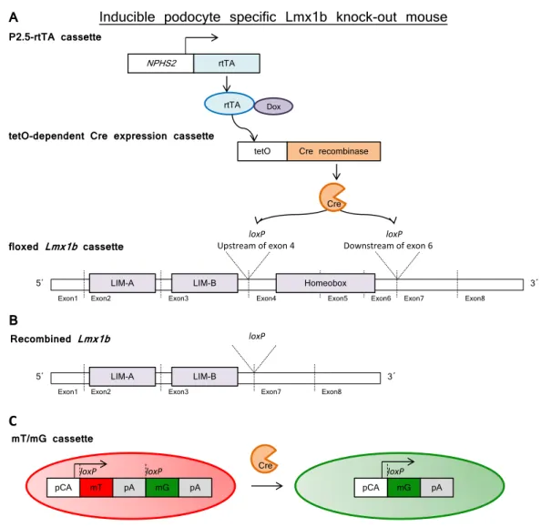
3.1.1 Inducible podocyte-specific Lmx1b knock-out mice . . . 60
3.1.2 Quadruple transgenic mice and “green podocytes” . . . 60
3.1.3 Microarray studies of glomeruli isolated from inducible podocyte- specific Lmx1b knock-out mice . . . 61
3.1.4 Stably transfected HeLa cell line synthesizing the human LMX1B in an inducible manner – HtTA-1/LMX1B cells . . . 63
3.1.5 Promoter binding studies of the putative LMX1B target genes . . 63
3.2 Functional analysis of the putative LMX1B target genes . . . 65 3.2.1 Optimization of the luciferase assay for promoter studies . . . 65 3.2.2 Transcriptional regulation of the human IL6, ABRA and ARL4C
genes by LMX1B . . . 66 3.3 Protein localization studies . . . 69
3.3.1 Subcellular localization of proteins encoded by putative LMX1B target genes in primary podocytes . . . 69 3.3.2 Abra, Arl4c, and Crct1 lead to the increased formation of F-actin 69 3.3.3 Subcellular localization of wild-type and mutant Arl4c proteins in
primary podocytes . . . 69 3.3.4 Mutant Arl4c proteins lead to the greater formation of F-actin . . 71 3.3.5 Ultrastructural localization of Abra, Arl4c and Crct1 in a human
podocyte cell line . . . 73 3.4 Actin polymerization studies . . . 79
3.4.1 LMX1B has no effect on the F- and G-actin content in the HtTA- 1/LMX1B cell line . . . 79 3.4.2 The expression of Abra, Arl4c, and Crct1 has no effect on the F-
to G-actin ratio in a murine podocyte cell line . . . 79 3.5 Expression of recombinant Abra, Arl4c and Crct1 proteins in Escherichia
coli . . . 80 3.6 Generation, affinity purification, and characterization of rabbit antibodies
directed against Abra, Arl4c, and Crct1 . . . 82 3.7 Expression of endogenous Abra, Arl4c and Crct1 after the podocyte-specific
inactivation of Lmx1b . . . 84 3.8 Paxillin expression is affected in conditionally immortalized murine podocytes
expressing Abra and Crct1, but not Arl4c . . . 84 3.9 Turnover of proteins involved in the formation of the actin cytoskeleton
and focal adhesions . . . 86 3.9.1 FRAP studies in the HtTA-1/LMX1B cell line . . . 87 3.9.2 FRAP studies in primary podocytes isolated from quadruple trans-
genic mice . . . 91
4 Discussion 95
4.1 Effect of LMX1B on its putative target genes . . . 95 4.2 Subcellular and ultrastructural localization of
Abra, Arl4c and Crct1 . . . 96 4.3 Regulation of the actin cytoskeleton by LMX1B and its target genes . . . 97 4.4 Glomerular distribution of Abra, Arl4c and Crct1 after the podocyte-
specific inactivation of Lmx1b . . . 100 4.5 Influence of the putative LMX1B target genes on cell-matrix contacts . . 102 4.6 Role of LMX1B in the regulation of cell-matrix adhesion dynamics . . . . 103 4.7 Summary . . . 104 4.8 Perspectives . . . 105
Bibliography 107
Glossary 120
Acknowledgements 125
Erklarung 126
Introduction
1.1 Inside the nephron
The nephron represents the basic structural and functional unit of the kidney. It filters the blood, reabsorbs useful substances, and excretes waste substances with the urine.
The nephron consists of the glomerulus and the tubule. The tubule is divided in three compartments: the proximal, the intermediate and the distal tubules. The glomerulus consists of three major cell types: the fenestrated endothelial cells, which build the capil- lary tuft located inside of Bowman’s capsule, the mesangial cells, which help to maintain the three-dimensional structure of the capillary tuft, and the podocytes. The specialized form of the basement membrane called glomerular basement membrane (GBM) is located between the endothelial cells and the podocytes (Figure 1.1) (Pozzi and Zent, 2012).
Loop of Henle
Afferent arteriole
Bowman capsule Parietal cell
GBM Mesangial cell Glomerular capilary
Podocyte Podocyte foot processes
Proximal tubule Proximal tubular cell
Ultrafiltrate
Efferent arteriole Endothelial cell
Smooth muscle cell
Glomerulus Nephron
Glomerulus Proximal convoluted tubule
Distal convoluted
tubule
Collecting duct Artery
Figure 1.1: Major structural components of the nephron and the glomerulus. Figures were modified from (Albiges et al., 2012) and (Pozzi and Zent, 2012), respectively. GBM stands for glomerular basement membrane.
The podocytes are highly differentiated cells with a unique cytoarchitecture. Many large protrusions, called primary or major processes, extend directly from the podocyte
5
cell body. Primary processes branch and finally end in many small extensions called foot processes. The foot processes from two adjacent podocytes interdigitate with each other thus forming the slit diaphragm (Mundel and Kriz, 1995). The endothelial cells, the glomerular basement membrane, and the slit diaphragm build the glomerular filtration barrier. Dysregulation of the glomerular filtration barrier on the molecular level may lead to renal diseases. One example of such a disease is nail-patella syndrome which affects the kidneys in approximately 40% of patients (Sweeney et al., 2003). The syndrome is described in detail in Section 1.2.
1.2 Nail-Patella Syndrome
Nail-patella syndrome (NPS), also known as ¨Osterreicher syndrome ( ¨Osterreicher, 1930), Turner-Kieser syndrome (Turner, 1933; Kieser, 1939), Fong disease (Fong, 1946) and hereditary osteo-onychodysplasia (Roeckerath, 1951) is a rare autosomal-dominant dis- order with an incidence of ∼1:50,000. As its name indicates, the typical clinical features of nail-patella syndrome include the deformation of finger- and toenails, the hypoplasia or absence of kneecaps, and iliac horns in 95.1%, 92.7% and 70-80% of cases, respec- tively [reviewed by (Bongers et al., 2002)]. Approximately 40% of NPS patients suffer from renal symptoms like proteinuria, hematuria or chronic renal failure (Sweeney et al., 2003; Witzgall, 2007). Electron microscopic studies of kidney biopsies from NPS patients demonstrate morphological abnormalities such as a thickened glomerular basement mem- brane with both fibrillar inclusions and electron-lucent areas, and loss of podocyte foot processes (Del Pozo and Lapp, 1970; Ben-Bassat et al., 1971; Rohr et al., 2002; Witzgall, 2007).
The first report about NPS dates back to 1820 (Roeckerath, 1951). In 1955 NPS was genetically linked to the ABO blood group locus by Renwick et al. and in 1969 to the adenylate kinase locus by Schleutermann et al. (Renwick and Lawler, 1955; Schleuter- mann et al., 1969). Only in 1998 mutations in the LMX1B gene, coding for the LIM homeobox transcription factor 1 beta, were identified in patients with NPS (Dreyer et al., 1998; McIntosh et al., 1998; Vollrath et al., 1998). LMX1B and its target genes are de- scribed in Section 1.3.
1.3 LMX1B and its target genes
LIM homeobox transcription factor 1 beta (LMX1B) is a transcription factor that belongs to the family of LIM-homeodomain proteins and plays a significant role in the normal development of the dorsal limb structures, the anterior segment of the eye, dopaminergic and serotonergic neurons and the podocytes in the kidney. The LMX1B gene gives rise to a protein of 395 and 402 amino acids (Dunston et al., 2004). The LMX1B protein contains two N-terminal cysteine-rich zinc-binding LIM-A and LIM-B domains mediating interactions with other proteins, one central homeodomain responsible for the binding of LMX1B to specific target genes, and a C-terminal glutamine- and serine-rich transcrip- tional activation domain (Bongers et al., 2002). LMX1B is exclusively expressed in the brain, developing limb and eye, cranial mesenchyme, and in the podocytes.
Since the genetic linkage of nail-patella syndrome to the LMX1B gene, more than 140 mutations including missense, splicing, insertion/deletion and nonsense alterations have been identified in NPS patients. Roughly 82% of such mutations identified in
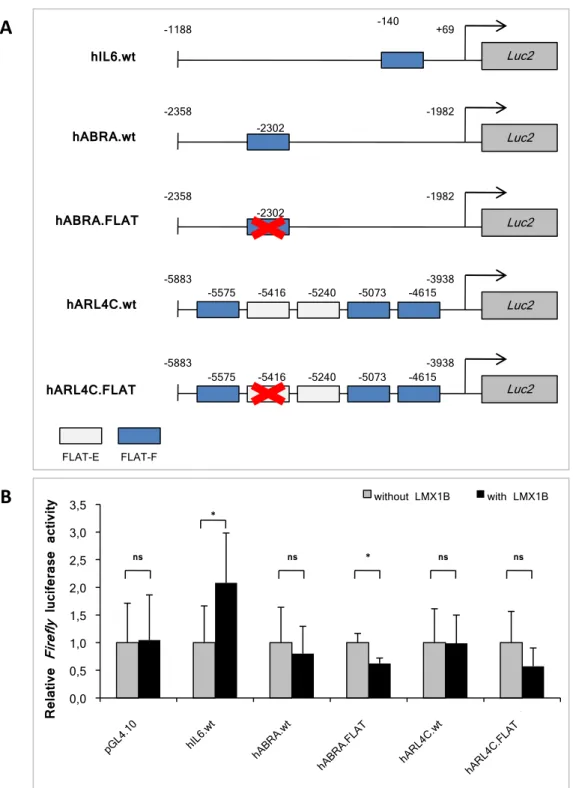
LMX1B are concentrated in the LIM-A and LIM-B domains, while only 18% are located in the homeodomain (Bongers et al., 2002). The binding of LMX1B to its target genes is mediated by the central homeodomain which specifically recognizes the so-called FLAT (FAR linked AT-rich) elements. These elements were characterized for the first time in the promoter sequence of the rat insulin I gene (German et al., 1992). FLAT elements can be subdivided into two types: FLAT-E (5’-TAATTA-3’) and FLAT-F (5’-TTAATA-3’).
To date several LMX1B target genes have been identified. Morelloet al. have demon- strated that expression of the α3 and α4, but not of the α1, α2 and α5 chains of col- lagen IV in the GBM of Lmx1b null mice was strongly reduced. Furthermore, LMX1B showed binding to a FLAT element within the first intron of both mouse and human COL4A4 and it upregulated reporter constructs containing this sequence (Morello et al., 2001). Moreover, LMX1B regulates expression of some genes involved in podocyte dif- ferentiation and function. Mineret al. have shown that Cd2ap and podocin mRNA and protein levels in Lmx1b−/− mice were significantly decreased. In cotransfection assays LMX1B showed binding to the FLAT elements within CD2AP and NPHS2 (podocin) promoters in vitro and activated transcription of these genes through FLAT elements (Miner et al., 2002). In contrast to what has been found in the conventional Lmx1b knock-out mouse, podocin and the α3 and α4 chains of collagen IV were still detected in constitutive podocyte-specific Lmx1b knock-out mice (Suleiman et al., 2007) and in kid- ney biopsies from NPS patients (Heidet et al., 2003). Functional studies using chromatin immunoprecipitation and luciferase reporter assays in HeLa cells revealed that LMX1B regulates the expression of NF-κB target genes, such as IL6 or IL8, by binding to the FLAT-F elements within the proximal promoter of these genes (Rascle et al., 2009).
Microarray studies of glomeruli isolated from inducible podocyte-specificLmx1bknock- out mice have shown a significant increase of the mRNA levels of several actin cytoskeleton- associated proteins, including Abra and Arl4c, as well as a protein of unknown func- tion, Crct1, in comparison to control mice. Chromatin immunoprecipitation experi- ments in conditionally immortalized human podocytes revealed that LMX1B binds to FLAT elements within the ABRA and ARL4C promoter regions (Burghardt et al., 2013). The aim of this PhD project was the characterization of putative target genes which have shown an upregulation in microarray studies of adult Lmx1b knock-out ani- mals. Therefore, the main properties of ABRA and ARL4C are summarized in Subsec- tions 1.3.1 and 1.3.2, respectively. Currently there is no biological information available regarding CRCT1 (Cysteine-rich C-terminal 1), making it attractive as a research object.
1.3.1 ABRA – Actin Binding Rho Activating
The actin binding Rho activating protein ABRA was identified in 2002 by two different laboratories and is also named STARS (for Striated Muscle Activator of Rho Signal- ing) and ms1 (for myocyte stress factor 1) (Arai et al., 2002; Mahadeva et al., 2002).
Initially, ABRA transcripts were discovered in human and murine cardiac and skeletal muscles (Arai et al., 2002; Mahadeva et al., 2002), and most recently in smooth mus- cles (Troidl et al., 2009). In rat primary cardiomyocytes ABRA showed localization to the I-band and was partially seen between Z-lines of the sarcomere. Binding of ABRA to F-actin and activation of Rho-signaling events was demonstrated in transfected COS-7 cells (Arai et al., 2002). Recently, Foglet al. characterized two independent F-actin bind- ing domains at the C-terminus of the protein – actin binding domains 1 and 2 (ABD1 and ABD2) located between amino acids 193-296 and 294-375, respectively. Moreover,
ABD1 showed higher affinity to F-actin in comparison to ABD2 (Fogl et al., 2012).
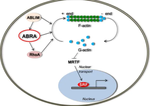
Two members of the actin-binding LIM (ABLIM) protein family, ABLIM-2 and ABLIM-3 were recently reported as interaction partners of ABRA. Upon ABLIM-2 and ABLIM-3 association with ABRA, the activation of downstream targets of the serum response factor (SRF) is initiated (Barrientos et al., 2007). Another pathway of ABRA- mediated SRF signaling includes depletion of G-actin and release of myocardin-related transcription factors (MTRFs) which after transport into the nucleus stimulate SRF tar- gets (Kuwahara et al., 2005). Finally, ABRA stimulates the SRF-dependent transcription via RhoA activation (Arai et al., 2002). A scheme of the ABRA-mediated regulation of actin dynamics and SRF signaling is shown in Figure 1.2.
F-actin
G-actin
+ end - end
ABLIM
ABRA
RhoA
MRTFNuclear transport
SRF Nucleus
Figure 1.2: A scheme of the ABRA-mediated regulation of actin dynamics and SRF sig- naling. ABRA stimulates F-actin polymerization directly or through activation of RhoA.
ABLIM proteins associate with ABRA and stimulate the ABRA-dependent activation of the downstream targets of SRF. Upon the ABRA-dependent polymerization of F-actin, MTRFs are released from the inhibitory influence of G-actin and imported into the nucleus, where they stimulate SRF transcriptional activity. Abbreviations: ABLIM – actin binding LIM domain protein, MTRF – myocardin-related transcription factor, RhoA – Ras homolog gene family, member A, SRF – serum response factor. Modified from (Kuwahara et al., 2005;
Barrientos et al., 2007).
1.3.2 ARL4C – ADP-Ribosylation Factor-Like 4C
The ADP-Ribosylation Factor-Like 4C (ARL4C), also known as ARL7, belongs to the ARF-related family of small GTP-binding proteins (Jacobs et al., 1999). As reported by Jacobs et al. transcripts of human ARL4C are highly enriched in the brain, and lower levels of ARL4C were detected in the spleen, thymus, esophagus, stomach, intestine and uterus (Jacobs et al., 1999). A few years after the work of Jacobs et al., Wei et al. demonstrated mRNA expression of ARL4C in human lung, brain, leukocytes and
placenta (Wei et al., 2009). Recent immunohistochemical analysis of mouse embryos on day 15 revealed that Arl4c was expressed in the brain, in some epithelial rudiments, and in the kidney, where Arl4c was detected in ureteric buds, pretubular aggregates, renal vesicles, and the comma-shaped body (Matsumoto et al., 2014).
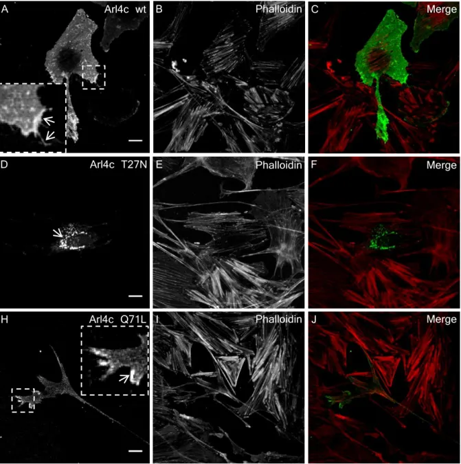
The subcellular localization of ARL4C depends on the binding to GTP or GDP. In overexpression studies the wild-type form of ARL4C and the active GTP-bound mutant of ARL4C were detected within the cytoplasm and at the plasma membrane of filopodia.
On the other hand, the inactive GDP-bound ARL4C mutant was detected in the per- inuclear region of the cell and was not associated with filopodia (Engel et al., 2004). A ARL4C mutant which lacked the myristoylation site lost its ability to bind to the plasma membrane (Engel et al., 2004).
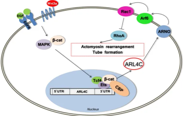
EGF
Wnt3a
β-cat MAPK
5΄UTR 3΄UTR
Tcf4Ets
ARL4C
ARNO Arf6
Rac1
RhoA Actomyosin rearrangement
Tube formation
ARL4C
Nucleus β-cat
Figure 1.3: Scheme of the Arl4c-mediated regulation of actomyosin rearrangement and epithelial tube formation after combinatorial induction of Wnt3a/EGF signaling based on the data from Matsumoto et al. (Matsumoto et al., 2014). The induction of Wnt3a/EGF activates the MAPK and β-catenin pathways, then a complex consisting of Ets/Tcf4/CBP forms at the Ets-binding site in the 3’ non-coding region of the Arl4c gene. Arl4c induces ARNO and Arf6, resulting in Rac1 activation. Rac1 induction leads to the inhibition of RhoA, rearrangement of the cytoskeleton and tubular structure formation. Abbreviations:
β-cat – β-catenin, ARF6 – ADP-ribosylation factor 6, ARNO – Arf nucleotide-binding site opener, CBP – cyclic AMP-responsive element binding protein [(CREB)-binding protein], EGF – epidermal growth factor, EGFR – epidermal growth factor receptor, MAPK – mitogen activated kinase-like protein, Rac1 – ras-related C3 botulinum toxin substrate 1, RhoA – Ras homolog gene family, member A, Tcf4 –T-cell factor 4, UTR – untranslated region, Wnt3a – wingless-type MMTV integration site family, member 3A.
The first reported interaction partner of ARL4C was α-tubulin. Wei et al. have shown that the association of both proteins leads to microtubule-dependent vesicular transport (Wei et al., 2009). Hofmannet al. demonstrated that Arl4c was able to recruit ARNO (Arf nucleotide-binding site opener) and Arf6 (ADP-ribosylation factor 6) to the
plasma membrane (Hofmann et al., 2007). Recent findings of Matsumoto et al. shed light on the interaction of Arl4c with other proteins (Matsumoto et al., 2014). It was demonstrated that the expression of Arl4c was induced by the combined action of Wnt3a and epidermal growth factor (Wnt3a/EGF). The Wnt3a/EGF signaling activated the MAPK and β-catenin pathways, then a complex of Ets/Tcf4/CBP formed at the Ets- binding site in the 3’ non-coding region of theArl4cgene (Matsumoto et al., 2014). Arl4c induced ARNO and Arf6, resulting in Rac1 activation. Finally, Rac1 activation led to RhoA inhibition, rearrangement of the cytoskeleton and tubular structure formation as shown in Figure 1.3 (Matsumoto et al., 2014).
1.4 The podocyte actin cytoskeleton
The cytoskeleton is a dynamic network of filaments with a large number of functions, including cell shape maintenance, cell migration, intracellular transport, cell division, etc. It consists of three main components: microfilaments, intermediate filaments, and microtubules. Microfilaments, or actin filaments, are polymeric structures with a diam- eter of 5-9 nm, formed by actin. Intermediate filaments, as its name indicates, are the fibers of the cytoskeleton with an intermediate size of about 10 nm. They are composed of intermediate filament proteins. The largest component of the cytoskeleton are the microtubules, which have a diameter of around 25 nm and are formed by a protein called tubulin (Alberts et al., 2008).
As mentioned previously, the following segments can be distinguished in podocytes:
the cell body, the large processes, and the foot processes (FPs). The cell body and the large processes contain all components of cell cytoskeleton, while the FPs are exclu- sively composed of actin filaments and actin-associated proteins (Drenckhahn and Franke, 1988). The membrane of FPs is functionally divided into three domains: the apical mem- brane domain (AMD), the basal membrane domain (BMD), and the slit diaphragm (SD), as shown in Figure 1.4 (Kerjaschki, 2001).
Figure 1.4: Scheme of three membrane domains of FPs: the apical membrane domain (AMD) (blue), the basal membrane domain (BMD) (red), and the SD (black). PAB stands for parallel actin bundles. The figure was taken from Greka and Mundel, 2012.
The disruption of any of the three FP domains leads to the reorganization of the actin cytoskeleton: parallel bundles of actin cytoskeleton change into a disordered struc- ture (Shirato et al., 1996; Kerjaschki, 2001), which is followed by the retraction of FPs and the loss of the slit diaphragm. This process is known as foot process effacement. Foot process effacement leads to proteinuria (Farquhar et al., 1957; Murphy et al., 1979) which is typical of numerous glomerular disorders, such as focal segmental glomerulosclerosis
(FSGS) (Grishman and Churg, 1975), minimal change disease (Farquhar et al., 1957), and diabetic nephropathy (Kerjaschki, 2001). The described morphological changes in the FP actin cytoskeleton were also observed in some NPS patients (Del Pozo and Lapp, 1970). Therefore, the purpose of Subsection 1.4.1 is to present the current knowledge in the (re)organization of the actin cytoskeleton during podocyte injury.
1.4.1 (Re)organization of the actin cytoskeleton after podocyte injury
It is now commonly accepted that podocytes play a key role in the maintenance of the glomerular filtration barrier. Injury of podocytes leads to proteinuria and development of glomerular diseases. During podocyte injury, changes in their shape are observed, which is called foot process effacement. The FPs of each podocytes are retracted, moreover the amount of filtration slits is reduced, so that FPs appears as a continuous cytoplasmic sheet along the GBM as shown in Figure 1.5 (Shirato, 2002).
To date many causes of podocyte injury have been described, including changes in the SD structure (Simons et al., 2001; Verma et al., 2003), interference with the GBM or the podocyte-GBM interaction (Noakes et al., 1995; Kreidberg et al., 1996; Raats et al., 1997; Regele et al., 2000; Kretzler et al., 2001), modulations of the negatively charged podocyte membrane (Orlando et al., 2001; Takeda et al., 2001; Galeano et al., 2007), alterations of the transcriptional regulation of podocytes (Quaggin, 2002; Rascle et al., 2007), and reorganization of the podocyte actin cytoskeleton (Smoyer and Mundel, 1998;
Kos et al., 2003; Jones et al., 2006; Verma et al., 2006).
Despite the increasing number of studies on actin-associated proteins in podocytes, the molecular mechanisms of the reorganization of the podocyte cytoskeleton remain un- clear. Upon podocyte injury the coordinated parallel bundles of actin filaments change into an interwoven network (Shirato et al., 1996; Kerjaschki, 2001). The expression of actin and the actin-filament cross-linking protein α-actinin is increased (Shirato et al., 1996). Studies by Smoyeret al. considerα-actinin-4 as initiator of FP effacement (Smoyer et al., 1997). Subsequently glomerular α3-integrin is induced (Smoyer et al., 1997). The importance of α-actinin-4 in podocyte injury was demonstrated by the fact that muta- tions in the ACTN4 gene coding for α-actinin-4 lead to an autosomal dominant form of FSGS (Kaplan et al., 2000; Pollak, 2002). Miao et al. discovered further cytoskele- tal proteins involved in the dynamic changes of foot processes. Transgelin, survivin, Arp2, cytokeratin7, and vinculin mRNA and protein levels were significantly increased in puromycin aminonucleoside nephropathy in rats and in patients with proteinuric re- nal diseases (Miao et al., 2009). Mutations in the MYH9 gene, which encodes another component of the FP cytoskeleton (non-muscle myosin heavy chain IIA), are associated with Epstein, Fechtner, and Sebastian syndromes, and May-Hegglin anomaly (Arrondel et al., 2002; Seri et al., 2003).
The Rho family of small GTPases takes part in many cellular aspects including regu- lation of actin cytoskeleton (Hall, 1998). Recent studies in podocytes indicate that signal- ing pathways regulated by the most studied members of Rho-GTPases (RhoA, Rac1 and Cdc42) are crucial for maintenance of structural and functional podocyte integrity. Ac- tivation of RhoA in podocytes leads to albuminuria accompanied by FP effacement (Zhu et al., 2011; Wang et al., 2012). Podocyte-specific deletion of Cdc42 causes proteinuria, FP effacement, and glomerulosclerosis, while deletion of Rac1 in podocytes prevents FP effacement (Blattner et al., 2013).
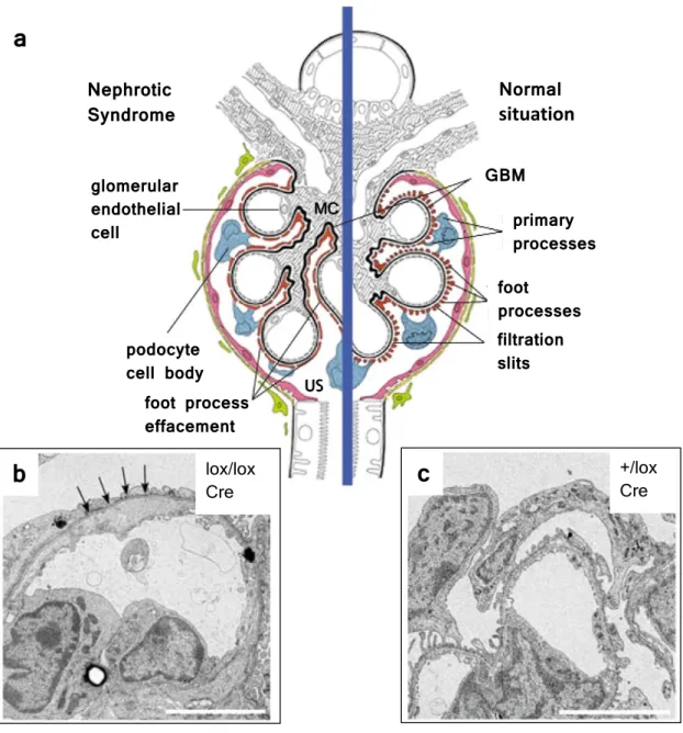
a
b
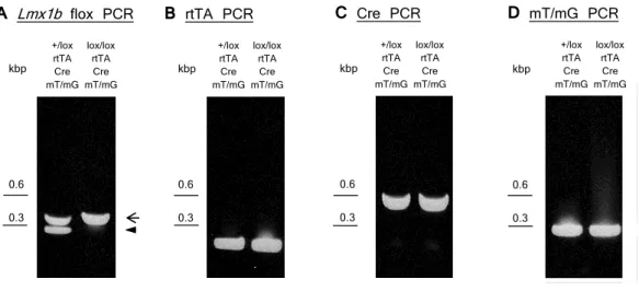
lox/lox Crec
+/lox CreNormal situation Nephrotic
Syndrome
GBM
primary processes foot
processes filtration slits foot process
effacement podocyte cell body glomerular endothelial cell
US MC
Figure 1.5: (a) The structure of the affected (left) and of the normal (right) glomeru- lus. Nephrotic syndrome leads to FP effacement. (b-c) Ultrastructural imaging taken from kidneys of mice with podocyte-specific inactivation of Lmx1b, where FP effacement and a thickened GBM are observed (arrows in b). Scale bars, 5 µm. Abbreviations: GBM, glomerular basement membrane; MC, mesangial cells; US, urine space; +/lox control mice with one wild-type and one floxedLmx1b allele;lox/lox, mice with two floxed Lmx1b alleles;
Cre, Cre transgene under control of the human NPHS2 promoter. Modified from (Somlo and Mundel, 2000; Suleiman et al., 2007).
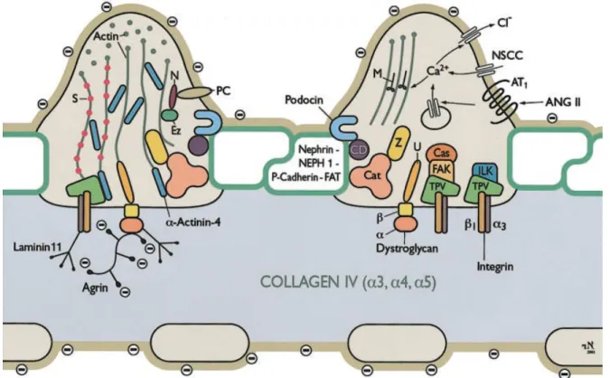
1.5 Actin and actin-associated proteins
One of the objectives of this study was to unveil the role of LMX1B for the maintenance of the podocyte actin cytoskeleton. Because of the fact that to date about 150 pro- teins involved in the formation of the actin cytoskeleton are known (Zaidel-Bar et al., 2007), only several proteins, including actin, α-actinin-1, α-actinin-4, focal adhesion ki- nase, non-muscle myosin heavy chain IIA, paxillin, talin, utrophin, vasodilator stimulated
phosphoprotein, vinculin, and zyxin were chosen for these studies. The essential infor- mation about the selected proteins will be presented in the following subsections. The interactions between the actin-associated proteins in the podocyte FPs are shown in Figure 1.6.
Figure 1.6: Scheme of the molecular composition of podocyte FPs. Abbreviations: Cas, p130Cas; Cat, catenins; CD, CD2-associated protein; Ez, ezrin; FAK, focal adhesion kinase;
ILK, integrin-linked kinase; M, myosin; N, NHERF2; NSCC, nonselective cation channel;
PC, podocalyxin; S, synaptopodin; TPV, talin, paxillin, vinculin; U, utrophin; z, ZO-1. The figure was taken from Pavenst¨adtet al., 2003.
1.5.1 Actin
Actin is the most abundant protein in many eukaryotic cells. It builds microfilaments, one of the major components of the actin cytoskeleton. Actin exists in two forms, monomeric G-actin (globular) and polymeric F-actin (filamentous). A multitude of actin-associated proteins is engaged in the maintenance of the actin cytoskeleton. Thus, the actin-binding protein formin promotes elongation of actin filaments, while actin-related protein 2/3 (Arp2/3) induces branched actin filaments. Profilin increases the elongation of actin fila- ments, and cofilin accelerates its disassembly. Another actin-associated protein,α-actinin, cross-links microfilaments into loose bundles (Alberts et al., 2008). As previously men- tioned, actin filaments in healthy podocytes are organized in parallel bundles. Podocyte injury causes the reorganization of actin filaments into a dense network (Shirato et al., 1996; Kerjaschki, 2001). Moreover, the expression of actin is increased (Shirato et al., 1996).
1.5.2 α-Actinins
α-Actinin was originally isolated from striated muscles (Ebashi and Ebashi, 1965). To date, four α-actinins have been reported, which are named α-actinin-1, -2, -3 and -4.
α-Actinins are dimeric proteins with an essential role in cross-linking actin filaments.
In vertebrates, α-actinin-1 and α-actinin-4 are expressed in most tissues and cell types, while expression ofα-actinin-2 andα-actinin-3 is restricted to muscles (Foley and Young, 2014). Despite the great sequence similarity between α-actinin-1 and α-actinin-4, muta- tions in these genes lead to different abnormalities. Thus, mutations in the α-actinin-1 gene can lead to congenital macrothrombocytopenia characterized by decreased numbers of thrombocytes (Gu´eguen et al., 2013). On the other hand, mutations in theα-actinin-4 gene are associated with different tumors, including breast cancer (Honda et al., 1998), ovarian cancer (Yamamoto et al., 2007), neuroendocrine lung cancer (Miyanaga et al., 2013), and many others. Additionally, mutations in the α-actinin-4 gene result in fo- cal segmental glomerulosclerosis characterized by proteinuria and kidney failure (Kaplan et al., 2000).
1.5.3 Focal Adhesion Kinase
Focal adhesion kinase (FAK), also known as protein tyrosine kinase 2 (PTK2), is a cy- toplasmic tyrosine kinase localized to focal adhesions (Schaller et al., 1992). It interacts with the integrin-binding proteins paxillin (Hildebrand et al., 1995), talin (Chen et al., 1995), and vinculin (Stevens et al., 1996). Fibroblast-like cells derived fromfak−/−murine embryos at day 8 showed decreased mobility (Ili´c et al., 1995). The importance of FAK in cell-matrix adhesion of podocytes during glomerular injury was demonstrated by Maet al.
As a consequence of glomerular injury FAK is activated in murine podocytes, which then leads to proteinuria and FP effacement. Inhibition of FAK with specific pharmacological agent protects against glomerular damage (Ma et al., 2010).
1.5.4 Non-muscle myosin heavy chain IIA
The non-muscle myosin heavy chain IIA (NMMHC-IIA) is a cytoskeletal protein encoded by theMYH9 gene. It plays a role in cell contractility, motility, morphology, cytokinesis, and polarity (M¨uller et al., 2011). Mutations in theMYH9 gene are associated with var- ious autosomal-dominant diseases such as Epstein, Fechtner, and Sebastian syndromes, and May-Hegglin anomaly. All of them are characterized by macrothrombocytopenia, hearing defects, cataracts, D¨ohle-like bodies in neutrophils, and glomerular injury (Seri et al., 2003). In adult human kidney MYH9 mRNA and protein are predominantly ex- pressed in podocytes (Arrondel et al., 2002). Recent studies by Johnstoneet al. revealed that the podocyte-specific deletion of Myh9 predisposes mice to glomerulopathy. Mice with the podocyte-specific deletion of Myh9 showed no renal insufficiency or proteinuria compared to control. However, mutant mice treated with doxorubicin hydrochloride de- veloped proteinuria and glomerulosclerosis, while control mice were resistant (Johnstone et al., 2011).
1.5.5 Paxillin
The cytoskeleton-associated protein paxillin was initially localized to the focal adhesions at the ends of actin stress fibers in chicken embryo fibroblasts (Turner et al., 1990). To
date the binding of paxillin to various focal adhesion proteins including vinculin (Turner et al., 1990), focal adhesion kinase (Hildebrand et al., 1995), and β1 integrin (Schaller et al., 1995) has been reported. Hagelet al. have demonstrated the necessity of paxillin for normal development of mice: no paxillin-deficient embryos were detected after embryonic day 9.5 (Hagel et al., 2002). During maturation of a healthy rat kidney the expression of paxillin is reduced and restricted to distal tubular cells (Matsuura et al., 2011).
1.5.6 Talin
The cytoskeletal protein talin identified in focal adhesions (Burridge and Connell, 1983a;
Burridge and Connell, 1983b) plays an essential role in linking the actin cytoskeleton to integrins (Horwitz et al., 1986; Critchley et al., 1999). Besides actin and integrin, talin is able to interact with other focal adhesion proteins such as vinculin (Critchley and Gingras, 2008). Recent studies of talin1 in murine podocytes revealed that talin1 is important for development of the glomerular filtration barrier and its maintenance (Tian et al., 2014). The podocyte-specific loss of talin1 in mice leads to severe proteinuria, foot process effacement, and kidney failure. The activation of β1 integrin as well as cell spreading and adhesion in the talin1-deficient podocytes were moderately reduced.
Nevertheless, significant changes in the actin cytoskeleton of podocytes without talin1 were observed (Tian et al., 2014).
1.5.7 Utrophin
Utrophin, also known as dystrophin-related protein (DRP), is a component of the dys- trophin-glycoprotein complex. Utrophin participates in linking the actin cytoskeleton to agrin, a major heparan sulfate proteoglycan in the glomerular basement membrane, through its interaction with β-dystroglycan. In the normal human and rat kidney utrophin was localized within glomerulus outlining the peripheral capillary loops (Raats et al., 2000; Regele et al., 2000). Detailed immunoelectron microscopy studies of the rat kidney demonstrated that utrophin was localized at the cytoplasm of some podocyte foot processes with the strongest intensity in the regions near the GBM (Raats et al., 2000).
1.5.8 Vasodilator stimulated phosphoprotein
Vasodilator stimulated phosphoprotein (VASP) was initially described in human platelets as a major protein phosphorylated in response to stimulation with vasodilators (Halbr¨ugge and Walter, 1989). VASP is known to interact with multiple actin-associated proteins. It is able to bind profilin, which as a consequence leads to actin polymerization (Reinhard et al., 1995). Its interaction with other cytoskeletal proteins, such as vinculin (Brindle et al., 1996), talin,α-actinin, and F-actin stabilizes focal adhesion and the connection with actin filaments (Hemmings et al., 1995). Recent studies by Harris et al. on conditionally immortalized podocytes revealed that proteases present in the plasma of focal segmen- tal glomerulosclerosis (FSGS) patients after transplatation lead to a podocin-dependent phosphorylation of VASP via protease-activated receptor-1. Additionally, FSGS plasma induced VASP phosphorylation-dependent motility of podocytes (Harris et al., 2013).
1.5.9 Vinculin
Vinculin was originally described as an intracellular protein localized at specialized sites where microfilaments terminate at cell membranes (Geiger et al., 1980). This cytoskele- tal protein consists of head and tail domains separated by a proline-rich region (Price et al., 1989). The head domain of vinculin is able to bind talin, which then leads to the recruitment of vinculin to focal adhesions, while the tail domain binds F-actin and pax- illin (Ziegler et al., 2008). Vinculin plays an essential role during embryonic development:
vinculin-deficient embryos die due to heart and brain defects (Xu et al., 1998). More- over, vinculin-deficient mouse embryonic fibroblasts show reduced adhesion and migration rates (Xu et al., 1998).
1.5.10 Zyxin
The integrin-associated linker protein zyxin was discovered at both cell-matrix and cell- cell junctions (Crawford and Beckerle, 1991). The interaction of zyxin with several focal adhesion proteins including α-actinin (Crawford et al., 1992) and VASP (Grange et al., 2013) was demonstrated. Zyxin is not absolutely essential for development: zyxin null mice showed no defects, they are viable and fertile (Hoffman et al., 2003).
1.6 Research goals
The main purpose of this study was to clarify the molecular pathway(s) regulated by LMX1B by focusing on the characterization of the putative LMX1B target genes which are upregulated after the inactivation of Lmx1b, and on the importance of LMX1B for the maintenance of the podocyte actin cytoskeleton. The specific goals of the studies were:
1) Identification of LMX1B binding elements (FLAT elements) within the promoter regions of its putative target genes.
2) Determination of the role of LMX1B in the transcriptional regulation of its putative target genes.
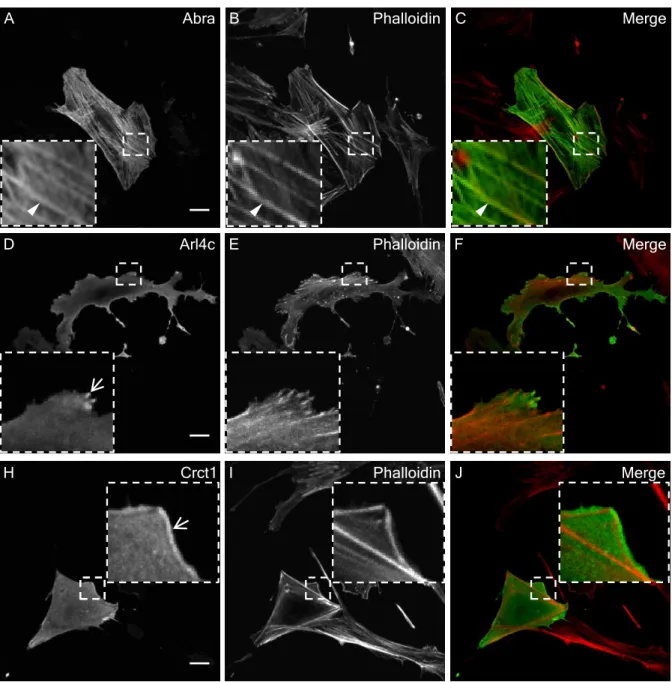
3) Demonstration of the subcellular localization of the corresponding proteins and their interaction with the actin cytoskeleton.
4) Determination of the effect of Abra, Arl4c and Crct1 on F-actin and paxillin.
5) Demonstration of the localization of Abra, Arl4c and Crct1 in the kidney and determination of their role in nail-patella syndrome.
6) Investigation of the role of LMX1B in the dynamics of the podocyte actin cytoskele- ton.
Materials and methods
2.1 Materials
2.1.1 Chemicals and reagents
Chemicals/reagents Producer
0.25% Trypsin-EDTA solution Sigma-Aldrich
4-(2-hydroxyethyl)-1-piperazineethanesulfonic acid (HEPES) Roth
5x Phusionr HF Reaction Buffer NEB
Acetic acid Merck
Acrylamid/bis solution 37.5:1 Serva
Agarose Roth
Albumin fraction V Roth
Amido black 10B Merck
Ammonium persulfate (APS) Fluka
Ampicillin sodium salt Roth
Aprotinin Roth
Bacto agar Becton Dickinson
Bacto tryptone Becton Dickinson
Bacto yeast extract Becton Dickinson
β-mercaptoethanol Merck
Bovine serum albumine (BSA) Sigma
Bromophenol blue Serva
Calcium chloride Roth
Cyanogen bromide-activated-Sepharoser 4B Sigma-Aldrich
DEAE-Dextran Sigma-Aldrich
Deoxynucleotide triphosphates (dNTPs) Fermentas
Dimethyl sulfoxide (DMSO) Sigma
Dipotassium phosphate Merck
Dithiothreitol (DTT) Roth
DMEM/Ham’s F12 PAA
Doxycycline hyclate Applichem
Dulbecco’s modified Eagle’s Medium (DMEM) - high glucose Sigma-Aldrich
Ethanol Sigma
Ethidium bromide Sigma
Ethylene glycol tetraacetic acid (EGTA) Serva 17
Ethylenediaminetetraacetic acid (EDTA) Roth
Fetal bovine serum (FBS) Gibco
Ficollr 400 Serva
Formaldehyde solution, 37% Sigma
G-418-sulfate Invitrogen
Glucose Merck
Glycerine/glycerol Roth
Glycine Merck
Hydrochloric acid Merck
Hygromycin B liquid PAA
Imidazole Merck
Immersol 518F for Microscope Carl Zeiss
Isopropanol (2-propanol) Merck
Isopropyl β-D-1-thiogalactopyranoside (IPTG) Fermentas
ITS supplement PAA
Kanamycin sulfate Roth
λ DNA/Hind III Marker, 2 Fermentas
Leupeptin Serva
Light cyclerr480 SYBR Green I master mix Roche
Magnesium chloride Sigma
Magnesium sulfate Merck
Methanol Merck
Monopotassium phosphate Merck
Nickel sulfate Merck
PageRuler prestained protein ladder ThermoScientific
Paraformaldehyde Merck
Penicillin-streptomycin Sigma-Aldrich
Phenylmethanesulfonylfluoride (PMSF) Sigma-Aldrich φX174 DNA/BsuRI (HaeIII) Marker, 9 Fermentas
Polyethylen glycol (PEG 3350) Sigma-Aldrich
Potassium acetate Merck
Potassium chloride Merck
Powdered milk Sucofin
Puromycine PAA
Rotir-Phenol (Phenol/chlorophorm/isoamylalcohol 25:24:1) Roth
Rotir-Quant Roth
RPMI-1640 with L-glutamine Sigma
Sodium acetate Roth
Sodium azide Merck
Sodium cacodylate trihydrate Fluka
Sodium chloride VWR
Sodium dodecyl sulfate (SDS) Serva
Sodium fluoride Merck
Sodium hydrogen carbonate Merck
Sodium hydroxide Merck
Tetramethylenediamine (TEMED) Serva
Thimerosal Sigma-Aldrich
Tris Affymetrix
Trisodium phosphate Merck
Triton X-100 Roth
Tweenr 20 Roth
Urea Merck
Western Lightning-ECL Enhanced Luminol Reagent Perkin Elmer Western Lightning-ECL Oxidizing Reagent Perkin Elmer
2.1.2 Consumables
Consumable Producer
15 ml tube Sarstedt
50 ml tube Sarstedt
96-well white non-binding plate for luciferase assay Greiner bio-one
Autoclave tape VWR
Cell scraper Sarstedt
Centrifuge tubes for centrifuge (20 ml) Beckman coulter GmbH
Chromotography paper “3MM Chr” Whatman
Cover glasses 24 x 40 mm Roth
Cover glasses 24 x 60 mm Roth
CryoPure tube 1.8 ml Sarstedt
Culture flasks Schott duran
Filter tips Sarstedt
Folded filters 90 mm Schleicher & Schull
Gene Pulserr Cuvette for electroporation Bio-Rad
Glass coverslips 12 mm R.Langenbrinck
Glass coverslips 22 mm R.Langenbrinck
Glass Pasteur pipettes VWR
Glassware (media bottles, beakers with scale, Erlenmeyer flasks etc.)
VWR
Light cycler 96-well plate Sarstedt
Membrane filters VSWP (0.025 µm) Millipore
Micro tube 0.2 ml Sarstedt
Micro tube 1.5 ml Sarstedt
Microscope slides Roth
Neubauer counting chamber depth 0.1 mm Marienfeld
Nitrile gloves Kimtech
Parafilm Pechiney Plastic
Packaging
Pipette tips Sarstedt
Polystyrene cuvettes for spectrophotometer U-2000 Sarstedt
PVDF transfer membrane (0.45 µm) Millipore
Scalpel Swann-Morton
Serological pipettes Sarstedt
Sterile filter 0.20 µm VWR
SuperFrostr Plus Microscope Slides R.Langenbrinck
Syringes Brown
Task wipes Kimtech
Tissue culture dishes Sarstedt
Tissue culture flasks Sarstedt
Tissue culture plates Sarstedt
2.1.3 Enzymes
A. Restriction enzymes
Enzyme Producer
BamHI-HF: 20 U/µl NEB
EcoRI-HF: 20 U/µl NEB
KpnI-HF: 20 U/µl NEB
MluI: 10 U/µl Fermentas
NheI-HF: 20 U/µl NEB
XhoI: 20 U/µl NEB
B. DNA and RNA modifying enzymes
Enzyme Producer
BAP (Bacterial alkaline phosphatase): 150 U/µl Invitrogen
T4 DNA ligase: 400 U/µl NEB
Phusionr DNA Polymerase: 2 U/µl Finnzymes
RNase A: 1µg/µl Serva
2.1.4 Equipment and instruments
Equipment/ Instrument Producer
Agarose gel documentation system “Gel Max” Intas
Agarose gel electrophoresis apparatus “Horizon 58” Gibco BRLTM
Autoclave “Systec 5050 ELV” Tuttnauer
Battery powered pipette filler for all pipetting from 0.1 ml to 200 ml Pipetusr-akku
Hitschmann Laborgerte
Bunsen burner Usbeck
Centrifuge “Heraeus Pico 17” with Ch.500005 PP rotor Thermo Scientific
Chemiluminescence system “Fusion Fx7” Vilber Lour-
mat Deutschland GmbH
CO2 incubator “APT.lineTMCB” Binder
Confocal microscope “LSM 510 Meta” Zeiss
Cryostat “Leica CM3050s” Leica
Electrophoresis power supply “PS 608” Gibco BRLTM Electrophoresis power supply “Standard Power Pack P25” Biometra Electroporator “Gene Pulser XcellTMElectroporation System” Bio-Rad Filter catridge set “Seralpur PRO 90 CN” Seral Fluorescence microscope “Axiovert 200” Zeiss Flow cytometry system “CyFlow Space” Partec
Freezers -20◦C Privileg
Freezers -80◦C “Herafreeze” Heraeus
Gel electrophoresis chamber “OwlTMEasyCastTMB2 Mini Gel Electrophoresis Systems”
Thermo Scientific
Heating block “VLM LS 1” VLM
Hot plate with a magnetic stirrer “MR 2002” and “MR 3001” Heidolph
Ice machine “AF-10” Scotsman
Incubator “Kelvitron t” Heraeus
Incubator “Unitron” Infors
Laboratory balance “KERN 770” KERN & Sohn
GmbH
Laboratory pH Meter “CG 842” SCHOTT Gerte
GmbH Laminar flow bench “Lamin Air HA 2448 GS” Heraeus
LightCyclerr 480 II Roche
Liquid nitrogen container “ARPEGE TP 170” Air Liquide Medi- cal GmbH
Microplate luminometer “MicroLumat Plus LB96V” Berthold Technolo- gies
Microtome “Leica RM2255” Leica
Microscope “Nikon Eclipse TS 100” Nikon
Microwave “Privileg 8016 G” Privileg
pH electrode “SenTix60” WTW
Pipette 2-20, 20-200 Labmate
Pipette P2, P10N, P1000 Gilson
Refrigerated centrifuge “Avantir J-26 XP” with rotor Ja-10 and Ja-25.50
Beckman Coulter Refrigerated centrifuge “Multifuge 3 L-R” with rotor Ch.
2454
Heraeus
Refrigerated centrifuge “Sigma 3K20” with rotor Nr. 12158 B.Braun, Labora- tory Centrifuges GmbH
Refrigerators Privileg
Rocking platform shaker “Duomax 1030” Heidolph
Rotating wheel for Eppendorf-tubes Workshop of Uni-
versity of Regens- burg NWF III
Spectrophotometer “U-2000” Hitachi
Tabletop high-speed micro centrifuge“HITACHI himac CT15RE” with rotor T15A61-1041
VWR
Thermal cycler “MyCycler” Bio-Rad
Thermomixer “5436” for Eppendorf tubes Eppendorf Trans-Blotr SD Semi-Dry ElectrophoreticTransfer Cell Bio-Rad Transmission electron microscope “EM Zeiss 902” Zeiss
Ultramicrotome “Ultracut E” Reichert Jung
UV-visible spectrophotometer “Evolution 201” Thermo Scientific
Vacuum gas pump VWR
Vertical gel electrophoresis cell “Mini Protean Tetra cell” Bio-Rad
Vortexer “Vortex-Genie 2” Scientific Industries
Weighing scale “BL 1500 S” Sartorius
2.1.5 Software
A. Programs
Program Purpose Company
Adobe Photoshop Digital image processing Adobe Systems Incorpo- rated
Bio1D Optical density Vilber Lourmat
FCS express version 3 FACS data analysis De Novo Software FileMaker Pro 6 Institute databases FileMaker, Inc.
Fusion version 15.18 WB imaging Vilber Lourmat
ImageJ 1.46r National Institutes of
Health LightCyclerr480 1.5.0 Real-time PCR Roche Microsoft Excel Data calculations, dia-
grams and tables
Microsoft
Microsoft Word Word processing Microsoft
SPOT Advanced 4.0.9 Image processing Diagnostic instruments, Inc.
Thermo Insight 1.4.40 DNA/RNA concentration measurement
Thermo Scientific
ZEN (blue edition) LSM images visualization Carl Zeiss MicroImaging GmbH
B. Internet databases and software
Name of the
database/software
Purpose Internet address
EMBOSS: stretcher Needleman-Wunsch alignment of two se- quences
www.emboss.bioinformatics.nl/cgi- bin/emboss/stretcher
Nucleotide Gene and transcript source
www.ncbi.nlm.nih.gov/nuccore
Primer3 Primer design www.bioinfo.ut.ee/primer3-
0.4.0/primer3
Protein Protein sequence source www.ncbi.nlm.nih.gov/protein
PubMed Biomedical literature
search
www.ncbi.nlm.nih.gov/pubmed RestrictionMapper Mapping sites for re-
striction enzymes in DNA sequences
www.restrictionmapper.org
UCSC Genome
Browser
Promoter studies www.genome.ucsc.edu Universal ProbeLi-
brary Assay Design Center
Primer design for RT- qPCR
www.lifescience.roche.com
2.1.6 Kits
Kit name Producer
E.Z.N.A.r Gel Extraction Kit VWR
iScriptTMcDNA Synthesis Kit Bio-Rad
NucleoSpinr RNA II Macherey-Nagel
Western Lightning Chemiluminescence Reagent Perkin Elmer Wizardr Plus Midipreps DNA Purification System Promega
2.1.7 Antibodies
A. Primary antibodies, peptides
Name Immunogen/Epitope Species Dilution Source
12CA5 HA-epitope Mouse mAb 1:30 WB, IF J. Kyriakis
Acti-stain- 555 Flu- orescent Phalloidin
F-actin - 1:500 IF Cytoskeleton
Acti-stain- 488 Flu- orescent Phalloidin
F-actin - 1:500 IF Cytoskeleton
Anti-Abra mouse Abra Rabbit, poly- clonal
1:100 WB this work 1:1 IF
Anti-Actin C-terminal actin frag- ment (C11 peptide at- tached to MAP back- bone)
Rabbit, poly- clonal
1:2500 WB Sigma-Aldrich
Anti-Arl4c aa of mouse Arl4c Rabbit, poly- clonal
1:30 WB this work undiluted IF
Anti-Crct1 Peptide mapping near N-terminus of NICE-1 of human origin
Goat, poly- clonal
1:500 WB Santa Cruz Biotechnology 1:2500 IF
Anti- GAPDH
314-333 aa of mouse GAPDH
Rabbit poly- clonal
1:10000 WB Sigma-Adrich
Anti-His6 Histidine-Tagged Pro- teins
Mouse mAb 1:100 WB Calbiochem
Anti-LMX1B 322-395 aa of human LMX1B, clone 193-67
Mouse mAb 1:10 WB D. Heudobler
Anti-Paxillin 1-557 aa of chicken paxillin
Mouse mAb 1:10000 WB BD Transduc- tion Laborato- ries
B. Secondary antibodies
Name Immunogen/Epitope Species Dilution Source Anti-goat
IgG FITC- conjugated
Goat IgG Donkey,
polyclonal
1:100 IF Santa Cruz Biotechnology
Anti-mouse IgG (Fab specific) peroxidase conjugate
Mouse IgG, Fab spe- cific
Goat, poly- clonal
1:10000 WB Sigma-Aldrich
Anti-mouse IgG CyTM3- conjugated
Mouse IgG (H&L) Goat, poly- clonal
1:400 IF Dianova
Anti-mouse IgG (whole molecule) FITC- conjugated
Mouse IgG Goat, poly-
clonal
1:150 IF Cappel
Anti-rabbit IgG (whole molecule) peroxidase conjugate
Rabbit IgG Goat, poly-
clonal
1:20000 WB Sigma-Aldrich
Anti-rabbit IgG CyTM3- conjugated
Rabbit IgG (H&L) Goat, poly- clonal
1:300 IF Dianova
Anti-
rabbit IgG DyLightTM405 conjugated
Rabbit IgG (H&L) Donkey, polyclonal
1:600 IF Rockland
2.1.8 Oligonucleotides
A. Oligonucleotides for PCR
Oligonucleotides for PCR were synthesized by Metabion (Planegg). “F” and “R” stands for “Forward” and “Reverse” primers, respectively. Tm is the melting temperature of oligonucleotides calculated with OligoAnalyzer 3.1.
Name Sequence [5’-3’] Tm[◦C]
Cloning to pGL4.10 NheI-
hABRA.wt F
ATT TGC GCT AGC TCA GAT GCC GTT GAA CTC TG 64.7
XhoI-
hABRA.wt R
GCA AAT CTC GAG GAG GCT GGA GTG CAG TGG 66.8
NheI-
hARL4C.wt F
ATT TGC GCT AGC AGC ATC TCC ATC CCA AAC AG 65.0
XhoI-
hARL4C.wt R
GCA AAT CTC GAG ACC CAG CTG AGA CCA GAG AA 65.1
NheI- hIL6.wt F
ATT TGC GCT AGC TGA GAC CAA GGA TCC TCC TG 65.4
XhoI- hIL6.wt R
GCA AAT CTC GAG AGT TCA TAG CTG GGC TCC TG 64.3 Cloning to pcDNA3.1
BamHI- Kozak-HA- mAbra F
ATT TGC GGA TCC GCC ACC ATG TAC CCA TAC GAC GTC CCA GAC TAC GCT ATG GCT CCA GGA GAA AGG GA
73.5
XhoI- mAbra R
ATT TGC CTC GAG TTA CTC AAG GAG AGT AAT C 59.3
BamHI- Kozak-HA- mArl4c F
ATT TGC GGA TCC GCC ACC ATG TAC CCA TAC GAC GTC CCA GAC TAC GCT ATG GGC AAC ATC TCC TCC AAC
72.9
XhoI- mArl4c R
ATT TGC CTC GAG TTA CCG CTT CTT CTT CTG C 63.3
BamHI- Kozak-HA- mCrct1 F
ATT TGC GGA TCC GCC ACC ATG TAC CCA TAC GAC GTC CCA GAC TAC GCT ATG TCT CAA CAG GGC GCC A
73.9
XhoI- mCrct1 R
ATT TGC CTC GAG TCA GCA GCC ACC GGA GCA G 69.3
Cloning to pET21a BamHI-
mAbra F
ATT TGC GGA TCC ATG GCT CCA GGA GAA AGG GA 66.9
XhoI- mAbra R
ATT TGC CTC GAG CTC AAG GAG AGT AAT CAC 61.4
BamHI- mArl4c F
ATT TGC GGA TCC AAG TCG CTG CCG GTG GCC G 71.4
XhoI- mArl4c R
ATT TGC CTC GAG CCG CTT CTT CTT CTG CTT G 65.2
BamHI- mCrct1 F
ATT TGC GGA TCC ATG TCT CAA CAG GGC GCC A 67.9
XhoI- mCrct1 R
ATT TGC CTC GCA GCC ACC GGA GCA GCA G 71.1
Cloning to pminiSOG-C1-mCherry XhoI-
mAbra F
ATT TGC CTC GAG TAA TGG CTC CAG GAG AAA GGG A
65.9
KpnI- mAbra R
ATT TGC GGT ACC TTA CTC AAG GAG AGT AAT CAC 60.7
XhoI- mArl4c F
ATT TGC CTC GAG TAA TGG GCA ACA TCT CCT CCA AC
65.2
KpnI- mArl4c R
ATT TGC GGT ACC TTA CCG CTT CTT CTT CTG CTT G
64.2
XhoI- mCrct1 F
ATT TGC CTC GAG TAA TGT CTC AAC AGG GCG CCA 66.8
KpnI- mCrct1 R
ATT TGC GGT ACC TCA GCA GCC ACC GGA GCA GCA GCA G
72.2
Cloning to pLVPT-rtTR-KRAB-2SM2 BamHI-
mAbra F
ATT TGC GGA TCC ATG GCT CCA GGA GAA AGG G 66.3
MluI- mAbra R
ATT TGC ACG CGT GGC TCA AGG AGA GTA ATC AC 64.7
BamHI- mArl4c F
ATT TGC GGA TCC ATG GGC AAC ATC TCC TCC AAC 66.1
MluI- mArl4c R
ATT TGC ACG CGT GGC CGC TTC TTC TTC TGC TTG 68.1
BamHI- mCrct1 F
ATT TGC GGC TCC ATG TCT CAA CAG GGC GCC 67.2
MluI- mCrct1 R
ATT TGC ACG CGT GGG CAG CCA CCG GAG CAG CAG
73.5
Oligonucleotides for PCR from genomic DNA
rtTA F CCC ACT TCT GAG ACA ACG 52.8
rtTA R GGT CAA AGT CGT CAA GGG 53.2
Tomato F CTC TGC TGC CTC CTG GCT TCT 61.6
Tomato R1 CGA GGC GGA TCA CAA GCA ATA 57.5
Tomato R2 TCA ATG GGC GGG GGT CGT T 62.3
Cre F TGG ACA TGT TCA GGG ATC GC 57.4
Cre R TCA GCT ACA CCA GAG ACG GA 57.4
Floxed Lmx1b F
AGG CTC CAT CCA TTC TTC TC 54.1
Floxed Lmx1b R
CCA CAA TAA GCA AGA GGC AC 54.1
Oligonucleotides for qPCR
luc2 F CAT GAC CGA GAA GGA GAT CG 54.8
luc2 R CAG CTT CTT GGC GGT TGT A 55.4
B. Oligonucleotides for site-specific mutagenesis
Oligonucleotides for PCR were synthesized by Metabion (Planegg). “F” and “R” stands for “Forward” and “Reverse” primers, respectively. Tm is the melting temperature of oligonucleotides calculated with OligoAnalyzer 3.1.
Name Sequence [5’-3’] Tm[◦C]
Abra-mut F CTG TAG ATT AAG TCT AGA ATC ACT TCC C 54.4 Abra-mut R GGG AAG TGA TTC TAG ACT TAA TCT ACA G 54.4 Arl4c-mut F GTC CAG CCA AGT AAG CTT GAT GAA ATG AAA ATA
ATA G
59.3
Arl4c-mut R CTA TTA TTT TCA TTT CAT CAA GCT TAC TTG GCT GGA C
59.3
C. Oligonucleotides for cloning of shRNAs into pInducer10 vector
The shRNAs were designed using the miR RNAi design option of the Block-iT RNAi Designer program. “T” and “B” stands for “Top” and “Bottom” strands, respectively.
Name Sequence [5’-3’]
XhoI-Arl4c- shRNA # 559 T
TCG AGT AAC GAT GTG CAG AGA CTG GAG TTT TGG CCA CTG ACT GAC TCC AGT CTG CAC ATC GTT AG EcoRI-Arl4c–
shRNA # 559 B
AAT TCT AAC GAT GTG CAG ACT GGA GTC AGT CAG TGG CCA AAA CTC CAG TCT CTG CAC ATC GTT AC XhoI-Arl4c-
shRNA # 850 T
TCG AGC AAA CTT GGT CAC CTT GTG CAG TTT TGG CCA CTG ACT GAC TGC ACA AGG ACC AAG TTT GG EcoRI-Arl4c–
shRNA # 850 B
AAT TCC AAA CTT GGT CCT TGT GCA GTC AGT CAG TGG CCA AAA CTG CAC AAG GTG ACC AAG TTT GC XhoI-Arl4c-
shRNA # 1037 T
TCG AGT CAT AGA GCT TGT CCA TGC CCG TTT TGG CCA CTG ACT GAC GGG CAT GGA AGC TCT ATG AG EcoRI-Arl4c–
shRNA # 1037 B
AAT TCT CAT AGA GCT TCC ATG CCC GTC AGT CAG TGG CCA AAA CGG GCA TGG ACA AGC TCT ATG AC
2.1.9 Vectors
A. Outside plasmids
Vector Selection
marker
Source
mEmerald-FAK kan M. W. Davidson (National High Mag-
netic Field Laboratory, Florida State University)
mEmerald-Talin kan M. W. Davidson (National High Mag- netic Field Laboratory, Florida State University)
mEmerald-VASP kan M. W. Davidson (National High Mag- netic Field Laboratory, Florida State University)
Lifeact-GFP kan R. Faessler, S. Wickstroem (Max Plank Institute of Biochemistry)
pcDNA3.1 amp Invitrogen
pcDNA6.2/N-EmGFP- MYH9
amp T. Vallenius (University of Helsinki)
pCMV delta R8.2 amp Addgene
pCMV-SPORT6.1/Crct1 amp Source BioScience
pCR4-TOPO/Abra kan Source BioScience
pCS2+/GFP-UtrCH amp E. Kerkhoff (The Molecular Cell Biol- ogy Laboratory, University of Regens- burg)
pDEST/Lifeact-mCherry-N1 kan Addgene
pEGFP-α-actinin1 kan C.A. Otey (Department of Cell Biol- ogy and Physiology, UNC, Chapel Hill, USA)
pEGFP-α-actinin4 kan C.A. Otey (Department of Cell Biol- ogy and Physiology, UNC, Chapel Hill, USA)
pET21a amp Novagen
pEYFP/Arl4c-wt kan Th. Engel (Leibniz Institute for Arte- riosclerosis Research, University Muen- ster)
pEYFP/Arl4c-T27N kan Th. Engel (Leibniz Institute for Arte- riosclerosis Research, University Muen- ster)
pEYFP/Arl4c-Q71L kan Th. Engel (Leibniz Institute for Arte- riosclerosis Research, University Muen- ster)
pGL3-Enhancer amp Promega
pInducer10 amp S.Elledge (Brigham and Women’s Hos-
pital, Boston)
pLVPT-rtTR-KRAB-2SM2 amp Addgene
pmCherry/Paxillin-C3 kan E. Kerkhoff (The Molecular Cell Biol- ogy Laboratory, University of Regens- burg)
pmCherry-C1/Zyxin kan E. Kerkhoff (The Molecular Cell Biol- ogy Laboratory, University of Regens- burg)
pMD2.G amp Addgene
pminiSOG-C1 kan M. W. Davidson (National High Mag-
netic Field Laboratory, Florida State University)
pYX-Asc/Arl4c amp Source BioScience
Vinculin-Venus amp Addgene
B. Own constructs
Name of construct Size of in- sert [kbp]
5’ cloning site
3’ cloning site
Resistance
pcDNA3.1/HA-mAbra 1.1 BamHI XhoI amp
pcDNA3.1/HA-mArl4c 0.6 BamHI XhoI amp
pcDNA3.1/HA-mCrct1 0.3 BamHI XhoI amp
pET21a/mAbra-His 1.1 BamHI XhoI amp
pET21a/mArl4c-His 0.4 BamHI XhoI amp
pET21a/mCrct1-His 0.3 BamHI XhoI amp
pGL4.10/hAbra.wt 0.4 NheI XhoI amp
pGL4.10/hArl4c.wt 1.9 NheI XhoI amp
pGL4.10/hIL6.wt 1.3 NheI XhoI amp
pInducer10/shRNA-Arl4c
#559
0.065 XhoI EcoRI amp
pInducer10/shRNA-Arl4c
#850
0.065 XhoI EcoRI amp
pInducer10/shRNA-Arl4c
#1037
0.065 XhoI EcoRI amp
pLVPT-rtTR-KRAB- 2SM2/mAbra-GFP
1.1 BamHI MluI amp
pLVPT-rtTR-KRAB- 2SM2/mArl4c-GFP
0.6 BamHI MluI amp
pLVPT-rtTR-KRAB- 2SM2/mCrct1-GFP
0.3 BamHI MluI amp
pminiSOG-C1- mCherry/mAbra
1.1 XhoI KpnI kan
pminiSOG-C1- mCherry/mArl4c
0.6 XhoI KpnI kan
pminiSOG-C1- mCherry/mCrct1
0.3 XhoI KpnI kan
2.1.10 Cells
A. Bacterial strains
Strain Antibiotic resistance
DH5α none
BL21(DE3) none
BL21(DE3)pLysS chloramphenicol Rosetta(DE3)pLysS chloramphenicol
TOP 10 none
B. Cell lines
Name Description Source
COS-7 African green monkey kidney fibroblast-like cell line
B. Royer-Pokora (Institute of Hu- man Genetics, Heidelberg)
HEK293T Human Embrionic Kidney 293 cells, which constitutively express the simian virus 40 (SV40) large T antigen
W. Nickel (University of Heidel- berg, Biochemistry Center)
Murine podocyte cell line
Podocytes isolated from H- 2kb-tsA58 mice, which carried temperature-sensitive SV40 large T antigen under control of the IFN-γ-inducible H-2kb promoter
K. Endlich (University of Heidel- berg, Institute of Anatomy and Cell Biology I)
Human podocyte cell line
conditionally immortalized hu- man podocyte cell line trans- fected with the temperature- sensitive SV40-7 gene
M.A. Saleem (University of Bris- tol)
HtTA- 1/myc- LMX1B clone #34
HtTA-1 transfected with pUHD10-3/myc-LMX1B
A. Rascle (University of Regens- burg)
C. Mouse strains
Name Description Source
Lmx1b2loxP LoxP sites are above and below of exons 4 and 6 respectively
R. Johnson (MD Anderson Can- cer Center, Houston, USA) P2.5-rtTA Gene for reverse tetracycline-
dependent transactivator (rtTA) under the control of 2.5 kbp of hu- man NPHS2 promoter
J.Kopp (NIH, Bethesda, USA)
LC-1 Expression of the luciferase and cre gene is regulated by the Tet system
H. Bujard (ZMBH, Heidelberg)
mT/mG Expression of membrane-targeted tandem dimer Tomato (mT) prior to Cre-mediated excision and membrane-targeted green fluores- cent protein (mG) after excision
T.Huber (Nephrology, University Medical Center, University of Freiburg)
2.1.11 Media, solutions, buffers
A. Media and solutions for work with bacteria
Medium/solution Components
LB Broth Amount per 1 liter:
10 g Bacto-Tryptone 5 g Yeast Extract 10 g NaCl
LB Broth with agar Amount per 1 liter:
(Agar plates) 10 g Bacto-Tryptone 5 g Yeast Extract 10 g NaCl
15 g Bacto Agar
After autoclaving medium was cooled to about 50◦C and appropriate antibiotics were added. Approximately 25 ml were poured into each plate. Inverted plates were stored at 4◦C.
Transformation and 10% PEG 3350 storage solution (TSS) 5% DMSO
25 mM MgSO4 LB medium
B. Solutions and buffers for DNA isolation, cloning and electrophoresis
Solution/buffer Components
Cell lysis solution 0.2 M NaOH/1% SDS (Alkaline SDS solution)
Cell resuspension solution 50 mM glucose (GTE buffer) 25 mM Tris, pH 8.0
10 mM EDTA pH 8.0 CutSmart buffer, 1× 50 mM Potassium acetate
20 mM Tris acetate
10 mM Magnesium acetate 100 µg/ml BSA
pH 7.9 at 25◦C
DNA loading buffer, 5× 0.125% bromophenol blue 15% Ficoll type 400 50 mM EDTA, pH 8.0 0.5% SDS
dNTP, 10 mM 10 mM dATP, 10 mM dTTP, 10 mM dCTP, 10 mM dGTP
NEBuffer 1.1, 1× 10 mM Bis Tris Propane-HCl 10 mM MgCl2
100 µg/ml BSA pH 7.0 at 25◦C
NEBuffer 2.1, 1× 50 mM NaCl
10 mM Tris-HCl 10 mM MgCl2
100 µg/ml BSA pH 7.9 at 25◦C NEBuffer 3.1, 1× 100 mM NaCl
50 mM Tris-HCl 10 mM MgCl2
100 µg/ml BSA pH 7.9 at 25◦C
Potassium acetate solution 5 M potassium acetate
pH 4.8 was adjusted with 5 M acetic acid Sodium acetate, 3 M 3 M sodium acetate
pH 5.2 was adjusted with glacial acetic acid Solution 1 for genomic DNA 10 mM Tris, pH 7.6
isolation from blood 10 mM KCl 10 mM MgCl2
2.4% NP-40
Solution 2 for genomic DNA 10 mM Tris pH 7.6 isolation from blood 10 mM KCl
10 mM MgCl2 0.5 M NaCl 2 mM EDTA 0.5% SDS T4 DNA ligase buffer 50 mM Tris-HCl
10 mM MgCl2 1 mM ATP 10 mM DTT pH 7.5 at 25◦C
TAE, 50× 2 M Tris
0.1 M EDTA
pH 8.0 was adjusted with acetic acid
C. Buffers for protein isolation
Buffer Components
G actin buffer 1% Triton X-100 20 mM Tris, pH 7.4 5 mM EGTA, pH 7.4 20 mM NaF
25 mM sodium pyrophosphate 10 mM DTT
0.5 mM PMSF 2 µg/ml leupeptin 2 µg/ml aprotinin prepared each time fresh Protein lysis buffer 1× PBS
1% Triton X-100 6 M urea
stored at 4◦C
D. Buffers for protein electrophoresis
Buffer Components
Laemmlie buffer, 5× 2% SDS
(SDS-PAGE loading buffer) 5% β-mercaptoethanol or 200 mM DTT 20% glycerol
0.1% bromophenol blue 62.5 mM Tris-HCl, pH 6.8 Lower gel buffer, 4× 1.5 M Tris, pH 8.8
0.4% SDS
SDS-PAGE Running buffer, 10× 0.25 M Tris base 1.92 M glycine 1% SDS
Upper gel buffer, 4× 0.5 M Tris, pH 6.8 0.4% SDS
Resolving gel components 10% 12% 15%
30% acrylamide/ 0.8% bisacrylamide 1.66 ml 1.98 ml 2.46 ml 4× lower gel buffer, pH 8.8 1.25 ml 1.25 ml 1.25 ml
deionized water 2.07 ml 1.75 ml 1.27 ml
TEMED 2.9 µl 2.9 µl 2.9 µl
10% APS 13.9 µl 13.9 µl 13.9 µl
Stacking gel components 4%
30% acrylamide/ 0.8% bisacrylamide 0.39 ml 4×upper gel buffer, pH 6.8 0.75 ml
deionized water 1.84 ml
TEMED 3 µl
10% APS 15 µl
E. Solutions and buffers for protein detection
Solution/buffer Components
Amido black destaining solution 50% methanol 10% acetic acid
Amido black staining solution 1.5 mM amido black 10 B 50% methanol
10% acetic acid
Blocking buffer 5% low-fat milk in PBST buffer
PBST 0.02% Tweenr 20
1× PBS
Semi-dry transfer buffer, 1× 25 mM Tris base 192 mM glycine 20% methanol 0.02% SDS
F. Buffers for recombinant protein purification
Buffer Components Binding buffer, 8× 40 mM imidazole
4 M NaCl
160 mM Tris-HCl, pH 7.9 Charge buffer, 8× 400 mM NiSO4·H2O Elute buffer, 4× 4 M imidazole
2 M NaCl
80 mM Tris-HCl, pH 7.9 SDS-sample buffer 125 mM Tris, pH 6.7
2.5% SDS 10% glycerol
2.5% β-mercaptoethanol 0.01% bromophenol blue
Strip buffer, 2× 200 mM EDTA
1 M NaCl
40 mM Tris-HCl, pH 7.9 Wash buffer, 4× 240 mM imidazole
2 M NaCl
80 mM Tris-HCl, pH 7.9
G. Buffers for coupling of proteins
Buffer Components
Coupling buffer 100 mM NaHCO3, pH 8.3 500 mM NaCl
Wash buffer 1 100 mM NaAc, pH 4.0 500 mM NaCl
Wash buffer 2 100 mM NaHCO3, pH 8.0 500 mM NaCl
H. Buffers for affinity purification of antibodies
Buffer Components
Wash buffer A 10 mM Tris-HCl, pH 7.5 170 mM NaCl
Wash buffer B 10 mM Tris-HCl, pH 7.5 170 mM NaCl
0.02% Tweenr 20 Wash buffer C 10 mM Tris-HCl, pH 7.5
500 mM NaCl 0.02% Tweenr 20
I. Media, solutions and buffers for work with cells Medium/solution/buffer Components
Ca2+-buffer 150 mM NaCl
(Krebs-Henseleit Buffer) 5 mM KCl
2.2 mM CaCl2·2H2O 1 mM MgCl2·6H2O
5 mM glucose (C6H12O6·H2O) 10 mM HEPES
DEAE-dextran/chloroquine 1× PBS
solution 10 mg/ml DEAE-dextrane
2.5 mM chloroquine
Solution was stored at 4◦C Freezing medium 10% DMSO in FCS
PBS, 10×, pH 7.5 Solution A:
1.4 M NaCl
100 mM K2HPO4·3H2O Solution B:
100 mM KH2PO4
Solution A was adjusted to pH 7.5 with so- lution B
![Figure 3.4: Induction of the HtTA-1/LMX1B cell line. (A) Scheme of the tet-off system [modified from (Kohan, 2008)]](https://thumb-eu.123doks.com/thumbv2/1library_info/5559044.1689327/66.892.148.742.568.773/figure-induction-htta-lmx-cell-scheme-modified-kohan.webp)