Regulation of Acute and Chronic Stress-Related Behavior and Physiology
DISSERTATION ZUR ERLANGUNG DES DOKTORGRADES DER
NATURWISSENSCHAFTEN (DR. RER. NAT.) DER FAKULTÄT FÜR BIOLOGIE UND VORKLINISCHE MEDIZIN DER UNIVERSITÄT REGENSBURG
vorgelegt von Daniel Peterlik aus Burglengenfeld
im Jahr 2016
Das Promotionsgesuch wurde eingereicht am: 19.02.2016
Die Arbeit wurde angeleitet von: Prof. Dr. rer. nat. Peter J. Flor
Unterschrift:
Durchgeführt am Institut für Zoologie der Universität Regensburg im Labor für molekulare und zelluläre Neurobiologie
unter Anleitung von
Prof. Dr. rer. nat. Peter J. Flor
Für meine Familie
Introduction ... 11
1. Stress and its physiological systems ... 12
1.1 Acute, repeated and chronic stress ... 14
1.2 Psychosocial stress ... 15
1.3 Established rodent models for the evaluation of stress... 16
1.3.1 Assessment of acute stress in rodents ... 17
1.3.2 Assessment of chronic stress in rodents ... 19
2. The glutamate system is implicated in stress-related physiology and disorders ... 25
2.1 Glutamate signaling via mGlu receptors in the CNS ... 27
2.1.1 Group I mGlu receptors: neurobiochemistry and distribution ... 28
2.1.2 Group II mGlu receptors: neurobiochemistry and distribution ... 29
2.1.3 Group III mGlu receptors: neurobiochemistry and distribution ... 30
2.2 Dysregulation of brain mGlu receptor gene expression in chronic stress models – state of the art ... 32
2.3 Current knowledge of mGlu receptor genetic and pharmacological modulation in acute and chronic stress ... 32
2.3.1 Role of group I mGlu receptors in stress physiology – focus on mGlu5 functional blockade ... 33
2.3.2 Role of group III mGlu receptors in stress physiology – focus on mGlu7 functional blockade ... 36
3. Concluding introductory remarks ... 39
4. Aims of the present thesis ... 41
Material and Methods ... 45
1. Material ... 46
1.1 Drugs ... 46
1.2 RNA processing, reverse transcription and quantitative PCR (qPCR) ... 47
1.3 Oligonucleotides ... 47
1.4 Blood hormone level analysis and adrenal ACTH stimulation in vitro ... 48
1.5 Analysis of immunological parameters ... 49
1.6 Chemicals, enzymes, reagents and technical equipment ... 50
1.7 Software ... 50
2. Methods ... 50
2.1 Animals ... 50
2.2 Drug treatment and surgical procedure ... 51
2.3 Acute behavioral tests ... 53
2.3.1 The TST ... 53
2.3.2 Cued fear conditioning and fear expression ... 53
2.3.3 The EPM test ... 54
2.3.4 The SIH test ... 55
2.4 The CSC paradigm ... 56
2.5 Trunk blood sampling ... 56
2.6 Determination of body and organ weight and brain dissection ... 57
2.7 ACTH stimulation of adrenal explants in vitro ... 57
2.8 ELISA for CORT and ACTH ... 58
2.9 Isolation and incubation of mesLNC ... 58
2.10 Determination of the histological damage score in the colon ... 59
2.11 Relative quantification of mGlu receptor mRNA via quantitative PCR (qPCR) 60 2.12 Receptor saturation analysis of mGlu5 ... 60
2.13 Statistics ... 60
Results ... 63
1. Acute pharmacological blockade of mGlu7 inhibits depressive-, anxiety-like and fear-related behavior ... 64
1.1 Attenuation of depressive-like behavior in the TST ... 64
1.2 Relief of innate anxiety in the EPM test ... 65
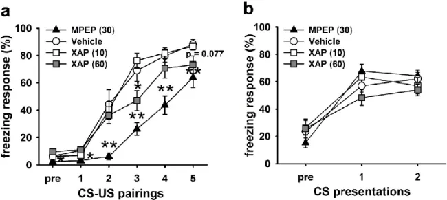
1.3 Reduction of cued fear conditioning/learned fear without affecting fear expression ... 65
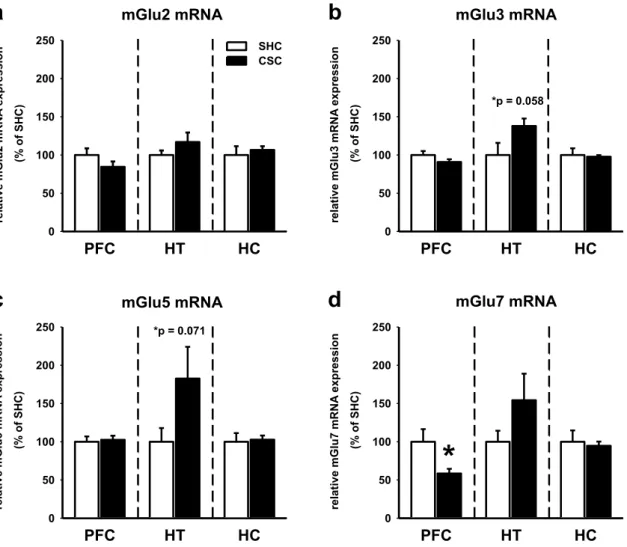
2. Chronic psychosocial stress in mice affects mGlu5 and mGlu7, but not mGlu2/3 mRNA expression in distinct brain regions ... 67
3. Chronic psychosocial stress-protective phenotype in mice lacking mGlu7 ... 69
3.1 CSC-induced changes do not differ between C57BL/6J and C57BL/6N mice .... 69
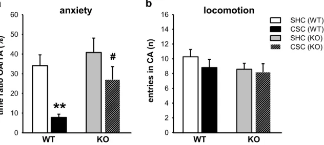
3.2 mGlu7 genetic ablation protects against the CSC-induced anxiety-prone phenotype ... 70
3.3 mGlu7 genetic ablation protects against CSC-induced physiological and neuroendocrine alterations ... 72
3.4 mGlu7 genetic ablation protects against CSC-induced immunological alterations ... 75
4. mGlu5 functional blockade relieves maladaptive stress consequences induced by CSC ... 77
4.1 Chronic stress-protective phenotype in mice lacking mGlu5 ... 77
4.2 Pilot-study: Chronic application of vehicle (PEG) and CTEP in naïve mice precludes any undesirable effects on typical CSC-affected parameters ... 79
4.3 Chronic pharmacological mGlu5 blockade reverses the CSC-induced decrease in body weight ... 80
4.4 Chronic pharmacological mGlu5 blockade corrects CSC-induced alterations in
HPA axis functionality in a dose-dependent manner ... 82
4.5 Chronic pharmacological mGlu5 blockade dose-dependently prevents CSC-
induced immunological alterations ... 85
4.6 Chronic pharmacological mGlu5 blockade has no influence on the CSC-induced anxiety-prone phenotype ... 88
4.7 Acute and sub-chronic pharmacological mGlu5 blockade induce anxiolytic-like effects in the SIH test... 89
Discussion ... 93
1. Acute pharmacological blockade of mGlu7 inhibits depressive-, anxiety-like and fear-related behavior ... 94
2. Chronic psychosocial stress in mice induces changes of specific mGlu receptors in the mouse brain ... 96
3. Chronic psychosocial stress-protective phenotype of mGlu7 KO mice ... 100
4. mGlu5 functional blockade relieves maladaptive stress consequences induced by CSC ... 103
4.1 Stress-protective phenotype in mice lacking mGlu5 ... 103
4.2 Stress-protective effects of chronic CTEP treatment ... 104
Future Outlook ... 113
Summary ... 117
German Summary ... 123
References ... 129
Abbreviations ... 167
First pages of first/joint-first author publications ... 175
Curriculum vitae, publications and awards ... 181
Acknowledgements - Danksagung ... 187
Author’s declaration – Eidesstaatliche Erklärung ... 190
Introduction
The Introduction section includes chapters taken and adapted from Peterlik et al. (2016a):
Peterlik, D., Flor, P.J., Uschold-Schmidt, N., 2016a. The Emerging Role of Metabotropic Glutamate Receptors in the Pathophysiology of Chronic Stress-Related Disorders. Curr.
Neuropharmacol., 14(5), 514–39
(Curr. Neuropharmacol. permits the author to include published journal articles in full or in part in the author’s dissertation)
Peterlik D. is responsible for design, content and writing of the first draft of the
manuscript.
12
1. Stress and its physiological systems
Hans Selye, the founder of the modern stress concept, stated “everybody knows what stress is and nobody knows what it is” (Selye, 1973). In spite of its frequent use, the word
“stress” is at best an ambiguous term. In fact, it has been used to describe both what creates
the stress and the response of the body to it. In order to circumvent this ambiguity, two
different terms have been introduced: “stressor” and “stress response”. A stressor is
defined as anything that disrupts physiological balance independent of whether it is an
actual or anticipated disruption of homeostasis or an anticipated threat to well-being
(Bartolomucci, 2007; Chrousos, 2009). On the other hand, the stress response is an
adaptive behavioral and physiological reaction aiming to re-establish homeostasis even in
the most demanding of circumstances (Chrousos, 2009; Dhabhar, 2002; McEwen, 2004),
thereby involving an efficient and highly conserved set of interconnected physiological
systems (Ulrich-Lai and Herman, 2009). In mammalian species, the two major stress
system mediating behavioral and physiological responses are the autonomic nervous
system (ANS), especially its sympathetic branch (SNS), and the hypothalamic-pituitary-
adrenocortical (HPA) axis. The activation of the SNS provides the most immediate
response to stressor exposure within seconds via exclusively neuronal pathways. These
pathways originate in the thoracolumbal regions of the spinal cord and in turn project to
end organs and to chromaffin cells in the adrenal medulla (Figure 1a). In the latter case,
they trigger the release of adrenaline/noradrenaline into the blood (Mason, 1968; Ulrich-
Lai and Herman, 2009). In contrast to the fast acting SNS, activation of the HPA axis,
which is driven by hormones, takes longer to develop. Stimulation of the HPA axis is
triggered by the secretion of corticotropin releasing hormone (CRH) and arginine
vasopressin (AVP) from the parvocellular region of the paraventricular nucleus (PVN) of
the hypothalamus into the portal circulation of the pituitary. By binding to respective
receptors expressed in the anterior pituitary, CRH promotes there the synthesis and
secretion of adrenocorticotropic hormone (ACTH) into the peripheral blood, which, in
turn, stimulates adrenal cortical cells to produce and secrete glucocorticoids (GC) into the
blood stream (Figure 1b). Termination of the stress response is achieved by negative
feedback inhibition via GC acting at glucocorticoid receptors (GR) and mineralocorticoid
receptors (MR) at different brain levels, e.g. the hippocampus and hypothalamus (Harris et
al., 2013; Keller-Wood, 2015; Lupien et al., 2009).
13
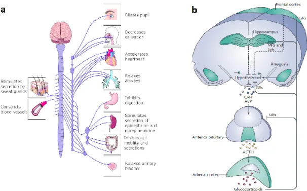
Figure 1. Schematic illustration of the sympathetic nervous system (SNS) and the hypothalamic- pituitary-adrenocortical (HPA) axis. The SNS (left-hand side; schematic overview adapted from http://wc1.smartdraw.com/cmsstorage/exampleimages/bf159507-dfa1-47d1-910e-f6903c30c35c.png) and the HPA axis (right-hand side; adapted from Lupien et al. (2009)) represent the primary mammalian systems responsible for maintaining or reinstating homeostasis during stress. (a) Stressor exposure results in activation of preganglionic sympathetic neurons in the intermediolateral cell column of the thoracolumbal region of the spinal cord. These preganglionic neurons project to pre- or paravertebral ganglia that in turn project to end organs and to chromaffin cells of the adrenal medulla. This sympathetic activation represents the classic “fight or flight” response that generally increases circulating levels of adrenaline (primarily from the adrenal medulla) and noradrenaline (primarily from sympathetic nerves), which in turn increases heart rate and force of contraction, peripheral vasoconstriction, and energy mobilization. In addition, the parasympathetic tone can also be modulated during stress. In the parasympathetic system (not shown), activation of craniosacral preganglionic nuclei activates postganglionic nuclei located in or near the end organs that they innervate; parasympathetic actions are generally opposite to those of the sympathetic system.
(b) For the HPA axis, stressor exposure activates hypophysiotrophic neurons in the paraventricular nucleus (PVN) of the hypothalamus that secrete releasing hormones, such as corticotropin-releasing hormone (CRH) and arginine vasopressin (AVP), into the portal circulation of the median eminence. These releasing hormones act in the anterior pituitary to promote the secretion of adrenocorticotropic hormone (ACTH), which in turn acts on the inner adrenal cortex (zona fasciculata) to initiate the synthesis and release of glucocorticoids (GC). Following activation of the system, and once the perceived stressor has subsided, feedback loops are triggered at various levels of the system in order to shut down the HPA axis and to return to a homeostatic point. Adapted from Ulrich-Lai and Herman (2009).
14 1.1 Acute, repeated and chronic stress
Although the term “stress” commonly bears a negative meaning, an acute stress response is one of the most important mechanisms of an organism to adapt appropriately to challenges and threats in the environment. An acute stress response results in immediate behavioral and physiological changes including enhanced attention and arousal, increased energy mobilization and increased cardiovascular and respiratory rates, whereas digestive and reproductive functions are inhibited (Charmandari et al., 2005). The fundamental process through which the organism actively adjusts to stressful challenges is referred to as
“allostasis” and represents an essential component of maintaining homeostasis (McEwen,
1998). When not overstrained, these changes are adaptive and beneficial as they increase
an individual’s chance of survival. In case of excessive and prolonged activation during
repeated or chronic stressor exposure, these adaptive systems may become overstimulated,
a condition termed “allostatic load” (McEwen and Stellar, 1993; McEwen and Wingfield,
2003; McEwen, 1998). Accordingly, inappropriate, severe or prolonged stress is associated
with changes in the brain that impair its ability to regulate appropriately physiological and
behavioral responses (Arnsten, 2009; McEwen, 2007). Thus, important criteria to
distinguish stress are its duration and intensity. While acute stress is defined to last for
minutes to hours, chronic stress persists for days to month (Dhabhar, 2000). On the other
hand, stress intensity can be measured by the magnitude of heart rate and blood pressure
and by stress hormone levels (GC, catecholamines) in blood. Acute stressor exposure
results in an increase of blood GC levels after 15-30 min and a decline to basal levels 60-
120 min later (de Kloet et al., 2005; Keeney et al., 2006). In contrast, chronic stressor
exposure may result in persistently elevated levels of plasma corticosterone (CORT). For
instance, sustained increased basal plasma CORT levels were reported following chronic
subordination for 14 days (Albeck et al., 1997) or repeated restraint stress for 7 days with
1.5 h/day (Zelena et al., 1999) compared to unstressed controls. Such prolonged elevations
of plasma CORT levels are considered the most toxic because they are most likely to result
in long-term or permanent changes in emotional, physiological and behavioral responses
that influence susceptibility to disease (Chrousos, 2009; de Kloet et al., 2005; Karatsoreos
et al., 2010). Interestingly, although HPA axis hyperactivity (hypercorticism) has been
generally linked to prolonged stressor exposure, there is accumulating evidence for even
opposite alterations (Heim et al., 2000). There are chronic or repeated stress models
leading even to reduced or unaffected plasma GC levels. For instance, chronic isolation
15 stress resulted in reduced basal plasma CORT (Djordjevic et al., 2012), whereas repeated exposure to noise did not affect basal plasma CORT levels (Armario et al., 1986).
Concurrently, chronic or repeated exposure to the same (homotypic) stressor is often associated with an adaptation of the stress response (Bartolomucci, 2007; Wood et al., 2010), a mechanism that enables the organism to habituate to innocuous, not life threatening challenges. However, despite adaptation to these familiar stressors, it is important for the individual to respond adequately to novel, possibly dangerous threats. So far, possible mechanisms underlying habituation to familiar and sensitization to subsequent novel stressors include functional changes at the level of the adrenal gland (Uschold- Schmidt et al., 2012) or altered negative feedback regulation of the HPA axis (Aguilera, 1994).
Of note, the consequences of chronic or repeated stressor exposure can vary based on individual differences. For example, while some individuals perceive a challenging situation as stressful, others that are resilient can actively cope with the same situation.
Evidence supports the hypothesis that genetic predisposition (DeRijk and de Kloet, 2005;
Gillespie et al., 2009) as well as adverse (early) life experiences (Eiland and McEwen, 2012; Gola et al., 2012; Veenema et al., 2008) strongly increase the individual’s vulnerability to stress-related pathological conditions.
Until now, numerous studies have focused on physiological and behavioral consequences of an acute stress response that are generally well understood. Instead, although chronic stress-induced physiological and behavioral alterations are likely to play a major role in the etiology of various diseases, the detailed underlying mechanisms are less well understood due to, at least in part, the lack of appropriate animal models (Langgartner et al., 2015).
1.2 Psychosocial stress
Hans Selye’s theory of the “general adaptation syndrome” involved the description of an
organism’s reaction to stressors of exclusively physical nature including intoxication or
cold (Selye, 1998). Even nowadays many physical stressors are frequently used in
laboratory such as restraint or forced swim stress. However, the major limitation these
stress paradigms have is that they do not mimic the animal’s natural situation, i.e. when
exposed to psychological and social stressors (Bartolomucci et al., 2005). More
importantly, physical stressors do not reflect the typical situations humans have to face in
today’s highly demanding society. Nowadays it is widely acknowledged that, for instance,
16 work-related stress represents a common feature of modern life at high costs of an individual’s health and performance. Most of the challenges and threats humans face today are of psychological nature, such as changing world of work, demand for flexible contracts accompanied with increased job insecurity as well as poor work-life balance. Concurrently, psychological stress occurs when an individual perceives that these environmental demands strain or exceed the own adaptive capacity to deal with (Cohen et al., 2007). Of note, most of these challenges include social interactions between individuals like competition for resources or social rank and workplace bullying. Accordingly, social stress represents one of the most potent but also naturalistic type of challenges (Koolhaas et al., 1997; Tamashiro et al., 2005). Psychosocial stress, the combination of psychological and social aspects of stress, is accepted as a major risk factor for the development of a wide variety of somatic and affective disorders in humans (Bennett et al., 1998; Heim and Nemeroff, 2001; Lupien et al., 2009; Sgoifo and Meerlo, 2002). In view of this, animal models utilizing a chronic psychosocial stress component represent the most promising approach on a preclinical basis to unravel the mechanism underlying chronic stress- promoted pathologies as they more accurately reflect the human situation. The chronic subordinate colony housing (CSC) model has emerged as one such appropriate model, as it combines chronic, psychological and social aspects of stress, and promotes the development of both somatic and affective pathologies (Langgartner et al., 2015; Peterlik et al., 2016a; Reber et al., 2007; detailed description see below).
1.3 Established rodent models for the evaluation of stress
Understanding the biological basis of the stress response is essential for a better
comprehension of the etiology of stress-related disorders. Animal models have turned out
to be instrumental in this respect and, like in humans, animals use coping strategies when
exposed to stress. They can express both active coping mechanisms manifested by
aggressive behaviors as well as exploratory activity or passive coping manifested by
freezing, immobility and submission (Franklin et al., 2012). All these behaviors can be
reliably measured in different animal models. In the following, a number of animal models
are discussed in which the animal’s stress response is reflected either upon exposure to
acute or to a chronic stressor. Paradigms that employ acute stressor exposure include
stress-induced hyperthermia (SIH), the forced swim test (FST), the tail suspension test
(TST), elevated plus maze (EPM) and learned helplessness (LH), to name a few. On the
17 other hand, chronic mild stress/chronic unpredictable stress (CMS/CUS), chronic social defeat stress (CSDS) and chronic subordinate colony housing (CSC) represent chronic stress models, all of which employ relatively long-term exposure to inescapable or uncontrollable stress events.
1.3.1 Assessment of acute stress in rodents The SIH Test
In general, SIH is known to be a physiological phenomenon when a mammalian organism is confronted with an either physical or psychological stressor (Adriaan Bouwknecht et al., 2007; Olivier et al., 2003; Vinkers et al., 2013; Zethof et al., 1994). Notably, the SIH test in rodents is also referred to a physiological animal model for anxiety (Adriaan Bouwknecht et al., 2007). Typical for the test is that, modified from the version originally described by Borsini et al. (1989), the basal temperature is measured rectally (T1) followed by a second rectal measurement 15 minutes later (T2). During these 15 minutes, the temperature usually rises due to the physical stress the animal is exposed to (handling, rectal measurement). Conveniently, potential anxiolytic-like effects of drugs are measured by a decrease in the SIH response (Adriaan Bouwknecht et al., 2007). Those measurements of the body temperature are not dependent on the animal’s motoric activity, which makes SIH different from other mild stress models/anxiety tests that depend on locomotor performance of the animal, for instance the EPM or open field test.
The FST and TST
The FST and TST are the two most widely used preclinical screening tests that allow rapid detection of substances with potential antidepressant-like activity (good predictive validity). In general, both tests are based on the same principle, which is the measurement of the duration of immobility while rodents are exposed to an acute, short-term (minutes) inescapable situation. In the FST, first described by Porsolt et al. (1977a,b), a mouse or a rat is placed in a water-filled cylinder, in which the animal is unable to escape from.
Following an initial period of escape-oriented movements, the animal will eventually
display an immobile posture, a passive behavior characterized by the absence of
movements except those necessary to keep the head above the water level. By contrast, in
the TST, immobility is scored while mice are suspended by their tails and, as water is not
required, this test is not confounded by challenges of thermoregulation. In both tests, the
18 immobility is typically interpreted as an expression of behavioral despair (Cryan et al., 2005a, 2005b; Lucki, 2001), which can be reversed by the acute administration of compounds with antidepressant potential. So, testing of new substances in these stress models allows a simple and rapid screening of potential antidepressant activity by the measurement of their acute effect on immobility. However, this poses a problem for the model, as antidepressants used in depressed humans in the clinics generally require many weeks of administration to elevate mood. Nevertheless, both the FST and TST are currently popular models mostly due to their low cost, their ease of use and their reliability across laboratories (Borsini and Meli, 1988; Holmes, 2003).
The EPM test
The EPM represents one of the most widely used anxiety models, in which anxiety-related behavior is typically measured by indices of open-arm avoidance and locomotor activity by the frequency of closed-arm entries (Lister, 1987; Rodgers et al., 1999). In principle, this test exploits the balance between the preference of rodents for avoiding open exposure to potential predators vs. exploration for possible rewards. When placed in the center portion of the plus-maze and allowed to explore each of the arms freely, animals with higher anxiety levels will show reduced open-arm activity and vice versa. This tendency can be, for instance, suppressed by anxiolytics and potentiated by anxiogenic agents (Belzung and Griebel, 2001). As short-term exposure of animals to heights and bright open spaces demonstrates an acutely stressful situation, the EPM can also be used and interpreted as a test for mild acute stressor exposure (Salomé et al., 2006).
The LH model
The LH model can be basically viewed as analogous to the abovementioned tests, with the difference that it involves a series of stressors over a few hours or even days (Nestler and Hyman, 2010). Following an uncontrollable stressor such as exposure to inescapable electric foot shocks, animals eventually will either display increased escape latency or completely fail to escape from a subsequent situation in which escape is possible (Seligman and Beagley, 1975; Seligman et al., 1975; Willner, 1984). Importantly, escape deficits can be reversed by a variety of antidepressants (Henn and Vollmayr, 2005).
Following one or more sessions of inescapable shock, animals have been shown to develop
persistent changes that are reminiscent of depression, including weight loss, alterations in
19 sleep pattern, changes in HPA axis activity and loss of spines in hippocampal regions (Cryan and Mombereau, 2004; Nestler et al., 2002a, 2002b). The attractiveness of LH is that the model is based on the consideration that cognitive functions (e.g. learning) are linked to other behavioral outcomes (e.g. neurovegetative modalities), and thus, this model helps to provide a reasonably integrated and broad picture of depressive symptomatology analogous to the human situation. However, the major drawback of the model is that most of the depression-like symptomatology does not persist beyond 2-3 days following cessation of the uncontrollable shock. Moreover, another limitation is – in contrast to the FST and TST – the difficulty to replicate between laboratories, particularly in mice.
Until today, these acute stress models clearly represent the first line of behavioral tests used to rapidly screen putative antidepressant and anxiolytic compounds and to phenotype transgenic animals. Even though direct links to emotional disorders in man are obviously weak due to utilizing only acute stressors and testing only acute antidepressant/anxiolytic responses, these acute stress models have helped enormously to reveal important molecular players within the CNS emotion circuitry (Krishnan and Nestler, 2011, 2010, 2008;
Krishnan et al., 2008).
1.3.2 Assessment of chronic stress in rodents
While acute stress paradigms are used broadly for their ease, automation potential, and rapid phenotyping abilities, they offer singular readouts that often cannot be unambiguously interpreted. For instance, increased immobility in the FST is typically interpreted as an expression of despair. However, it can also be understood as a successful and adaptive behavioral response that functions to conserve energy (West, 1990). Today’s chronic stress models are distinguished by their remarkable ability to simultaneously produce a set of behavioral alterations with strong face validity for depression and anxiety disorders (behavioral manifestations that should be similar to the symptoms observed in affected humans). Based on the clinical evidence that chronic stress significantly increases pathogenesis of affective diseases, these stress models are potentially of high value to better understand the underlying physiological mechanisms (Krishnan and Nestler, 2008;
Nestler and Hyman, 2010). Basically, they are composed of repeated and/or permanent
applications of uncontrollable and unpredictable stressors that are associated with
quantifiable molecular, behavioral and physiological changes.
20 The CMS model
In the CMS model, also referred to as chronic unpredictable stress (CUS) paradigm (Willner, 1997; Willner et al., 1992), rodents are exposed to a variety of relatively mild, mostly physical, stressors such as restraint, isolation housing, disruption of light-dark cycles, intermittently for relatively prolonged time periods (e.g. several weeks). Typically, a variety of stressors is used within the CMS schedule in order to prevent or delay habituation, which can occur rapidly when a single stressor is presented repeatedly (Willner, 1997; Willner et al., 1992). In addition to a reduction in sucrose preference (Muscat and Willner, 1992), CMS has also been shown to result in a number of other changes that are difficult to objectively quantify, such as grooming deficits and changes in aggressive and sexual behavior. Interestingly, many of these changes can be reversed by chronic antidepressants applied either during the stress procedure or as a post-stress treatment (Guidotti et al., 2013; Strekalova et al., 2006). However, as the CMS model only employs physical stressors and often lacks cross-laboratory reliability, other approaches to develop chronic psychosocial stress-based models, more reminiscent of human depression, have emerged in recent years.
The CSDS model
The stress models described above are based exclusively on physical stressors, and thus, are lacking the relevance of mimicking most important situations that human beings encounter in everyday life – i.e. social interactions (Brinkborg et al., 2011; DeVries et al., 2007; Kouzis and Eaton, 1998; Vega et al., 2004). As opposed to CMS, the CSDS model clearly includes an important social stress component, and thus displays remarkable strength as it relies on innate social behavior. The model is based on the principle that two animals interact socially and physically such that one achieves dominant status and the other becomes subordinate. Much of the preclinical aggression research has been conducted so far in territorial male resident rats or mice confronting an intruder conspecific. As a consequence of territoriality, the resident will attack unfamiliar males intruding in its home cage. However, there are many versions of CSDS for mice and rats.
For example, a typical procedure in mice lasts for 21 days where the experimental animal
is exposed repeatedly to 10 intermittent bouts (5-10 min, once daily) of social defeat. Here,
the experimental mice are forced to intrude into cage space occupied by a larger mouse of
a more aggressive strain, leading to subordination of the experimental mice. In addition to
21 the short-time physical stress during direct contact with the dominant male, the experimental mice are exposed to additional psychological stress in form of prolonged “not physical” contact by housing them for 24 h in the same cage as the residents, but with a transparent partition allowing only sensory interaction (Wagner et al., 2014, 2011). Other laboratories expose the experimental mice also daily to 10 min of physical interaction with a resident, followed by 24 h of sensory contact, but only for 10 consecutive days (Berton et al., 2006; Razzoli et al., 2011; Tsankova et al., 2006). For rats there are protocols where the experimental animals are placed in a resident’s home cage for 5 min physical interaction followed by 10 min of sensory threat for 4 consecutive days (Duclot and Kabbaj, 2013; Hollis et al., 2010), or where the intruders are placed in the cage of the resident for intermittent physical interaction until submission followed by 30 min of sensory threat for 1 to 3 consecutive days (Razzoli et al., 2009, 2007, 2006). Following repeated exposure to a dominant encounter, independent of the protocol used, animals reliably show decreased sucrose preference, indicative of an anhedonia-like state, and show reduced social interaction/sociability as well as alterations in HPA axis and autonomic function (Avgustinovich et al., 2005; Hollis and Kabbaj, 2014; Krishnan et al., 2007). Importantly, many of these changes can be reversed by chronic, but not acute, antidepressant drug administration, illustrating pharmacological validity of this stress model (Balsevich et al., 2014; Der-Avakian et al., 2014; Venzala et al., 2012).
The CSC model
The CSC paradigm was established by Reber et al. (2007) and represents a chronic
psychosocial stress model with similarities to the CSDS model, but with the difference of
applying psychosocial stress not only intermittently but permanently over a period of 19
days (24 h per day; Figure 2a). This chronic stress model is a very reliable animal model in
combining chronic, psychological and social aspects of stress. In doing so, and as
compared to the other stress models of above, it more comprehensively mimics the type of
health compromising stressors that humans are exposed to in daily life. Typically, four
male mice are housed together with a larger male resident in its homecage for 19
consecutive days. This results in immediate subordination of the four intruder CSC mice,
and a hierarchy within each colony is formed, in which the resident clearly obtains the
dominant position. To avoid habituation to the dominant mouse, the four CSC mice are
transferred into the homecage of a novel larger male resident mouse on days 8 and 15.
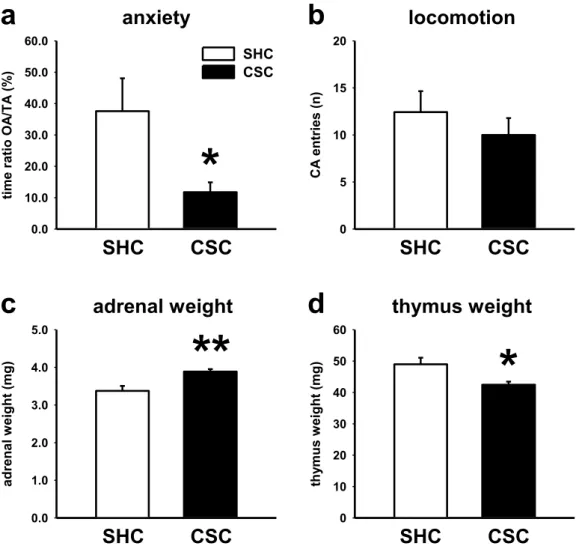
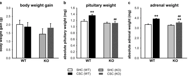
22 Single-housed animals that remain undisturbed serve as unstressed controls (SHC; Reber and Neumann, 2008). Importantly, studies by the group of Reber S.O. clearly demonstrate that CSC exposure leads to the development of affective and somatic changes and also results in reduced GC signaling (Langgartner et al., 2015; Figure 2b), and thus provides a powerful experimental tool to study the mechanisms underlying several relevant stress- induced conditions. In detail, is has been shown that exposure to CSC alters several parameters indicative of chronic stress, including reduced body weight gain, decreased thymus weight and increased pituitary and adrenal weights (Reber et al., 2007; Uschold- Schmidt et al., 2012). The latter finding is accompanied by a reduced responsiveness of adrenal explants to an ACTH challenge in vitro. Importantly, adrenal ACTH sensitivity seems to be not only diminished under in vitro conditions, as CSC mice show unaffected basal morning plasma CORT despite elevated plasma ACTH levels in comparison with SHC mice. Moreover, CSC mice show basal evening hypocorticism, suggested by decreased basal evening plasma CORT levels compared to SHC mice (Reber et al., 2007;
Uschold-Schmidt et al., 2013, 2012). The decline in GC signaling is further amplified by a reduced GC sensitivity seen in lipopolysaccharide-stimulated splenocytes (Reber et al., 2007) and plate-bound anti-CD3-stimulated T helper (Th) 2 cells from peripheral lymph nodes (Schmidt et al., 2010) of CSC compared to SHC mice. These are interesting findings, as an insufficient GC signaling can be observed in numerous affective and somatic disorders in man following chronic psychosocial stressor exposure (Caplan et al., 1979; Heim et al., 2000; Yehuda, 1997a, 1997b). In addition, CSC-stressed mice develop spontaneous colonic inflammation, indicated by an increased secretion of proinflammatory cytokines from mesenteric lymph node cells in vitro and an increased histological damage score of colonic tissue (Peters et al., 2012; Reber et al., 2007; Veenema et al., 2008).
Moreover, CSC exposure was also shown to increase the risk for the development of inflammation-induced colorectal cancer (CRC), indicated by the development of macroscopic suspect lesions, as well as a trend towards an increased incidence of low- and/or high-grade colonic dysplasia (Peters et al., 2012). Interestingly, in humans, inflammatory bowel disease (IBD) has been shown to be a consequence of chronic life stress and colorectal cancer poses one of the most serious complications in these patients (Cairns et al., 2010; Eaden, 2004, 2003; Eaden et al., 2000; van Hogezand et al., 2002).
Furthermore, IBD was also shown to be comorbid in patients suffering from depression
(Bitton et al., 2003; Duffy et al., 1991; Levenstein et al., 2000). These findings further
indicate that the CSC paradigm is an appropriate model of chronic psychosocial stress with
23 high construct validity (i.e. high disease relevance of methods by which the animal model is constructed; Nestler and Hyman, 2010). With respect to their behavior, CSC mice show reduced open-arm activity on the EPM and reduced center-activity in an open field after 19 days of CSC, further illustrating that chronic stressor exposure increases anxiety-related behavior, a phenomenon that co-occurs with depression in humans (Kennedy, 2008).
Moreover, CSC mice spend a similar time investigating an empty cage and a cage with an unknown conspecific during the social preference/avoidance test (SPAT) on day 20 of CSC, suggesting a lack of social preference (Slattery et al., 2012). In addition, CSC mice also show an increased ethanol (EtOH) preference and total intake, which was already shown following 14 days of CSC exposure (Peters et al., 2013). Interestingly, in humans, chronic psychosocial stress represents a strong risk factor for the development of substance abuse such as alcoholism, which is often co-morbid with anxiety disorders (Boger et al., 2014; Cooper et al., 2014; Morley et al., 2014). Taken together, the CSC paradigm represents an animal model that utilizes a chronic psychosocial stress component and results in decreased GC signaling and concomitant affective and somatic pathologies, in particular the stress-induced anxiogenic phenotype and the systemic proinflammatory phenotype (Figure 2b). Thus, the CSC model is likely to have more translational value than other stress models in animals, e.g. when compared to the CMS or CSDS model, which lack either social or truly chronic components. Most interestingly, CSC exposure is associated with hypocorticism, a phenomenon that is not apparent after CMS or CSDS, but occurs in humans suffering from mood disorders. In contrast, CMS and CSDS are associated with greatly elevated plasma CORT levels (hypercorticism). Interestingly, in addition to mice, the CSC paradigm was also established in rats (Nyuyki et al., 2012).
However, to date the effects of CSC exposure in rats are not that well characterized as
compared to mice. Overall, the CSC paradigm is a promising animal model that makes it
also possible to gain further insight into how stress-induced pathophysiological changes
(e.g. HPA axis alterations) eventually lead to affective and somatic disorders in man.
24
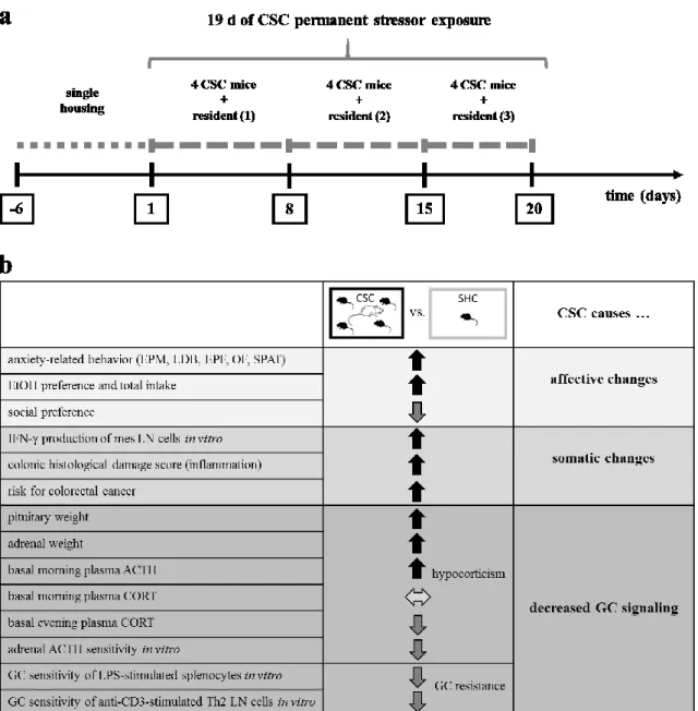
Figure 2. Schematic illustration of the experimental design of the chronic subordinate colony housing (CSC) paradigm in mice and a summary of the main effects of CSC on behavioral, immunological and physiological parameters. (a) In the CSC paradigm, male mice weighing 19-21 g are housed singly for one week before they are assigned to the single-housed control (SHC) or the CSC group in a weight-matched manner. In order to induce chronic psychosocial stress, CSC mice are housed together with a larger dominant male for 19 consecutive days. In detail, four experimental CSC mice are put into the homecage of resident (1) on day 1 of CSC, resulting in immediate subordination of the four intruder CSC mice. The latter are then housed together with this dominant resident (1) for 7 consecutive days. On day 8, and again on day 15 of CSC, the four experimental CSC mice are transferred into the homecages of resident (2) (day 8) and resident (3) (day 15), respectively, in order to avoid habituation. On day 19, CSC and SHC mice are usually tested for their innate or physiological anxiety and on day 20 immunological and physiological parameter are assessed.
(b) Compared to SHC mice, CSC mice show affective and somatic changes and develop decreased glucocorticoid (GC) signaling. Thus, the CSC paradigm represents a promising animal model to mimic
25
diseases in which decreased GC signaling is a core feature and to unravel the underlying mechanisms of stress-related pathology in humans. Abbreviations: EPM, elevated plus-maze; LDB, light-dark box; EPF, elevated platform; OF, open field; SPAT, social preference/avoidance test; mes LN cells, mesenterial lymph node cells; ACTH, adrenocorticotropic hormone; CORT, corticosterone; LPS, lipopolysaccharide; Th2, T helper 2; Adapted from Peterlik et al. (2016a).
Taken together, the rodent models explained above can be performed in mice as well as in rats. Which species to use probably depends on the particular research question to be answered. However, in case of the CSC model, mice are the first choice to gain more knowledge about the role of the brain glutamatergic neurotransmitter system in the development of the resulting affective and somatic changes, as the effects of chronic psychosocial stressor exposure using this model are much better characterized and robust in mice. Other advantages of mouse models are lower costs and less space necessary.
Moreover, mouse models also offer the possibility to use transgenic animals, e.g. knockout mice, in order to analyze the underlying mechanisms.
2. The glutamate system is implicated in stress-related physiology and disorders
Major depression, anxiety, and drug abuse disorders represent the most prevalent stress- related psychiatric conditions and are an enormous health concern worldwide (Aas et al., 2012; Cryan and Holmes, 2005; Schneiderman et al., 2005). The etiology of these pathologies is complex, with chronic psychosocial stressors being the most acknowledged risk factors (Caspi et al., 2003; Cryan and Holmes, 2005; Cryan and Slattery, 2007; de Kloet et al., 2005; Schneiderman et al., 2005; Virtanen and Kivimäki, 2012; Virtanen et al., 2013). These factors include continuing adverse conditions, such as social decline along with poverty, or life events that possess a high degree of chronic threat (e.g. medical disabilities), long lasting negative emotions and experience of personal loss (de Kloet et al., 2005; Hammen, 2005; Virtanen and Kivimäki, 2012; Virtanen et al., 2013). The majority of these types of factors has been demonstrated to increase both anxiety- and depression-related behavior, but also alcohol and drug abuse (Blanchard et al., 2005, 2006;
Kotov et al., 2010; Schmidt et al., 2007). Indeed, and not surprisingly, there’s a high
comorbidity between anxiety and depression/mood disorders, with approximately half of
26 the patients suffering from major depression also meeting criteria for comorbid anxiety (Kennedy, 2008).
As disorders of mood and emotion may show a common excessive or inappropriate brain excitability within crucial brain circuits, the L-glutamate system, which represents the primary excitatory neurotransmitter system in emotion and cognition circuits, is increasingly considered to play an important role in mental disease etiology and persistence. Several lines of evidence from human clinical studies link dysfunction in the L-glutamate system to the pathogenesis of psychiatric disorders (Cortese and Phan, 2005).
For instance, changes in glutamate levels have been found in plasma, cerebrospinal fluid (CSF), and in the brain of patients suffering from mood and anxiety disorders (Levine et al., 2000; Mauri et al., 1998; Mitani et al., 2006). Interestingly, recent postmortem studies showed significant increases in glutamate levels in the frontal cortex and dorsolateral prefrontal cortex of depressed and bipolar patients, respectively (Hashimoto et al., 2007;
Lan et al., 2009). Furthermore, various clinical neuroimaging studies have consistently demonstrated volumetric changes in brain regions, in which glutamatergic neurons predominate, such as the hippocampus, amygdala and several cortical regions (Lorenzetti et al., 2009).
The L-glutamate neurotransmitter system of the emotion and cognition circuitry of mammalian brains is composed of a large diversity of genetically regulated factors, including a group of vesicular, glial and synaptic glutamate transporters (Sheldon and Robinson, 2007) as well as two families of glutamate receptors: ligand-gated ionotropic glutamate receptors (iGlu) comprising (2R)-2-(methylamino)butanedioic acid (NMDA)-, 2-amino-3-(5-methyl-3-oxo-2,3-dihydro-1,2-oxazol-4-yl)propanoic acid (AMPA)- and (2S,3S,4S)-3-(carboxylatomethyl)-4-(prop-1-en-2-yl)pyrrolidine-2-carboxylate (kainate, KA)-receptors (Nakanishi, 1992; Planells-Cases et al., 2006; Willard and Koochekpour, 2013; Xiao et al., 2001), and the G protein-coupled metabotropic glutamate receptor (mGlu) subtypes -1 to -8 (mGlu1-8; Conn, 2003; Nicoletti et al., 2011; Pinheiro and Mulle, 2008; Willard and Koochekpour, 2013).
Throughout the last three decades, several drug discovery efforts were made targeting iGlu receptors, with preclinical and clinical data demonstrating NMDA and AMPA receptors to be promising targets in controlling cognitive and emotional changes observed in stress- related disorders (Boyce-Rustay and Holmes, 2006; Graybeal et al., 2012; Kim et al., 1996;
Magariños and McEwen, 1995). In this regard, a major breakthrough came from clinical
studies using the NMDA receptor antagonist ketamine by showing clinical efficacy in
27 treatment-resistant depression (TRD) and major depressive disorder (MDD) patients.
Interestingly, ketamine administered intravenously showed strong decreases in the Hamilton Depression Rating Scale (HDRS) analysis, with an improvement observed 2 h after infusion that remained significant for over 1 week (Berman et al., 2000; Zarate et al., 2006). However, this iGlu receptor-based strategy is not devoid of limitations and risks, as ketamine administration has also been shown to be associated with cognitive and dissociative adverse effects, which thus limits ketamine’s widespread application for the treatment of mood disorders (Pilc et al., 2008; Witkin et al., 2007). In contrast, therapeutic strategies targeting mGlu receptors represent a more subtle alternative in regulating excitatory (and possibly inhibitory) neurotransmission and are therefore considered to have a more favorable side-effect profile than ligand-gated ion channel modulation (Cartmell and Schoepp, 2000; Niswender and Conn, 2010). Indeed, signaling via mGlu receptors is slower and longer lasting than via iGlu receptors, allowing fine-tuning of glutamate regulation and its cellular responses, which could eventually avoid the adverse effects associated with direct modulation of iGlu receptors. In addition to that, growing evidence gives rise to mGlu-based compounds to be effective in regulating iGlu receptor signaling, further emphasizing the modulatory potential of mGlu receptors.
2.1 Glutamate signaling via mGlu receptors in the CNS
The existence of neuromodulatory glutamate receptors, namely the mGlu receptors, provides a mechanism by which binding of glutamate, in contrast to the fast synaptic responses mediated by iGlu receptors, slowly modulates cell excitability, synaptic neurotransmission and plasticity. mGlu receptor-induced modulation occurs via second messenger signaling pathways and their interactions with ion channels (C. H. Kim et al., 2008; Rondard and Pin, 2015; Willard and Koochekpour, 2013; Yin and Niswender, 2014).
According to sequence homology, second messenger coupling and pharmacological properties, the mGlu receptor family is subdivided into three groups. The group I members, mGlu1 and mGlu5, are coupled to G
q/11proteins and primarily elevate {[(1R,2S,3R,4R,5S,6R)-2,3,5-trihydroxy-4,6-
bis(phosphonooxy)cyclohexyl]oxy}phosphonic acid (IP
3), diacylglycerol (DAG), and Ca
2+signal transduction. In general, these receptor subtypes function to enhance glutamate-
mediated postsynaptic excitation (Abe et al., 1992; Bordi and Ugolini, 1999; Cartmell and
Schoepp, 2000; S. J. Kim et al., 2008; D. Youn, 2014; D.-H. Youn, 2014). In contrast,
28 group II (mGlu2 and mGlu3) and group III (mGlu4, -6, -7, and -8) receptors inhibit adenylyl cyclase activity and other effector proteins via coupling to G
i/oproteins and thereby negatively modulate excitatory neurotransmitter efflux and neuronal excitability upon activation (Cartmell and Schoepp, 2000; Kintscher et al., 2012; Lavreysen and Dautzenberg, 2008; McMullan et al., 2012; Mercier and Lodge, 2014; Schoepp, 2001;
Tang et al., 2013). Interestingly, various mGlu receptors are expressed in both neurons and glial cells of the CNS, as well as in peripheral tissue like the enteric nervous system or adrenal gland cells (Julio-Pieper et al., 2011). In neurons, group I receptors show predominantly postsynaptic location and modulate cell excitability, while group II and III members are mainly expressed at the presynapse and are involved in regulating neurotransmitter release, mostly inhibiting release (Conn and Pin, 1997; Swanson et al., 2005). As they are members of class C GPCR, all mGlus are characterized by a large extracellular N-terminal “Venus flytrap domain” (VFTD), which is known to serve as the orthosteric ligand binding site and shows abundant homology between the different mGlu receptor subtypes. The binding site for allosteric modulators of the mGlus is located topographically distinct within the transmembrane domain (Christopoulos and Kenakin, 2002; Flor and Acher, 2012; Gregory et al., 2013; Nickols and Conn, 2014; Wood et al., 2011; Wu et al., 2014). As the allosteric binding site contains a higher level of sequence diversity between the receptor subtypes, allosteric ligands typically show greater subtype selectivity (Conn et al., 2009a, 2009b). Importantly, the widespread distribution of mGlu subtypes suggests that these modulatory receptors have the ability to participate in a broad array of physiological functions throughout the CNS and may represent suitable targets for therapeutic intervention in a variety of CNS disorders. Thus, the therapeutic potential of the mGlu receptors is increasingly receiving attention as possible treatment strategies for CNS diseases such as Parkinson’s disease (PD), Fragile X syndrome (FXS), schizophrenia, addiction and in particular depression and anxiety-related disorders (Herman et al., 2012;
Nicoletti et al., 2015; Picconi and Calabresi, 2014; Pomierny-Chamioło et al., 2014; Scharf et al., 2014).
2.1.1 Group I mGlu receptors: neurobiochemistry and distribution
In general, the group I members mGlu1 and mGlu5 couple to G
q/11proteins and activate
phospholipase C, a process that results in the formation of IP
3and DAG (Figure 3). This
classical pathway leads to intracellular Ca
2+mobilization and activation of protein kinase C
29 (PKC). Apart from this, group I receptors can also activate a range of further downstream effectors, most notably proteins involved in synaptic plasticity, such as mitogen-activated protein kinase/extracellular receptor kinase (MAPK/ERK) and a mammalian target of rapamycin (mTOR) (Fieblinger et al., 2014; Pałucha-Poniewiera et al., 2014b; Zhang et al., 2015). With respect to their distribution, expression of both mGlu1 and mGlu5 primarily overlaps within brain regions implicated in mood disorders (Witkin et al., 2007). However, there are also distinct differences, with mGlu1’s expression being abundant in cerebellum, olfactory bulb, the CA3 region of the hippocampus, in thalamus, dentate gyrus and substantia nigra, whereas mGlu5 is highly expressed in telencephalic regions, CA1 and CA3 regions of the hippocampus, septum, basal ganglia, striatum, amygdala and nucleus accumbens (Ferraguti and Shigemoto, 2006; Pilc et al., 2008). In addition, mGlu5 is also expressed in glial cells (Figure 3), particularly in astrocytes, where its expression has been highlighted with respect to a number of potential physiological roles, e.g. neuroprotection (Bruno et al., 2001a, 2001b, 1999; D’Antoni et al., 2011; Verkhratsky and Kirchhoff, 2007; Wierońska and Pilc, 2009).
2.1.2 Group II mGlu receptors: neurobiochemistry and distribution
In contrast to group I, the group II members mGlu2 and mGlu3 are coupled predominantly
to G
i/oproteins, negatively modulating adenylyl cyclase activity and directly regulating ion
channels and other downstream signaling components via the release of the G
βγsubunit. In
addition, group II mGlus also couple to MAPK and phosphatidyl inositol 3- (PI3-) kinase
pathways which are implicated in synaptic plasticity (D’Onofrio et al., 2001; Lin et al.,
2014; Niswender and Conn, 2010). Both mGlu2 and mGlu3 are localized presynaptically
in rather preterminal than terminal axonal portions, distant from the active zone of
neurotransmitter release, where they are potentially activated by synaptic glutamate
spillover (Tamaru et al., 2001). mGlu2 mRNA is observed to be highly expressed in
pyramidal neurons in the enthorhinal and parasubicular cortical regions and in granule cells
of the dentate gyrus (Hayashi et al., 1993; Ohishi et al., 1993a). In contrast, mGlu3 mRNA
is highly expressed in neurons of the cerebral cortex and the caudate-putamen and in the
granule cells of the dentate gyrus (Ohishi et al., 1993b; Tanabe et al., 1993). In addition,
the mGlu3 receptor subtype (Figure 3) is also prominently expressed in glial cells
throughout the whole brain, and its activation provides robust neuroprotection in vitro and
in vivo (Battaglia et al., 2014; Ciceroni et al., 2010; Durand et al., 2014, 2013).
30 2.1.3 Group III mGlu receptors: neurobiochemistry and distribution
Group III represents the largest family of mGlu receptors and comprises the subtypes mGlu4, mGlu6, mGlu7 and mGlu8. Like group II, group III members are predominantly expressed presynaptically (Shigemoto et al., 1997; Figure 3) where they regulate neurotransmitter release (Antflick and Hampson, 2012; de Rover et al., 2008; Grueter and Winder, 2005; Panatier et al., 2004; Pinheiro and Mulle, 2008; Woodhall et al., 2007). By coupling to G
i/oproteins, their activation inhibits cAMP formation and indirectly affects synaptic transmission and neurotransmitter release by modulating membrane Ca
2+- and K
+- channels (Lavreysen and Dautzenberg, 2008; Mercier and Lodge, 2014; Pin and Duvoisin, 1995). As with group II mGlu receptors, the group III subtypes also couple to other signaling pathways, including MAPK and PI3-kinase, providing further complexity to the mechanisms by which they regulate synaptic transmission (Iacovelli et al., 2014, 2004, 2002). Except for the mGlu6 receptor subtype, whose expression is restricted to the retina, all other group III mGlus are widely expressed throughout the mammalian brain and also in peripheral tissue (Julio-Pieper et al., 2011; Pilc et al., 2008). In detail, in the CNS the mGlu4 receptor subtype is highly expressed in the cerebellum, the olfactory bulb and thalamus as well as in the hippocampus, the cerebral cortex and basal ganglia in pre- and post-synaptical position (Corti et al., 2002; Kinoshita et al., 1996; Shigemoto et al., 1997).
In addition, widespread peripheral mGlu4 expression has been shown also in the pancreas, adrenal glands and gastrointestinal tract (Akiba et al., 2009; Brice et al., 2002; Chang et al., 2005; Sarría et al., 2006). The mGlu7 receptor is highly localized in the presynaptic active zone and abundantly expressed in brain regions such as neocortex, hippocampus, amygdala, locus coeruleus, thalamus and hypothalamus (Kinoshita et al., 1998; Kosinski et al., 1999; Ngomba et al., 2011). In the periphery, mGlu7 expression has been reported in the adrenal glands, the colon and stomach, among other areas (Julio-Pieper et al., 2010;
Nakamura et al., 2010; Scaccianoce et al., 2003). The mGlu8 receptor is found
predominantly in the CNS in presynaptic terminals in the olfactory bulb, hippocampus,
cerebellum and cortical areas (Ferraguti and Shigemoto, 2006), but also in peripheral tissue
such as pancreas and testis (Julio-Pieper et al., 2011). Interestingly, general expression
levels of mGlu8 receptors seem considerably lower than those of mGlu4 and mGlu7
(Niswender and Conn, 2010).
31
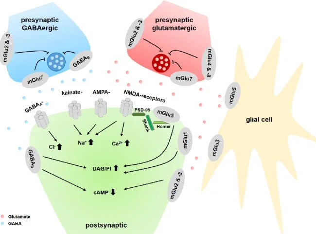
Figure 3. Schematic representation of mGlu receptors at the synapse. In general, group I mGlu subtypes are localized postsynaptically, whereas group II and III receptors are localized mainly in presynaptic locations. While the mGlu7 receptor subtype is localized in the active zone, mGlu subtypes 2, 3, 4, and 8 are generally found in perisynaptic locations on the presynapse. Group II and III receptors modulate the release of glutamate (right, red circles) or 4-aminobutanoic acid, GABA (left, blue circles). At the postsynaptic terminal, the ionotropic (2R)-2-(methylamino)butanedioic acid (NMDA)-, 2-amino-3-(5-methyl-3-oxo-2,3- dihydro-1,2-oxazol-4-yl)propanoic acid (AMPA)- and (2S,3S,4S)-3-(carboxylatomethyl)-4-(prop-1-en-2- yl)pyrrolidine-2-carboxylate (kainate, KA)-receptors respond to glutamate with increases in intracellular sodium or calcium, promoting cell excitability. Group I mGlus signal via Gq/11 proteins to increase diacylglycerol (DAG) and phosphatidylinositol (PI). Importantly, mGlu5 and NMDA receptors are closely linked to each other via Shank, Homer and PSD-95 (postsynaptic density-95) proteins. Postsynaptic mGlu2/3 and GABAB receptors couple to cAMP inhibition. Instead, GABAA chloride channels modulate intracellular chloride levels. Expression of mGlu3 and mGlu5 on glial cells has emerged as another key site for regulation of synaptic activity, however, the consequences of receptor activation on these cells and the exact signaling pathways are presently not well understood. Adapted from Peterlik et al. (2016a).