Hydrogel Wound Dressings for the
Bioactive Treatment of Acute and Chronic Wounds
DISSERTATION ZUR ERLANGUNG DES DOKTORGRADES DER NATURWISSENSCHAFTEN (DR. RER. NAT.) DER
FAKULTÄT FÜR CHEMIE UND PHARMAZIE DER UNIVERSITÄT REGENSBURG
vorgelegt von Julia Köhler aus Würzburg
Mai 2017
Die Arbeit wurde von Herrn Prof. Dr. Achim Göpferich angeleitet.
Promotionsgesuch eingereicht am: 09. Mai 2017 Datum der mündlich Prüfung: 23. Juni 2017
Prüfungsausschuss: Prof. Dr. Joachim Wegener (Vorsitzender)
Prof. Dr. Achim Göpferich (Erstgutachter)
PD Dr. Stephan Schreml (Zweitgutachter)
Apl. Prof. Dr. Rainer Müller (Drittprüfer)
Your assumptions are your windows on the world.
Scrub them off every once in a while, or the light won't come in.
Isaac Asimov
Table of Contents
Chapter 1 Introduction and Goal of the Thesis ... 1 Chapter 2 Antimicrobial Interpenetrating Polymer Network Hydrogels for an
Application in Acute Wound Care ... 31 Chapter 3 pH-Modulating Poly(ethylene glycol)/Alginate Hydrogel Dressings
for the Treatment of Chronic Wounds ... 59 Chapter 4 Systematic Structural Modification of the Interpenetrating Polymer
Network System Poly(ethylene glycol)/Acrylic Acid/Alginate ... 89 Chapter 5 Alkaline Poly(ethylene glycol)-Based Hydrogels for a Potential Use
as Bioactive Wound Dressings ... 109 Chapter 6 Buffering Hydrogel Wound Dressing Materials for a Universal
Application onto Chronic Wounds ... 137 Chapter 7 Summary and Conclusion ... 167 Appendix Abbreviations ...
List of Publications ...
Acknowledgements ...
Statement in Lieu of an Oath ...
177
181
183
185
Chapter 1
Introduction and Goal of the Thesis
Based on this chapter, a review article was submitted: Koehler J, Brandl FP, and
Goepferich AM. Hydrogel wound dressings for a bioactive treatment of acute and chronic
wounds. Eur. Polym. J. 2017; Manuscript Number: EUROPOL_2017_1345.
1. Introduction 1.1 Wound Healing
An injury of the skin is a severe intervention to the normal function of the human body, as thermal insulation, body fluid retention, protection from exogenous pathogens, and hosting of different sensing receptors are only some of the many important functionalities of the intact skin barrier.
1To enable fast recovery, the endogenous healing process starts almost instantly.
2The blood flow is stopped within minutes by the aggregation of platelets and fibrin clot formation (Figure 1A). Thereby released growth factors (GFs) and cell mediators recruit inflammatory cells like neutrophils and monocytes to the wound site (Figure 1B). The aim of the following inflammatory response is the removal of foreign bodies, bacteria, and damaged endogenous tissue.
3Figure 1. Schematic illustration of the healing process, including the coagulation (A), the inflammation (B), the proliferation (C), and the remodeling (D) phase.4 The upper light pink layer represents the epidermis; the lower pink layer represents the dermis.
Towards the end of the inflammatory phase, macrophage GFs induce the proliferation and
migration of fibroblasts and epithelial cells into the wound (Figure 1C). During the so-
called proliferative phase, new blood vessels are formed (in red), the synthesis of
strengthening collagen fibers begins (in blue), and granulation tissue (in yellow) is built
from epithelial cells, fibroblasts, and keratinocytes. Complete wound healing takes several
weeks or month (Figure 1D).
4The wound finally contracts and the granulation tissue is
transformed into the more stable extracellular matrix (ECM).
5The overall duration of the
healing process depends on the age and the health status of the patient (impairing factors are e.g. diabetes or venous insufficiency), and external factors such as the presence of foreign bodies or infections in the wound. In acute wounds, the healing process follows the above described phases and the wound closure occurs after 8 to 12 weeks.
6In contrast, chronic wounds mainly remain in the inflammation phase, accompanied by large numbers of wound exudate, heavy infections, pain, and tissue necrosis. Thus, chronic wounds fail to heal over a period of minimum 12 weeks, sometimes it even takes several months or years to full recovery.
1.2 Wound Dressing Requirements
Chronic wounds are a big burden for the patients themselves, facing reduced quality of life, connected to frequent dressing changes, numerous hospital stays, and disabilities in daily life. Furthermore, the economic burden for the health care system is immense.
7Approximately 20 million patients around the world suffer from chronic wounds and the global wound care market revenue rose to more than 20 billion dollars in 2016.
8,9Therefore, appropriate wound dressings are required for an unproblematic healing process.
Acute wounds should heal in a moist environment for reduced scar formation and
facilitated epithelization and cell migration into the wound.
10Furthermore, the wound
dressing should serve as a barrier to external threads like microbes, foreign bodies, or
tissue damaging forces. Sufficient mechanical stability (under pressure and tension) is
required during the application, the wearing, and the removal of the dressing.
11Yet, an
elastic texture must be maintained, as the dressing should adapt to the specific wound
profile and high dressing flexibility is necessary when the patient is moving. The
requirements of chronic wound dressings are even more challenging. These dressings
additionally have to deal with a high volume of wound exudate; up to 12 L·m
–2per day
were described for example for venous leg ulcers.
12Moreover, low adherence to the
underlying wound is necessary to protect newly formed tissue from destruction during
frequent dressing changes. Especially for the treatment of chronic wounds, but also for the
treatment of more complex acute wounds, an active intervention in the healing process is
also required.
6The active wound healing capacity of dressing materials can be based on
different components, such as released drugs, the dressing material itself, or incorporated
cells. A detailed description of important approaches to bioactive wound dressings can be
found below. In any case, the chosen wound dressing should be as supportive to healing as
possible, but at the same time it must be as cost-effective as possible for adoption in clinics.
1.3 Hydrogel Wound Dressings
There is a wide range of different wound dressing types and material compositions on the market, covering the requirements of various kinds of wounds.
13Traditional dressings like gauze and tulle mainly have a covering effect while maintaining proper gaseous exchange.
However, their strong adherence to the wound site causes pain and further lesions during dressing changes. A modern dressing type that combines numerous advantageous properties in one single material is the hydrogel-based wound dressing. Hydrogels consist of around 90% water and 10% natural or synthetic polymers. Because of the high water content, hydrogel dressings are suitable for dry and necrotic wounds. The created moist environment enhances the healing process.
14Furthermore, it enables a debridement of necrotic tissue, likewise leading to improved healing. On the other hand, hydrogel dressings are also able to absorb high amounts of liquid in contact with exuding wounds.
Dependent on the hydrogel composition, a liquid uptake of up to 1000 gram per gram of dressing is described.
15The permeable hydrogel structure further enables an undisturbed gaseous exchange of CO
2, O
2, and water vapor. In clinical studies, hydrogel dressings were found to reduce the pain for treated patients, induced by a cooling effect of the material, and by its non-adhering nature. The similarity of the hydrogel structure to the structure of the ECM, which is characterized by a vast (polymer) network in an aqueous environment, allows the establishment of a new and very effective wound healing feature, namely the incorporation of cells and biomolecules into the hydrogel polymer network. Further bioactive wound healing properties such as controlled drug release can easily be achieved by a precise chemical modification of the polymer network. For this purpose, various natural and synthetic precursors can be combined, and hydrogel wound dressings with defined wound healing properties can be developed.
1.3.1 Available Hydrogel Base Materials
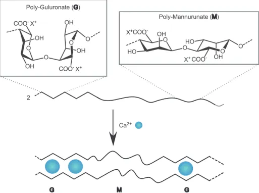
A widely used hydrogel component with a biological origin is the polysaccharide alginate
(Figure 2). Alginate forms hydrogels by ionic cross-linking via –COO
–or, when
structurally modified, by chemical cross-linking between additional side chains.
16Figure 2. Representative parts of the alginate structure and its ionic cross-linking reaction.
Alginate dressings can absorb high amounts of wound exudate, because of their hydrophilic nature. In contact with bleeding wounds they exhibit a hemostatic effect. But most importantly, they can actively support the wound healing process. Alginate has been proven to enhance cell migration into the wound, to increase the angiogenesis and the production of collagen I, and to reduce the concentration of proinflammatory cytokines in chronic wounds.
17,18The advantageous material properties of alginate are reflected in the high number of commercially available alginate containing dressings. Alginate dressings are available as various hydrogels (e.g. Nu-Gel
®(Systagenix), Tegagel
®(3M GmbH)), cryogels (e.g. Algosteril
®(4M Medical GmbH), Curasorb
®Alginate (Medtronic), Sorbsan
®(B. Braun Melsungen AG)), or hydrocolloids (e.g. Comfeel Plus Flexible
®(Coloplast AG), Kendall™ Hydrocolloid Dressing (Medtronic)).
Another suitable polysaccharide for medical application is chitosan. Animal studies and
clinical trials detected an overall faster healing in chitosan treated wounds.
19–21Chitosan is
hemostatic, bacteriostatic, and fungistatic. Furthermore, it encourages cell proliferation and
collagen and hyaluronic acid (HA) formation. However, there are only a few commercial
chitosan containing wound dressings, e.g. KytoCel
®(MasterCare Medical GmbH), a
chitosan cryogel, and Chitoderm
®plus (Trusetal Verbandstoffwerk GmbH), a chitosan
coated dressing. The big discrepancy between the scientific interest in chitosan dressings
and its commercialization might be explained by the animal origin of chitosan; it is a
derivate from chitin, found in shrimp and crab shells. As a natural product, however, it bears a high risk of batch variations, for example concerning the molecular weight.
22Yet, the biological activity of chitosan is dependent on the molecular weight of the macromolecules.
23–26Additional drawbacks are the low elasticity of chitosan materials and the difficulty in producing fibrous wound dressings.
1The protein collagen is, together with alginate, one of the most frequently used materials in wound coverage. In the human body, collagen occurs inter alia in the ECM, in blood vessels, in bones, and in tendons.
27The three main forms, Collagen I, II and III, constitute around 75% of dry human skin. Therefore, it is a well-tolerated material that is particularly suitable for wound dressing and tissue engineering applications. For medical use, the main sources are bovine, porcine, and avian derived collagen.
28Collagen dressings are applied in the form of hydrogels (e.g. CellerateRX
®(Wound Care Innovations LLC), Regenecare
®Wound Gel (MPM Medical Inc.), Wun’Dres
®(Coloplast AG)), fibrous cryogels (e.g.
Biobrane
®(Smith & Nephew), CollaSorb
®(Paul Hartmann AG), Fibracol
®(Acelity)), and grounded cryogels (e.g. Medifil
®(Human Bio Science, Inc.), Stimulen
™(Southwest Technologies, Inc.)). Collagen is chosen for its high liquid absorbance capacity and its mechanical strength.
29Furthermore, it plays an active role in wound healing. By recruiting fibroblasts, endothelial cells, and keratinocytes, the vascularization, granulation tissue formation, and collagen deposition is enhanced.
30,31Moreover, scaring and the rate of bacterial infections is reduced. In current studies, two major issues of collagen are faced;
enzymatic degradation of collagen leads to a fast loss of dressing stability and shape, and dependent on the source of the collagen, there is a risk of pathogen transmission.
32–34In contrast, wound dressing materials from synthetic precursors are non-infective, they have well defined chemical structures, and their properties can precisely be modified to fulfill the desired material requirements.
13However, synthetic hydrogels do not actively participate in the wound healing process. Therefore, combinations of natural and synthetic polymers are usually preferred. Examples of available (partially) synthetic wound dressings are the polyacrylamide/polysaccharide-based FlexiGel
®(Smith & Nephew), the poly(ethylene glycol) (PEG)/oakin-based Oakin
®hydrogel wound dressing (Amerigel), and the polyurethane (PU)-based AquaClear
®dressing (Paul Hartmann AG).
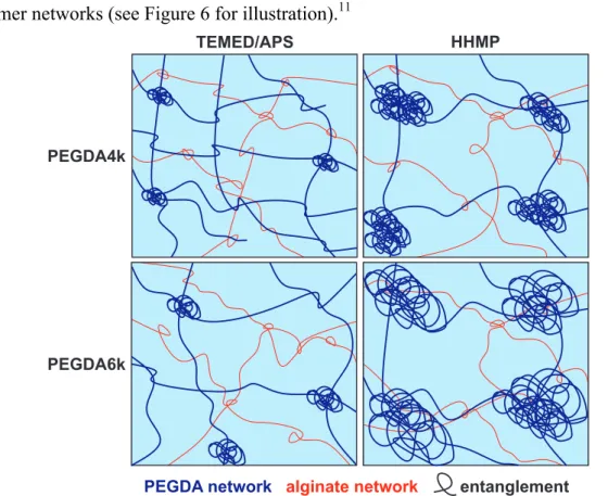
1.3.2 Double Network Hydrogels
A general issue of hydrogel materials is their relatively low mechanical stability, which is
for example related to structural inhomogeneities in the polymer network and the low
friction between single polymer chains, as hydrogels typically have a polymer content of only 5 to 10%.
35Yet, this drawback can be avoided by proper selection of the monomers, by chemical modifications of the precursor molecules, and in particular by physical modification of the hydrogel system. A physical hydrogel network modification, particularly aimed at enhancing mechanical resistance, was first described by Gong et al.
The group developed hydrogels that consist of two separate polymer networks with internetwork entanglements, so called “double network” (DN) hydrogels (Figure 3).
36Figure 3. Schematic structure of a double network hydrogel with the covalent network in dark blue and the ionic network in red. Dots represent the cross-linking points.