1
The role of liver p38α in acute and chronic inflammatory reactions
Inaugural-Dissertation zur
Erlangung des Doktorgrades
der Mathematisch-Naturwissenschaftlichen Fakultät
der Universität zu Köln
vorgelegt von Jan Heinrichsdorff
Köln 2010
Berichterstatter: Prof. Manolis Pasparakis Prof. Jens C. Brüning
Tag der mündlichen Prüfung: 25.10.2010
Part of this work was published in the following journal:
Heinrichsdorff, J., T. Luedde, et al. (2008). "p38 alpha MAPK inhibits JNK activation and collaborates with IkappaB kinase 2 to prevent endotoxin-induced liver failure." EMBO Rep 9(10): 1048-54.
3 Acknowledgments
During my four years as a member of the Pasparakis Lab, I have acquired a vast range of experiences and knowledge, not only in a scientific manner, but also personally. These experiences have allowed me to grow and become more mature in many aspects of my life.
For this reason, I would like to thank all the people from inside and outside the Pasparakis lab that contributed in making these four years a life changing experience.
Most of all, I would like to thank Manolis, for giving me the opportunity to perform my PhD work in his laboratory. Manolis is a great supervisor that on the one hand demands a high degree of commitment, but is also rewarding and permitting the acquisition of a vast range of knowledge and skills through the many resources available in his lab. I strongly believe that during my time in this laboratory I have learned what it really means to be a scientist performing basic research.
Furthermore, I would like to thank Dr. Tom Lüdde and Dr. Hanno Ehlken who, through excellent supervision at the beginning but also throughout the duration of my PhD, guided me in the right direction concerning my research projects. In addition, I would like to thank all the members of the laboratory, for valuable discussions and technical help.
I would also like to thank my parents for giving me great support throughout the whole time of my PhD work.
Die Rolle der MAP Kinase p38α in akuten und chronischen Entzündungsreaktionen Jan Heinrichsdorff, Institute für Genetik, Universität zu Köln, Deutschland
Die Leber spielt eine wichtige Rolle für das Gleichgewicht im Körper da sie wichtige Stoffwechselfunktionen ausübt. Neben synthetischen und exkretorischen Funktionen hat die Leber auch wichtige immunologische Aufgaben. Diese Funktionen werden vor allem von Zytokinen gesteuert. Während der nuclear factor-kB (NF-κB) Signalweg vor allem anti apoptotische Funktionen hat, und Zellen vor TNF induziertem Tod schützt, hat der c-Jun amino- terminal kinase (JNK) Signalweg proapoptotische Funktionen.
Die Kinase p38α ist ein Mitglied Familie der MAP Kinasen und wird durch diverse entzündliche Reaktionen aktiviert. Darüber hinaus wurde gezeigt, dass p38α eine Funktion in der Zellproliferation, in der Differenzierung von Zellen und beim programmierten Zelltod hat. Ziel dieser Promotionsarbeit war es, die Rolle von p38α in akuten und chronisch entzündlichen Reaktionen in der Leber zu untersuchen. Hierzu wurden leberspezifische p38α knockout Mäuse generiert und in einem Model LPS/TNF induzierter akuter Leberentzündung sowie einem Model von Diät induzierter Fettleibigkeit untersucht.
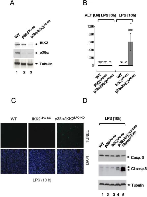
Nach LPS Injektion führt die leberspezifische Deletion von p38α zu einer verstärkten Aktivierung der MAP Kinase JNK. Die verstärkte Aktivierung von JNK führt nicht zu einer erhöhten Sensitivität für LPS/TNF induzierte Leberschädigung. Im Gegensatz dazu, führt eine LPS Injektion in Mäusen mit einer leberspezifischen Deletion für p38α und die IκB kinase 2 (IKK2) zu akuten Leberversagen. Folglich führt eine partielle Inhibierung von NF-κB in Kombination mit p38α Defizienz zu akuter Leberschädigung. Die Ergebnisse zeigen eine neue Rolle von p38α das zusammen mit IKK2 LPS/TNF induzierte Leberschädigungen verhindert.
Darüber hinaus führt die Deletion von p38α in der Leber zu erhöhter Insulinresistenz in einem Model von Diät induzierter Fettleibigkeit und Insulinresistenz. p38α defiziente Lebern und primäre Hepatozyten zeigen eine Einschränkung von Insulin vermittelten Signalen. Dagegen hat der p38α Knockout keinen Einfluss auf Quantität und Qualität von Leberlipiden. Auch eine erhöhte Expression von entzündlichen Zytokinen in der Leber wurde nicht detektiert. Eine erhöhte Aktivität von JNK und seinen Aktivatoren MKK4, TAK1 und ASK1 scheint für die eingeschränkte Ansprechbarkeit von p38α defizienten Zellen durch Insulin verantwortlich zu sein.
Folglich scheint die Kinase p38α in der Leber eine wichtige Funktion bei der Regulation der Glukosehomeostase zu haben, da sie eine Reihe von Kinasen reguliert die negativen Einfluss auf Insulin vermittelte Signale nehmen können.
5 The role of liver p38α MAP kinase during acute and chronic inflammatory states
Jan Heinrichsdorff, Institute for Genetics, University of Cologne, Germany
The liver plays an important role in body homeostasis due to its inflammatory, metabolic and synthetic functions. Cytokines control these functions by activating various intracellular signaling pathways. Activation of the nuclear factor-kB (NF-κB) pathway protects cells from TNF-induced apoptosis by inducing the expression of anti-apoptotic proteins, while sustained c-Jun amino- terminal kinase (JNK) is important for TNF-induced killing of NF-κB-deficient cells.
The p38 mitogen-activated protein kinase (p38) is a member of the MAP kinase family and is activated by various inflammatory stimuli. p38 was shown to regulate cellular responses to stress and is implicated in cell proliferation, differentiation and apoptosis. To investigate the function of p38α in the liver during acute and chronic inflammatory conditions, mice with a liver parenchymal cell specific ablation of p38α were generated. Two models of inflammation, a model of acute LPS/TNF induced liver toxicity and a model of diet induced obesity were applied.
Upon challenge with bacterial lipopolysaccharide (LPS), liver parenchymal cell specific ablation of p38α results in excessive activation of JNK. Despite increased JNK activity, p38α deficient hepatocytes are not sensitized to LPS/TNF induced apoptosis, suggesting that JNK activation is not sufficient to mediate TNF-induced liver damage. By contrast, LPS injection caused liver failure in mice lacking both p38α and IκB kinase 2 (IKK2) in liver parenchymal cells through decreased level of the anti apoptotic molecule c-FLIP (L). Thus, hyperactivation of JNK combined with partial inhibition of NF- κB sensitizes the liver to cytokine-induced damage, revealing a new function of p38α in collaborating with IKK2 to protect the liver from LPS/TNF-induced failure.
Furthermore, deletion of the p38α MAPK in liver parenchymal cells results in increased insulin resistance in obese mice. p38α deficient livers and isolated primary hepatocytes show increased insulin resistance while liver lipids remain unaffected ruling out a possible accumulation of lipid intermediates that could negatively regulate insulin signaling. Moreover, an elevated expression of inflammatory cytokines was not detected. Increased activation of kinases including JNK, MKK4, TAK1 and ASK1 is likely to be responsible for the increased insulin resistance in p38α deficient hepatocytes. Thus, in obese mice, liver p38α might have an important function regulating glucose homeostasis by controlling the activity of kinases that might be responsible for the negative regulation of insulin signaling.
Content
1. Introduction ... 14
1.1 The liver ... 14
1.2 Tumor Necrosis Factor ... 15
1.3 TNF induced signaling in hepatocytes ... 17
1.3.1 NF‐κB signaling in the liver ... 18
1.3.2 MAP kinase cascades ... 20
1.3.3 JNK signaling in the liver ... 22
1.3.4 p38 MAPK ... 25
1.4 The role of p38α in acute and chronic inflammatory diseases ... 29
1.4.1 LPS/TNF induced acute liver failure ... 29
1.4.2 Chronic inflammatory states in the liver: diet induced obesity and insulin resistance . 30 1.5 The cre/ loxP system ... 33
1.6 Objectives ... 34
2 Material and Methods ... 36
2.1 Material ... 36
2.2 Methods ... 41
2.2.1 Molecular biology ... 41
2.2.1.1 Preparation of genomic DNA from tail biopsies of mutant mice ... 41
2.2.1.2 Polymerase chain rection (PCR) ... 41
2.2.1.3 Agarose gel electrophoresis and DNA gel extraction ... 41
2.2.1.4 RNA isolation ... 41
2.2.1.5 cDNA synthesis ... 42
2.2.1.6 Quantitative PCR ... 42
2.2.2 Cell biology ... 43
2.2.2.1 Isolation of primary hepatocytes ... 43
2.2.2.2 Hepatocyte culture ... 43
2.2.3 Biochemistry ... 44
2.2.3.1 Lysis of tissue samples with NP‐40 Buffer ... 44
2.2.3.2 Lysis of hepatocytes with high salt RIPA buffer ... 44
7
2.2.3.3 Bradford assay ... 44
2.2.3.4 Immunoblot analysis ... 45
2.2.3.5 Stripping of PVDF membranes ... 45
2.2.3.6 Electromobility shift assay (EMSA). ... 45
2.2.3.7 Histology ... 46
2.2.3.8 TUNEL assay ... 47
2.2.3.9 Lipid analysis (Thin layer Chromatography) ... 47
2.2.4 Mouse analysis ... 48
2.2.4.1 Animal care ... 48
2.2.4.2 Generation of conditional knockout mice ... 48
2.2.4.3 Genotyping of mutant mice ... 48
2.2.4.4 Liver injury models ... 49
2.2.4.5 Antioxidant treatment ... 49
2.2.4.6 Submandibular bleeding ... 50
2.2.4.7 Serum analysis ... 50
2.2.4.8 Glucose tolerance test ... 51
2.2.4.9 Insulin tolerance test ... 51
2.2.4.10 Insulin Signaling, in vivo ... 51
2.2.4.11 Measurement of serum insulin concentrations. ... 51
2.2.4.12 Analysis of body composition by NMR ... 52
2.2.5 Statistics. ... 52
3 Results ... 53
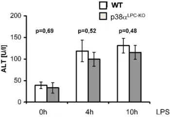
3.1 Generation of mice with a liver parenchymal cell specific deletion of p38α ... 53
3.2 The role of liver p38α in a model of LPS/TNF induced liver toxicity ... 55
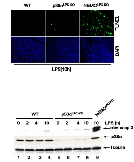
3.2.1 p38αLPC‐KO mice are not sensitive to LPS‐induced liver failure ... 55
3.2.2 Increased activation of JNK in p38α‐deficient livers ... 56
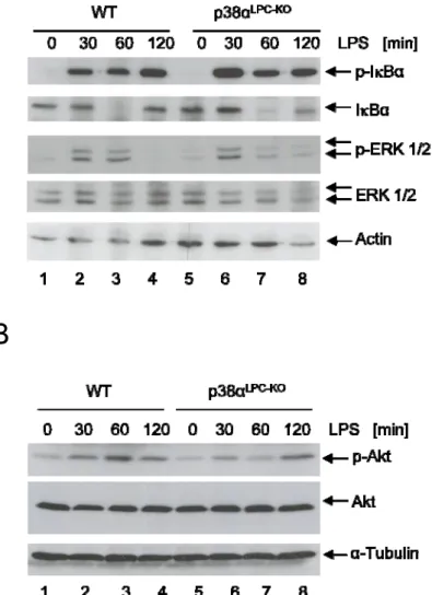
3.2.3 LPS does not induce increased activation of the NF‐κB, ERK and AKT survival pathways lllllllllllllllll in p38αLPC‐KO livers ... 58
3.2.4 Decreased c‐Flip (L) levels in the livers of NEMOLPC‐KO mice, but not in p38αLPC‐KO mice .. 59
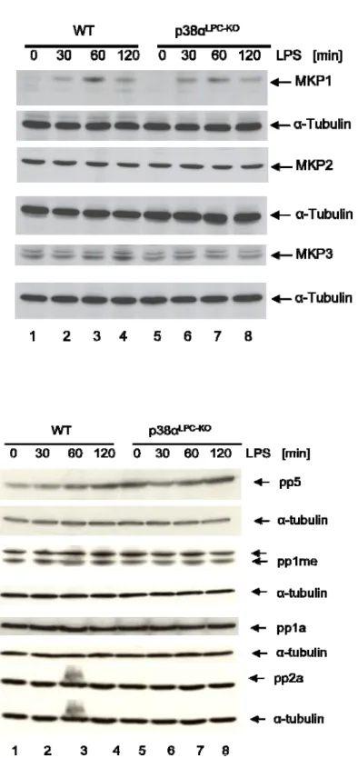
3.2.5 Oxidative stress and MAP kinase phosphatases are not involved in the hyperactivation
lllllllllllllllll of JNK in livers of LPS injected p38αLPC‐KOmice ... 60
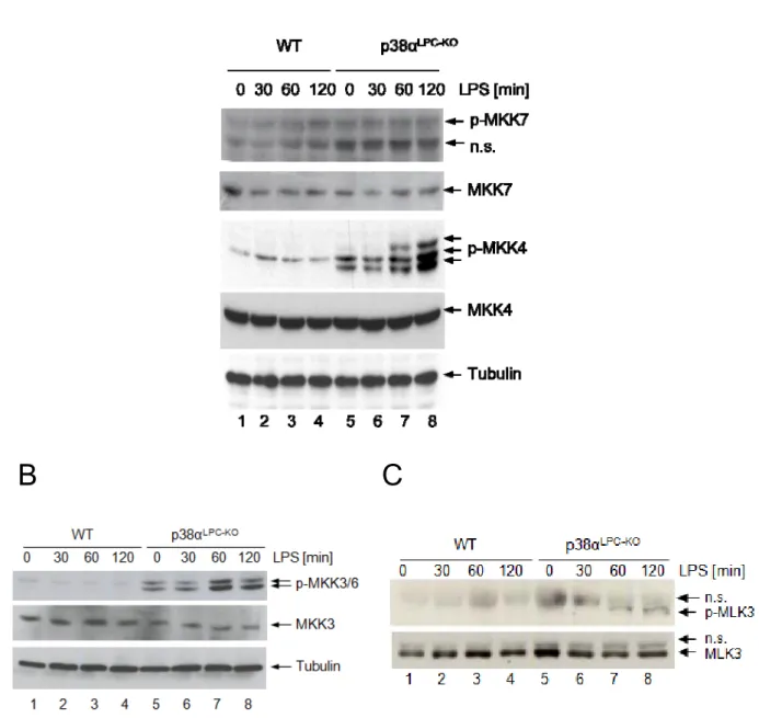
3.2.6 LPS induced hyperactivation of MKK4, MKK3/6 and MLK3 in livers of p38αLPC‐KO mice . 62 3.2.7 p38α and IKK2 collaborate to prevent liver failure ... 63
3.2.8 p38α/IKK2LPC‐KO mice fail to resynthesize c‐FLIP ... 65
3.3 The Role of liver p38α in chronic low grade inflammatory states. A model of high fat diet lllllllllllll induced obesity and insulin resistance. ... 67
3.3.1 p38αLPC‐KO mice do not gain more weight on a high fat diet compared to control mice 67 3.3.2 The deletion of p38α in liver parenchymal cell does not lead to an alteration in blood lllllllllllllllll glucose, serum insulin, cholesterol and triacylglyceride levels ... 67
3.3.3 Obese p38αLPC‐KO mice are more insulin resistant than control mice ... 70
3.3.4 p38α deficient livers show decreased insulin stimulated signaling ... 73
3.3.5 p38α deficient primary hepatocytes show decreased insulin stimulated signaling ... 74
3.3.6 Liver parenchymal cell specific knockout of p38α does not alter the liver lipid content . llllllllllllllllllllllllllllllllllllllllllllllllllllllllllllllllllllllllllllllllllllllllllllllllllllllllllllllllllllllllllllllllllllllllllllllllllllllllllllllllllllll75 3.3.7 The deletion of p38α in liver parenchymal cells does not alter the expression of TNF, llllllllllllllllllIL‐6 and IL1‐β in liver ... 78
3.3.8 Unaltered expression of inflammatory cytokines in adipose tissue taken from p38αLPC‐KO llllllllllllllllllllllland control mice. ... 79
3.3.9 Increased activation of JNK in the livers of obese p38αLPC‐KO mice ... 81
3.3.10 Inhibition of JNK can only partially restore insulin signaling in p38α deficient llllllllllllllllllhepatocytes ... 83
3.3.11 Increased phosphorylation of Ser307 in p38α deficient hepatocytes ... 84
3.3.12 p38α negatively regulates JNK and upstream kinases through an unknown mechanism lllllllllllllllll and therefore positively regulates insulin signaling ... 85
4. Discussion ... 86
4.1 The role of liver p38α in acute inflammatory reactions ... 86
4.1.1 Despite hyperactivation of JNK, p38αLPC‐KO mice are not sensitive to LPS‐induced liver lllllllllllllllll failure 86
4.1.2 Oxidative stress and MAP kinase phosphatases are not involved in the hyperactivation lllllllllllllllll of JNK on p38α deficient livers. ... 87
9 4.1.3 p38α and IKK2 collaborate to prevent LPS/TNF induced liver failure ... 89
4.2 The role of liver p38α in chronic low grade inflammatory states. A model of high fat diet llllllllllllll induced obesity and insulin resistance. ... 91
4.2.1 p38αLPC‐KO are more insulin resistant than control mice ... 91
4.2.2 Lipids and the expression of inflammatory cytokines do not contribute to the llllllllllllllll llllllllllllllll development of insulin resistance ... 92 4.2.3 Increased activation of JNK in the livers of p38αLPC‐KO mice ... 93
4.2.4 Inhibition of JNK can only partially restore insulin signaling in p38α deficient
llllllllllllllllhepatocytes ... 94 4.2.5 Increased phosphorylation of Ser307 in p38α deficient hepatocytes ... 94 5. Perspectives ... 96
Abbreviations
Acyl-CoA Acyl-Coenzym A
AKT Proteinkinase B
ALFP alpha fetoprotein
AP-1 activator protein 1
ASK Apoptosis Signal Regulating Kinase Atf2 activating transcription factor
Atf2 Activating transcription factor 2
ATP Adenosine Triphosphate
BAFF cell activating factor
BHA butylated hydroxyanisole
BSA Bovine Serum Albumin
CaCl2 Calcium Chloride
cDNA Complementary DNA
c-Flip cellular FLICE-inhibitory protein
Cre Cyclization Recombination
CREB response element-binding protein DAG Diacylglyceride
DANN Deoxyribonucleic Acid
DAPI 4'-6-Diamidino-2-phenylindole
DD Death Domain
DEN diethylnitrosamine
DLK leucine zipper-bearing kinase
DMEM Dulbecco’s Modified Eagle Medium
DTT Dithiothreitol
dUTP Deoxyuridine Triphosphate
EDTA Ethylene Diamine Tetraacetate
eIF-4E eukaryotic initiation factor-4e
ELISA Enzyme-linked Immunosorbent Assay EMSA Electromobility shift assay
ER endoplasmatic reticulum
ERK extracellular signal related kinases FADD Fas Associated Death Domain protein
FCS Fetal Calf Serum
FRT FLP recombinase target
GalN D-Galactosamine
11
GSK3 Glykogen Synthase Kinase 3
GTT Glucose tolerance test
H&E Hematoxylin & Eosin
HEPES N-2-Hydroxyethylpiperazine-N’-2-Ethane Sulfonic Acid
HFD high fat diet
HRP Horseradish Peroxidase
HSP27 shock protein 27
IKK1 IκB kinase 1 IKK2 IκB kinase 2
IL-1 interleukin-1
IL-6 interleukin 6
IR Insulin receptor
IRS insulin receptor substrate
ITT Insulin tolerance test
IVC individual ventilated cages
IκB inhibitor of κB protein
JIP JNK Interacting Proteins
JNK c-Jun N-terminal kinase
KCL Potassium Chloride
kDa Kilo Dalton
KO Knockout
LoxP locus of X-over P1
LPA Lysophosphatidic acid
LPC-KO liver parchenchymal cell speciffic knockout LPS lipopolysaccharide
MAP3K MAP Kinase Kinase Kinase
MAPK Mitogen-activated protein kinase MCP-1 monocyte chemotactic protein-1 MK2 kinase- activated protein kinase 2
MKP MAP kinase phospahatase
MLK Mixed Lineage kinases
mRNA Messenger RNA
mTOR mammalian Target of Rapamycin
NaCL Natrium Chloride
NaCL Sodium Chloride
NaF Sodium Fluoride
natural killer NK
NCD normal chow diet
NEMO nuclear factor-kappa B essential modulator
NF-kB nuclear factor-kappaB
NMR Nuclear magnetic resonance
P Phospho
PA Palmatic acic
PBS Phosphate-Buffered Saline
PCR Polymerase Chain Reaction
PFA Paraformaldehyde RIP1 Receptor Interacting Protein 1
RNA Ribonucleic Acid
ROS Reactive Oxygen Species
Rpm Rounds per Minute
RT Reverse transcriptase
RT Room Temperature
Sap1 SRF accessory protein
TAK1 TGF-b-activated kinase 1
TAO Thousand and one amino acid kinase
TBS Tris buffered saline
TCF Ternary Complex Factor
TNFR II TNF-receptor II
TNFR-I TNF-receptor I
Tpl-2 Tumor progression locus-2
TRADD TNFR I Associated Death Domain protein TRAF2 TNF Receptor Associated Factor 2
TUNEL
Terminal Deoxynucleotidyl Transferase Mediated dNTP Nick End Labelling
TNF tumor-necrosis factor alpha
WAT white adipose tissue
WB Western Blot
WT Wild-Type
13
„… because the liver is a source of many diseases, and it is a noble organ that serves many other organs, almost all of them: so it suffers, it is not a small suffering, but a great manifold one“
Paracelsus (1494-1541)
1. Introduction
1.1 The liver
The liver is the largest solid organ present in vertebrates. It has a wide range of functions keeping the body in homeostasis. The liver plays a major role in glucose and glycogen metabolism, lipid and lipoprotein synthesis, hormone production, decomposition of red blood cells and detoxification. It produces bile, an alkaline compound which aids in digestion and detoxification, via the emulsification of lipids. The liver also performs and regulates a wide variety of high-volume biochemical reactions requiring highly specialized tissues, including the synthesis and breakdown of small and complex molecules, many of which are necessary for normal vital functions. It is absolutely essential for survival; there is currently no way to compensate for the absence of liver function as opposed to - for example – blood dialysis in kidney dysfunction.
With respect to its anatomical location the liver has a unique position in the body. Situated below the diaphragm in the thoracic region of the abdomen it allows continuous blood flow from the gastrointestinal tract through the sinusoids. A human liver consists of 2 lobes, comprising 1/50 of the body weight of an adult. In contrast, a mouse liver consists of 4 lobes (Martins, Theruvath et al. 2008).
In terms of its cellular composition, the liver is a heterogeneous organ. Circulating blood cells, from the innate or adaptive immune system and a variety of intrahepatic cell populations such as parenchymal liver cells (hepatocytes 60-65% and biliary epithelial cells 3,5% off all liver cells), the sinusoidal cell compartment including endothelial cells (15-20%), liver-resident macrophage (Kupffer cells 8-12%), lymphocyte [mainly natural killer T (NKT) cells] populations and hepatic stellate cells (3-8%) steer the different hepatic functions (Racanelli and Rehermann 2006).
The function of the diverse cell populations in the liver is orchestered by cytokines, which are key mediators within the complex interplay of intrahepatic immune cells and hepatocytes.
Cytokines can activate effector functions of immune cells, as well as intracellular signaling pathways within the hepatocytes, acting as key players controlling cellular homeostasis.
Serving as autocrine, paracrine or endocrine messengers, cytokines are essential mediators that influence various liver functions including inflammatory and metabolic responses (Luedde, Liedtke et al. 2002).
15 In the liver, Kupffer cells and liver-infiltrating monocyte-derived macrophages are primary sources of cytokines, such as tumor-necrosis factor alpha (TNF) and Interleukin-6 (IL-6).
Especially TNF seems to play an outstanding role controlling liver homeostasis and is therefore involved in important functions in liver physiology and pathology. TNF activates specific intracellular pathways in hepatocytes that influence cell fate in different manners, through the activation of proapoptotic signals via caspase or Mitogen-activated protein kinase (MAPK) cascades, but also survival pathways, like the nuclear factor-kappaB (NF-κB) pathway. Recent experiments with genetically modified mice demonstrated important and partly opposing functions induced by TNF receptor (TNFR) signaling. While on the one hand TNF is able to promote cell survival and liver regeneration, on the other hand it can act as a death inducing cytokine. Therefore, the dissection of hepatocyte responses to TNF in homeostasis and injury could potentially identify novel targets for the treatment of many acute and chronic liver diseases.
1.2 Tumor Necrosis Factor
TNF was initially described 30 years ago as an endotoxin induced serum factor that induces death in BALB/c sarcomas (Carswell, Old et al. 1975). Independently, TNF was described as a factor mediating differentiation of hematopoetic cells into the monocyte/macrophage linage (Takeda, Iwamoto et al. 1986) and as a hormone produced by activated macrophages, responsible for hypertriglyceridemia and wasting of adipose tissue in cancer patients and patients suffering from infections (Beutler, Greenwald et al. 1985; Fried and Zechner 1989). It was shown to control a diverse range of physiological functions, many of which are related to the immune response and inflammation. TNF has been shown to play an important role in leukocyte migration, tissue resorption, and fever response (Beutler and Cerami 1989).
TNF binds to two plasma membrane receptors, TNF-receptor I (TNFR I) and TNF-receptor II (TNFR II) (Brockhaus, Schoenfeld et al. 1990; Dembic, Loetscher et al. 1990; Schall, Lewis et al. 1990). Whereas TNFR I is the receptor for soluble TNF, TNFR II mediates effects through the membrane bound TNF precursor and seems mostly restricted to T cells (Zheng, Fisher et al. 1995; Kim and Teh 2001; Depuydt, van Loo et al. 2005). The majority of the biological effects induced by TNF seem to be mediated by TNFR I through the activation of NF-κB and the initiation of MAPK cascades as well as cell death inducing caspase cascades.
The diversity of the different pathways that are activated by TNFR I is mediated through different adaptor proteins recruited to the cytoplasmic part of the receptor. The cytoplasmic part of the TNFR I contains a Death Domain (DD) that is shared by a number of receptors promoting cell death (Tartaglia, Rothe et al. 1993). The Death Domain creates a binding
scaffold for downstream mediators of TNFR I signaling such as TNFR I Associated Death Domain protein (TRADD), Fas Associated Death Domain protein (FADD), Receptor Interacting Protein 1 (RIP1), TNF Receptor Associated Factor 2 (TRAF2) and other factors (Hsu, Xiong et al. 1995; Hsu, Huang et al. 1996; Hsu, Shu et al. 1996). According to the current model, two complexes are built upon receptor activation. Complex I leads to the activation of NF-κB, the MAP kinase pathways c-Jun N-terminal kinase (JNK) and p38. TNF- mediaded activation of NF-κB is initiated by the recruitment of the adaptor molecules TRADD, TRAF2, and Rip to the cytoplasmic part of TNFR I. Once this complex is formed, TRAF2 and RIP1 cooperate to recruit the TGFβ-activated kinase 1 (TAK1). TRAF2 contains ubiquitin ligase activity and leads to the addition of Lys-63 ubiquitin chains to RIP1. These ubiquitin chains are recognized by TAB2/3 which both mediate the recruitment of TAK1 (Chen and Goeddel 2002; Chen 2005). Activated TAK1 phosphorylates the critical residues on IKK2 and leads to the activation of NF-κB signaling (Wang, Deng et al. 2001; Sato, Sanjo et al. 2005; Shim, Xiao et al. 2005). TAK1 is a member of the MAP3K family. Therefore, recruitment of TAK1 to the TNFR I does not only cause TNF induced activation of NF-κB, it also causes activation of the MAP kinases JNK and p38. The mechanisms leading to temporal and spatial separation of MAPK and NF-κB signaling downstream of TNFR I are currently under investigation (Matsuzawa, Tseng et al. 2008). In contrast, Complex II, which forms after dissociation of the membrane proximal complex I from TNFR I, induces hepatocyte apoptosis by recruiting the adaptor molecules FADD and TRADD. FADD contains a death effector domain by which it can recruit procaspase 8. The clustering of procaspase 8 in the DISC complex leads to its autoactivation. Active caspase 8 is then able to proteolytically activate other effector caspases initiating apoptosis (Micheau and Tschopp 2003). In cells that lack substantial NF-κB activity, formation of complex II results in caspase- 8 activation and apoptotic cell death (Muppidi, Tschopp et al. 2004) However, if NF-κB has been activated during complex I formation, it prevents caspase 8 activation by the DISC complex through induction of antiapoptotic genes like the cellular FLICE-inhibitory protein (c- FLIP), an adaptor protein with a pseudocaspase domain that specifically inhibits caspase-8 (Thome and Tschopp 2001). Thus, NF-κB may negatively regulate TNF induced apoptosis through regulation of c-FLIP levels and its activation serves as a checkpoint that determines whether the DISC complex will signal cell death or not (Muppidi, Tschopp et al. 2004).
17 1.3 TNF induced signaling in hepatocytes
In the healthy liver, cytokine production is absent or very low. During liver inflammation cells are exposed to increased levels of TNF. TNF induction is known as one of the earliest events of hepatic inflammation triggering a cascade of other cytokines that cooperate in recruiting inflammatory cells, killing of hepatocytes and initiating wound healing responses (Bradham, Plumpe et al. 1998). Depending on the cellular context, exposure of hepatocytes to TNF induces signals that mediate cell death or survival pathways, which allow hepatocytes to survive and recover from tissue damage (Akerman, Cote et al. 1992; Yamada, Kirillova et al.
1997; Yin, Wheeler et al. 1999; Rudiger and Clavien 2002). Moreover, in addition to its role in triggering life and death decisions in hepatocytes, TNF has also been shown to affect important metabolic functions in the liver. TNF expression is upregulated in the fat of obese rodents and humans and is elevated in the serum of patients with type 2 diabetes.
Additionally, it has been shown to cause insulin resistance in hepatocytes in vivo and in vitro (Uysal, Wiesbrock et al. 1997; Hotamisligil 1999)
As a result of this complex interplay between different signaling cascades activated downstream of the TNFR I, there are different levels of regulation determining the biological outcome of the TNF induced signaling. Various conflicting signaling pathways lead to a wide spectrum of TNF induced responses ranging from cell survival to cell death. These signals also influence cell metabolism, gene expression, differentiation, adhesion and mortality (Shaulian and Karin 2002). Especially NF-κB and MAPK mediated responses to TNF signaling in the liver were a subject of intense investigation in the last decade.
Graph 1: TNFR I signaling TNFR1 activation leads to formation of complex I at the cytoplasmic tail of the receptor resulting in JNK1 and IKK activation. Complex I dissociates from the receptor. Complex II, which includes FADD, is formed. If c-FLIP (L) levels are high (high NF-κB, low JNK activation), caspase-8 recruitment to FADD and its subsequent activation are inhibited and the cells survive. If c-FLIP (L) levels are low (low NF-κB, high JNK), caspase-8 is recruited to FADD and activated, leading to apoptotic cell death. Image taken from Chang, L et al. 2006 Cell.
1.3.1 NF-κB signaling in the liver
NF-κB was identified about 20 years ago as a transcription factor that binds to the intronic enhancer of the kappa light chain gene in B cells (Sen and Baltimore 1986; Sen and Baltimore 1986). The family of NF-κB transcription factors consists of p50 and its precursor p105, p52 and its precursor p100, RelA (p65), c-Rel and RelB all of which share a Rel homology domain responsible for homo and heterodimerisation as well as for sequence specific DNA binding. RelA, c-Rel and RelB also contain a C-terminal transcription activation domain, whereas p50 and p52 do not and rely on interactions with other partners to positively regulate transcription (Hayden and Ghosh 2008). In resting cells, NF-κB transcription factors are normally kept in an inactive state in the cytoplasm through the binding to inhibitors of κB proteins IκBs (IκBα, IκBβ, IκBγ) and the precursor proteins p100 and p105.
19 Many different stimuli can activate NF-κB transcription factors and induce their nuclear accumulation (Ghosh and Karin 2002; Bonizzi and Karin 2004) which is controlled by the IκB kinases (IKKs).
The major and most well known pathway to activate NF-κB is the so called canonical NF-κB pathway. Activation is controlled by the IKK complex consisting of IKK1, IKK2 and the regulatory subunit nuclear factor-kappa B essential modulator (NEMO). Upon cell stimulation the IKK complex phosphorylates the IκB proteins that keep the NF-κB transcription factors in the cytoplasm. This leads to ubiquitination and degradation of the IκB proteins. As a result the NF-κB transcription factors enter the nucleus and activate gene transcription. In the case of canonical NF-κB signaling, mainly RelA:p50 and c-Rel:p52 heterodimers translocate to the nucleus (Mercurio, Zhu et al. 1997; Woronicz, Gao et al. 1997; Yamaoka, Courtois et al.
1998).
The so called non-canonical NF-κB signaling pathway relies on activation through receptors that are mainly involved in developmental processes like the lymphotoxin beta receptor or B cell activating factor (BAFF). Here, IKK1 acts independent from IKK2 and NEMO (Senftleben, Cao et al. 2001; Derudder, Dejardin et al. 2003) and leads to the phosphorylation and proteolytic processing of p100 to p52 which leads to the nuclear accumulation of p52:RelB dimers (Senftleben, Cao et al. 2001).
Healthy hepatocytes are resistant to cytotoxic effects of TNF. NF-κB signaling may give an essential survival stimulus that balances the effects of the cell death machinery that are activated by TNF. If NF-κB signaling is impaired as reported in RelA, NEMO and IKK2 deficient mice, embryonic lethality results due to TNF induced degradation of the fetal liver (Beg, Sha et al. 1995; Li, Van Antwerp et al. 1999; Rudolph, Yeh et al. 2000). Additional deletion of TNF or TNFR I rescues the embryonic lethality of these mice (Doi, Marino et al.
1999). This in vivo evidence showing that NF-κB prevents TNF mediated cell death is also supported by in vitro studies with hepatocytes which, if transfected with a dominant negative IκBα, are sensitive to TNF induced apoptosis (Bellas, FitzGerald et al. 1997).
The activation of NF-κB influences the pathogenesis of several TNF-mediated hepatic diseases. In TNF induced liver injury, wild type mice are only sensitized to TNF induced cell death upon co-administration of D-Galactosamine (GalN). GalN is a hepatotoxin that blocks transcription specifically in hepatocytes, including the transcription of anti-apoptotic NF-κB target genes, and therefore sensitizes mice to TNF induced lethality (Decker and Keppler 1974). To the same extent GalN sensitizes mice to bacterial Lipopolysaccaride (LPS) a strong inducer of TNF. Moreover, in liver regeneration after partial hepatectomy, TNF induced NF-κB plays an important role due to the induction of interleukin 6 (IL-6) (Cressman,
Greenbaum et al. 1996). Liver NF-κB also seems to play a role in carcinogenesis. IKK2 liver deficient mice develop more hepatocellular carcinomas after diethylnitrosamine (DEN) treatment due to a higher rate of hepatocyte apoptosis and compensatory proliferation (Maeda, Kamata et al. 2005). Furthermore, in mice lacking NEMO specifically in the liver, cell death and the spontaneous development of chronic hepatitis result in the development of hepatocellular carcinomas. This process is initiated by hypersensitivity of NEMO deficient hepatocytes to oxidative stress dependent, FADD mediated apoptosis, triggering compensatory cell proliferation, inflammation and activation of progenitor cells (Luedde, Beraza et al. 2007). Similar to the DEN model, hepatocyte specific deletion of NEMO highlights the essential anti-apoptotic role of NF-κB avoiding cell death followed by compensatory proliferation and cancer. NF-κB also processes inflammatory responses since its activation does not only lead to the expression of anti-apoptotic mediators but also to the expression of inflammatory cytokines. Indeed, deletion of IKK2 in the liver seems to prevent liver insulin resistance due to a lower expression of inflammatory cytokines that affect insulin signaling (Arkan, Hevener et al. 2005) while liver specific hyperactivation of IKK2 has the opposite effect (Cai, Yuan et al. 2005).
1.3.2 MAP kinase cascades
Besides NF-κB signaling, ligand binding to TNFR I also activates Mitogen-activated protein kinases (MAPKs), in particular c-Jun n-terminal kinases (JNK) and p38.
MAPKs are evolutionary conserved serine-threonine kinases that connect cell surface receptors to critical regulatory targets within the cells. Mammals express at least four distinctly related groups of MAPKs, (1) extracellular signal related kinases (ERK) 1/2, (2) Jun n-terminal kinases (JNK) 1/2/3, (3) p38 proteins α, β, γ, δ and (4) ERK 5.
MAPKs are activated by phosphorylation cascades. These cascades consist of a set of three evolutionarily conserved, sequentially acting kinases: (1) a MAPK, (2) a MAPK kinase (MAP2K), and (3) an MAPK kinase kinase (MAP3K). MAP3Ks, are Ser/Thr kinases that are activated via phosphorylation or through interaction with small GTP proteins of the Ras/Rho family in response to a wide range of extracellular stimuli. MAP3K activation leads to phosphorylation and activation of a downstream MAP2K. MAP2Ks are dual specificity kinases that can phosphorylate MAPK on both threonine and tyrosine on a conserved Thr-x- Tyr motif to activate them. The Thr-x-Tyr phosphorylation motif is localized in an activation loop near the ATP- and substrate-binding sites. Once activated, MAPKs phosphorylate their target substrates on serine or threonine residues only if the amino acid residues are followed
21 by proline. A wide variety of MAPK substrates exists, among them transcription factors, other kinases (MAPK activated kinases or MKs) or proteins like cytoskeletal proteins. Each of these cascades is named after the subgroup of its MAPK component ERK1/2, JNK, p38 or ERK 5. In mammals 20 genes encoding for MAP3Ks have been reported, followed by 7 MAP2Ks and 11 MAPKs genes (Uhlik, Abell et al. 2004). Different extracellular signals can produce stimulus- and tissue-specific responses by activating one or more MAPK pathways.
In addition, MAPK-activated protein kinases (MKs), the downstream targets of MAPKs, further contribute to additional specificity, diversity and amplification in the MAPK cascades.
Graph 2: MAPK signaling. (A) MAPK signaling pathways mediate intracellular signaling that can be initiated by a variety of extracellular or intracellular stimuli. Initially, MAP3Ks, which are activated by MAP4Ks or GTPases, mediate phosphorylation and activation of MAP2Ks, which in turn phosphorylate and activate MAPKs. Activated MAPKs phosphorylate various substrate proteins including transcription factors, resulting in regulation of a variety of cellular activities including cell proliferation, differentiation, migration, inflammatory responses, and death. (B) The p38 and JNK pathways are activated by a MAP3K such as ASK1, MEKK1, or MLK3 as well as a MAP2K such as MKK3 or MKK6 for the p38 pathway and MKK4 or MKK7 for the JNK pathway. Pictures taken from Kyung, E et al. 2010 Biochimica et Biophysica Acta.
One of the most explored functions of MAPK signaling modules is the regulation of gene expression in response to extracellular stimuli (Treisman 1996). MAPKs function inside the nucleus and target transcription factors that are bound to DNA. It has also been reported that MAPKs regulate gene expression through post-transcriptional mechanisms involving cytoplasmic targets. (Chen, Gherzi et al. 2000).
A B
The biological outcomes of MAPK signaling include a wide range of functions including cell proliferation, differentiation, survival, death and transformation. These functions are particularly important in an organ like the liver which has a high proliferative and regenerative capacity and always balances between cell survival and cell death. In contrast to NF-κB, MAPK signaling is considered to be rather diverse with pro-apoptotic but also anti-apoptotic functions.
Two members of the MAPK family, JNK and the p38 signaling pathway, can be activated by TNF in response to cellular stress such as genotoxic, osmotic, hypoxic or oxidative stress.
1.3.3 JNK signaling in the liver
The family of JNK protein kinases is encoded by 3 different genes. The JNK 1 and JNK 2 genes are expressed ubiquitously while JNK 3 expression has a limited pattern and is restricted to the brain, heart and testis (Gupta, Barrett et al. 1996).
JNK is activated by the dual specificity MAP2Ks MKK4 and MKK7, which are activated by their upstream MAP3Ks through phosphorylation on serine or threonine residues in the activation loop. A wide range of MAP3Ks have neen described, which most likely act in a cell type and stimulus-specific manner (Davis 1995). The first and most potent JNK-activating MAP3K identified was MEKK1 (Gerwins, Blank et al. 1997). However, numerous other MAP3Ks, which can activate JNK were identified, including: MEKK2 and MEKK3 (Blank, Gerwins et al. 1996), MEKK4 (Gerwins, Blank et al. 1997), Mixed Lineage kinases 2 and 3 (MLK2 and MLK3) (Hirai, Izawa et al. 1996; Tibbles, Ing et al. 1996), Dual leucine zipper- bearing kinase (DLK) (Fan, Merritt et al. 1996), Tumor progression locus-2 (Tpl-2) (Salmeron, Ahmad et al. 1996), TGF-β-activated kinase (TAK1) (Yamaguchi, Shirakabe et al.
1995), Apoptosis Signal Regulating Kinases 1 and 2 (ASK1 and ASK2) (Ichijo, Nishida et al.
1997), and Thousand and one amino acid kinases 1 and 2 (Tao1 and Tao2) (Chen, Hutchison et al. 1999). The large number of MAP3Ks that can activate the JNK cascade have made the identification of the individual family member responsible for JNK activation by distinct stimuli extremely difficult.
The organization of MAPK cascades as distinct signaling modules allows their highly coordinated and specific activation response to extracellular stimuli (Davis 2000). Thus, the presence of a large number of JNK-activating MAP3Ks in mammals may provide the benefit of improved signal specificity to diverse types of physiological and stress stimuli.
23 Another level of regulation adding specificity to JNK signaling is the scaffolding of proteins, which includes the JNK Interacting Proteins (JIP) like scaffolds (Davis 1999) and the MKKs.
(Xia, Wu et al. 1998; Morrison and Davis 2003). Besides the purpose of specificity, scaffolding might also serve the purpose of amplifying a signal.
Graph 3: JNK Scaffolding JIP-1 can act as a scaffold assembling specific components of JNK activation including the upstream kinase HPK-1, the MAP3K MLK1, the MA2K MKK7 and the MAPK JNK into a signaling module. Picture taken from Chang L et al. 2001 Nature.
JNK proteins phosphorylate a wide range of transcription factors including c-Jun which is phosphorylated at Ser 63 and 74 leading to increased transcriptional activity (Pulverer, Kyriakis et al. 1991) and AP-1 proteins like JunB, JunD and Atf2 (Auer, Contessa et al. 1998;
Ip and Davis 1998). JNK appears to be important for AP-1 target gene activation caused by stress and cytokines but is not required for AP-1 transcriptional activation by other stimuli (Yang, Tournier et al. 1997).
In recent years, JNK isoforms have been recognized as critical regulators of various aspects of mammalian physiology, including: cell proliferation, cell survival, cell death, DNA repair and metabolism. Upon TNF signaling, the duration and magnitude of JNK activation seems to be determined by the balance between activating MKKs. Only a modest and transient JNK activation is required for TNF-induced cell proliferation, whereas an intense and prolonged JNK activation mediates cell death (Kamata, Honda et al. 2005) highlighting the critical balance of TNF induced JNK activation for the outcome of the biological response.
A
After partial hepatectomy, which induces TNF, JNK is activated and is instrumental for liver regeneration (Schwabe, Bradham et al. 2003). On the other hand, in livers and hepatocytes unable to activate NF-κB, JNK activity controls TNF-induced death through the proteasomal processing of c-FLIP (L) (Chang, Kamata et al. 2006). Moreover, JNK plays a role in DEN induced hepatocarcinogenesis. In hepatocytes, NF-κB activation promotes sustained JNK activation in cells exposed to TNF, whose expression is induced by DEN. JNK activity is required for normal hepatocyte proliferation and liver regeneration. Mice lacking JNK1 were much less susceptible to DEN-induced hepatocarcinogenesis. The decreased tumorigenesis in JNK1-/- mice was shown to correlate with decreased expression of cyclin D and vascular endothelial growth factor, diminished cell proliferation, and reduced tumor neovascularization (Sakurai, Maeda et al. 2006). JNK also seems to be involved in the negative regulation of insulin signaling. It has been demonstrated in vitro that JNKs can phosphorylate the insulin receptor substrate (IRS1) on serine 307, which correlates with reduced Insulin receptor mediated IRS1 tyrosine phosphorylation leading to a decreased transduction of the insulin stimulus (Aguirre, Uchida et al. 2000). Moreover, JNK isoforms undergo activation in response to pro inflammatory cytokines, such as TNF, which is elevated during obesity (Kallunki, Su et al. 1994). Indeed, it was observed that consumption of high fat diet (HFD) or genetic obesity results in JNK activation (Hirosumi, Tuncman et al. 2002) and that JNK1-/- mice are protected from diet induced obesity and insulin resistance, even though in this model the tissue specific contributions of JNK1 to this phenotype were not addressed.
Furthermore, treatment of obese mice with a cell permeable peptide inhibitor of JNK was found to result in improved insulin sensitivity (Bogoyevitch, Boehm et al. 2004). It is also of interest that mutations in the Jip1 gene have been found in patients with a genetic form of type II diabetes (Waeber, Delplanque et al. 2000). This suggests that a JIP1-organized JNK1 cascade plays an important role in the regulation of insulin signaling.
In contrast to JNK, which has been studied in various different models in the liver, the functions of p38, another MAPK activated by TNF signaling, are far less understood.
25 1.3.4 p38 MAPK
Human p38 was originally discovered as the molecular target of the pyridinyl imidazole class of compounds that were known to inhibit biosynthesis of inflammatory cytokines such as interleukin-1 (IL-1) and TNF in lipopolysaccharide (LPS) stimulated human monocytes (Lee, Laydon et al. 1994). Later, the murine p38α MAPK was identified as a major phosphoprotein activated by LPS and inflammatory cytokines (Han, Lee et al. 1994). So far, four isoforms of p38 MAPK have been identified. Of these, the ubiquitously expressed p38α is the best characterized and perhaps the most physiologically relevant kinase involved in inflammatory responses. Besides p38α, p38β was identified which has >70% identity to p38α (Jiang, Chen et al. 1996; Kumar, McDonnell et al. 1997). Later, p38γ (also known as SAPK3 and MAPK12) and p38δ (also known as SAPK4 and MAPK13) (Li, Jiang et al. 1996; Mertens, Craxton et al. 1996) were identified. These kinases have ~60% identity to p38α and much less is known about their function.
p38 MAPK is activated though a classical MAPK activation cascade involving a MAP3K, that activates a MAP2K which then activates the MAPK p38. Activation of p38 occurs by dual phosphorylation on Thr180 and Tyr182 by an upstream MAP2K termed MKK6. Other MAP2Ks, such as MKK3 have also been suggested to activate p38 MAPK. MKK6 is activated by several MAP3Ks, which are in turn activated by a wide variety of stimuli including MAP3Ks activated in response to various physical and chemical stresses, such as oxidative stress, UV irradiation, hypoxia, ischemia, and various cytokines, including interleukin-1 (IL-1) and TNF (Adams, Badger et al. 2001; Chen, Gibson et al. 2001; Kyriakis and Avruch 2001).
Graph 4: p38 MAPK pathway activators and substrates Environmental stress and/or inflammatory cytokines activate MAP3Ks through a variety of signaling mechanisms. Activated MAP3Ks subsequently activate MAP2Ks, which in turn activate MAPKs. Activated MAPKs then phosphorylate several substrates, such as transcription factors, other kinases and cytosolic proteins. Picture taken from Kumar et al. 2003 Nat. Rev.
MEK4 (also known as MKK4 and Sek1) is a known JNK kinase that possesses some MAP2K activity toward p38, suggesting that MEK4 represents a site of integration for the p38 and JNK pathways. While MKK6 activates all p38 isoforms, MKK3 is somewhat selective, as it preferentially phosphorylates the p38α and p38β isoforms.
The first p38α substrate identified was the MAP kinase- activated protein kinase 2 (MAPKAPK2 or MK2) (Freshney, Rawlinson et al. 1994; Rouse, Cohen et al. 1994;
McLaughlin, Kumar et al. 1996). MK2, along with its closely related family member MK3,
27 were both shown to activate various substrates including small heat shock protein 27 (HSP27) (Stokoe, Engel et al. 1992), lymphocyte-specific protein 1 (LSP1) (Huang, Zhan et al. 1997), cAMP response element-binding protein (CREB) (Tan, Rouse et al. 1996), transcription factor ATF1, SRF (Heidenreich, Neininger et al. 1999), and tyrosine hydroxylase (Thomas, Haavik et al. 1997). MNK1 is another kinase substrate of p38 whose function is thought to reside in translational initiation due to the observation that MNK1 and MNK2 can phosphorylate eukaryotic initiation factor-4e (eIF-4E) (Waskiewicz, Flynn et al. 1997).
Another group of substrates that are activated by p38 comprises transcription factors. Many transcription factors encompassing a broad range of action have been shown to be phosphorylated and subsequently activated by p38. Examples include activating transcription factor 1, 2 & 6 (ATF-1/2/6), SRF accessory protein (Sap1), CHOP (growth arrest and DNA damage inducible gene 153, or GADD153), p53, C/EBPβ, myocyte enhance factor 2C (MEF2C), MEF2A, MITF1, DDIT3, ELK1, NFAT, and high mobility group-box protein 1 (HBP1) (Hazzalin, Cano et al. 1996; Wang and Ron 1996; Han, Jiang et al. 1997; Janknecht and Hunter 1997; Whitmarsh, Yang et al. 1997; Huang, Ma et al. 1999; Zhao, New et al.
1999; Yee, Paulson et al. 2004). An important cis-element, AP-1 appears to be influenced by p38 through several different mechanisms. ATF-2 is known to form heterodimers with Jun family transcription factors thereby directly associating with the AP- 1 binding site. Another possible mechanism comes from the observation that a component of AP-1 is c-fos. c-fos is known to be SRE dependent and SRE is able to bind Ternary Complex Factor (TCF).
Ternary Complex mFactor is comprised of Sap-1a, a protein that is phosphorylated by p38.
Thus, p38 indirectly regulates AP-1 activity.
Considering the wide range of substrates linked to p38 MAPKs, it is a natural consequence that this kinase should also be involved in a wide and diverse variety of functions. Indeed, roles for p38 MAPKs have been described in processes such as cell differentiation and migration, cell transformation, cell survival, inflammation, angiogenesis, myogenesis and others.
The fact that p38-/- mice are lethal at embryonic day 10.5 has deprived researchers of an important genetic tool for functional studies in vivo. Four groups have reported the generation of these mice (Adams, Porras et al. 2000; Allen, Svensson et al. 2000; Mudgett, Ding et al.
2000; Tamura, Sudo et al. 2000). Insufficient vascularization during placental development was described as a possible cause of death. Mudget et al. found that in addition to the placental defect, angiogenesis was also abnormal in the yolk sac and the embryo itself.
Adams et al. reported massive reduction of the myocardium and malformation of blood vessels in the head region. However, this defect appears to be secondary to insufficient
oxygen and nutrient transfer across the placenta. Finally, fetuses surviving to relatively late stages of development are anemic and have a deficiency of erythropoietin (Epo) mRNA expression in their liver. Therefore, the studies conducted on p38 function have been restricted to in vitro assays and stimulus specific functions in the context of a specific organ like the liver could not be addressed so far.
Among other processes, p38 seems to play a role in inflammation. Rheumatoid arthritis, Alzheimer’s disease and inflammatory bowel disease are all postulated to be regulated in part by the p38 pathway (Perregaux, Dean et al. 1995; Johnson and Bailey 2003;
Hollenbach, Neumann et al. 2004). p38 signaling seems to play an essential role in the production of proinflammatory cytokines (IL-1β, TNF and IL-6) (Guan, Buckman et al. 1998).
Moreover, p38 has a role in the expression of intracellular enzymes such as iNOS, a regulator of oxidation (Da Silva, Pierrat et al. 1997; Craxton, Shu et al. 1998), induction of VCAM-1 and other adherent proteins along with other inflammatory related molecules (Pietersma, Tilly et al. 1997).
The role of p38 in apoptosis seems to be cell type and stimulus dependent. While p38 signaling has been shown to promote cell death in some cell lines, in other cell lines p38 has been shown to enhance survival, cell growth, and differentiation. There is evidence that p38 might act up and downstream of caspases in apoptosis (Ziegler-Heitbrock, Blumenstein et al.
1992; Cardone, Salvesen et al. 1997).
Furthermore, the participation of p38 in cell growth has been observed in yeast and mammals (Takenaka, Moriguchi et al. 1998). Overexpression of p38 in yeast led to significant attenuation of proliferation, while treatment in mammalian cells with p38α/β inhibitor SB203580 slowed proliferation as well. p38 has been implicated in G1 and G2/M phases of the cell cycle in several reports (Molnar, Theodoras et al. 1997; Wang, McGowan et al. 2000).
p38α and p38β have been implicated in the differentiation of certain cell types. Differentiation of 3T3-L1 cells into adipocytes and PC12 cells into neurons requires p38α and/or β (Engelman, Lisanti et al. 1998). More recently, a cross-talk model between the p38 pathway and phosphatidylinositol 3-kinase (PI3 kinase)/Akt in the orchestration of myoblast differentiation has been proposed (Iwasa, Han et al. 2003).
Moreover, p38 seems to have a role in tumorigenesis and sensescence. There have been reports that activation of MKK6 and MKK3 led to a senescent phenotype dependent upon p38 MAPK activity. Also, p38 MAPK activity was shown responsible for senescence in response to telomere shortening, H2O2 exposure, and chronic RAS oncogene signaling (Wang, Chen et al. 2002).
p38 might also be involved in the energy metabolism of certain organs. Here, most studies are restricted to in vitro work. The few in vivo studies were carried out with p38 inhibitors. In
29 the liver, p38 might participate in the processes to increase blood glucose levels through reducing glycogen synthesis and increasing hepatic gluconeogenesis (Cao, Collins et al.
2005). p38 appears to prevent fat storage by inhibiting hepatic lipogenesis and promoting fatty acid oxidation in the liver. Additionally, p38 may play a critical role in cholesterol metabolism by regulating expression of the LDLR gene and bile metabolism, but many functions remain controversial (Cao, Collins et al. 2005; Cao, Xiong et al. 2007).
1.4 The role of p38α in acute and chronic inflammatory diseases
As mentioned before, a strong link has been established between the p38α pathway and inflammation. Rheumatoid arthritis, Alzheimer’s disease and inflammatory bowel disease are all postulated to be regulated in part by the p38 signaling pathway (Johnson and Bailey 2003;
Hollenbach, Neumann et al. 2004). Furthermore, the activation of the p38 pathway plays an essential role in the production of proinflammatory cytokines like IL-1β, TNF and IL-6 (Guan, Buckman et al. 1998). All three cytokines were reported to play important roles in liver homeostasis and disease. In contrast to NF-κB and JNK, the tissue specific role of liver p38α during various inflammatory conditions remains to be investigated in vivo. Thus, as a central topic of this thesis, the role of liver p38α in acute and chronic inflammatory reactions was investigated. In order to do this, two mouse models of inflammation were chosen. For the role of liver p38α in acute inflammatory reactions, a model of LPS/TNF induced acute liver toxicity was applied. To investigate the role of p38α in chronic low grade inflammatory reactions a model of diet induced obesity and insulin resistance was utilized.
1.4.1 LPS/TNF induced acute liver failure
The liver is the central metabolic organ in the body fulfilling important functions ranging from carbohydrate and lipid metabolism, to synthetic and excretory functions. Due to these central functions processed in the liver, diseases of this organ can lead to severe symptoms. The liver has an outstanding potential to regenerate and is able to compensate lack of function for an extended period of time. Therefore, symptoms arise very late during the course of disease, which makes diagnosis and therapy of liver diseases extremely difficult. Often, liver damage is recognized at a very late stage (Sass and Shakil 2003).
Acute liver damage can be caused by a wide range of substances, including toxins and drugs like Paracetamol or Halothan. In addition, sepsis or ischemic hepatitis after surgery can lead to acute liver failure. The liver is able to tolerate and compensate chronic sickness like viral infections, parasites or metabolic disorders to a certain degree. Nevertheless, once
the amount of intact hepatocytes falls below a certain threshold, acute liver failure is the consequence.
Two molecular mechanisms were identified to cause liver failure: (1) Necrosis. Due to disruption of cellular metabolism caused by various insults, acute lack of ATP and dysregulation of ion homeostasis lead to cell swelling and rupture of the cell membrane. As a result intracellular proteins, ions and metabolites leak into the surrounding tissue causing recruitment of immune cells and inflammatory reactions (Bauer 2002). (2) The second molecular mechanism that can induce liver failure is programmed cell death (Apoptosis).
Apoptosis is an essential process during embryonic development and tissue remodeling.
Cells die without affecting the surrounding tissue since they are phagocytozed by immune cells. Intracellular pathways like the caspase cascades lead to rapid apoptosis of cells (Neuman 2001). In the liver, apoptosis is initiated during viral infections or by inflammatory cytokines like TNF when necessary survival stimuli are not present. TNF seems to have an outstanding role in liver failure caused by ischemia/reperfusion injury (Colletti, Remick et al.
1990), alcohol induced hepatitis (Felver, Mezey et al. 1990), chronic viral hepatitis (Gonzalez-Amaro, Garcia-Monzon et al. 1994) and poisoning by toxins (Leist, Gantner et al.
1997). Elevated serum levels of TNF were shown in patients with liver failure (Muto, Nouri- Aria et al. 1988). Patients that died from acute liver failure showed significantly higher levels of TNF in their serum than patients that survived (Bird, Sheron et al. 1990). The amount of apoptotic or necrotic hepatocytes can be directly correlated to the expression of TNF and underlines the outstanding role TNF has during the development of liver failure (Streetz, Leifeld et al. 2000). In light of the importance that TNF has in the development of acute liver failure it is essential to understand the molecular mechanisms that underlie this process. TNF is known to activate MAP kinases including p38α. In order to find new targets for medical intervention in cases of acute liver failure it is important to gain more knowledge about the molecular events that precede this severe medical condition.
1.4.2 Chronic inflammatory states in the liver: diet induced obesity and insulin lllllllllllllllresistance
The incidence of obesity worldwide has increased drastically during recent decades.
Consequently, obesity and associated disorders now constitute a serious threat to the current and future health of the world population. The World Health Organization estimates that more than 1 billion adults worldwide are overweight, 300 million of who are clinically obese (World Health Organization, 2002 Geneva). With obesity, comes a variety of adverse health outcomes, such as high blood pressure, insulin resistance, and type 2 diabetes (Mokdad, Ford et al. 2003; Reaven 2005).
31 The metabolic disorder diabetes mellitus is characterized by chronic hyperglycemia, caused by an absolute or relative insulin deficiency. Continuously high blood sugar levels interfere with numerous metabolic processes and, as a consequence, lead to severe tissue damage.
Only a minority of patients suffers from an absolute insulin deficiency. This so-called Type 1 diabetes is caused by the complete absence of insulin secondary to autoimmune destruction of the insulin-producing islet β-cells in the pancreas (Eisenbarth, Nayak et al. 1988). In the vast majority of patients, diabetes is triggered by a combination of genetic and environmental factors, which interfere with the body’s ability to react to insulin (type 2 diabetes).
Insulin resistance, a hallmark of Type 2 diabetes is defined as an inadequate response of insulin target tissues, such as skeletal muscle, liver, and adipose tissue, to the physiologic effects of circulating insulin. The hallmarks of impaired insulin sensitivity in these three tissues are decreased insulin-stimulated glucose uptake into skeletal muscle, impaired insulin-mediated inhibition of hepatic glucose production in the liver, and a reduced ability of insulin to inhibit lipolysis in adipose tissue. Pancreatic β-cells counteract insulin resistance by increasing the production and release of insulin resulting in a compensatory hyperinsulinemia (Perley and Kipnis 1966; Polonsky, Given et al. 1988; Kahn, Prigeon et al.
1993). With time, the progressive deterioration in β-cell function contributes to the characteristic relative insulin deficiency and aggravates hyperglycemia (DeFronzo 1997).
Type 2 diabetes is a well characterized disease and has been known for many years.
Nevertheless, the molecular mechanisms underlying the development of insulin resistance are still not fully understood. During the past decade, it became clear that inflammation is a key feature of obesity and Type 2 diabetes (Wellen and Hotamisligil 2005). Currently, there are two main theories that explain the molecular mechanisms underlying the development of insulin resistance.
1.Inflammation. Weight gain during the course of obesity induces low grade chronic inflammation in white adipose tissue leading to local insulin resistance and the release of pro-inflammatory cytokines such as TNF and IL-6 into the circulation (Hotamisligil, Shargill et al. 1993). Inflammatory cytokines inhibit insulin action in classical insulin target tissues such as skeletal muscle, liver and adipose cells, thereby resulting in impaired glucose homeostasis (Hotamisligil, Budavari et al. 1994; Plomgaard, Bouzakri et al. 2005).
2.Lipotoxicity. Insulin resistance in the adipose tissue leads to elevated release of lipids into the circulation. Once the capacity of the skeletal muscle and liver to dispose fatty acids via oxidation or storage is exceeded, various fatty acid intermediates, including Diacylglycerols and Ceramides, accumulate in skeletal muscle and liver and negatively regulate insulin
action (Bachmann, Dahl et al. 2001; Morino, Petersen et al. 2006; Summers 2006). Nutrient sensors like p70S6K and the PKC pathway are activated and lead to an impairment of insulin signaling in concert with a subsequent activation of inflammatory signaling pathways like JNK and NF-kB (Sathyanarayana, Barthwal et al. 2002; Jean-Baptiste, Yang et al. 2005;
Sampson and Cooper 2006; Wang, Devaiah et al. 2006).
Graph 5: Obesity, tissue inflammation, and insulin resistance Picture taken from Olefsky et al. JCI 2008
In summary, inflammatory cytokines including TNF and lipids seem to play an important role initiating a chronic low grade inflammatory state in peripheral insulin sensitive organs. Among the insulin sensitive tissues, the liver is of central importance for glucose homeostasis. It is able to store excess glucose after food intake in the form of glycogen and releases glucose produced by glycogenolysis or gluconeogenesis during fasting in order to maintain steady blood glucose levels (Gribble 2005). Moreover, the liver is able to inhibit glycogenolysis and gluconeogenesis by controlling gene expression or activity of rate limiting enzymes.
Dysregulation of insulin action in the liver is a key component in the pathogenesis of Type 2 diabetes. (Saltiel and Kahn 2001). Therefore, it is important to understand the molecular basis of chronic inflammatory reactions and the mechanisms that impair insulin signaling, especially in the liver.
33 1.5 The cre/ loxP system
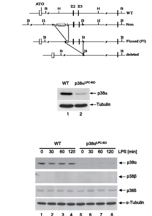
As described before, p38α-/- mice are embryonic lethal. To investigate the role of p38α in the liver a conditional mouse model had to be utilized which leads to the deletion of p38α after embryonic development. Therefore, mice with a liver parenchymal cell specific deletion of p38α were generated using the Cre/LoxP system. The Cre/LoxP system is derived from the bacteriophage P1 and allows site specific recombination of DNA strands. The system consists of an enzyme (Cre) that catalyses recombination of DNA between specific sites (LoxP sites). A loxP site is 34 base pairs long and consists of an asymmetric 8 base pair sequence that is flanked by 13 base pair palindromic sequences on both sides. These palindromic sequences act as binding sites for the Cre recombinase. The recombination then occurs in the asymmetric sequence. As a result a reciprocal recombination event may take place between two LoxP sites if cre is expressed in a cell. The asymmetric sequence gives directionality to the LoxP site. Important for the outcome of the recombination is the spatial orientation of two LoxP sites one against the other. If two sites are placed on the same arm of the chromosome and have the same direction, the recombination will result in a deletion of the DNA region that was located between the LoxP sites. This effect is commonly used in genetics in order to introduce mutations or delete gene regions in a controlled and inducible manner. The loxP sites are placed at the desired location of the genome by homologous recombination. The presence of the mutation or deletion is regulated via transducing Cre into cells or via expressing Cre under the control of an inducible or tissue specific promoter.
In order to delete p38α specifically in hepatocytes p38αFL mice were crossed with mice carrying the Alfp cre transgene (Reichardt, Kellendonk et al. 1999). The p38αFL mice carry loxP sites flanking exons 2 and 3, which include the ATP-binding site of the kinase domain.
1.6 Objectives
In terms of its cellular composition the liver is a heterogeneous tissue and cytokines are key messengers orchestrating the numerous important functions to keep the body homeostasis.
TNF is an important molecule in this complex interplay of different cell types. Therefore, a healthy balance of TNF induced signaling is crucial to maintain liver functions. Dysregulated cytokine signaling can shift biological responses in an unfavorable direction. This can lead to serious impairment in liver functions having an impact on whole body metabolism or lead to liver failure. Understanding the functional complexity of the downstream pathways activated by TNF signaling could potentially identify novel targets for the treatment of many acute and chronic liver diseases.
Important progress has been made in order to gain some understanding of the functions that the signaling cascades activated by TNFR I signaling process. Many functions of the NF-κB signaling pathway and JNK MAPK cascade in the liver have been investigated in detail. The functions of p38, another MAPK activated upon TNF signaling, are less understood. This is partly due to the fact that p38α-/- mice are embryonic lethal, preventing extensive in vivo studies.
Making use of conditional gene targeting, the aim of the thesis was to study the role p38α in the liver during acute and chronic inflammatory states, in vivo. Due to the outstanding role of TNF in tissue homeostasis, two models that induce significantly elevated levels of circulating TNF were chosen to study the role of p38α in different inflammatory conditions in the liver.
To study the role of the MAPK p38 during the development of acute liver failure, a model of bacterial Lipopolyscaacride (LPS) was applied. LPS is one of the most potent TNF inducers but usually does not cause hepatocyte apoptosis unless it is co-administered with D- Galactasoamine (GalN) which specifically blocks transcription in the liver (Decker and Keppler 1974; Bradham, Plumpe et al. 1998). The aim of the first part of this thesis was to analyze the LPS/TNF induced apoptotic signals in mice specifically lacking p38α in liver parenchymal cells.
In order to study the role in chronic low grade inflammatory states, a model of high fat diet induced obesity and insulin resistance was utilized. Type 2 diabetes is a complex multifactorial disease triggered by the combination of β- cell dysfunction and insulin resistance of target tissues. Insulin resistance results from impaired activation of the enzymatic cascade of phosphorylation events in the insulin signaling pathway. Although a huge variety of processes involved in the development of insulin resistance has already been
35 described, the underlying exact molecular mechanisms are still to be investigated. Therefore the objective of the second part of this thesis was to analyze the role of liver p38α in chronic sub-acute inflammatory states caused by high fat diet feeding and its influence on glucose homeostasis and insulin sensitivity.
2 Material and Methods 2.1 Material
Solutions:
Hepatocyte culture medium: High Glucose DEMEM, 5% FCS, Pen/Strep.
Ketamin-Rompun solution: 5ml Ketamin (10mg/ml), 230µl 2% Rompun.
Loading Dye DNA: 15% Ficoll 400 in ddH2O, heat to 50
°
C then add Organe G ad libidum, autoclave.Loading Dye Protein 5x (10ml): 1,1ml Tris HCL pH 6,8, 2ml Glycerol, 3,8ml 10% SDS, 800µl ß-mercaptoethanol.
NP-40 Lysis Buffer (500ml): 452,5ml ddH2O, 25ml 1M Tris HCL pH 7,5, 15ml 5M NaCL, 2,5ml NP-40, 1,05g NaF. Before use add 1 tablet Protease and Phospahtase inhibitor to 10 ml of buffer.
Running buffer (1l): 30,25g Tris Base, 144,2g Glycine, 10g SDS. pH was not adjusted.
Starving medium: High Glucose DEMEM, Pen/Strep
TAE Buffer 25x (10l): 1210g Tris, 0.5l EDTA pH 8,0 500 m, 285,5mL Acetic acid
Tail Lysis Buffer (1l): 100ml Tris HCL pH 8,5, 20ml 10% SDS, 11,68 NaCL, 1,86g EDTA.
TBS (10x) 1l: 24,2g Tris Base, 80g NaCl. Adjust pH to 7,6 with 37% HCL
TBS/T: add 0,1% of Tween to TBS
Transfer Buffer (1l): 3g Tris Base, 14,3g Glycine, 20% Methanol. Do not adjust pH.
Oil Red O Solutin: 1,5g Oil Red O dissolved in 300ml Isopropanol over night, then 200ml of ddH2O are added.
37 RIPA buffer high salt (50ml): 1ml 1M HEPES, 3,5ml 5M NaCL, 10ml Glycerol, 50µl 1M MgCL2, 50µl 500mM EDTA, 50µl 0,1M EGTA, add ddH2O to 50ml. Before lysis add 1 tablet Protease and Phospahtase inhibitor to 10 ml of buffer, 1% NP-40, 0,1mM NaF.
Material
Acrylamide: BioRad 30% Acrylamide/Bis solution Ref: 161-0158
Agarose: BioZym Ref: 840004
Amonium Persulfate: Sigma, Ref:
Bornyle: Adsyte Pro, 22 GA 0,9 x 25mm, BD, Ref: 388714
Chloroform: Applichem
Collagenase: Collagenase Type II, Worthington, Ref: 4176 Lot: 47D9570
Culture dish (Collagen coated) 10cm:
DMEM: Gibco: 4,5g/l glucose, Ref: 41965
DNA Ladder: PeqLab, Ref: 25-2000
DNase: Qiagen RNase free DNase set Ref: 79254
EBSS -CA, -MG: Gibco, Ref: 14155
EBSS +CA, +MG: Gibco, Ref: 24010
ECL solution: GE Healthcare, Ref: PRN2209
ECL plus: GE Healthcare, Ref: PRN2132
EGTA: AppliChem
Ficoll 400: AppliChem A4969,0100, 100g
Films: Amersham Hyperfilm ECL, Ref: 28906837