Cullmann and Dick: A chromogenic assay for a2-macroglobulin 287
J. Clin. Clicm. Clin. Biochem.
Vol. 19, 1981, pp. 287-290
A Chromogenic Assay for Evaluation of a2-Macroglobulin Level in Serum and Cerebrospinal Fluid
By W. Cullmann1) and W. Dick
Zentrallabor des Lukaskrankenhauses Neuß (Received August 8/December 17, 1980)
Summary: A photometric assay using the chromogenic substrate carbobenzoxy-valyl-glycyl-arginine-p-nitroanilide acetate (CHROMOZYM TRY®) for the determination of a2-macroglobulin which binds trypsin as a trypsin-protein complex, thus preserving its amidolytic activity, even in the presence of aprotinin, has been studied in serum as well as in cerebrospinal fluid (CSF). Normal range in sera was found to be = 10.2 ±3.3 kU/1 (male), = 10.9 ± 3.2 kU/1 (female), and = 11.9 ± 5.0 U/l in CSF. Within-run precision in sera was ascertained to be C.V. = 2.06%, and in CSF C.V. = 2.15%, respectively. There was a good correlation with immunochemical methods: r = 0.974 in 60 sera (p < 0.001) and r = 0.976 in 16 pathological samples of CSF (p < 0.001).
Bestimmung von ot2'Makroglobulin im Serum und Liquor mit einem chromogenen Substrat
Zusammenfassung: Es wurde ein photometrisches Verfahren mit dem chromogenen Substrat Carbobenzoxy-valyl- glycyl-arginin-p-nitranilidacetat (CHROMOZYM TRY®) zur Bestimmung des Gehalts an a2-Makroglobulin, das Trypsin als Trypsin-Protein-Komplex bindet, der auch in Gegenwart von Aprotinin amidolytische Aktivität behält, sowohl im Serum als auch im Liquor untersucht. Für den Normalbereich wurden folgende Werte gefunden: im Serum
= 10,2 ± 3,3 kU/1 (männlich) und = 10,9 ± 3,2 kU/1 (weiblich), sowie im Liquor = 11,9 ± 5,0 U/l. Für die Präzi- sion in der Serie wurde im Serum ein VK von 2,06% und im Liquor von 2,15% erhalten. Weiterhin bestand eine aus- gezeichnete Übereinstimmung zu immunchemischen Methoden: r = 0,974 in 60 Seren (p < 0,001) sowie r = 0,976 in
16 pathologischen Liquorproben (p < 0,001).
Introduction
a2-Macroglobulin has proved to be a sensitive parameter in various neurological diseases (2,3). It is well estab- lished that the serum-CSF gradients of albumin and a2-macroglobulin cover a wide range of serum-CSF con- centration ratios (3,4, 5). The analysis of serum cannot be regarded as sufficient by itself, as the serum-CSF gradient appears unpredictable (6). Immunochemical procedures often require a concentration step prior to the analysis of CSF. The recent development of a chromogenic substrate, highly sensitive to trypsin, allows the determination of a2-macroglobulin as a trypsin- protein-complex which retains amidolytic activity, even in the presence of aprotinin (7). In this study the optimal conditions for a chromogenic assay will be evaluated and compared to immunodiffusion, the most widespread immunochemical method for determination of a2-macro- globulin.
1) Reported (1) in part at the Joint Congress of the Scandinavian and German Societies of Clinical Chemistry, Hamburg, Octo- ber 1980.
Materials and Methods
Samples
60 sera and 60 samples of CSF, representing unselected clinical material sent to our laboratory, were investigated. For deter- mination of within-run precision and kinetic parameters a pool serum of 20 healthy donors and CSF from 20 donors were taken at random. All samples were immediately frozen at - 20 °C and analyzed within 4 weeks. There was no decrease in the a2 -macro- globulin level during storage for 3 months at - 20 °C. Prior to the photometric assay, all sera were diluted 1 + 25 with 9 g/l sodium chloride.
Reagents and procedures
Protein measurements in sera as well as in CSF were carried out using standard procedures (8, 9). Cell count was estimated from a microscopic count. Immunochemical levels of c^-macroglobulin were calculated from immunodiffusion (M-Partigen and LC-Parti- gen, Behring Institute, Marburg FRG) (10).
For the chromogenic assay, all reagents (CHROMOZYM TRY®, trypsin (33 U/mg protein with benzoyl-arginine-p-nitroanilide as substrate), and aprotinin (140 U/mg protein)) of analytical grade were kindly supplied by Boehringer Mannheim (FRG). Plasmin (300 CU/l, 15 CU/mg protein) was a commercial preparation from KABI (Stockholm, Sweden) and thrombin (4000 NIH U/l) from Boehringer Mannheim (FRG). The buffer consisted of 0.05 mol/1 triethanolamine and of 0.15 mol/1 sodium chloride, adjusted to pH 8.O.
The following CSF values were considered normal: protein level below 500 mg/1, cell count below 3 cells/mm3, no red cells on microscopic examination.
0340-076X/81 /OO19-0287 $02.00
© by Walter de Gruyter & Co. · Berlin · New York
288 Cullmann and Dick: A chromogenic assay for a2-macroglobulin
Results
Methodological studies
A linear slope function representing the splitting of the chromogenic substrate by trypsin was obtained over a wide range (fig. 1).
The time for the trypsin to react with serum protein before addition of aprotinin was varied between 5 and 60 seconds. It is evident that trypsin reacts rapidly: half- rate was determined to be 3.47 seconds (fig. 2).
Trypsin is known to bind to other antiproteases like arantitrypsm, therefore the amount of trypsin, required for this assay, was determined (fig. 3).
A 5 minute pre-incubation of the diluted samples with other proteases (plasmin (final concentration 240 CU/1) or thrombin (6400 NIH U/l)) showed no influence on the assay.
However, attention has to be drawn to the necessity to add an exact amount of aprotinin, as higher amounts of aprotinin (addition of the 10-fold amount as used in our assay) cause a non-competitive inhibition of the amidoly- tic activity of the trypsin-a2-macroglobulin complex (unpublished observations).
All this considered, the photometric assay can be sum- marized as follows:
0.50 ml buffer with trypsin (85 U/l) 0.01 ml sample dilution (1 + 25)
l min incubation
0.01ml aprotinin (14 KU/1) l min incubation
0.05 ml CHROMOZYM TRY® (5 mmol/1) increase in absorbance at 405 nm; 37 °C.
20 t is] 60
Fig. 2. Variation of the incubation time of trypsin with a pre- diluted pool serum (standard conditions).
100Ã
Trypsin [U/l] 150 200 Fig. 3. Evaluation of the amount of trypsin required for the assay
(a pool serum was used, standard conditions).
Trypsin (U/l]6 8 10 12
Fig. 1. Splitting of the chromogenic substrate CHROMOZYM TRY® by free trypsin (standard conditions, determination of trypsin activity with benzoyl-arginine-p-nitroanilide as substrate).
With the assay, a linear range for the splitting of the chromogenic substrate was observed up to ÄÁ/min = 0.120.
For analysis of CSF, the assay was slightly modified:
only 0.4 ml of buffer was taken and 0.1 ml of CSF. With Tris buffer (tris hydroxymethyl-aminomethane), a pre- cipitation of proteins in CSF was observed. As the CSF samples were taken without dilution, the unspecific splitting of the chromogenic substrate was evaluated in the CSF samples: in the normal range no splitting could be detected, and in the pathological range unspecific splitting did not exceed 0.5% of total splitting.
Normal range
With the chromogenic assay, the normal range of a2- macroglobulin in sera was found to be 10.2 ±3.3 kU/1 (male, n = 30, total protein 74 ± 6,5 g/1) and 10.9 ± 3.2 kU/1 (female, n = 30, total protein 71 ± 7.6 g/1). In CSF, the normal range was found to be 11.9 ±, 5.0 U/l;
no sex differences were found.
J. Gun. Chem. CUn. Biochem. / Vol. 19,1981 / No. 5
Cullmann and Dick: A chro m ge nie assay for a2-macroglobuljn 289
Within-run precision and detection limit of the assay in CSF
Within-run precision was ÷ = 16.6 ± 0.342 kU/1 (C. V. = 2.04%) for pool serum and ÷ = 24.2 ± 0.52 U/l (C. V. = 2.15%) for a CSF pool. Detection limit in CSF was 1.04 U/l.
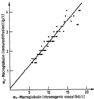
Correlation with immunodiffusion
There was a good correlation of the chromogenic assay with immunodiffusion in sera as well as in CSF (r = 0.974 for 60 sera, ñ < 0.001, and r = 0.976 for
16 samples of CSF, p < 0.001) (fig. 4 and 5). Linear regression analysis showed the equation y = 0.236 ÷ + 0.093 for sera (fig. 4), and y = 0.28 ÷ + 4.9 for CSF (fig. 5).
In table 1 the results obtained from analysis of CSF are summarized. It is evident that immunodiffusion can only be used when markedly high levels are expected.
"300
I 200 åå
¼ 100
"ó»ï
_L I é
100 200 500 1000 a2-Macroglobulin (chromogenic assay)(U/l ]
Fig. 5. Comparison of the chromogenic assay of a2-macroglobulin with immunodiffusion in 16 pathological samples of CSF (y = 0.28 ÷ + 4.9, r = 0.976, p < 0.001).
I z
.0Βo>
§1ó
Tab. 1. Determination of ^-macroglobulin level in CSF with the chromogenic assay and by immunodiffusion.
J_ _L
5 10 15 20
a2-Macroglobulin (chromogenic assay) [kU/U
Fig. 4. Comparison of the chromogenic assay of a2-macroglobulin with immunodiffusion in 60 sera at random (y = 0.236 ÷ + 0.093, r = 0.974, ñ < 0.001).
Discussion
Our results strongly suggest that the chromogenic assay allows a rapid and reliable determination of ai-macro- globulin in sera as well as in CSF. Sensitivity of the chromogenic assay in CSF is 30 times better than that of immunodiffusion. The normal range obtained with the chromogenic assay confirms previous reports using immunochemical procedures (2, 3). Furthermore, ex- cluding an impaired barrier function, the serum-CSF
Range of a2-macroglobulin
Normal (n = 26)
Moderately high levels
o2 -Macro- «2 -Macro- globulin globulin (chromo- (immuno- genic assay) diffusion) (U/I) (mg/1)
11.9 ±5.0 -*
22.8 ± 14.2 -*
Total Protein
(mg/1) 371 ±113 844 ± 346 (n-18)
Markedly high levels 155 ±226 48 ± 66 1442 ±661
* not detectable
concentration ratio can be calculated at 800, which agrees with the findings of Felgenhauer (3).
It is known that chromogenic substrates are also split by the majority of proteases. Concerning this assay, dilution of the sample on the one hand and addition of aprotinin prior to the onset of the reaction on the other hand will prevent an unspecific splitting. It could be demonstrated that the proteolytic activity in CSF is negligible. More- over, there was no influence by other proteases (plasmin and thrombin) on the assay.
All these considerations make it apparent that an amido- lytic assay of a2-macroglobulin in serum and in CSF can with advantage be used for the evaluation of the serum- CSF barrier gradient.
J. Clin. Chexn. Clin, Biochem. / Vol. 19,1981 / No. 5
290 Cullmann and Dick: A chromogenic assay for
References
1. Cullmann, W. & Dick, W. (1980), J. Clin. Chem. Clin. Bio- 6. Weisner, B. & Bernhardt, W. (1978), Fortschr. Med. 96, ehem. 18, 671. 1865-1870.
2. Felgenhauer, K. (1974), Klin. Wochenschr. 52,1158-1164. 7. Ganrot, P. O. (1966), Clin. Chim. Acta 14, 493-501.
3. Felgenhauer, K., Schliep, G. & Rapic, N. (1976), J. Neurol. 8. Weichselbaum, B. (1946), Am. J. Clin. Pathol. 10, 10-21.
Sei. 30, 113-128. 9. Lowry, 0. H., Rpsebrough, N. J., Farr, A. L. & Randall, R. J.
4. Schliep, G. & Felgenhauer, K. (1974), J. Neurol. 207, 171- (1951), J. Biol. Chem. 193, 265-275.
181. 10. Mancini, G., Carbonara, A.. Q. & Heremanns, J. F. (1965), 5. Schliep, G. & Felgenhauer, K. (1978), J. Neurol. 218, 77-96. Immunochemistry 2, 235-254.
Dr. W. Cullmann
Lehrstuhi für Med. Mikrobiologie und Immunologie der Ruhruniversität D-4630 Bochum-Querenburg
J. Clin. Chem. Clin.. Biochem. / Vol. 19,1981 / No. 5