J. Clin. Chem. Clin. Biochem.
Vol. 28, 1990, pp. 407-411
© 1990 Walter de Gruyter & Co.
Berlin · New York
A Colorimetric Assay
for the Determination of Acid Nucleoside Triphosphatase Activity
By M. Kirchgesser and N. Dahlmann
Institut für Klinische Biochemie der Universität Bonn (Received February 26, 1990)
Summary: A photometric method for the determination of the acid nucleoside triphosphatase (EC 3.6.1.—)
is described, in which inorganic phosphate is liberated from ATP or other nucleoside triphosphates. Colori- metric determination of liberated phosphate is based on the formation of a green complex of phosphomolyb- date and malachite green hydrochloride. Optimal test conditions were evaluated as well as the sample preparation. The enzyme activities measured in 100 normal human sera are in the range of 0.5 to 9.0 U/l with an average of 4.0 U/l for men and 3.8 U/l for women.
Introduction
Recently we reported preliminary studies on a novel enzyme with an unusual pH optimum of 3.0 (1). This enzyme, acid nucleoside triphosphatase (EC 3.6.1. —), has been partly purified and characterised. The en- zyme hydrolyses all nucleoside triphosphates only to their corresponding diphosphates and inorganic phos- phate (2). Because of its putative lysosomal origin we have chosen myocardial infarction as a model of wound repair to investigate some aspects of its bio- logical function. Enzymatic activity was maximally elevated six weeks after myocardial infarction (3), indicating that the enzyme did not originate from the myocardial tissue itself but probably from activated macrophages, which infiltrate the scar at that time (4).
The activity of the enzyme has so far only been measured by using radiolabeled nucleoside triphos- phates as a substrate (1, 2), a method which is specific, but expensive and inconvenient for routine laboratory use. The assay presented here is based on a colori- metric determination of inorganic phosphate released by the acid nucleoside triphosphatase, using a sensi- tive method developed by Lanzetta et al. (5) and adapted to the actual assay conditions. In the present assay, phosphate forms a complex with ammonium
molybdate and malachite green hydrochloride. The absorbance of the resulting green complex can be estimated at 640 —660 nm.
Materials and Methods Chemicals
All tritium-labelled nucleotides were obtained from Amersham- Buchler, unlabelled nucleotides from Boehringer-Mannheim.
Malachite Green and dithiothreitol were purchased from Sigma Chemical Co., Rheomacrodex 10% was from Schiwa GmbH, Glandorf. All other chemicals were supplied by E. Merck, Darmstadt, in the highest quality grade commercially available.
Solutions
Unless otherwise indicated distilled water was used in the prep- aration of all solutions.
Dialysis buffer: 150 mmol/1 NaCl, 10 mmol/1 Tris-HCl pH 7.5, Rheomacrodex 10%, volume fraction 0.25.
Reaction buffer: The following three stock solutions, (1) 100 mmol/1 citric acid - NaOH, pH 3.0, (11) 40 mmol/1 EDTA, disodium salt,
(111) 40 mmol/1 dithiotreitol, were mixed in a ratio of 1 + 1 + 1.
Colour reagent: Mix one part of 42 g/1 ammonium molybdate in 4mol/l HC1 with 3 parts of 0.45 g/1 Malachite Green, stir the mixture for at least 20 min, then filter through a Whatman No. 5 filter paper. The ready-to-use reagent should not be stored for more than two weeks.
Stabilizer: 340 g/1 sodium citrate.
Stop reagent: 250 g/1 trichloroacetic acid.
Pi standards: Na2HPO4 in the range 0-250 μιηοΐ/ΐ (0-50 μηιοΐ/l final).
Substrate solution: Nucleoside triphosphate, 1.25 mmol/1 (250 μπιοΐ/l final). Store at -20 °C.
Sample preparation: 250 — 500 μΐ of serum are intensively dia- lysed for 12 — 24 hours in micro-collodion-bags (Sartorius GmbH, G ttingen) at 4 °C against the dialysis buffer.
Nucleoside t r i p h o s p h a t a s e test
Samples and reagents are pipetted according to the scheme in table 1. The total volume of the assay is 250 μΐ. After incubation for 30 min at 30 °C, stop reagent (50 μΐ) is added, the mixture vortexed, then centrifuged for 3 min (8000 min"1) to pellet the protein. Supernatant (200 μΐ) is transferred to a new vial, followed by 1 ml of colour reagent. After l min of colour development, 100 μΐ of stabilizer are added and mixed thor- oughly. Absorbance is determined at 650 nm, using water as the blank. The colour is stable for at least 4 hours.
The substrate blank and the serum blank are both subtracted from the sample values and the resulting value is referred to the PJ standard curve. One international unit (U) represents the production of 1 μηιοί Pj per minute.
Radioisotopic nucleoside triphosphatase assay This assay, developed by Dahlmann et al. (1), uses radiolabeled nucleoside triphosphate as substrate. After incubation, nucleo- side mono-, di- and triphosphates are separated by thin-layer chromatography, and radioactivity is measured by liquid scin- tillation counting.
Results
Dialysis
Because of the high Pi concentration in human serum (1—2 mmol/1), this metabolite must be removed by dialysis to about 1/1000 of the physiological concen- tration. In order to minimise volume changes of the samples during dialysis, an osmoticum should be added to the dialysis buffer. We chose a dextran solution in a final concentration of 2.5%. This dialysis buffer has the average osmolarity of human serum.
Serum blanks after extensive dialysis showed an ab- sorbance (χ ± s) of 0.01 ± 0.002 making spot checks of 2 or 3 serum blanks sufficient for an entire series.
Standard curves
Since proteins in serum are capable of binding ions, we investigated the effect of serum protein on the recovery of inorganic phosphate. Pj standards in a final concentration of 0 — 50 μιηοΐ/ΐ were estimated in assay mixtures containing either buffer, serum, sub- strate, or combinations of these; there were no sig- nificant differences in Pi recovery between them.
Therefore, serum seems to have no matrix effect on Pi recovery, and the Pj standard curve can be deter- mined in reaction buffer without the addition of pro- tein. An absorbance of 1.0 corresponds to a Pi con- centration of 60 — 80 μηιοΐ/ΐ.
Substrate stability
To distinguish between enzymatic turnover and un- specific hydrolysis, the stability of different substrates was tested at various temperatures and pH values with a substrate concentration of 250 μηιοΐ/ΐ. For these experiments background values of the substrates were not subtracted. A non-enzymatic hydrolysis value of 5% corresponds to an absorbance of 0.15.
The stability of 2'-deoxy thymine triphosphate (dTTP) was monitored at four different temperatures at pH 3.0. The maximal hydrolysis rate was found to be 0.5%, measured over a period of 30 minutes at 37 °C, whereas at 0 °C no hydrolysis was observed (fig. 1).
An incubation temperature of 30 °C is a compromise between the optimal temperature of 43 °C for the enzymic activity (2) and a temperature that gives a low substrate blank value.
Since the assay is conducted at pH 3.0 and stopped by the addition of trichloroacetic acid, the effect of pH on the substrate stability was assessed. The non- enzymatic hydrolysis rate of 2'-deoxyadenosine tri- phosphate (dATP) and dTTP was measured at pH 1.0 and pH 3.0 for 60 min at 30 °C (fig. 2). The results indicate that, at pH 3.0, both dTTP and dATP were stable with a rate of hydrolysis of less than 0.5%
within one hour. However, the hydrolysis rate in- creased significantly at pH 1.0 with a rate of 2% for dATP and 3% for dTTP. Samples should therefore be kept on ice after the addition of stop reagent, and absorbance determined within 30 minutes.
Tab. 1. Pipetting scheme for nucleoside triphosphatase test (volumes in μΐ).
Reaction buffer Water
Serum
Substrate solution Pi standards
Water blank 150100
——
—
Substrate blank Serum blanks Samples (serum) Standard curve 15050
_ 50—
15050 50—
—
150— 5050
—
15050
—— 50
3-
CO
>%
Νc
<p cο
2 -
15 30
t[min]
60
CΟ
6 -
l 3
N
1-
15 30
t[min] 45 60
Fig. 1. Non-enzymatic hydrolysis of dTTP was measured for Fig. 2. Non-enzymatic hydrolysis of dATP (·, o) and dTTP 60 min at pH 3.0 and four different temperatures: 0 °C (Α, Δ) was measured at pH 3.0 (·, A) and pH 1.0 (o, (·), 20 °C (o), 30 °C (A), and 37 °C (Δ). Δ) for 60 min at 30 °C.
Tab. 2. Non-enzymatic hydrolysis of different substrates (250 μιηοΐ/ΐ) under standard assay conditions (30 min at 30 °C and pH 3.0)
Substrate ATP dATP dTTP dCTP dGTP dUTP
Hydrolysis (%) 0.15 0.44 2.64 2.90 2.42 2.82
To compare the stability of different substrates, non- enzymatic hydrolysis was measured for 30 min at 30 °C and pH 3.0 at a final concentration of 250 μηιοΐ/ΐ of various nucleotides. Results are depicted in table 2 and indicate that ATP is about 3 times more
0.8 ·
0.6-
< gO.4
0.2
30
60
t[min]
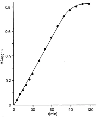
90 120Fig. 3. Linearity of the assay at different incubation times. A pool of 20 sera was incubated with 250 μιηοΐ/ΐ dATP at pH 3.0 and 30 °C.
stable than dATP and 15 times more stable than all other deoxynucleotides. Therefore, we consider ATP to be the most suitable substrate. Although nucleo- tides can be stored at — 20 °C, repeated freezing and thawing increases the rate of non-enzymatic hydrol- ysis, making the inclusion of a substrate blank una- voidable.
Substrate concentration
As the substrate concentration is increased towards approximate saturation of the enzyme (V
max), the sub- strate blank increases faster than the sample values.
It is therefore not possible to determine acid nucleo- side triphosphatase activity at an optimal substrate concentration. At ATP concentrations up to 300 μηιοΐ/ΐ, the substrate blank does not exceed an ab- sorbance of 0.03. A concentration of 250 μηιοΐ/l is recommended as an optimum. This is about 6 times more than the K
mvalue of ATP, sustaining a rate that is 85% ofV
max.
Time course of the reaction
As the amount of substrate is the limiting factor of
linearity, the time course was monitored at the sub-
strate concentration of 250 μιηοΐ/ΐ ATP. Under these
conditions the change of absorbance remained linear
for 90 minutes (fig. 3).
Linearity and sensitivity
Under standard assay conditions the colorimetric as- say was linear to an absorbance of at least 0.8, and sensitive to about 0.3 μηιοΐ/ΐ Pj. Sensitivity can be increased by changing the ratio of assay supernatant to colour reagent. Maximum sensitivity is achieved when 500 μΐ of the supernatant are mixed with 1 ml of colour reagent, but 200 μΐ of supernatant are usu- ally sufficient.
Influence of anticoagulants
Possible effects of anticoagulants on acid nucleoside triphosphatase activity were assessed under standard assay conditions. No significant differences were found for plasma containing heparin (25000 U/l), EDTA (1 g/1), citrate (7.6 g/1), fluoride (2 g/1), or oxalate -h fluoride (3.4 + 3.4 g/1) when compared with the untreated serum.
2.5-
2.0-
£ f 1.5 H
si 8-g
« I 1.0 H
11
0.5-
0.5 1.0 1.5 2.0 Acid nucleoside triphosphatase (radioisotopic assay) [U/l]
2.5
Fig. 4. Comparison of the catalytic activity of acid nucleoside triphosphatase as determined by the colorimetric and by the radioisotopic assay.
Effect of storage
Aliquots of a pool serum were stored at —20, + 4 and + 20 °C. Acid nucleoside triphosphatase activity was determined over a period of 10 days. The data confirm that serum can be stored without loss of activity at — 20 °C, + 4 °C, or even room temperature for at least 8 days (data not shown).
Reference values of nucleoside triphosphc tase
Nucleoside triphosphatase activity was determined in 100 sera of healthy blood donors (64 men, 36 women) under standard assay conditions with 250 μηιοΐ/ΐ ATP as substrate. The mean + standard deviation was 4.0
±1.6 U/l for men, and 3.8 + 1.9 U/l for women.
Precision
Precision data are listed in table 3.
Tab. 3. Precision data of the assay (n = 20).
Within-series precision Mean
(U/l) 2.531.28
SD (U/l) 0.033 0.060
CV 2.62.4
Day-to-day precision Mean
(U/l) 0.642.10
SD (U/l) 0.025 0.097
CV 4.63.8
Correlation between photometric and ra- dioisotopic test
To evaluate the specificity of the present assay, acid nucleoside triphosphatase activity was determined in 50 sera of healthy blood donors, using the radioiso- topic test as reference. Substrate concentration was 60 μιηοΐ/ΐ dTTP according to 1. c. (1). The correlation found between the two methods was excellent with a correlation coefficient of 0.97 (fig. 4).
Discussion
In order to expand our knowledge of the clinical
importance of acid nucleoside triphosphatase, a sim-
ple and rapid assay for a large number of patients'
samples is required. Before the development of this
photometric assay, the enzyme activity had only been
measured using a radioisotopic assay involving thin-
layer chromatography, which is time-consuming, ex-
pensive, and permitting only a few samples to be
analysed simultaneously. However, it is easier to de-
termine the enzymic production of inorganic phos-
phate. A number of methods (6, 7) as well as com-
mercial kits were found unsuitable because of their
low sensitivity, whereas the assay methods of Lanzetta
(5) and Bencini (8, 9) are sensitive enough for the
estimation of a Pi concentration in the micromolar
range. The method developed by Bencini, however,
uses short wavelengths, and therefore the presence of
proteins interferes with the assay. Other assay meth-
ods for determining inorganic phosphate are unsuit-
able if labile phosphate esters such as nucleoside tri-
phosphates are present in high concentrations (10).
The present photometric method is simple, economi- cal and a large number of samples can be analysed within a few hours. Its specificity has been proved in
comparison with the radioisotopic assay, which meas- ures the specific reaction product, nucleoside diphos- phate. Precision is excellent, with extensive linearity.
References
1. Dahlmann, N. & Ueckermann, C. (1982) Properties of Four Different Deoxy-Thymidine-5'-Triphosphate Hydrolyzing Enzymes in Human Serum. Biochem. Int. 5, 185 — 192.
2. Dahlmann, N. & Kirchgesser, M. (1990) Acid Nucleoside Triphosphatase: Partial Purification and Characterisation of a New Enzyme from Human Serum. Biochem. Int. 20,
317-327.
3. Dahlmann, N., Hobel, E. & Steinhagen-Thiessen, E. (1985) A New Acid Nucleoside Triphosphatase as a Tool in Mon- itoring the Follow-up of Chronic Inflammation. European Society for Clinical Investigation, Toulouse p. 46.
4. Becker, A. E., Anderson, R. H. & Braunwald, E. (1985) Cardiac Pathology, 1st edn., pp. 356 — 361, Thieme Verlag, Stuttgart.
5. Lanzetta, P. A., Alvarez, L. J., Reinach, P. S. & Candia, O. A. (1979) An Improved Assay for Nanomole Amounts of Inorganic Phosphate. Anal. Biochem. 100, 95 — 97.
6. Berti, G., Fossati, P., Tarenghi, G. & Musitelli, C. (1988) Enzymatic Colorimetric Method for the Determination of Inorganic Phosphorus in Serum and Urine. J. Clin. Chem.
Clin. Biochem. 26, 399-404.
7. Van Zanten, A. P. & Weber, J. A. (1987) Direct Kinetic Method for the Determination of Phosphate. J. Clin. Chem.
Clin. Biochem. 25, 515-517.
8. Bencini, D. A., Shanley, M. S., Wild, J. R. & O'Donovan, G. A. (1983) New Assay for Enzymatic Phosphate Release:
Application to Aspartate Transcarbamylase and Other En- zymes. Anal. Biochem. 132, 259 — 264.
9. Bencini, D. A., Wild, J. R. & Donovan, G. A. (1983) Assay of Inorganic Phosphate, Total Phosphate and Phospha- tases. Anal. Biochem. 132, 254-258.
10. Ames, B. N. (1966) Assay of Inorganic Phosphate, Total Phosphate, and Phosphatases. Meth. Enzymol. VIII, pp.
115-118.
Prof. Dr. N. Dahlmann
Institut für Klinische Biochemie Sigmund-Freud-Straße 25 D-5300 Bonn l