J. Clin. Chem. Clin. Biochem.
Vol. 18,1980, pp. 17-21
A Kinetic Test for the Assay of the C1 Esterase-inhibitor
1)
By F. P. Schena, C Manno,
2nd Clinica Medica, University of Bari, R. D'Agostino. G. Bruno, F. Cramarossa,
Chimica Generale e Inorganica, University of Bari and
L· Bonomo,
2nd Clinica Medica, University of Bari, Bari, Italy (Received February 2/July 9,1979)
Summary: The most satisfactory diagnostic procedure for hereditary angioneurotic oedema is the demonstration of low serum levels of Cl esterase-inhibitor. A modified method for the assay of this protein is described. It is based on the kinetic measurement of the Cl esterase-inhibitor when it inhibits the hydrolysis of N-acetyl-L-tyrosine-ethyl ester by Cl esterase. The relative Cl esterase-inhibitor concentration is based on the initial hydrolytic velocity, which can be evaluated from the pH change in a short time and within a small range. High reproducibility, cheap instrumentation and short time of analysis are some of the favorable aspects of this method in comparison with the 'end point titri- metric' method. Furthermore, this paper describes the mechanism of inhibition of Cl esterase by Cl esterase-inhibitor.
The results are indicative of a non-competitive mechanism. The value of the Michaelis-Menten constant, Kmy is 0.017 ± 0.001 mol/1 at 37 °C, in the optimum pH range 7.2—7.4. An estimate of K\ in arbitrary units is also given.
Kinetischer Test für die Bestimmung des Cl-Esterase-Inhibitors
Zusammenfassung: Das zufriedenstellendste Verfahren zur Diagnostik des hereditären angioneurotischen Ödems ist der Nachweis der erniedrigten Konzentration des Cl-Esterase-Inhibitors im Serum. Eine modifizierte Methode zur Bestimmung dieses Proteins wurde erarbeitet. Sie beruht auf der kinetischen Messung des Cl-Esterase-Inhibitors, der die von Cl-Esterase katalysierte Hydrolyse des Substrats N-Acetyl-i-tyrosin-ethylester hemmt. Die relative Konzen- tration des Cl-Esterase-Inhibitors kann aus der Anfangsgeschwindigkeit der Hydrolyse, die in kurzer Zeit und inner- halb eines geringen Bereichs aus der pH-Zeitkurve berechnet wird, ermittelt werden. Hohe Reproduzierbarkeit, billige Ausrüstung und kurze Analysendauer sind einige wichtige Aspekte dieser Methode im Vergleich zur Endpunkt- Titrationsmethode. Ferner wird der Mechanismus der Hemmung der Cl-Esterase durch C l-Esterase-inhibitor beschrie- ben. Die Ergebnisse weisen auf einen nichtkompetitiven Mechanismus hin. Die Michaelis-Menten-Konstante Km be- trägt 0,017 ± 0,001 mol/1 bei 37 °C im optimalen pH-Bereich von 7,2-7,4. Ein Schätzwert für die Inhibitorkonstante KI in freigewählten Einheiten wird angegeben.
Introduction * levels of Cl esterase-inhibitor (1), which modulates the
„ . - . - . - activation of the complement classical path way. In its Hereditary angioneurotic oedema is a well defined clinical afesence Cl activation eds autocatalytically and syndrome characterized by a repeated occurrence of acute feacts ^ ^ substrates C4 ^ C2 The attacks of oedema in any organ, skin and mucosae of the atic assay of esterase-inhibitor is to be upper respiratory and gastrointestinal tracts. ^^ ^ ^ ^™ochenucal method, since a The most satisfactory diagnostic procedure for hereditary variant of hereditary angioneurotic oedema is known, angioneurotic oedema is the demonstration of low serum which is due to a functional deficiency of the inhibitor
. (2,3,4). The enzymatic measurement was performed by i) This study was supported in part by Grant No. 78.02281.04 of evaluating the esterolytic activity of the Cl esterase on
Consiglio Nanpnale delle Ricerche, Rome the synthetic substrate, N-acetyl-i-tyrosine-ethyl ester, 0340-076X/80/0018-0017S2.00
©by Walter de Gruyter & Co. · Berlin · New York
18 Schena, Manno, D'Agostino, Bruno, Cramarossa and Bonomo: Kinetic test for the assay of the Cl esterase-inhibitor by microformol titration, as described by Levy &
Lepow (5). Other authors (6,7) have subsequently modified this method based upon the hydrolytic property of Cl esterase on N-acetyl-Z-tyrosine- ethyl ester. Cl esterase is added to the inhibitor, until hydrolysis is complete, the reaction being followed to the end point with a pH-stat automatic titrator.
This paper describes the kinetic mechanism of the in- hibitory effect of the Cl esterase-inhibitor on the hydro-
lysis of N-acetyl-I-tyrosine-ethyl ester catalysed by Cl esterase. Furthermore, it presents a modified technique for the assay of Cl esterase-inhibitor. High reproducibility, cheap instrumentation and quick
analysis are some of the favorable aspects of this method.
Materials
The reactor is stirred magnetically and thermostated by means of a water jacket at 37 ± 0.1 °C for 15 min. The pH is measured and registered with a microelectrode and a pH-meter (E 436 Metrohm Herisau).
Enzyme', purified Cl esterase (Cordis, Miami), activated with an equal volume of saline solution at 37 °C for 15 min. The amount of enzyme was expressed in terms of its activity. One unit (U) of Cl esterase is defined as the amount which liberates 1 μιηοΐ of acid per min (IUB), under the conditions defined below. The concentration of the activated solution of enzyme is 150 mU/1.
Substrate'. N-acetyl-I-tyrosine-ethyi ester 1 mol/1 in methyl cellosolve (2-methoxy ethanol).
Inhibitor: two types of inhibitor have been used: 1. pool of normal human sera of blood donors stored at -70 °C in a refrigerator (S10) with liquid nitrogen; this preservation is very important, owing to the easy degradation of Cl esterase- inhibitor, 2. purified Cl esterase-inhibitor (Cordis, Miami).
Method
A defined amount (1100-1200 μΐ) of saline solution is added to the reactor together with normal human serum (0-40 μΐ) or purified Cl esterase-inhibitor (0-120 μΐ); N-acetyl-L-tyrosine- ethyl ester 1 mol/1 is slowly added until the final concentration (0.005-0.067 mol/1) is attained; during these operations the pH is maintained at the optimum value in the range 7.2-7.4 by adding small amounts of 0.02 mol/1 NaOH. Finally, all the activated enzyme (300-400 μΐ) is added and the pH is registered.
From the slopes of the pH/time curves the instantaneous velocity of the reaction, d[H*]/dt, can be calculated. Reaction rates are determined with the test serum and with a pool of normal sera, and inhibition is expressed as a percentage of that observed with normal serum.
Results
Hydrolysis of the synthetic substrate (S) initiated by Cl esterase (E) was studied with normal human serum containing Cl esterase-inhibitor (I), and with a purified inhibitor. The results are illustrated in figures 1 and 4 for the normal human serum and in figure 2 for the purified Cl esterase-inhibitor.
As shown in figure 1 the amount of hydrogen ions freed, at given substrate and enzyme concentrations, is a linear function of time for a wide range of added amounts of normal human serum (0-40 μΐ). Identical results were obtained using scale dilutions of purified Cl esterase-inhibitor instead of normal human serum.
The slope of the straight lines of figure 1 represents the instantaneous velocity of the hydrolytic reaction, d[H+]/dt [mol/1 · s], which is effectively independent of time for the first few minutes of the reaction. A longer lasting autocatalytic effect of Cl esterase present in the normal
Fig. 1. Hydrolysis of the N-acetyl-Z,-tyrosine-ethyl ester 0.05 mol/1 as a function of (t), at various amounts of normal human serum (μΐ), containing Cl esterase-inhibitor, by Cl esterase (left: Cl esterase=300 μΐ; right: Cl esterase=400 μΐ, corresponding to 30 and 40 mU/1 in the final solutions, respectively).
J. Clin. Chem, Clift; Biochem. / Vol. 18,1980 / No. 1
0.20-
K/UJ HO
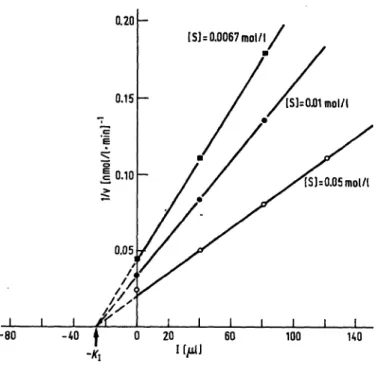
Fig. 2. Inverse of the rate of hydrolysis versus inhibitor (Ι=μ1 of ' purified Cl esterase-inhibitor) at various concentrations of substrate, for calculation of K\. Cl esterase concen- tration is 40 mU/1.
the other hand when analysing with normal human serum, we utilized a substrate concentration [S] = 0.05 mol/1, as ' suggested by Haines & Lepow (10). The relationship 1/v versus I is also valid at high substrate concentrations, as shown in figure 2; in this study a concentration of [S]
= 0.025 mol/1 gave approximately the same results.
The results are also reported in figure 3 in terms of the direct-linear plot of Eisenthal & Cornish-Bowden (11).
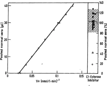
This figure shows that the inhibition mechanism is non- competitive and provides an accurate estimation of Km. The linear correlation between 1/v and the amount of purified Cl esterase-inhibitor, expressed in μΐ, also holds when the inhibitor is present in the normal human serum (fig. 4). Samples of scale quantities of normal human serum (0—40 μΐ) were tested at an enzyme concentration corresponding to 40 mU/1. Figure 4 represents the calibration plot of the inhibition percentage in respect to pooled normal sera. Twenty normal subjects were tested and they showed a range of variability of 83-126% pooled normal sera (x ± 2SD).
Cl esterase-inhibitor in the serum of a patient with hereditary angioneurotic oedema was 43%; it was 18%
in serum when Cl esterase-inhibitor was inactivated by heating for 30 min at 59 °C. This latter value confirmed the fact that Cl esterase-inhibitor is thermolabile (12).
human serum is to be expected (8,9). This effect, if present, is negligible in our experimental conditions, since no autoacceleration was detected, as clearly shown in figure 1. It should be noted that the rate of the hydro- lytic reaction is proportional to the enzyme concen- tration (30-40 mU/1), whereas the presence of the in- hibitor has a negative influence on the rate of hydrolysis.
The effect of the substrate concentration on the rate of hydrolysis was extensively studied with normal humari serum and with or without purified Cl esterase- inhibitor. The double-reciprocal plot showed a linear dependence of 1/v on 1/[S], for [S] < 0.025 mol/1.
When 0.025 < [S] < 0.05 mol/1, the velocity attains a broad plateau and then decreases. This phenomenon is probably due to the difficulty in keeping the system homogeneous at high concentrations of the substrate.
Similar results can be obtained by plotting the inverse velocity against scale amounts of purified Cl esterase- inhibitor at different concentrations of the substrate (fig. 2). Here again 1/v and Ϊ are linearly dependent and the slope is influenced by the concentration of S.
It should be emphasized at this point that an inhibitor concentration cannot be expressed in terms of inhibited units per liter due to the reversibility of the equilibria involved. The amounts of inhibitor are expressed ih terms of j d of inhibiting solutions; in fact figure 2 shows that 1/v is linearly correlated to I; consequently U is hyperbolicafly correlated to I. The equilibrium constant with the inhibitor,.K\, can be calculated in μΐ units by extrapolating the different lines onto the abscissa axis. Ori
-0.01
Fig. 3. DirecMinear plot of Eisenthal & Cornish-Bowden. Each intersection provides an estimate of V and Kj^. The shifts of the intersections at the same value of ATm indicate a non-competitive mechanism.
J. Clin, Chem. Clin. Biochem. /Vol. 18,1980 /No. 1
20 Schena, Manno, D'Agostino, Bruno, Cramarossa and Bonomo: Kinetic test for the assay of the Cl esterase-inhibitor
!20
a.§10
I J_
Δ _
0.05 0.1 1/v lnmol/1-min]"1
0.15 a-Esterase Inhibitor
:140
120
80
"I
20 0
Fig. 4. Calibration plot of percentage inhibition with respect to pooled normal sera. The amount of Cl esterase-inhibitor (μΐ of pool of normal sera) as a function of 1/v. 1/v is the inverse of velocity of N-acetyl-Z-tyrosine-ethyl-ester (0.05 mol/1) hydrolysis by Cl esterase (40 mU/1). The scale of % pooled normal sera is shown on the right, where 100 % corresponds to 30 μΐ; also the range of variability (x ± 2SD) determined from 20 blood donors (·); (Δ) patient with hereditary angioneurotic.oedema; (o) human serum heated at 59 °C for 30 min.
Discussion
The experimental results compare well with the results reported by Haines & Lepow (10), who studied the enzyme kinetics in absence of the inhibitor.
Due to various factors in our system and the way the velocity was measured, the effect of pH on the rate of hydrolysis should be taken into consideration. In the experimental pH-range (7.2-7.4) the activity of the enzyme and the inhibitor are on a plateau (6.7—8.0).
In fact, Haines & Lepow (10) have shown that when the pH is varied between 6.7 and 8.0, the Cl esterase activity towards N-acetyl-L-tyrosine-ethyl ester is optimal; Pensky et al (12) found a greater pH-range for the optimum of Cl esterase-inhibitor function. On the other hand, the results of this study showed that the pH had no significant effect on the system under the experimental conditions.
Concerning the relationship between the velocity of esterolytic reaction and the inhibitor concentration, the data obtained show that 1/v is linearly related to the μΐ of normal human serum or the μΐ of purified C1 e.sterase- inhibitor. This reaction can be expressed by the equation:
1/v = K! + K2 [I], which allows us to measure the in- hibition, expressed as % pooled normal sera. The prin- ciple on which this assay is based derives from the mechanism of inhibition. In 1975 Harpel & Cooper (13) studied the molecular basis of the interaction between
Cl esterase-inhibitor and Cl ester se. With SDS gel- aciylamide electrophoresis they saw that the enzyme- inhibitor complex was in equilibrium with the free enzyme and free inhibitor; no cleavage peptides of the Cl esterase-inhibitor were found as a consequence of the action of Cl esterase, suggesting that this was not the real substrate for the enzyme. However, these findings were not sufficient to determine the type of inhibition and the equilibrium present in this system. Although other authors (10,12) had studied the kinetic characteristics of Cl esterase and some properties of Cl esterase- inhibitor, these aspects of the problem had not been taken into consideration. The enzymatic dosage suggested by thenij which gave way to successive methods, was based on the general ability of Cl esterase-inhibitor to diminish the hydfolytic activity of the Cl esterase on N-acetyl-L-tyrosine-ethylester while the kinetic problem was not duly taken into consideration. The experiments carried out in our laboratories with purified Cl esterase- inhibitor have confirmed the results with human serum, and they have permitted the characterization of the mechanism of Cl esterase inhibition, and the determina- tion of the Michaelis-Menten constant, Km, and the con- stant of equilibrium with 19Κ\. The results are indicative of a non competitive mechanism of inhibition, as clearly shown in the Dixon plot (fig. 2) as well as in the Eisen- thal & Cornish-Bowden plots (fig. 3). With this direct- linear plot the value of Km is easily calculated as the average of each series of points. The non-competitive mechanism is confirmed by the shift of the common intersection points at a practically constant value of Km = 17 mmol/1. This value can be compared to
19 mmol/1, obtained by Haines &Lepow (10).
Based on non-competitive inhibition, a new kinetic assay is suggested in place of the previously reported method (8), which seems to be less rigorous and reproducible.
The processes involved in the method reported by Lachmann et al. (8) are in fact complicated by the presence of the phosphate buffer, whose basic component (HP04~) reacts with the H+ freed in the enzymatic reac- tion, when repeated amounts of Cl esterase are added.
This implies that the 'end point' claimed by L chmann et al. (8) is attained when enough Cl esterase is added to overcome the effect of HPO^", in addition to that of the inhibitor alone. It is worth noting that one should keep in mind the exact quantity of HPOj" present. This factor has been explicitly considered by the authors. Moreover, the repeated sampling of Cl esterase contributes to the systematic errors, and it decreases the rapidity of the method by a factor of 5. hi contrast, our method is highly reproducible as is shown in the calibration plot of figure 4, where the regression factor is 0.998. Further- more pur method is more economic, since it is not necessary to use a complicated pH-stat, but simply a pH- meter connected to a suitable recorder.
J. Clin. Chem. Clin. Biochem. / Vol. 18,1980 / No. 1
References
1. Donaldson, V. H. & Evans, R. R. (1963), Amer. J. Med. 35, logy, (Weir, D. M., ed.) 2nd ed., BlackweU Scientific Publ., 37-44. pp. 5.13-5.14.
2. Rosen, F. S., Alper, C. A., Pensky, J., Klemperer, M. R. & 9. Lepow, I. H., Naff, G. B. & Pensky, J. (1963), Mechanism Donaldson, V. H. (1971), J. Clinjnvest. 50, 2143-2149. of activation of Cl and inhibition of C'l esterase. In CIBA 3. Axelsson, U. & Laurell, A. B. (1971), Clin. Exp. Immunol. Foundation Symposium Complement, Churchill, London.,
*, 511-516. pp. 74-98.
4. De Marchi, M., Jacot-Guülarmod, H., Ressa, T. G. & 10. Haines, A. L. & Lepow, I. H. (1964), J. Immunology 92, Carbonara, A. O. (1973), Clin. Genetics 4t 229-235. 456-467.
5. Levy, L. R. & Lepow, L H. (1959), Proc. Soc. Exp. Biol. Med. 11. Eisenthal, R. & Cornish-Bowden, A. (1974), Biochem. J. 139, 707,608-611. 715-720.
6. Laurell, A. B. & Silboo, R. (1966), Acta Pathol. Microbiol. 12. Pensky, J., Levy, L. R. & Lepow, I. H. (1961), J. Biol. Chem.
Scand. 68, 230-242. 236,1674-1679.
7. Hadjiynnaki, K. & Lachmann,P. J. (1971), Clin. Allergy 7, 13. Harpel, P. C. & Cooper, N. R. (1975), J. Clin. Invest. 55, 221-233. 593-604.
8. Lachmann, P. J., Hobart, M, J. & Aston, W. B. (1973), Com-
plement technology. In: Handbook of Experimental Immuno- Prof. F. P. Schena Clinica Medica II Policlinico University of Bari 1-70124 Bari
J. Clin. Chem. Clin. Biochem. / Vol. 18,1980 / No. 1