Eur. J. Cliii. Chem. Clin. Biochem.
Vol. 32, 1994, pp. 797-803
© 1994 Walter de Gruyter & Co.
Berlin · New York
Assay of Endotoxin in Human Plasma Using Immobilized Histidine, Limulus Amoebocyte Lysate and Chromogenic Substrate
By Satoshi Minobe, Masahiro Nawata, Noriko Shigemori and Taizo Watanabe
Research Laboratory of Applied Biochemistry, Tanabe Seiyaku Co., Ltd., Osaka, Japan (Received November 29, 1993/July 7, 1994)
Summary: The Limulus amoeboeyte lysate test for endotoxin is inhibited or enhanced by many substances. It is particularly difficult to determine endotoxin in plasma. In order to overcome this problem, we have modified the specific endotoxin assay method by using a membrane fllter unit, a chromogenic Limulus amoeboeyte lysate rea- gent, and immobilized histidine (which is a specific adsorbent for endotoxins). This immobilized histidine method consists of the endotoxin adsorption Step on immobilized histidine, the Separation Step, in which Limulus amoebo- eyte lysate-interfering substances are removed, and the Limulus amoeboeyte lysate test. Preheating of plasma sam- ples (40-fold dilution with distilled water, at 100 °C for 7.5 min) was necessary, and it was necessary to dilute the sample more than 100-fold for the adsorption Step. Under these conditions, the fraction of endotoxin recovered frorii plasma by the immobilized histidine method was almost l. Moreover, by increasing the sample volume and extending the Limulus amoeboeyte lysate reaction time, the sensitivity could be increased. By using the immobilized histidine method, 50—200 units/1 of endotoxin in plasma samples can be accurately assayed. The method was used for the determination of plasma endotoxin in rabbits.
Introduction
The Limulus amoeboeyte lysate test is widely used for the detection of bacterial endotoxin (also called lipo- polysaccharide, pyrogen) in a variety of industrial, phar- maceutical, and research samples. However, this test, which is based on the endotoxin-induced coagulation re- action (1), has certäin shortcomings. Unfortunately, the ß-l,3-Z?-glucan-indüced cascade also occurs, and this causes a false positive result. Moreover, the test is inhib- ited or enhanced by many substances such äs antibiotics, hormones, heavy metäls, amiiiö acids, alkajoids, carbo- hydrates, plasma proteins, enzymes and electrolytes in the sample solution (2). Thus, in order to overcome these problems, we have ättempted to develop new spe- cific assay methods for endotoxins using immobilized histidine, and two of these methods have been reported.
One is a method usiüg an ültrafiltration unit, a fluoro- metric Limulus amoeboeyte lysate reagent, and immobi- lized histidine (3), and the other is a method using a membrane filter unit, a chromogenic Limulus amoebo- eyte lysate reagent, and immobilized histidine (4).
Immobilized histidine adsorbs endbtoxins in Solutions contaiiiing Limulus amoeboeyte lysate-inhibiting or -en- hancing substances. Thus, it is possible to use the adsor- bent äs a tool for separating endotoxins from Limulus amoeboeyte lysate-interfering substances. After Separa- tion, the activity of the endotoxins adsorbed on immobi- lized histidine can then be directly assayed by the Limu- lus amoeboeyte lysate test without any Inhibition or en- hancement. This procedure had a high reproducibility and accuracy, and had certäin advantages for the deter- mination of endotoxins in a solution containing Limulus amoeboeyte lysate-inhibiting or -enhancing substances, compared with the more common gel-clot technique (3,4).
We reported the use of this immobilized histidine method for the determination of endotoxin in industrial and pharmaceutical samples (3, 4). However, it is more difficult to determine endotoxin in plasma samples than in industrial or pharmaceutical samples, and endotoxin Eur. J. Clin. Chem. Clin. Biochem. / Vol. 32, 1994 / No. 10
assay in plasma is important for tlie diagnosis of Gram- negative sepsis.
Plasma contains many inhibitors. To overcome this in- hibitory activity, several methods of treating plasma be- fore using it for the Limulus amoebocyte lysate test have been described. These methods include treatment of plasma by dilution-heating (5-8), Chloroform extraction (9), and acid oxidation with perchloric acid (10, 11).
However, these methods do not ftilly satisfy the basic requirement for elimination of interference. We there- fore investigated the use of immobilized histidine, with a membrane filter unit and a chromogenic Limulus amoebocyte lysate reagent, for the determination of en- dotoxin in plasma samples.
In this paper, we describe the pretreatment conditions of plasma sample for the immobilized histidine method, the assay procedure, and the recovery of spiked endotoxin in plasma samples firom healthy volunteers. The results are compared with those from the dilution-heating pro- cedure and the new perchloric acid treatment. Moreover, we increased the sensitivity of the method, and exam- ined the effects of globulin prepar tions on the pyro- genic reaction and plasma endotoxin concentration in rabbits administered endotoxin.
Methods
Materials
United States Standard endotoxin (Lot EC-5, from Escherichia coli UKT-B) and Limulus HS II-Test wako (manufactured by Associates of Cape Cod, Inc.) were purchased from Wako Pure Chemical In- dustries, Ltd. (Osaka, Japan). The Toxicolor test was from Seika- gaku Kogyo Co., Ltd. (Tokyo, Japan); QCL-1000 (chromogenic Limulus amoebocyte lysate reagent) was from BioWhittaker, Inc.
(Walkersville, MD). Immobilized histidine (sold s PyroSep) and PyroSep-C (endotoxin adsorbent) were produced by Tanabe Sei- yeku Co., Ltd. (Osaka, Japan). Ultrafree CL-GV Retention (a unit composed of a filter cup with a membrane filter (pore size: 0.22 μπι) and a tube) was purchased from Nihon Millipore, Ltd. (Tokyo, Japan). Heparin sodium injection (1000000 U/l) was from Mochida Pharmaceutical Co., Ltd. (Tokyo, Japan). Venoglobulin- IH (globulin preparation) was from Green Cross (Osaka, Japan).
Pyrogen-free water was a product of Tanabe Seiyaku Co., Ltd.
(Osaka, Japan). All other chemicals were of analytical reagent grade.
Preparation of endotoxin-spiked plasma samples Blood (40 ml) was drawn from healthy volunteers into a pyrogen- free syringe containing 1600 U of heparin-suitable for intraven us administration (pyrogen-free). Platelet-rich plasma was obtained by centrifligation at 1000 min""1 for 10 min at 4 °C. Endotoxin stock Solutions with a wide r nge of concentrations were prepared in pyrogen-free water. Known concentrations of endotoxin in plasma (spiked plasma) were then prepared by addition of l volume of the appropriate endotoxin stock solution to 9 volumes of plasma in an ice bath.
Preparation of pyrogen-free buffers
Each buffer was treated with PyroSep-C s follows. To each buffer^
PyroSep-C was added at a concentration of 100 g (wet weight) per
litre. The Suspension was stirred for l h, then flltered through a membrane filter (pyrogen-free).
Depyrogenation of filter cup
Filter cups were soaked in hydrogen peroxide solution (l .47 mol/1), heated at 70 °C for 3 h, then dried at 60 °C.
Assay of endotoxin in human plasma by the immobilized histidine method
Pretreatment of plasma samples
Plasma samples were diluted 40-fold with pyrogen-free water in an ice bath, and heated in boiling water for 7.5 min.
Assay
The assay of endotoxin by the immobilized histidine method was carried out by the modified method of Nawata et al. (4) s follows.
In a filter cup were placed 600 μΐ of immobilized histidine suspen- sion (150g (wet weight) per litre in sodium acetate buffer, pH 5.5, ionic strength of 0.17) and 400 μΐ of sample solution, and the Suspension was shaken at 25 °C for 15 min using a Micromixer MT (Taitec Co., Tokyo, Japan). After shaking, the filter cup was drained by suction for 2 min using a Milliliter vac urh holder (Millipore Corp., Bedford, MA) and the filtrate was discarded. To the filter cup, l ml of sodium Chloride splution (0.02 mol/1) was added and shaken for 5 min using a Micromixer MT. After shaking, the filter cup was drained by suction for 5 min. One vial of Sub- strate (QCL-1000) was dissolved in 13 ml of pyrogen-free water.
One vial of Limulus amoebocyte lysate (QCL-1000) was dissolved in 1.4 ml of Tris-HCI buffer (0.4 mol/1, pH 8.0) containing magne- sium Chloride (0.04 mol/l) and sodium ehloride (0.1 mol/1) (pyro- gen-free). The filter cup containing the endotoxins adsorbed on the adsorbent was preinc bated at 37 °C for 10 min. After preincub ^ tion, 300 μΐ of the mixture of Limulus amoebocyte lysate solution and Substrate solution (1:2) were added to the filter cup, and the Suspension was incubated at 37 °C for 40 min using a QA thermo- mixer Model DP-110 (Erma Inc., Tokyo, Japan) with shaking. The reaction was then stopped by adding 200 μΐ of acetic acid solution (4.37 mol/1) and the mixture was filtered with suction for 2 min.
The absorbance of the filtrate at 405 nm was measured. All deter- minations were performed in duplicate. The recovery of efidotoxin in plasma samples was calculated by the Standard curve produced by the immobilized histidine method using Standard endotoxin so- lution in water instead of plasma sample solution.
Pretreatment of plasma samples by the new perchloric acid procedure
Pretreatment of plasma samples by the new perchloric acid pro- cedure was carried out by the method oflnada et al. (11). To 100 μΐ of plasma sample, 100 μΐ of sodium hydroxide solution'(0.18 mol/1) were added and incubated at 37 °C for 5 min. After incub - tion, 100 μΐ of perchloric acid solution (0.32 mol/1) were added to the mixture and incubated at 37 °C for 10 min. After incubation, 200 μΐ of sodium hydroxide solution (0.18 mol/1) and 500 μΐ of Tris-HCI buffer (0.2 rnol/1, pH 8.0) were added tp the mixture in that order.
Pretreatment of plasma sample by the dilution-heating procedure
To 0.1 ml of plasma sample, 0.9 ml of pyrogen-free water was added in an ice bath. The sample was then heated in boiling water for 10 min. „*
E r. J. Clin. .Chern. Clin. Biochem. / Vol. 32, 1994 / No. 10
Assay of endotoxin by the QCL-1000
One vial of Substrate was dissolved in 13 ml of pyrogen-free water.
One vial of Limulus amoebocyte lysate was dissolved in l .4 ml of Tris-HCl buffer (0.4 tnol/1, pH 8.0) containing magnesium Chloride (0.04 mol/1) and sodium chloride (0.1 mol/1) (pyrogen-free). A 0.3 ml aliquot of the mixture of Limulus amoebocyte lysate solution and Substrate solution (1:2) was added to 0.1 ml of sample, and the mixture was incubated at 37 °C for 40 min. After incubation, 0.2 ml of acetic acid solution (4.37 mol/1) was added to the reaction mixture, then the absorbance of the mixture at 405 nm was mea- sured. All determinations were performed in duplicate. The recov- ery of endotoxin in plasma sarnples was calculated from the Stan- dard curve prepared in water.
Assay of endotoxin by the Toxicolor test
A 0.1 ml aliquot of the Toxicolor test was added to 0.1 ml of sample, and the mixture was incubated at 37 °C for 30 min. After incubation, 0.5 ml of sodium nitrite solution (4.7 mmol/1) in hydro- chloric acid (0.48 mol/1), 0.5 ml of ammonium sulphamate solution (0.026 mol/1), and 0.5 ml of N-1-naphthylethylenediamine dihydro- chloride solution (2.7 mmol/1) were added to the reaction mixture for diazotization. The absorbance of the mixture at 545 nm was then measured. All determinations were performed in duplicate.
The recovery of endotoxin in plasma samples was caluclated from the Standard curve prepared in water.
Assay of endotoxin by the Toxinometer
One vial ofLimulus HS II-Test wako was dissolved in 5 ml of Tris- HCl buffer (0.1 mol/1, pH 7.3, pyrogen-free). Sample soiution (100 μΐ) and Limulus HS Π-Test wako solution (100 μΐ) were placed in a test tube, and the endotoxin concentration was assayed by the turbidimetric method using the Toxinometer. All determinations were performed in duplicate. The recovery of endotoxin in plasma samples was calculated from the Standard curve prepared in water.
Increase of sensitivity of the endotoxin assay by the immobilized histidine method
Sample solution
An endotoxin-spiked plasma sample (EC-5: 1000 endotoxin units per litre) was diluted 40-fold with pyrogen-free water in an ice bath, and heated in boiling water for 7.5 min.
Assay
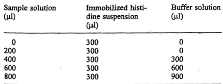
Sample vplurne was increased s follows. In a filter cup, 300 μΐ of immobilized histidine Suspension (150 g (wet weight) per litre in sodium acetate buffer, pH 5.5, ionic strength of 0.17), 0-900 μΐ of sodium acetate buffer '(pH 5.5, ionic strength of 0.17), and 0—800 μΐ of sample solution were placed according to table l, and
Tab. l Composition of sample solution, immobilized histidine Suspension, and buffer solution for the endotoxin assay by the inv mobilized histidine method (increase of sensitivity).
Sample solution (μΐ)
2000 400600 800
Immobilized histi- dine Suspension (μΐ)
300300 300300 300
Buffer solution (μΐ)
00 300600 900
the Suspension was shaken at 25 °C for 15 min using a Micromixer MT. The filter eup was then drained by suction for 2 min using a Milliliter vacuurn holder, and the filtrate was discarded. To the filter cup, l ml of sodium chloride solution (0.02 mol/1) was added and shaken for 5 min using a Micromixer MT. The filter cup was then drained by suction for 5 min. The filter cup containing the endotoxins adsorbed on the adsorbent was preincubated at 37 °C for 10 min. After preincubation, 150 μΐ of the mixture of Limulus amoebocyte lysate solution and Substrate solution (l : 2) were added to the filter cup, and the Suspension was incubated at 37 °C for 40 min using a QA thermomixer with shaking. The reaction was stopped by adding 100 μΐ of acetic acid solution (4.37 mol/1) and the mixture was filtered with suction for 2 min. The absorbance of the filtrate (200 μΐ) at 405 nm was measured with a THERMO- max microplate reader (Molecular Devices Corp., Menro Park, CA). All determinations were performed in duplicate.
High sensitive immobilized histidine method
The assay was carried out by the above method using 1.2 ml of immobilized histidine Suspension (37.5 g (wet weight) per litre), 0.8 ml of sample solution, and 60 min for the Limulus amoebocyte lysate reaction time.
Recovery of endotoxin in various plasma samples by the high sensitivity immobilized histidine method Sample Solutions
Plasma samples and the endotoxin-spiked plasma samples (EC-5:
500 endotoxin units per litre) were diluted 50-fold with pyrogen- free water in an ice bath, and heated in boiling water for 7.5 min.
Assay
The modified high sensitivity immobilized histidine method was used s follows. In a filter cup were placed l ml of immobilized histidine Suspension (45 g (wet weight) per litre in sodium-potas- sium phosphate buffer, pH 6.0, ionic strength of 0.16) and l ml of sample solution, and the Suspension was shaken at 25 °C for 15 min using a Micromixer MT. The filter cup was drained by suction for 5 min using a Milliliter vacuum holder and the filtrate was discarded. To the filter cup, l ml of sodium chloride solution (0.02 mol/1) was added and shaken for 5 min using a Micromixer MT.
The filter cup was then drained by suction for 5 min. The filter cup containing the endotoxins adsorbed on the adsorbent was preincu- bated at 37 °C for 10 min. After preincubation, 150 μΐ of the mix- ture of Limulus amoebocyte lysate solution and Substrate solution (1:2) were added to the filter cup, and the Suspension was incu- bated at 37 °C for 60 min using a QA thermomixer with shaking.
The re ction was stopped by adding 100 μΐ of acetic acid solution (4.37 mol/1) and the mixture was filtered with suction for 2 min.
The absorbanee of the filtrate (200 μΐ) at 405 nm was measured with a THERMOmax microplate reader. All determinations were performed in duplicate. The recovery of endotoxin in plasma sam- ples was calculated from the Standard curve produced by the modi- fied high sensitivity immobilized histidine method, using Standard endotoxin solution in water instead of plasma sample solution.
Effects of globulin preparation on the pyrogenic reaction and plasma endotoxin concentration in rab'bits administered endotoxin
Endotoxin from Escherichia coli UK.T-B was dissolved in saline at a concentration of 50 μ^. Japanese white rabbits (weight 2.8-3.2 kg) were administered saline (5 ml/kg) or Venoglobulin-IH (5 ml (250 mg)/kg) by intravenous injeetion, and each of these treatments was followed by the intravenous injeetion of endotoxin solution (l ml/kg) (n = 3), Body temperature of the rabbits was monitorcd every 10 min from 1.5 h before administration to 3.5 h after admin- Eur. J. Clin. Chem. Clin. Biochem. / Vol. 32, 1994 /No. 10
istration. Blood (about l ml) was drawn from each rabbit, at time of 0, l, and 3 h after adminislration, into a pyrogen-free syringe containing heparin. Platelet-rich plasma was obtained by centrifu- gation at 1000 min"1 for 10 min at 4 °C. Plasma samples were diluted 40-fold with pyrogen-free water in an ice bath, and heated in boiling water for 7.5 min. The endotoxin concentrations of pre- treated plasma samples were assayed by the high sensitivity immo- bilized histidine method.
Results
Optimal conditions for the pretreatment of plasma samples in the immobilized histidine method
For a quantitative assay of endotoxin using immobilized histidine, it is necessary that endotoxins in plasma sam- ple be adsorbed quantitatively by the immobilized histi- dine.
First, we tried to assay endotoxin in plasma without any pretreatment. In this case, the recovery of endotoxin was not good, and some non-specific amidases were also ad- sorbed on the adsorbent. We then found out that heat treatment in boiling water for 5—10 min after 40-fold dilution with distilled water gave good results.
Optimal conditions for the adsorption of endotoxin in plasma samples by immobilized histidine
Next, the optimal pH, ionic strength, and dilution of sample for the adsorption of endotoxin in plasma sam- ples by immobilized histidine were investigated. Opti- mal conditions were found to be pH 5.5—6.0, ionic strength 0.1, and more than 100-fold dilution.
Assay of spiked endotoxin in plasma by various endotoxin assay methods
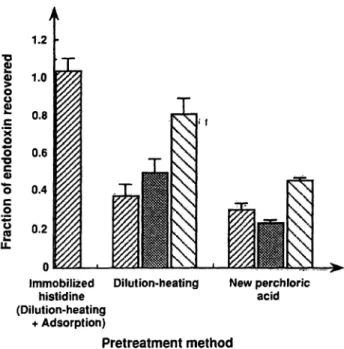
The immobilized histidine method was compared with the new perchloric acid treatment and the dilution-heat- ing procedure. Normal platelet-rich plasma samples were spiked with EC-5 (1000 endotoxin units per litre), and assayed by the immobilized histidine method, or the QCL-1000, the Toxicolor test, or the Toxinometer after pretreatment by the new perchloric acid procedure or the dilution-heating procedure. As shown in figure l, when the immobilized histidine method was used, the fraction of endotoxin recovered was almost 1. On the other hand, when the new perchloric acid procedure or the dilution- heating procedure were used, the fraction of endotoxin recovered was lower than that obtained with the immo- bilized histidine method.
•i
oo
l
o 0.2Immobilized Dilution-heating New perchloric histidine acid (Dilution-heating
+ Adsorption)
Pretreatment method
Fig. l Comparison of the three different pfetreatments and three different assay methods for the assay öf the spiked endotoxin in plasma. Normal platelet-rich plasma samples (n == 10) were spiked with EC-5 (1000 endotoxin units per litre), treated by the three different procedures äs described in the text, then assayed by the QCL-1000 (H), the Toxicolor test (EE), the Toxinometer (S) äs de- scribed in the text. The data were expressed äs means ± Standard error.
Increase of the sensitivity of the assay by the immobilized histidine method
A inore sensitive assay might be required for the deter- mination of endotoxin in clinical samples. We therefore investigated the following methods for increasing the sensitivity of the immobilized histidine method. First, the sample volume was increased, and second, the Limu- lus amoebocyte lysate reaction time was extended.
The maximum volume that can be held in the filier cup is 2 ml. Therefore, to increase sample volume, immobi- lized histidine, the mixture of Limulus amoebocyte ly- sate and Substrate, and acetic acid solution were halved.
As shown in figure 2, the absorbance increased linearly up to 800 of sample (4-fold volume). Thus, the sensi- tivity can be increased 4-fold by the increase of sample volume. Moreover, by extension of the Limulus amoebo- cyte lysate reaction time from 40 min tp 60 minvthe sensitivity can be increased about 2.5-fold. As shown in figure 3, the calibration curve obtained between 0 and 5 endotoxin units per litre (200 endotoxin units per litre in plasma samples) was well correlated.
Recovery of endotoxin in various plasma samples by the high sensitivity immobilized histidine method
Platelet-rich plasma samples from 5 healthy volunteers were spiked with EOS (50Q /endotoxin units per litre)
Eur. J. Clin. Cfaeni. Clin. Biochem. / Vol. 32, 1994 /No. 10
0.4
8 l
0.2 0.4 0.6
Sample volume [ml]
1.0
Fig. 2 Relationship of sample volume and absorbance in the im- mobilized histidine method.
Effects of globulin preparation on the pyrogenic reaction and on the plasma endotoxin concentration in rabbits administered endotoxin
Japanese white rabbits were administered saline and en- dotoxin or Venoglobulin and endotoxin, and the body temperature and the plasma endotoxin concentration of the rabbits were monitored. Increase of body temper- ature is shown in figure 4. Administration of endotoxin caused an increase in the body temperature of rabbits.
Venoglobulin tended to depress the second peak of increase of body temperature caused by the endotoxin.
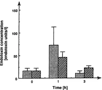
Figure 5 shows the plasma endotoxin concentration after administration of saline and endotoxin or Venoglobulin and endotoxin. Plasma endotoxin concentrations tended to increase l h after the administration.
0.1
2 4 6
Endotoxin concentration [endotoxin units/l]
Fig. 3 . Standard curve of the high sensitivity immobilized histi- dine method.
Tab. 2 Recovery of the spiked endotoxin in various plasma sam- ples by the modified high sensitivity immobilized histidine method.
Plasma sample Fraction of endotoxin recovered AB
CD E
1.121 0.967 0.723 0.853 0.815
and assayed by the modified high sensitivity immobi- lized histidine method. Table 2 shows the recovery of the spiked endotoxin in plasma sainples. The spiked en- dotoxins in every plasma sample showed a good recov- ery.
Discussion
Various pretreatments of plasma for the assay of endo- toxin by the Limulus amoebocyte lysate test have been described (5 — 11). However, these methods are not used widely for the endotoxin assay in clinical samples be- cause of their inaccuracy, that is, false positive or false negative results. On the other hand, the necessity for endotoxin assays in patient plasma has been increasing for diagnosis of Gra/w-negative septic syndrome. Anti- endotoxin monoclonal antibodies, which neutralize en- dotoxin activity, have been developed äs the drug for sepsis (12). Thus, endotoxin assay in plasma is required
P
i
100 200
Time [min]
300
Fig. 4 Effect of globulin preparation on the pyrogenic reaction in rabbits administered endotoxin. Japanese white rabbits were ad- ministered saline and endotoxin (·) or Venoglobulin and endotoxin (O), and the body temperature of the rabbits was monitored äs described in the text. The data were expressed äs means ± Stan- dard error.
Eur. J. CM. Chem. Clin. Biochera. / Vol. 32, 1994 / No. 10
150
II 8g
o.i
50 LU
Time [h]
Fig. 5 Endotoxin concentrations in plasma samples from rabbits administered saline and endotoxin or Venoglobulin and endotoxin.
Endotoxin concentrations in plasma samples from rabbits adminis- tered saline and endotoxin (E3) or Venoglobulin and endotoxin (H) were assayed äs described in the text. The data were expressed äs means ± siandard enror.
to select the patients to whom these drugs should be administered.
We have developed a specific assay method for endotox- ins using a membrane filter unit, a chromogenic Limulus amoebocyte lysate reagent, and immobilized histidine (4). We therefore investigated the suitability of this method for the determination of endotoxin in plasma samples. Preliminary experiments showed that pretreat- ment of plasma samples was necessary because some non-specific amidases were also adsorbed on immobi- .lized histidine, and some endotoxins were bound to some proteins in plasma. Previously, we had tried to remove endotoxin from serum by using immobilized histidine, and found that dilution of serum was neces- sary for the adsorption of serum endotoxin by immobi- lized histidine (unpublished data). Berger et al. reported the ability of human Gc-globulin and transferrin to bind endotoxin (13). This binding might interfere with the adsorption of endotoxin by immobilized histidine. We found 40-fold dilution with distilled water, followed by heat treatment in boiling water for 5-10 min gave good results. The optimal conditions for adsorption of endo- toxin in plasma samples were pH 5.5-6.0, ionic strength of 0.1, and more than 100-fold dilution. These conditions are thought to be necessary for the dissoci- ation of endotoxins from proteins.
Among the various plasma treatments used before the Limulus amoebocyte lysate test, the dilution-heating procedure (10-fold dilution and boiled for 10 min) (5, 6) and the new perchloric acid treatment (11) gave rela- tively good results. We therefore compared the immobi- lized histidine method with the dilution-heating pro-
cedure and the new perchloric acid treatment for the re- covery of the spiked endotoxin in plasma samples from healthy volunteers. It is clear that the immobilized histi- dine method is superior to the other methods (fig. 1).
In the determination of endotqxin concentratiori in pa- tient plasma, it is important that different endotoxins from various bacterial species are detected by the assay.
We have reported that different eridotoxins from various bacterial species are quantitatively adsorbed from water by immobilized histidine, and are assayed by the immo- bilized histidine method (4). Different endotoxins from various bacterial species are also assayed in plasma by the immobilized histidine method. These results will be reported elsewhere.
We next tried to increase the sensitivity of the immobi- lized histidine method. This method is based on the ad*
Sorption of endotoxin by immobilized histidine. An increase of sample volume at the adsorption step there- fore results in an increase of sensitivity. When a 4-fold volume of sample solution was used, the absorbance increased 4-fold (fig. 2). The Limulus amoebocyte lysate reaction is an enzyme reaction. An increase of the reac- tion time therefore results in an increase of sensitivity.
By using a 4-fold volume of sample solution and a 60 min reaction time, the sensitivity was increased about 10-fold (fig. 3). The spiked endotoxin in plasma samples was well recovered by the modified high sensitivity im- mobilized histidine method (tab. 2).
Furthermore, we investigated the effect of globulin prep- arations on the pyrogenic reaction and plasma eridotoxin coricentration in rabbits administered endotoxin. Veno- globulin tended to depress the second peak of increase of body temperature caused by endotoxin administration (fig. 4). One hour after the administration of endotoxin, plasma endotoxin concentrations tended to increase (fig.
5). Thus, it is considered that the immobilized histidirie method is also useful for determination of the plasma endotoxin concentration of rabbits.
However, to prove the usefulness of the immobilized his- tidine method, many poiiits must be clarified. For exam- ple, various native endotoxins should be tested, äs well äs the Standard endotoxin. It has been reported that the endo- toxin recovery in patient plasma differs from that in nor- mal plasma (14). The correlation betweeri the endotoxin concentration in patiemt plasrna and the clinical Symptoms should be clarified. These studies are in progress.
Acknowledgement
We thank T. Fukui for technical assistance.
Eur. J. Clin/Chem. Clifu Biochem. / Vol. 32, 1994 /No. 10
References
1. Morita, T., Tanaka, S., Nakamura, T. & Iwanaga, S. (1981) A new (l—>3)-ß-D-glucan-mediated coagulation pathway found in Limulus amebocytes. FEBS Lett. 129, 318-321.
2. Pfeiffer, M. & Weiss, A. R. (1987) Removal of LAL-test in- terfering low molecular weight substances by Ultrafiltration.
In: Detecüon of Bacterial Endotoxins with the Limulus Amebocyte Lysate Test (Stanley, W. W., Levin, J. & Novitsky, T., eds.) pp. 251-262, Alan R. Liss, New York.
3. Minobe, S., Nawata, M., Watanabe, T., Sato, T. & Tosa, T.
(1991) Specific assay for endotoxin using immobilized histi- dine and Limulus amebocyte lysate. Anal. Biochem. 198, 292-297.
4. Nawata, M., Minobe, S., Hase, M., Watanabe, T., Sato, T. &
Tosa, T. (1992) Specific assay for endotoxin using irnrnobi- lized histidine, Limulus amoebocyte lysate and a chromogenic Substrate. J. Chromatogr. 597, 415-424.
5. Yokota, M., Kambayashi, J., Tanaka, T., Tsujinaka, T, Sakon, M. & Mori, T. (1989) A simple turbidimetric time assay of the endotoxin in plasma. J. Biochem. Biophys. Methods 18, 97-104.
6. Kambayashi, J., Yokota, M., Sakon, M., Shiba, E., Kawasaki, T., Mori, T, Tsuchiya, M., Oishi, H. & Matsuura, S. (1991) A novel endotoxin-specific assay by turbidimetry with Limulus amoebocyte lysate containing ß-glucan. J. Biochem. Biophys.
Methods 22, 93-100.
7. Peason, F. C., Dubczak, J., Weary, M., Bruszer, G. & Donohue, G. (1985) Detection of endotoxin in the plasma of patients with Gram-negative bacterial sepsis by the Limulus amoebo- cyte lysate assay. J. Clin. Microbiol. 21, 865-868.
8. Roth, R. L, Levin, F. C. & Levin, J. (1990) Optimization of detection of bacterial endotoxin in plasma with the Limulus test. J. Lab. Clin. Med. 116, 153-161.
9. Levin, J., Tomasulo, P. A. & Oser, R. S. (1970) Detection of endotoxin in human blood and demonstration of an inhibitor.
J. Lab. Clin. Med. 75, 903-911.
10. Obayashi, T. (1984) Addition of perchloric acid to blood sam- ples for colorimetric Limulus test using chromogenic Substrate:
Comparison with conventional procedures and clinical appli- cations. J. Lab. Clin. Med. 104, 321-330.
11. Inada, K.., Yoshida, M., Takahashi, T, Tamura, S., Tanaka, S., Endo, S., Yoshida, T., Suda, H. & Komura, T. (1990) A new perchloric acid treatment of human plasma for detection of endotoxin by an endotoxin-specific chromogenic test. In: En- dotoxin (Friedman, H., Klein, T. W., Nakano, M. & Nowotny, A., eds.) pp. 225-229, Plenum Press, New York.
12. Warren, H. S., Danner, R. L. & Munford, R. S. (1992) Sound- ing Board. Anti-endotoxin monoclonal antibodies. N. Engl. J.
Med. 326, 1153-1157.
13. Berger, D. & Beger, H. G. (1987) Evidence for endotoxin bind- ing capacity of human Gc-globulin and transferrin. Clin. Chim.
Acta 163, 289-299.
14. Fukui, H., Brauner, B., Bode, J. C. & Bode, C. (1989) Chromo- genic endotoxin assay in plasma — Selection of plasma pre- treatment and production of Standard curves. J. Clin. Chem.
Clin. Biochem. 27, 941-946.
Satoshi Minobe Research Laboratory of Applied Biochemistry
Tanabe Seiyaku Co., Ltd., 16-89 Kashima-3-chome, Yodogawa-ku Osaka 532
Japan
Eur, J. Clin. Chem. Clin. Biochem. / Vol. 32,1994 / No. 10