Gyrate atrophy-like phenotype with normal plasma ornithine and low plasma taurine
Abstract
We present the case of a 39-year-old male with sectoral chorioretinal atrophy similar to that seen in gyrate atrophy (GA) but with a normal
Aubhugn T. Labiano
1Milagros H. Arroyo
1plasma ornithine level. Unlike previously reported cases of GA, he had below-normal plasma taurine concentration. Much remains unknown
about the pathophysiologic mechanism underlying gyrate-like chorio- 1 Department of
Ophthalmology and Visual retinal atrophy. The concomitant occurrence of GA-like phenotype and
Sciences, Sentro hypotaurinemia suggests a possible future avenue for research on this
matter. Oftalmologico Jose Rizal,
University of the Philippines Keywords:gyrate atrophy, chorioretinal atrophy, ornithine, taurine Manila – Philippine General Hospital, Manila, Philippines
Introduction
Gyrate atrophy of the choroid and retina (GA) is a rare autosomal recessive metabolic disorder characterized by progressive chorioretinal degeneration, with the atrophic lesions having scalloped or garland-like borders, hence the term “gyrate” [1], [2]. It has been associated with elevated levels of the amino acid ornithine in plasma as well as in aqueous humor, cerebrospinal fluid, and urine [3] . The underlying cause of the hyperornithinemia is a deficiency in the mitochondrial matrix enzyme ornithine aminotransferase (OAT) [4], [5]. The typical clinical history begins in childhood with a decrease in visual acuity associated with night blindness and poor peripheral vision [1], [6]. At the onset of visual symptoms, sharply defined round areas of atrophy are seen in the retinal midperiphery. As these atrophic areas enlarge, coalesce, and spread to the far periphery, the peripapillary area, and even the foveal area, the visual symptoms worsen, the visual fields progressively constrict, and blindness may ensue.
Occasional reports of cases having fundus findings resem- bling those typically seen in GA but with normal or mildly elevated plasma ornithine appear in the literature. These patients have been referred to as having normoornith- inemic GA, GA-like phenotype, or atypical GA [7], [8], [9], [10], [11].
We present a case with sectoral chorioretinal atrophy similar to that seen in GA but with a normal plasma ornithine level. Our patient also had a low plasma taurine concentration. To our knowledge, this is the first reported
case of a gyrate atrophy-like phenotype associated with hypotaurinemia.
Case description
A 39-year-old male presented with the complaint of blurred vision, described as haziness, in his left eye of two months’ duration. He acknowledged that he had ex- perienced blurring of vision of both eyes 12 years ago, and that his vision improved and stabilized after using unrecalled topical medications. He reported little or no difficulty with night vision. Aside from occasional tearing, the patient had no other associated ocular symptoms.
He had no known comorbid conditions and no systemic complaints. He denied any of his immediate family experiencing similar symptoms. He also denied consan- guinity of his parents.
Snellen uncorrected visual acuity in the right eye was 20/20, and the manifest refraction was plano. Snellen uncorrected visual acuity in the left eye was 20/100, which was best corrected to 20/20 with –1.25 D sphere
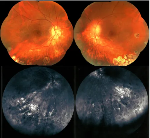
=–0.75 D cylinder x90°. Color vision, gross eye examina- tion findings, and intraocular pressures were within nor- mal limits. Slit lamp examination showed a small and thin posterior subcapsular cataract in the left eye. Dilated fundus examination revealed in both eyes roundish gray patches of chorioretinal atrophy, some coalescing to form a gyrate shape, mostly located in the inferior retinal midperiphery and far periphery (Figure 1). No such lesions were seen in the macula. There was also peripapillary atrophy.
Figure 1: Fundus photographs showing patches of chorioretinal atrophy in the inferior retina of both eyes
Figure 2: Pattern deviation probability maps showing superior visual field constriction in both eyes
On fluorescein angiography, the patches appeared as hypofluorescent areas with faintly hyperfluorescent bor- ders. Choroidal vessels were prominent within the lesions.
Spectral-domain optical coherence tomography (OCT) of the macula revealed normal central foveal thickness in both eyes and perifoveal thinning, which was more pro- nounced in the left eye than in the right. Horizontal scans of the macula showed no abnormalities. Testing of the central 30 degrees of the visual field with the Octopus 300 perimeter (Haag-Streit AG, Köniz, Switzerland) re- vealed in each eye a superior arcuate scotoma and en- largement of the blind spot (Figure 2). There was inferior retinal nerve fiber layer (RNFL) thinning in the right eye
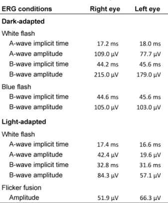
on OCT of the optic nerve head and RNFL. Full-field elec- troretinogram (ERG) responses in light- and dark-adapted conditions showed A and B waveforms with amplitudes and implicit times that were generally within the normal range (Table 1). Waveform amplitudes in the left eye were mostly lower than those in the right eye. Multifocal ERG, which is more sensitive than full-field ERG in detecting local retinal dysfunction, was not available in our center and in other centers in the country and thus was not performed.
Quantitative plasma amino acid analysis done after an overnight fast revealed a normal plasma ornithine level at 61 µmol/L (reference interval: 48–195 µmol/L). Levels
Table 1: Full-field electroretinogram results
of the other amino acids, with the exception of taurine, were within normal limits. Plasma taurine concentration was below normal range at 36 µmol/L (reference interval:
54–210 µmol/L). The amino acid analysis was done using ultrahigh performance liquid chromatography – Waters MassTrak amino acid analysis system. Reference intervals were adapted from the Princess Margaret Hospital for Children, Perth, Australia. Testing for OAT activity and OAT genetic testing were unavailable in our center and in nearby institutions and thus were not done. Nerve con- duction studies revealed motor potentials within normal limits and normal F-waves but with reduction of sensory potentials of the right median nerve, indicative of mild carpal tunnel syndrome. Monopolar needle studies on the muscles of the right paracervical and paralumbar areas showed no signs of acute denervation.
Based on the history, fundus findings, and results of an- cillary tests, our patient was diagnosed as having a gyrate- atrophy-like phenotype. The patient was advised spectacle correction of his error of refraction, increased intake of taurine-rich foods, and once- or twice-yearly follow-up for monitoring of the lesions and possible repeat plasma amino acid analysis. Ophthalmologic examination of his parents and siblings was also recommended.
Discussion
Differential diagnoses for gyrate atrophy include cho- roideremia and myopic fundus changes [7], [12]. Cho- roideremia is a rare X-linked recessive disorder. The earliest symptom is night blindness in the first or second decade of life, and there is eventual progressive visual field constriction and visual acuity loss. Early fundus
manifestations of choroideremia consist of pigment clumping at the level of the retinal pigment epithelium (RPE) and peripapillary atrophy [13]. In the advanced stage of the disease, the loss of RPE and choriocapillaris can form confluent scalloped areas resembling those seen in the later stages of GA. The well-demarcated round lesions observed in our patient are not characteristic of choroideremia. Patients with high myopia may present with peripheral clusters of atrophic areas or pavingstone degeneration that may resemble the lesions seen in GA [12], [14]. The manifest refraction of our patient did not show a high degree of myopia. Pavingstone degeneration is typically found between the ora serrata and the equator [15].
Few reports of atypical GA or GA-like cases appear in the literature. Similar to our patient, they were noted to have the lesions characteristically seen in GA and had normal plasma ornithine levels. Bargum reported on the case of a 67-year-old female patient with late onset of nyctalopia and blurred vision and a mild and protracted clinical course [7]. Kellner et al. described six patients, three from the same family, and three simplex cases, who presented with the typical lesions of gyrate atrophy. The presence of similar signs and symptoms among three members from the same family consisting of a 70-year- old father, the 41-year-old son and the 39-year-old son suggested an autosomal dominant disorder [8]. Saito et al. reported on the case of a 44-year-old woman of con- sanguineous parentage. Amino acid analysis of her urine revealed increased excretion of proline, hydroxyproline, and glycine [9]. Mishima et al. presented the case of a 3-year-old boy who was diagnosed with the Fukuyama type of congenital muscular dystrophy and high myopia [10]. None of these cases featured a plasma taurine level below the normal range.
Hypotaurinemia has also not been previously reported in association with the typical gyrate atrophy with hyper- ornithinemia. Berson studied the plasma amino acid levels of 41 patients with hereditary retinal degenerative diseases, among which five had gyrate atrophy, six rela- tives of patients diagnosed with GA, and 13 normal sub- jects. Mean plasma ornithine was found to be significantly elevated among the GA patients and, to a lesser extent, among their relatives. Mean plasma lysine level among GA patients was found to be significantly lower than that among the normal subjects. In all subjects with gyrate atrophy as well as in all other study participants, plasma taurine levels were within normal limits [16].
The pathophysiologic mechanism of the retinal degenera- tion seen in GA with hyperornithinemia is not yet fully understood. It has been hypothesized that this is due to the toxicity of high ornithine levels to chorioretinal tissue [17], [18]. Decreased levels of Δ1-pyrroline-5-carboxylate, proline, or phospocreatine have also been proposed to underlie the chorioretinal degeneration in GA [18], [19], [20]. Much less is known about the pathophysiology of GA-like lesions associated with normal ornithine levels.
Kellner et al. posit that there may be a common but still unknown disease process at play in hyperornithinemic
and normoornithinemic cases or that diverse conditions may result in the same chorioretinal degeneration [8].
Saito et al. suggest that a deficiency in proline may be relevant in the development of atypical and typical GA [9]. Determining the link between low plasma taurine and GA-like phenotype, if it at all exists, is beyond the scope of this case report. Future research on the possible asso- ciation of these two findings is suggested.
Taurine or 2-amino-ethanesulfonic acid is a free non- protein sulfur-beta-amino acid. It is a non-essential amino acid that can be synthesized from L-methionine and L-cysteine, although most taurine comes from the diet.
It is the most abundant amino acid in the mammalian retina. Of all tissues, the retina has the highest concen- tration of taurine. The specific function played by taurine in the retina is still unclear. Taurine is considered to be an antioxidant after being found to directly and indirectly neutralize reaction oxygen species. It could play a role in decreasing oxidative stress, which has been implicated in the development of retinal degenerative disorders. It is also considered to be neuroprotective because it acts to regulate the cellular concentration of calcium and could prevent glutamate excitotoxicity [21].
In cats, taurine deficiency has been associated with ret- inal degeneration, although not in the pattern and location of the lesions seen in gyrate atrophy. The initial fundus finding is a hyperreflective granular zone in the area centralis, which corresponds to the human macula. Mi- croscopically, there is photoreceptor outer segment de- generation in that area. Subsequently, there is progressive loss of the entire photoreceptor population [22]. Children on long-term parenteral nutrition with mean plasma taurine levels less than half that of the control group have been found to have delayed photopic and scotopic b-wave ERG implicit times and some have been observed to have RPE granularity in the posterior pole [23]. Taurine defi- ciency has been implicated as a cause of the retinal tox- icity induced by vigabatrin, which is known to cause visual field constriction [24]. Vigabatrin-treated rats have been found to have photoreceptor damage and retinal ganglion cell loss [25].
Conclusions
We described the case of a patient with chorioretinal le- sions characteristic of gyrate atrophy but with normal plasma ornithine level. Unlike other reported cases of GA, our patient had below-normal plasma taurine concen- tration. The concomitant occurrence of GA-like phenotype and hypotaurinemia may be more than fortuitous and suggests a future avenue for research on the patho- physiology of gyrate-like chorioretinal atrophy.
Notes
Competing interests
The authors declare that they have no competing in- terests.
References
1. Takki K. Gyrate atrophy of the choroid and retina associated with hyperornithinaemia. Br J Ophthalmol. 1974 Jan;58(1):3-23. DOI:
10.1136/bjo.58.1.3
2. Takki K, Simell O. Genetic aspects in gyrate atrophy of the choroid and retina with hyperornithinaemia. Br J Ophthalmol. 1974 Nov;58(11):907-16. DOI: 10.1136/bjo.58.11.907
3. Simell O, Takki K. Raised plasma-ornithine and gyrate atrophy of the choroid and retina. Lancet. 1973 May;1(7811):1031-3.
DOI: 10.1016/s0140-6736(73)90667-3
4. Valle D, Kaiser-Kupfer MI, Del Valle LA. Gyrate atrophy of the choroid and retina: deficiency of ornithine aminotransferase in transformed lymphocytes. Proc Natl Acad Sci USA. 1977 Nov;74(11):5159-61. DOI: 10.1073/pnas.74.11.5159 5. Kaiser-Kupfer MI, Valle D, Del Valle LA. A specific enzyme defect
in gyrate atrophy. Am J Ophthalmol. 1978 Feb;85(2):200-4. DOI:
10.1016/s0002-9394(14)75948-3
6. Takki KK, Milton RC. The natural history of gyrate atrophy of the choroid and retina. Ophthalmology. 1981 Apr;88(4):292-301.
DOI: 10.1016/s0161-6420(81)35031-3
7. Bargum R. Differential diagnosis of normoornithinaemic gyrate atrophy of the choroid and retina. Acta Ophthalmol (Copenh).
1986 Aug;64(4):369-73. DOI: 10.1111/j.1755- 3768.1986.tb06937.x
8. Kellner U, Weleber RG, Kennaway NG, Fishman GA, Foerster MH.
Gyrate atrophy-like phenotype with normal plasma ornithine.
Retina (Philadelphia, Pa). 1997;17(5):403-13. DOI:
10.1097/00006982-199709000-00008
9. Saito T, Hayasaka S, Yabata K, Omura K, Mizuno K, Tada K.
Atypical gyrate atrophy of the choroid and retina and iminoglycinuria. Tohoku J Exp Med. 1981 Nov;135(3):331-2.
DOI: 10.1620/tjem.135.331
10. Mishima H, Hirata H, Ono H, Choshi K, Nishi Y, Fukuda K. A Fukuyama type of congenital muscular dystrophy associated with atypical gyrate atrophy of the choroid and retina. A case report. Acta Ophthalmol (Copenh). 1985 Apr;63(2):155-9. DOI:
10.1111/j.1755-3768.1985.tb01528.x
11. Bhakhri R, Ridder WH 3rd. Gyrate Atrophy-Like Phenotype: Normal Plasma Ornithine and Retinal Crystals. Optom Vis Sci. 2016 09;93(9):1173-80. DOI: 10.1097/OPX.0000000000000906 12. Arshinoff SA, Leung K, Strube YNJ. Gyrate atrophy (Chapter 25).
In: Tasman W, Jaeger EA, editors. Duane's Ophthalmology.
Philadelphia: Lipincott, Williams & Wilkins; 2006.
13. Khan KN, Islam F, Moore AT, Michaelides M. Clinical and Genetic Features of Choroideremia in Childhood. Ophthalmology. 2016 10;123(10):2158-65. DOI: 10.1016/j.ophtha.2016.06.051 14. Karlin DB, Curtin BJ. Peripheral chorioretinal lesions and axial
length of the myopic eye. Am J Ophthalmol. 1976 May;81(5):625- 35. DOI: 10.1016/0002-9394(76)90129-x
15. O’Malley P, Allen RA, Straatsma BR, O’Malley CC. Paving-stone degeneration of the retina. Arch Ophthalmol. 1965 Feb;73:169- 82. DOI: 10.1001/archopht.1965.00970030171006
16. Berson EL, Schmidt SY, Rabin AR. Plasma amino-acids in hereditary retinal disease. Ornithine, lysine, and taurine. Br J Ophthalmol. 1976 Feb;60(2):142-7. DOI: 10.1136/bjo.60.2.142 17. Kuwabara T, Ishikawa Y, Kaiser-Kupfer MI. Experimental model
of gyrate atrophy in animals. Ophthalmology. 1981 Apr;88(4):331-5. DOI: 10.1016/s0161-6420(81)35027-1 18. Kaiser-Kupfer MI, Valle DL. Clinical, biochemical, and therapeutic
aspects of gyrate atrophy. Prog Retin Res. 1987;6:179-206. DOI:
10.1016/0278-4327(87)90023-X
19. Saito T, Omura K, Hayasaka S, Nakajima H, Mizuno K, Tada K.
Hyperornithinemia with gyrate atrophy of the choroid and retina:
a disturbance in de novo formation of proline. Tohoku J Exp Med.
1981 Dec;135(4):395-402. DOI: 10.1620/tjem.135.395 20. Sipilä I, Simell O, Arjomaa P. Gyrate atrophy of the choroid and
retina with hyperornithinemia. Deficient formation of guanidinoacetic acid from arginine. J Clin Invest. 1980 Oct;66(4):684-7. DOI: 10.1172/JCI109905
21. Froger N, Moutsimilli L, Cadetti L, Jammoul F, Wang QP, Fan Y, Gaucher D, Rosolen SG, Neveux N, Cynober L, Sahel JA, Picaud S. Taurine: the comeback of a neutraceutical in the prevention of retinal degenerations. Prog Retin Eye Res. 2014 Jul;41:44- 63. DOI: 10.1016/j.preteyeres.2014.03.001
22. Hayes KC, Carey RE, Schmidt SY. Retinal degeneration associated with taurine deficiency in the cat. Science. 1975
May;188(4191):949-51. DOI: 10.1126/science.1138364 23. Geggel HS, Ament ME, Heckenlively JR, Martin DA, Kopple JD.
Nutritional requirement for taurine in patients receiving long- term parenteral nutrition. N Engl J Med. 1985 Jan;312(3):142- 6. DOI: 10.1056/NEJM198501173120302
24. Jammoul F, Wang Q, Nabbout R, Coriat C, Duboc A, Simonutti M, Dubus E, Craft CM, Ye W, Collins SD, Dulac O, Chiron C, Sahel JA, Picaud S. Taurine deficiency is a cause of vigabatrin-induced retinal phototoxicity. Ann Neurol. 2009 Jan;65(1):98-107. DOI:
10.1002/ana.21526
25. Jammoul F, Dégardin J, Pain D, Gondouin P, Simonutti M, Dubus E, Caplette R, Fouquet S, Craft CM, Sahel JA, Picaud S. Taurine deficiency damages photoreceptors and retinal ganglion cells in vigabatrin-treated neonatal rats. Mol Cell Neurosci. 2010 Apr;43(4):414-21. DOI: 10.1016/j.mcn.2010.01.008
Corresponding author:
Aubhugn T. Labiano
Department of Ophthalmology and Visual Sciences, Sentro Oftalmologico Jose Rizal, University of the Philippines Manila – Philippine General Hospital, Taft Avenue, Manila, Philippines
atlabiano@gmail.com
Please cite as
Labiano AT, Arroyo MH. Gyrate atrophy-like phenotype with normal plasma ornithine and low plasma taurine. GMS Ophthalmol Cases.
2020;10:Doc04.
DOI: 10.3205/oc000131, URN: urn:nbn:de:0183-oc0001314
This article is freely available from
https://www.egms.de/en/journals/oc/2020-10/oc000131.shtml Published:2020-02-27
Copyright
©2020 Labiano et al. This is an Open Access article distributed under the terms of the Creative Commons Attribution 4.0 License. See license information at http://creativecommons.org/licenses/by/4.0/.