Dick and Culimann: Amidolytic assay for determination of -antitrypsin 57
J. Gin. Chem. Clin. Biochem.
Vol. 20,1982, pp. 57-60
An Amidolytic Assay for Determination of a, -Antitrypsin in Serum and in Cerebrospinal Fluid
By W. Dick1) and W. CulimannZentrallabor des Lukaskrankenhauses Neuss, F.R. G.
(Received April 10/July 21,1981)
Summary: An amidolytic assay using the chromogenic Substrate tosyl-glycyl-prolyl-lysine-4-nitranilide acetate (Chromozym PL®) has been studied for the determination of ^ -antitrypsin in serum and in cerebrospinal fluid (CSF). It was found that binding of trypsin to a2-macroglobulin has to be taken into consideration when 0^- antitrypsin is determined by an amidolytic assay. In serum this accounts for 7.4 ± 3.2% and in CSF 2.8 ± 1.8%
of the total antitrypsin capacity. The reference ränge of ÄJ -antitrypsin in sera was found to be 3T= 60.3 ± 20.8 kIU/1, and in CSF = 186 ± 99 IU/1. Within-run precision showed a C.V. of l.36% in sera, and a C.V. of l .86%
in CSF. There was a good correlation with immunochemical methods: r = 0.946 in 65 sera (p < 0.001) and r = 0.986 in 55 samples of CSF (p < 0.001).
Bestimmung von oti-Antitrypsin in Serum und Liquor mit einem chromogenen Substrat
Zusammenfassung: Es wurde ein photometrisches Verfahren mit dem chromogenen Substrat Tosyl-glycyl-prolyl- lysin-4-nitranilidacetat (Chromozym PL®) zur Bestimmung des o^ -Antitrypsingehaltes im Serum und im Liquor untersucht. Dabei ist bei der Bestimmung des c^-Antitrypsins die Bindung von Trypsin an a2-Makroglobulin zu berücksichtigen. Im Serum wurde dafür ein Anteil von 7,4 ± 3,2% und im Liquor von 2,8 ± 1,8% an der gesamten Antitrypsinkapazität ermittelt. Als Referenzbereich von c^ -Antitrypsin wurden folgende Werte gefunden: im Serum
= 60,3 ± 20,8 kIU/1 und im Liquor = 186 ± 99 IU/1. Für die Präzision in der Serie wurde im Serum ein VK von 1,36 und im Liquor von 1,86% erhalten. Weiterhin bestand eine ausgezeichnete Übereinstimmung zu immun- chemischen Methoden: r = 0,946 in 65 Seren (p < 0,001) sowie r = 0,986 in 55 Liquorproben (p < 0,001).
Introduction
Evaluation of the seruin-CSF barrier gradient of ax -anti- trypsin has received increasing attention within recent years (2,3). However, the determination of AI-anti- trypsin in serum with an amidolytic assay provides several problems. It is known from the literature that an increase in the dilution of serum or plasma results in a greater Inhibition of trypsin activity (4). A dilution of one hundred times exhibits the largest degree of in- hibition. Further dilution of rnore than five hundred times leads to a marked decrease in the Inhibition of trypsin activity (4). The development of a number of chromogeiyc Substrates has provided a Substrate for
the determination of dj -antitrypsin according to the äbove conditions. Mqreover, cerebrospinal fluid (CSF) should be assayed without ä preparation step prior to the photometric assay. It is well established that trypsin also binds to other antiproteases, like c^-macroglobulin.
*) Presented in part at the XL International/IV. European Congress of Clinical Chemistry, Vienna, Austria, Sept. 1981 (1)
2) STJ: soybean trypsin inhibitor
Inter-a-trypsin-inhibitor e)diibits a lower affinity to trypsin than other antiproteases and is therefore of minor importance (5). However, the influence of a2- macroglobulin on the assay has to beconsidered. The reaction of the assay can be scheduled äs follows:
l a. Trypsin + &i -antitrypsin -^"- trypsin-^-antitrypsin complex (inactive) + trypsin (free, active)
l b. Trypsin + a2 -macroglobulin-^· trypsin-a2 macro- globulin complex (active) + trypsin (free, active) Trypsin (free,active)+STI2)-=^ trypsin-STI complex
(inactive)
2. Chromozym PL® ^-(active) - peptide -H 4.nitro- aniline
(active trypsin = free trypsin + trypsin-a2- macroglobulin complex)
It was the purpose of this study to evaluate optimal conditions for such an amidolytic assay.
0340-076X/82/0020-005 7$02.00
© by Walter de Gruyter & Co. · Berlin · New York
jg Dick and Cullmj
Materials and Methods ^ Samples
65 sera and 55 samples of cerebrospinal fluid, representing ,_, unselected clinical material which had bcen sent to our labor- ·| 10 atory , wcre investigated. For determination of kinetic para- ^ meters, within-run, and day to day precision a pool serum of e 20 healthy donors and a CSF pool of 20 samples selected at —·
random were taken. All samples were immediately frozen at 5 - 20 °C and analyzed within 4 weeks. α ι -antitrypsin is known
to remain stable within this period (6).
Prior to the photometric assay, sera were diluted 1 + 100 with a solution of 9 g/l of sodium Chloride.
mn: Amidolytic assay for determination of α ι -anti trypsin
x'
,··*s ti
Υ 1 1 1 1 ... 1 1 ^ 2 4 6 θ 10 12
t [min]
Reagents and procedures
Protein was measured in sera and in CSF by Standard pro- cedures (7, 8). Cell count in CSF was estimated from a micro- scopic count. Immunochemical levels of a\ -antitrypsin were calculated from immunodiffusion (NOR-Partigen and LC- Partigen, Behring Institute Marburg, F.R.G.) (9). For the amidolytic assay, the reagents were commercial preparatipns of analytical grade, purchased from Boehringer Mannheim, F.R.G. (Chromozym TH®, Chromozym TRY®, and Chromozym PL®, trypsin (33 U/mg protein with benzoyl-/)>Z,-arginine-4- nitroanilide s Substrate), and soybean trypsin Inhibitor (STI)).
The buffer consisted of 0.05 mol/1 of triethanolamine and 0.15 mol/1 of sodium chloride, adjusted to pH 8.0. The following CSF values were considered normal: protein level below 500 mg/1, cell count below 3 cells/mm3, no red cells on microscopic examihation. For the calculation of the serum/CSF barrier gradient, sera exhibiting c^-antitrypsin levels below 2 g/l and more than 4 g/l were excluded. "m-values were calculated from Eadie-Hofstee plots (10,11).
Results
Methodological studies
The chromogenic Substrates Chromozym TH®, Chromo- zym TRY®, and Chromozym PL® were evaluated for determination of o^-antitrypsin in serum. The Substrates Chromozym TH® and Chromozym TRY®, both very sensitive to trypsin, require a dilution of sera of at least 600 fold; the Substrate Chromozym PL®, however, required only a dilution of one hundred fold (high levels of <*!-antitrypsin in serum require a 200 fold dilution) and corresponded to the conditions mentioned above.
This dilution resulted in a change of absorbance of ΔΑ/min = 0.4 for the chromogenic Substrate Chiomo- zym PL®; for the Substrates Chromozym TH® and TRY®, however, a change of absorbance of ΔΑ/min
= 2.6 was observed.
A linear slope furiction representing the Splitting of the Substrate Chromozym PL® by trypsin was obtained over a wide r nge. A change of absorbance of ΔΑ/min
= 0.350 corresponded to a final concentration of 25 U/l trypsin. In further experiments it was revealed that the Substrate Chromozym PL® is split by the trypsin- a2-macroglobulin complex, s already described for other Substrates (12,13).
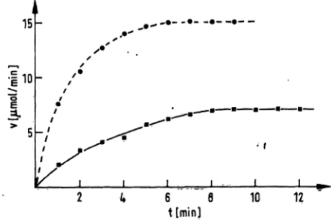
Kinetics of Inhibition of the amidolytic activity of trypsin by o^ -antitrypsin were evaluated at 25 °C and 37 °C (fig. 1). It is evident that trypsin reacts slowly with c*!-antitrypsin: half-rate at 25 °C was determined to be 142 seconds and at 37 0C to be 59 seconds (fig. 1).
Fig. 1. Variation of the incubation time of trypsin with a pre- diluted serum at 25 °C (a) and at 37 °C (·) (Standard conditions). Prolongation of incubation leads to decrease of free .trypsin.
Stepwise addition of diluted serum leads to a n n- competitive inhibition of the amidolytic activity of trypsin (fig. 2). For the Splitting of the chromogenic Substrate Chrornpzym PL® by trypsin, the Km was 2 Χ l O"5 mol/1, independent of the amount of diluted serum in the assay.
1.5
l U
10 20 30 ~ 4"0~
γτ\ [10"3-l/mm-I ]
50 60* 70
Fig. 2. Non-competitive inhibition of the amidolytic activity of trypsin by stepwise addition of diluted pool serum (Standard conditions) (·: no addition of diluted serum, o: addition of pool serum, dil ted l :400,
·: addition of pool serum, diluted 1:200, X: addition of pool Serum, diluted l:100;i : ainount of α ι -anti- trypsin in Inhibitory Units).
Tab. 1. Contribution of a2-macroglobulin to the total antl·
trypsin capacity, depending ori the orj-antitrypsin levels in serum and in CSF.
Level of antitrypsin in serum % of the total antitrypsin capacity due
globulin
< 2.5 g/l (n = 19) 2.5-4.0 g/l (n =
> 4.0 g/l (n = 18) Total (n g 65)
28) 13.5 ± 9.2
7.7 ± 2.6 4.2 ±1.8 7.4 ± 3.2
Levels of antitrypsin in CSF % of the total antitrypsin capacity due to ofc-rnacio- globulin
< 20 mg/1 (n = 32) 20-50 mg/1 (n = 14)
> 50 mg/1 (n = 9) Total (n = 55)
3.4 ±1.7 1,8 ± 0.9 2.5 ± 1.9 2.8 ± 1.8
J. din. Cherri. C n. Biochem. / Vol. 20,1982 / No. 2
Dick and Cullmann: Amidolytic assay for determination of arantitrypsin 59
In simultaneous assays, the contribution by a2-macro- globulin to the total antitrypsin amount was deter- mined s previously described (12,13), using the ability of a2-macroglobulin to form a trypsin-protein com- plex, which retains its amidolytic activity, also in the presence of soybean trypsin Inhibitor. These results are summarized in table 1. It is evident that in the case of a low α ι -antitrypsin level, a considerable part of trypsin will be bound to a2-macroglobulin.
All this considered, the photometric assays can be summarized s follows:
assay a)
0.40 ml of buffer with trypsin (25 U/l)
0.02 ml of sample (serum dilution 1 + 100 or CSF) 6 min incubation
0.05 ml of Chromozym PL® (3.2 X l (T4 mol/1) increase in absorbance at 405 nm; d = l cm, 37 °C;
assay b)
0.40 ml of buffer with trypsin (25 U/l)
0.02 ml of sample (serum dilution 1 + 100 or CSF) l min incubation
0.01 ml soybean trypsin inhibitor (5 X 10"^ mol/1) 0.05 ml of Chromozym PL® (3.2 Χ l O"4 mol/1) increase in absorbance at 405 nm; d = l cm, 37 PC.
Calculation was performed s follows:
AA/mintrypsin
- AA/minsample(assay a)
— ΔΑ/mina^acjogfcbuKn (assay b)
= ΔΑ/mintrypsin inhibited by *i -antitrypsin
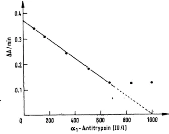
Using a pool serum the assay revealed a good linearity over a wide r nge (fig. 3). In the case of serum, the assay should be earried out in the r nge from 400 to 800 IU/13) (sera should be diluted 100 fold or 200 fold, respectively); evaluation of the CSF levels, how- ever, covers the total r nge.
(H
. 0.3
0.2
0.1
J_
200 WO 600 800 ar Antitrypsin [IU A]
1000 Fig. 3. Linearity of the assay, obtained with a pool serum
(Standard conditions; IU: amount of <*i -antitrypsin in Inhibitory Units).
As the CSF samples were taken without dilution, the unspecific Splitting of the chromogenic Substrate was evaluated in the CSF samples: even in the pathological r nge unspecific Splitting did not exceed 0.5% of total Splitting.
Reference r nge
With the assay, the normal r nge of c^ -antitrypsin in sera was found to be 60.3 ± 20.8 kIU/1 (n = 65) (total protein concentration 72 ± 9.2 g/l). In CSF, the refer- ence r nge was 186 ± 99 IU/1 (n = 55) (total protein did not exceed 500 mg/1); no sex differences were observed.
Within-run precision and detection limit Within-run precision was χ = 52.3 ± 0.71 kIU/1 (C.V. = l .36%) for pool serum and χ = 326 ± 6 IU/1 (C.V. = 1.86%) for a CSF pool. Detection limit in CSF was 12 IU/1.
Day to day precision
For serum, day to day precision was found to be χ = 55.1 ± 1.53 kIU/1 (C.V. = 2.78%) and for CSF χ = 319 ± 11 IU/1 (C.V. = 3.45%).
Correlation^with immunodiffusion
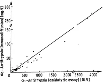
There was a good correlation with immunodiffusion in sera s well s in CSF (r = 0.946 for 65 sera (p < 0.001), and r = 0.986 for 50 samples of CSF (p < 0.001), fig. 4 and 5). Linear regression analysis showed the equation y = 0.054 χ + 0.05 for sera and y = 0.070 χ - 3.2 for CSF. 5 samples of CSF had to be excluded because of their low o^ -antitrypsin levels (not detectable with immunodiffusion).
Serum-CSF barrier gradient
Using the sera that exhibited an oti -antitrypsin level from 2 to 4 g/l and the CSF samples that were considered normal, the serum-CSF barrier gradient was calculated.
Using the amidolytic assay, a gradient of 276 was found, while immunodiffusion gave a value of 269.
l 8 · 0
6.0
4.0
2.0 l
3) l IU = l Inhibitor Unit = the amount of α ι -antitrypsin, which inhibits l U umol/min) of trypsin (Chromozym PL® s the Substrate) ·*'·
S 0 20 40 60 80 100 120 140 ce^Antitrypsin (amidolytic assay) [kIU/1]
Fig. 4. Comparison of the amidolytic assay ofaj-antitrypsin with immunodiffusion in 65 sera at random
(y = 0.054 χ + 0.05, r = 0.946, p < 0.001; IU: amount of <*i-antitrypsin in Inhibitory Units).
J. Clin. Chem. C h. Biochem, / Vol. 20,1982 / No. 2
60 Dick and Cullmann: Amidolytic assay for determination of -antitrypsin
1000 1500 2000 3500 A000 a1 -Antitrypsin (omidolytic ossoy) [1U/1]
Fig. 5. Comparison of the amidolytic assay of cq-antitrypsin with immunodiffusion in 50 CSF samples at random (y = 0.070 - 3.2, r = 0.986, p < 0.001; IU: amount of<*i-antitrypsin in Inhibitory Units).
Discussion
It is well known that o^ -antitrypsin can be regarded the major Inhibitor of trypsin in serum. However, the amount of trypsin bound to o^-macroglobulin is variable.
Consequently, an exact determination of o^ -antitrypsin using a functional assay requires exploration of the a2- macroglobulin level. On the other hand, in CSF the low levels of a2macroglobulin are negligible.
It is well known that the chromogenic Substrate Chromozym PL® is also split by other serine pro- teases like plasmin. As plasmin generally does not occur äs a free protease in plasina or serum, an un- specific Splitting of the chromogenic Substrate by free plasmin can be ruled out.
Our results suggest a serum-CSF barrier gradient of ca. 270 for oti -antitrypsin, whidi agrees with the Undings of Felgenhauer (14).
It has already been repörted that c^-antitrypsin reacts slowly with trypsin (4). In a previous study, we demon- strated that trypsin reacts rapidly with ai-macroglobiilin (13). Up to ftow, the understandihg of the physiological relevance of these pbservatiöns is very pob'r. Takada et al. (4) assumed that binding of trypsin to o^-macror globulin may have a protective effect, thus preserving the efficacy of the proteinase in breaking down physiö- logically active substances such äs peptide hormones.
Two recent reports have pointed to the diagnostic relevance of the determination of aA -antitrypsin in CSF.
In patients with intracranial tumours, raised levels could be observed (3). On the other hand, markedly lowered levels were repörted in patients suffering from multiple sclerosis (2). It is interesting that in the case of multiple sclerosis, serum levels remain unaffected, while the decrease of the -antitrypsin level in CSF is evident (2).
These findings suggest that in these cases j -antitrypsin is involved in the local immune respönse. Data, recently presented by Arora et al (15), iiidicate that %-anti- trypsin is an effector of irnmunolbgical stasis, (Ki-änti- tiypsin was fpund to suppress the in^vitro and in-vivo immune response: iricreasing amounts of -antitrypsin resulted in even,greater suppression of immune response (15).
All these cönsiderations make it apparent that ä deter- mination of ÄJ -antitrypsin can be used with advantage in the diagnpsis of various neurological diseases. As lowered levels of a^ -antitrypsin in CSF cannot be exactly quantified by immunodiffusion, an amidolytic assay might be useful.
References
1. Dick, W. & Cullmann, W. (1981) J. Clin. Chem. Clin. Bio- chem. 79, 652.
2. Price, P. & Cuzner, M. (1979) J. NeuroL Sei. 42, 251-259.
3. Salvez, S., Farcas, A. & Monari, M. (1979) Clin. Chim. Acta 97,191-196.
4. Takada, A., Fukuda, Sh. & Takada, Y. (1979) Thromb.
Res. M 413-422.
5. Lambin, P., Fine, J. M. & Steinbuch, M. (1978) Thromb.
Res. 13,563-568.
6. Hoffmann, J. J. M. L., van den Broek, W. G. M. & Jansen, A. P. (1976) Clin. Chim. Acta 77, 251-259.
7. Weichselbaum, B. (1946) Am. J. Clin. Pathol. 10 10-21 8. Lowry, O.H., Rosebrough, N. J., Farr, A. L. & Randall, R. J
(1951) J. Biol. Chem. 795, 265-275
9. Mancini, G., Carbonara, A. O. & Heremänns, J. F. (1965) Immunoehemistry 2, 235-254.
10. Hofstee, B. H. J. (1959) Natuie 184,1296-1298.
11. Ohlenbusch, H. D. (1966) Hoppe Seyler's Z. Physiol. Chem.
243,210-217.
12. Ganrot, P. O. (1966) qin. Chim. Acta 14,493-501.
13. Cullmann, W. & Dick, W. (1981) J. din. Chem. Clin. Bio- chem. 9, 287-290.
14. Felgenhauer, K. (1974) Kün. Wochenschr. 52,1158-1164.
15. Arora, P. K., Mülei, H. C. & Aronson, L. D. (1978) Natuie 274,589-590.
Dr. W, Dick Zentrallabor des
Lukaskrankenhauses Neuss Preußenstraße 84
D-4040 Neüss
J. ain. Chem. Clin. Biochem. / VSoJ. 20,1982 / No. 2