Gressner and Peltzer: Determination of αι-proteinase Inhibitor and a2-macroglobulin in scrum 633 J. Gin. Chem. Clin. Biochcm.
Vol. 22, 1984, pp. 633-640
Amidolytic and Immuno-Nephelometric Determination
of oti-Proteinase Inhibitor and az-Macroglobulin in Serum with Calculation of Specific Inhibitor Activities in Health and Disease
By A. M. Gressner
Department of Clinical Chemistry and Central Laboratory, Philipps University, .Marburg and Birgit Peltzer
Department of Clinical Chemistry and Pathobiochemistry, RV/TH-Klinikum, Aachen
(Received March 2/May 17, 1984)
Summary: In sera of healthy persons (n = 50) and patients with a variety of diseases (n = 197) the two major proteinase inhibitors, αι-proteinase inhibitor (oti-antitrypsin) and ai-macroglobulin, were measured by two methods: a chromogenic (amidolytic) Substrate assay to assess the functional activities, and a laser nephelo- metric method to determine the immunoreactive concentrations of the respective proteins. The specific pro- teinase inhibitor activities defined s the number of inhibitor units per g inhibitor protein were calculated.
The precision and accuracy of both assays were found to be similar, showing a satisfactory correlation of results for the sera of healthy persons (r ?= 0.916 for cta-macroglobulin and 0.988 for cti-proteinase inhibitor).
In diseased individuals the correlation was lower than in normal persons (0.862 for a2-macroglobulin and 0.907 for αϊ-proteinase inhibitor). A poor correlation was obtained in patients with liver diseases (r = 0.586 for αι-proteinase inhibitor and 0.852 for ai-macroglobulin).
Reference ranges were established for functional and immunological concentrations and for specific inhibitor activities, respectively. Normal values followed a Gaussian distribution.
In patients with various diseases including those with acute phase response, the specific inhibitor activities of ctrproteinase inhibitor re reduced significantly; this is because inhibitor activity shows a smaller relative increase than immunoreactivity. Among the various diseases, no significant differences were noted. The spe- cific inhibitor activity of az-macroglobulin changed significantly only in patients with carcinoma, liver diseases and trauma. Follow up of some patients shows also intraindividual Variation of specific proteinase inhibitor ac- tivities.
Amidolytische und immun-nephelometrische Bestimmung von a/-Proteinase-Inhibitor und <X2-Makroglobulin im Serum mit Berechnung der spezifischen inhibitorischen Aktivit ten bei Gesunden und Kranken
Zusammenfassung: In Sera von gesunden Personen (n = 50) und Patienten mit verschiedenen Erkrankungen (n = 197) wurden die beiden wesentlichen Proteinase-Inhibitoren αι-Proteinase-Inhibitor (ai-Antitrypsin) und ei2-Makroglobulin mit einem chromogenen (amidolytischen) Substrat-Test zur Ermittlung der funktio- nellen Aktivit t und mit einer lasernephelometrischen Methode zur Quantifizierung der immunreaktiven Konzentration bestimmt. Die spezifischen Proteinase-Inhibitor-Aktivit ten, definiert als die Zahl der Inhibi- tpreinheiten pro Gramm Inhibitorprotein, wurden berechnet.
Pr zision und Richtigkeit beider Bestimmungsprinzipien sind vergleichbar. Die Korrelation der Ergebnisse mit Seren von gesunden Personen ist zufriedenstellend (r = 0,916 f r aa-Makroglobulin und r = 0,988 f r cxr Proteinase-Inhibitor). Bei Erkrankungen ist diese Korrelation geringer als bei Gesunden (r = 0,862 f r ct2-
J. Clin. Chem. Olim Biochem. / Vol. 22,1984 / No. 10
634 Gressncr and Pcltzer: Determination of -proteinase inhibitor and ct2-macroglobulin in serum
Makroglobulin und r = 0,907 für cti-Proteinase-Inhibitor). Eine geringe Korrelation wurde vor allem bei Patienten mit Lebererkrankungen (r = 0,586 für -Proteinase-Inhibitor und r = 0,852 für az-Makroglobulin festgestellt.
Referenzbereiche wurden erstellt für funktionelle und immunreaktive Konzentrationen sowie für spezifische Inhibitoraktivitäten. Die Normalwerte folgen einer Gflwss'schen Verteilung. ,,
Bei Patienten mit verschiedenen Erkrankungen einschließlich solcher mit Akutphase-Reaktion ist die spezi- fische Inhibitoraktivität des -Proteinase-Inhibitors signifikant vermindert, was auf einen relativ geringeren Anstieg der Inhibitoraktivität im Vergleich zur Immunreaktivität zurückzuführen ist. Zwischen den ver- schiedenen Erkrankungen ergeben sich keine Unterschiede.
Die spezifische Inhibitoraktivität des ci2-Makroglobulins verändert sich signifikant nur bei Patienten mit Car- cinomen, Lebererkrankungen und Traumen. Verlaufskontrollen bei einigen Patienten weisen auch auf in- traindividuelle Variation der spezifischen inhibitorischen Aktivität hin.
Introduction
Nine proteinase inhibitors, mainly glycoproteins in the relative molecular mass ränge between 54000 (a r proteinase, inhibitor) and 725000 (ci2-macro- globulin), have so far been purified from human plasma and characterized by physico-chemical crite- ria (1—4). They play an important role in controlling the action of proteinases in fluids and tissues and guarantee homepstasis of enzyme Systems which in- volve the specific and limited action of certain pro- teinases, e. g. in coagulation and fibrinolysis, comple- ment cascade and proenzyme activation, and inflam- matory processes with connective tissue destruction by leukocyte elastase and collagenase (5). The two major proteinase inhibitors in human serum are cii- proteinase inhibitor (aj-antitrypsin) and az-macro- globulin. cci-Proteinase inhibitor consists of a single, glycosylated polypeptide chain and inactivates only serine proteinases by forming a reversible equimolar enzyme-inhibitor complex which results in a com- plete inactivation of the proteinase (e.g. trypsin, chymotrypsin, elastase, collagenase, plasmin) (l, 2).
a2-Macroglobulin is a high molecular weight protei- nase inhibitor protein which entraps the proteinase irreversibly by proteinase-induced change of its con- formation (6—9). Its broad inhibitor specificity is di- rected towards nearly all endoproteinases. In the complex with ai-macroglobulin the proteinase re- tains activity against low molecular weight Substrates (10). The pathogenetic and diagnostic significance of the two proteinase inhibitors necessitates simple, fast, reliable and sensitive assay procedures. The two proteins are determined routinely with imrnunologic techniques like radial immuno-diffusion, rocket im- muno-electrophoresis, turbidimetry, and nephe- lometry (11). More recently, chromogenic Substrates have been introduced which offer the possibility of determining the functional, i.e. the proteinase inhib- itor activity, of the two proteins (12-15). Although
the amidolytic assay with the chromogenic Substrates has been evaluated and compared with immunologi- cal methods in sera of healthy persons (13^15), pa- thologic specimens have not been studied extensive- ly so far. This, however, is important fof routine clin^
ical-chemical analysis and might give some hints an the pathophysiological role of proteinase inhibitor in serum. Therefore we compared both methods in a large number of pathological sera and calculated the specific proteinase inhibitor activities, i,e. the number of inhibitor units per g inhibitor protein, un- der normal and pathological conditions. The results point to significant changes of the specific inhibitor activity in certain diseases and intraindividually dur- ing the course of a disease.
Materials and Methods Materials
All reagents (LN-antisera, contfol and Standard sera) for laser im- munonephelometric determinations were purchased from Beh- ring Werke AG, Marburg; PreciChrom I/II was from Boehringer, Mannheim.
Specimens
Sera were obtained ffom the routine clinical-chemical analysis program of the central labofatory and classified according to the following disease categories: cafcinoma (mainly breast cancer and bronchial careinoma), diabetes mellitus, myocardial infarction, liver diseases (acute and chronic hepatitis, cholestasis, liver cir- rhosis), trauma, and renal diseases (nephrotic syndrome, glome- rulonephritis, renal insufficiency). The number of pajients studied is listed in table 3 and 4. Biochemically and clinically healthy blood donors (n = 50), mainly male individuals ranging from 18 to 35 years, served äs reference popuIation. The specimens were stored for a maximum of 3 weeks at -20 °C before use.
Immunological deierminaüon of -proteinase inhibitor and 0.2- macroglobulin
tBoth proteins were determined laser nephelometrically by use of a fully automated He-Ne laser nephejorneter (Behring Werke AG) (16, 17). The assays were peifpfmed according to the in- structions of the manufacturer.
J. Clin. Chem. Clin. Biochem. / Vol. 22,1984 / No, 10
Gressner and Peltzer: Determination of α ι-proteinase inhibitor and a2-macroglobulin in serum 635
Funclional delermination of u ι-proteinase Inhibitor and <X2-macro- globulin
Both proteins wcre determined by a chromogenic subslrate assay (Boehringer Mannheim), the principles of which have bccn de- scribed in detail previously (12—15, 18).
(ti-Proteinase inhibitor was assayed in 1:11 diluted serum with N- benzoyl-D^-arginine-p-nitroanilide (ΒΑΡΑ) s chromogenic Substrate. The method was adapted to the analyser ACP 5040 Eppendorf but otherwise performed according to the instructions of the manufacturer.
cxz-Macroglobulin was determined in 1:21 diluted serum with car- bobenzoxy-valyl-glycyl-arginine^/7-nitroanilide (Chromozym TRY) s chromogenic Substrate. The assay was performed manu- ally with an Eppendorf Photometer 1101 M. The test volume was reduced by 50%, but otherwise the instructions of the manufac- turer were followed.
The functional concentrations of the proteinase inhibitor are ex- pressed s IU/1. One inhibitor unit (IU) inhibits l U of the respec- tive proteinase, l U of the proteolytic enzyme turns over l iimol Substrate per min at 25 °C.
The calculated specific proteinase inhibitor activity is defined s the number of inhibitor units (IU) per g inhibitor protein.
Determination of enzyme catalytic activities
The catalytic activities of alanine aminotransferase (EC 2.6.1.2) (19), aspartate aminotransferase (EC2.6.1.1) (19), alkaline phos- phatase (EC 3.1.3.1) (19) and γ-glutamyltranspeptidase (EC 2.3.2.2) (20) were determined according to optimized Standard conditions adapted to an Eppendorf enzyme automate ACP 5040.
Statistical analyses
The significance of differences was tested with the Μαηη-Whil- ney-Wilcoxon procedure. The distribution mode of normal values was checked with the Kolmogorov-Smirnov test (21).
Results
Analytic criteria
Whereas the antigenic properties of αϊ-proteinase inhibitor and a2-macroglobuiin afe relatively stable, the inhibitory activities of these two proteins ( s measured by the amidolytic Substrate assay) are markedly influenced by the storage conditions of the sera. The functional activity of a2-macroglobulin and to a lesser extent of αϊ-proteinase inhibitor declines strorigly and rapidly d ring storage at room temper- atf re (+20 °C). This effect was less marked at lower storage temperatures (fig. 1). D ring 10 days at +4 °C and about 3 weeks at -20 °C f ll recovery of ai-macroglobulin is guaranteed. αϊ-Proteinase inhi- bitor always proved to be more stable than a^- macroglobulin. It is noteworthy that after 3 weeks at -20°C the functionat activity of ao-rpacrogl bulin begins to decline continuously whereas that of ar proteinase inhibitor remains constant for at least 20 weeks. The within-run and day-to-day precision of both the amidolytic and Immunologie determina-
1 5 10 1517 3 5 10 15 20 t[d] t [weeks]
Fig. 1. Effect of storage time and temperature (+20 °C, +4°C
—20°C) on the functional activity of human serum cxi- proteinase inhibitor (O—O) and <X2-macroglobulin (O—·), determined with the chromogenic Substrate as- say.
tions of the proteinase inhibitors are summarized in table l. The precision of the functional assay of 0*2- macroglobulin was lower than that of α ι-proteinase inhibitor, which might be due to the manual proce-
J. Ciin. Chem. Clin. Bipchetn. / Vol. 22, 1984 / No. 10
636 Gressner nnd Pcltzcr: Determination of αι-proteinase inhibitor and a2-macrogiobulin in serum
dure of the former and the mechanization of the lat- ter, With lascr nephelometry the determination of αι-proteinase inhibitor proved to be more precise than that of ch-macroglobulin, whereas no signifi- cant difference in the precision of both assay princi- ples was found.
The accuracies of both the functional and immuno- logical assays are similar. The inaccuracies were l to 2.8% with only minor differences between cti-pro- teinase inhibitor and a2-macroglobulin.
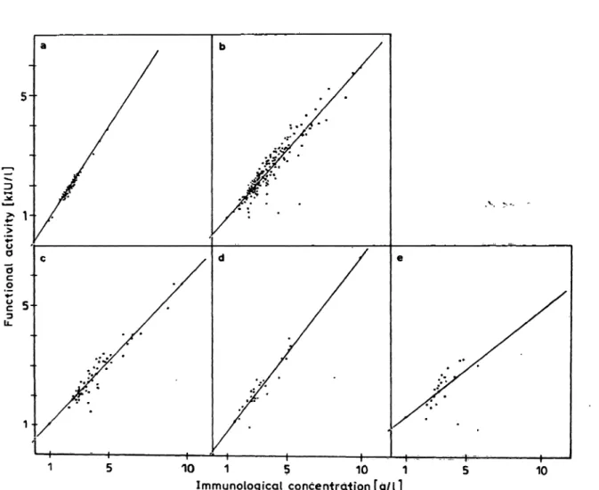
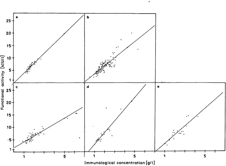
In sera of normal persons the coefficients of correla- tion between the amidolytic and functional assay of αι-proteinase inhibitor and ctz-macroglobulin were r = 0.988 and r = 0.916, respectively (fig. 2a, 3a). It should be noted, and will be shown in more detail below, that the correlations are w rse for patients than for healthy persons (fig. 2b, 3b).
Tab. l. Within-run (n = 20) and day-to-day (n = 20) imprecision of the functional and immunological determination of cxj- proteinase inhibitor and az-macroglobulin. Precichrom I was used for functional determinations the human control plasma, and human serum for the immunological deter- mination. αι-Proteinase inhibitor was assayed with the analyser ACP 5040 (EppendorQj az-macroglobulin was determined manually with an Eppendorf Photometer.
Immunological determinations were pefformed with a laser nephelometer.
αι-Proteinase inhibitor ai-Macroglobulin Assay procedure χ s CV χ s CV Functional
within-fun day-to-day Immunologie within-day day-to^day
(kIU/1) (kIU/1) (%) 1.42 0.03 2.07 1.32 0.06 4.24 (g/l) (g/0 (%) 2,48 0.07 2.70 2.30 0.11 4.86
(kIU/1) (kIU/1) (%) 4.43 0.20 4.6 4.68 0.32 6.8 (g/0 (g/0 (%) 1.73 0.08 4.8 1.76 0.11 6.3
5-
"5c
o
1 1 r—
10 1 5 10 1 Immunological concentration [g/l]
10
Fig. 2. Statistical correlation between the functional activity (chromogenic Substrate assay) and immunological concentration (laser nephelometry) of αι-proteinase inhibitor in human serum from
(a) normal persons (n = 50, r = 0.988, y = 0.760x + 0.054), (b) all patients (n = 183, r = 0.907, y = 0.559x H- 0.394),
(c) patients with carcinoma (n = 61, r = 0.942, y = 0.525x + 0.507), (d) renal diseases (n = 29, r = 0.933, y = 0.647x + 0.157), and
(e) liver diseases (n = 22, r = 0.586, y = 0.394x + 0.928). *'
J. CUn. Chem. Clin. Biochem. / Vol. 22, 1984 / No. 10
Gressner and Peltzer: Determination of ui-proteinase Inhibitor and a2-macroglobulin in serum 637
Reference ranges offunctional concentration and spe- cific Inhibitor activity of a/-proteinase Inhibitor and oL2'macroglobulin
The functional and immunological concentrations and the specific inhibitor activities (proteinase inhib- itor activity/g protein) of αϊ-proteinase inhibitor and ai-macroglobulin in sera of normal persons (n = 50) exhibit a Gaussian distribution (not shown). The ref- erence ranges (± 2 S.D.) of the concentrations and of calculated specific inhibitor activities are listed in table 2.
Functional concentration and specific inhibitor activi- ties of ai-proteinase inhibitor and ai-macroglobulin in patients with various diseases
Using sera from patients afflicted with carcinoma, diabetes mellitus, renal and liver diseases, myocar-
Tab. 2. Reference ranges (± 2 S. D.) of functional (proteinase in- hibitor) activity, immunological concentration, and spe- cific inhibitor activity of α ι -proteinase inhibitor and ua- rnacroglobulin in serum (n = 50).
Proteinase inhibitor Functional Immuno- Specific activity logical con- inhibitor
centration activity (kIU/1) (g/l) (klU/g) ui-Proteinase inhibitor 1.17-2.78 1.48-3.58 0.73-0.84 u2-Macroglobulin 3.59-9.14 1.03-2.36 3.12-4.53
dial infarction and trauma, the two proteinase inhibi- tors were measured with the amidolytic and immu- nologic method and the specific inhibitor activity was calculated. As summarized in table 3 the specific in- hibitor activity of αι-proteinase inhibitor showed a statistically significant (p < 0.05) decrease in all dis-
25-
1 5
Immunological concentration [g/l]
Fig. 3. Statistical correlation between the functional activity (chromogenic Substrate assay) and immunological concentration (lascr nephelometry) of oi2-macroglobulin in human serum from
(a) normal persons (n = 50, r = 0.916, y = 4.025x - 0.316), (b) all patients (n = 153, r = 0.862, y = 3.36x + 0.259),
(c) patients with carcinoma (n = 49, f = 0.858, y = 2.4 H χ 4- 1.694), (d) renal diseases (n = 23, r = 0.978, y = 4.773x - 1.730), and (e) liver diseases (n = 19, r = 0.852, y = 3.616x - 0.380).
J. Clin. Chem. Clin. Biochem. / Vol. 22, 1984 / No. 10
638 Grcssncr and Pcltzcr: Determination of cu-protcinase inhibitof and nu-macroglobulin in seruni Tab. 3. Mcan values ± s. D. f functional activity (kU/1), immu-
nological concentrations (g/l) and specific inhibitor activ- ity (klU/g) of αι-proteinase inhibitor in healthy persons and patients with various diseases. The statistical signifi- cance of the differences between healthy persons and pa- tients was tested (n.s. = not significant).
Disease category
Healthy persons Allpatients
Carci- noma Diabetes mellitus Renal disease Liver disease Myocar- dial in- farction Trauma
Method
functional immunologic specific inhibitor act.
functional immunologic specific inhibitor act.
functional immunologic specific inhibitor act.
functional immunologic specific inhibitor act.
functional immunologic specific inhibitor act.
functional immunologic specific inhibitor act.
functional immunologic specific inhibitor act.
functional immunologic specific inhibitor act.
Num-ber
5050 50 183197 183 6761 61 18 1515 3029 29 2222 22 2223 22 2223 22
Meanvalues
2.531.97 0.78 2.493.80 0.67 2.634.07 0.66 2.11 3.190.71 2.223.22 0.70 2.323.53 0.67 2.42 3.610.67 2.82 4.290.68
S.D.
0.40 0.530.03 0.871.39 0.09 0.87 0.081.49 0.62 0.890.05 0.731.06 0.09 0.590.87 0.13 0.951.32 0.09 1.02 0.061.58
Level of signifi- cance
p < 0.001
< 0.001
< 0.001 p < 0.001
< 0.001
< 0.001 n. s.
p < 0.01
< 0.001 n. s.
p < 0.01
< 0.001 p < 0.01
< 0.001
< 0.001 n. s.
p < 0.001
< 0.001 p < 0.05
<0.01
< 0.001
Tab. 4. Mean values ± S.D. of functional activity (kIU/I), immu- nological concentrations (g/l) and specific inhibitof activ- ity (klU/g) of aa-macroglobulin in healthy persons and patients with various diseases. The statistical significance of the differences between healthy persons and patients . was tested (n.s. = not significant).
Disease category
Healthy persons Allpatients
Carci- noma Diabetes mellitus Renal disease Liver disease Myocar- dial in- farction Trauma
Method
functional immunologic specific inhibitor act.
functionai immunologic specific inhibitor act.
functional immunologic specific inhibitor act.
functional immunologic specific inhibitor act.
functional immunologic specific inhibitor act.
functional immunologic specific inhibitor act.
functional immunologic
specific inhibitor act..
functional immunologic specific inhibitor act.
NunvMean S.D.
ber values
50 5050 153195 153 67 4949 1814 14 3023 23 2219 19 23 . 2020 23 1717
6.501.69 3.83 6.791.92 3.49 6.52 3.361.91 7.011.87 3.62 7.74 3.781.90 8.192.37 3.44 5.59 3.551.65 5.361.60 3.31
0.331.46 0.35 2.640.70 0.62 0.691.93 0.57 2.180.47 0.46 0.833.96 0.49 3.230.77 0.72 0.631.85 0.62 0.411.75 0.64
Level of signifi- cance
n. s.
n. s.
< 0.001 n. s.
n. s.
P n.
n.n.
n.
n.n.
P
n.P n.
Pn.
<
s.s.
s.
s.s.
<
s.<<
<
s.s.
0.001
0.050.01 0.05 0.05
<0.01 s.
P < 0.01
ease conditions, whereas the mean concentration and activity were increased in all groups. It is ob- vious that the elevation of the functional activity is less pronounced than that of the immunological con- centration. Extreme reductions of the specific inhibi- tor activity of cti-proteinase inhibitor (0.24 klU/g) were found in patients with acute pancreatitis and liver cirrhosis. However, this finding is not typical for these diseases, since other patients with similar le- sions did not show such a marked reduction of the specific activity. No significant differences of the specific inhibitor activities were found between the various disease groups.
The statistic correlation of the functional and immu- nologic assay of αι-proteinase inhibitor in diseased individuals is lower than that in healthy persons (fig. 2). A marked discrepancy in the results of both meth ds was observed in patients with acute and chronic liver diseases (r == 0.586). The specific inhib- itor activities of a2-macroglobulin and of ai-protein- ase inhibitor were decreased in rriost patients but sta-
tistically significant changes (p < 0.05) were found only in carcinoma, liver diseases, and trauma (tab. 4). Very large decreases of specific inhibitor ac- tivity were measured in sera of some patients with acute pancreatitis (2.22 klU/g) and CCU poisoning (2.23 klU/g), whereas highly elevated specific inhib- itor activities were found in patients with Morbus Hodgkin (4.98 klU/g), sepsis (4.87 klU/g), and nephrotic syndrome (4.81 klU/g). However, it should be emphasized that these changes are neither disease-typical nor disease-obligatory laboratory findings.
The statistical correlation between both assay princi- ples was lower in patients than in healthy persons.
This effect is pronounced in individuals fflicted with liver diseases (r = 0.852) and carcinoma (r = 0.858)
. 3).
0 In some cases, concentration, inhibitor activity and specific inhibitor activity of both proteinase inhibi- tors were monitored during the course of a disease.
J. Cliii. Chem. Clin. Biochem. / Vol. 22,1984 / No. 10
Gressner and Peltzcr: Determination of u ι-proteinase inhibitor and U2-macroglobulin in serum 639 A patient with acute carbon tetrachloride poisoning,
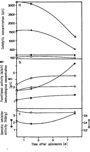
showing extremely elevated catalytic activities of both aminotransferases in serum, exhibited a rather constant specific activity of cn-proteinase inhibitor in the normal r nge. The specific activity of aa-macro- globulin, however, was initially significantly reduced (2.2 klU/g) but increased to normal values when the aminotransferases declined (fig. 4). This change is due to a selective, strong increase of the inhibitor activity of a2-macroglobulin, with only a slight in- crease in the concentration of the protein.
1 3 5 7
Time fter qdmission [d]
Fig. 4. Follow up of the functipnal (inhibitor) activities and im- munological concentrations of αι-proteinase inhibitor and ctz-macroglobulin in serum of a patient with acute CCU poisoning.
(a) catalytic concentrations of alanine-aminotransferase (B—B), aspartate-aminotransferase (A—A), alka- line phosphatase (·—·) and γ-glutamyltfanspepti- dase (O—O) are shown,
(b) functional activity (kIU/1) (·, O) and immunological concentration (g/l) (B, D), of αι-proteinase inhibitor (O, D) and ctz-macroglobulin (·, B).
(c) calculated specific inhibitor activity of α ι-proteinase inhibitor (O—O) and cta-macroglobulin (·—·).
Similar, but uncharacteristic changes of the specific inhibitor activity of ct2-macroglobulin were also no- ticed in the postoperative course and in patients with hepatocellular carcinoma (not shown). These longi- tudinal studies show that the immunoreactive con- centrations and functional activities of the proteinase inhibitors do not behave strictly in parallel during the course of a disease in an individual.
Discussion
The introduction of chromogenic Substrates provides an alternative, i.e. functional approach to the deter- mination of cti-proteinase inhibitor and o^-macro- globulin which is suitable for routine clinical chemi- cal analyses (12—15). The analytic criteria of the amidolytic assay are comparable to those of methods measuring the antigenic levels of the proteinase in- hibitors. In our study the precision of oi2-macroglob- ulin determination was lower than that of arpro- teinase inhibitor, irrespective of the assay principle;
but other authors have reported precision data which are similar to those found for α r proteinase inhibitor (15). The storage conditions of the sera must be con- trolled carefully if both proteins are determined ami- dolytically. cxz-Macroglobulin is obviously more sus- ceptible to in vitro inactivation than αι-proteinase inhibitor. By use of the amidolytic assay additional Information on the functional and pathogenetic sig- nificance of the two proteinase inhibitors can be ob- tained. Whereas immunologic procedures are unable to distinguish between active, non-active and inacti- vated protein molecules the chromogenic Substrate assay measures specifically the biologically relevant, i.e. the antiproteolytic active fraction of the protein.
In agreement with previous reports, healthy persons showed a satisfactory statistical correlation between the immunoreactive and functional concentrations of the proteins (13—15), and the reference r nge for the functional concentration of aa-macroglobulin was similar to that reported previously (15). In pa- tients, however, the correlations are lower, especial- ly in those suffering from acute and chronic liver dis- eases (a r proteinase inhibitor and ci2-macroglobu- lin) and carcinoma (a2-macroglobulin). The reason for this discrepancy is not known at present but vari- able degrees of endogenous complex formation be- tween the inhibitor protein and certain proteinases in serum might be relevant.
Data summarized in table 3 and 4 support the view that a2-macroglobulin, in contrast to α ι-proteinase inhibitor, does not behave like an acute phase reac- tant in humans (22). The specific inhibitor activity of the macroglobulin is changed only insignificantly un-
J. Clin. Chem. Clin. Bipchem. / Vol. 22,1984 / No. 10
640 Gressncr and Pcltzer: Determinalion of tt|-proteinase Inhibitor and <X2-macroglobulin in serurn
der various disease conditions, whereas that of otr proteinase Inhibitor is significantly reduced in all dis- ease categories. This is due to differences between the relative increase of the functional activity and immunoreactive concentration in acute phase reac- tions (e.g. trauma, carcinoma, myocardial infarc- tion). The finding might be explained by two mecha- nisms.
a) accumulation of functional inactive fraction of -proteinase inhibitor, which retains füll immu- noreactivity under these conditions. The inacti- vation of ctrproteinase inhibitor might be pro- duced by oxidation of a methionine residue near the proteinase inhibitory site, possibly by oxygen free radicals (23—25). It is known that oxygen free radicals generated by complement activated neutrophils or other processes are important me- diators of tissue injury in a great variety of dis- eases (26, 27).
b) Variable degrees of endogenous Saturation of the proteinase inhibitor capacity. cti-Proteinase inhi- bitor constitutes 90% of human serum elastase inhibitor capacity (4). This proteinase (together with others) is released from neutrophils duririg inflammation and is coftsequ£ntly trapped by ar proteinase inhibitor, leaving it aiitigeriically in- tact but functiorially inactive.
It is not possible to decide which of the two mecha- nisms is operative under various disease conditions.
The relative constancy of the specific inhibitor activi- ty of (Xa-macroglobulin can be explained by the effec- tive hepatic clearance of even low concentrations of
<X2-macroglobülin-proteinase complexes from the circulation with a half-life in the order of 3 to 4 min- utes (28,29), whereas that of a2-macroglobuliii itself is much longer (30). This mechanism prevents the accumulation of proteinase-burdened, i.e. inactive ct2-macroglobulin in the circulation.
References
J. Laskowski, M., Jr. & Kato, I. (1980) Ann. Rev. Biochem. 49, 593-626.
2. Travis, J. & Salvesen, G. S. (1983) Ann. Rev. Biochem. 52, 655-709.
3. Schwick, H. G. & Haupt, H. (1980) Angew. Chemie 92, 83- 95.
4. Heimburger, N., Haupt. H. & Schwick, H. (1971) In: Pro ceed. Internati. Res. Conference on Proteinase Inhibitors (Fritz, H. & Tschesche, H., eds.) pp. 1-21, de Gruyter, Ber- lin.
5. Laurell, C.-B. & Jeppsson, J.-O. (1975) In: The Plasma Pro- teins: Structure, Function, and Genetic Control (Putnam, F.
W., ed.) 2nd ed., Vol. l, pp. 229-264. Academic Press New York.
6. van Leuven, F. (1982) Trends in Biochemical Sciences 7, 185-187.
7. Starkey, P. M. (1979) In: The Physiological Inhibition of Blood Coagulation and Fibrinolysis (Collen, D., Wimand, B.
& Verstraete, M., eds.) pp. 221-230, EIsevier/North Hol- land, Amsterdam.
8. Harpel. P. C. (1979) In: The Physical Chemistry, Molecular Biology and Physiological Function of Human Plasma Pro- teins (Bing, D. H., ed.) pp. 385-399, Pergamon, .New York.
9. Starkey, P. M. & Barrett, A. J. (1977) In: Proteinases in Mammalian Cells and Tissues (Barrett, A. J., ed.) pp. 663- 696, North Holland, Amsterdam.
10. Barrett, A. J. & Starkey, P. M. (1973) Biochem. J. 133, 709- 724.
11. Cullmann, W. (1982) Lab. Med. 6, 187-191.
12. Ganrot, P. O. (1966) Clin. Chim. Acta 14, 493-501.
13. Dick, W. & Cullmann, W. (1982) J. Clin. Chem. Clin. Bio- chem. 20, 57-60.
14. Cullmann, W. & Dick, W. (1981) J. Clin. Chem. Clin. Bio- chem. 19, 287-290.
15. Witt, I. & Tritschler, W. (1983) J. Clin. Chem. Clin. Bio- chem. 27, 429-436.
16. Conrad, A., Schürmann, J., Kreutz, F. H. & Sieber, A.
(1978) J. Clin. Chem. Clin. Biochem. 76, 299-305.
17. Sieber, A. (1977) Laboratoriurnsblätter 27, 109-118.
18. Hoffmann, J. J. M. L., van den Broek, W. G. M. & Jansen, A.
P. (1976) Clin. Chim. Acta 77, 251-259.
19. Recommendations of the German Society for Clinical Chem- istry (1972) Z. Klin. Chem. Kim. Biochem. 10, 281-291.
20. Persijn, J. P. & van der Slik, W. (1976) J. Clin. Chem. Clin.
Biochem. 14, 421-427.
21. Sachs, L. (1972) Statistische Auswertungsmethoden. Sprin- ger Verlag, 3. Auflage.
22. Koj, A. (1974) In: Structure and Function of Plasma Proteins (Allison, A. C., ed.), pp. 73-125. Plenum Press, London.
23. Carp, H. & Janoff, A. (1979) J. Clin. Invest. 63, 793-797.
24. Janoff, A., Carp, H., Lee, D. K. & Drew, R. f. (1979) Science 206, 1313-1314.
25. Gadek, J. E., Fellis, G. A. & Crystal, R. G. (1979) Science 206, 1315-1316.
26. Ward, P. A., Till, G. O., Kunkel, R. & Beauchamp, C. (1983) J. Clin. Invest. 72, 789-801.
27. Freeman, B. A. & Crapo, J. D. (1982) Lab. Invest. 47, 412- 426.
28. Ohlsson, K. (1971) Acta Physiol. Scand. 81, 269-272.
29. Ohlsson, K. (1971) Scand. J. Clin. Lab. Invest. 25,219-223.
30. Imber, M. J. & Pizzo, S. V. (1981) J. Biol. Chem. 256,8134- 8139.
Prof. Dr. A. M. Gfessner Abteilung für Klinische Chemie und Zentrallaboratorium
Klinikum der Philipps Universität Marburg Baidingerstraße
D-3550 Marburg/Lahn
J. Clin. Chem. Clin. Biochem. / Vol. 22, 1984 / No. 10