Mäder and Dickmann: Choline acetyltransferase activity in human cerebrospinal fluid and serum 857 J. Clin. Chem. Clin. Biochem.
Vol. 26, 1988, pp. 857-861
© 1988 Walter de Gruyter & Co.
Berlin · New York
A Reevaluation of Choline Acetyltransferase Activity in Human Cerebrospinal Fluid and Serum
By M. Mäder and -U. Dickmann
Neurologische Klinik der Universität Göttingen
(Received November 4, 1987//March 31/September 12, 1988)
Summary: Choline acetyltransferase (EC 2.3.1.6) was studied in human cerebrospinal fluid and blood.
Sensitivity, precision and reproducibility of the radioactive assay were evaluated with a homogenate from pig brain and with partially purified enzymes from bovine brain and human placenta. Contrary to other reports, with the assay used in the present study, choline acetyltransferase activity was not detectable in the cerebro- spinal fluid or blood of 50 patients with various neurological disorders. Only a very slight but significant non-enzymatic formation of a radioactive product by concentrated cerebrospinal fluid was observed. Non- enzymatic but enzyme-like formation of acetylcholine by thiol reagents and other compounds is discussed.
Introduction
So far, only a few attempts, with conflicting results, have been made to detect choline acetyltransferase activity in human cerebrospinal fluid and blood (1 —3). As choline acetyltransferase is regarded to be specifically localized in cholinergic neurons, its changes in cerebrospinal fluid and serum might reflect destruction of nervous tissue (4).
Previously, choline acetyltransferase activity was termined preferentially by radipchemical methods.
Labeled acetylcholine is formed from labeled acetyl- CoA and choline (5—7). The labeled product has to be separated from the labeled substrate. This was achieved by ion exchange chromatography, two phase extraction and by combining both techniques. These procedures are time consuming and cumbersome, which may well explain the lack of investigations of choline acetyltransferase activity in human body fluids (4).
In 1975 Fonnum (8) published an improved method for choline acetyltransferase measurement which in- volves liquid cation exchange in a two-phase system with direct counting. This is achieved by using the toluene scintillation-mixture as an extraction solvent and by carrying out the extraction directly in the
scintillation vial. This method was supposed to be rapid, precise and sensitive (8). We considered it worthwhile to attempt the detection of choline ace- tyltransferase activity in cerebrospinal fluid and serum with the improved method.
Materials and Methods
Chemicals
Choline bromide, eserine (physostigmine) and tetraphenylboron were purchased from Sigma (München). EDTA (ethylenedini- trilo) tetraacetic acid, sodium salt, (Titriplex III), acetonitrile, toluene and salts for preparing buffers were purchased from Merck (Darmstadt). Cibacron Blue F3GA (C. I. Reactive Blue, Procion Blue H-BS) was purchased from Serva (Heidelberg).
Acetyl-Coenzyme A (acetyl-CoA) was purchased from Boeh- ringer (Mannheim). Coenzyme A (acetyl-l-14C) acetyl (1.48
— 2.22 GBq/mmol) and Omnifiuor (pre-mixed LSC Powder) were purchased from NEN (New England Nuclear, Dreieich).
Choline acetyltransferase partially purified from bovine brain (lyophilized powder containing 5—20% protein) and from hu- man placenta (suspension in 2.8 mol/1 (NH4)2SO4) were pur- chased from Sigma (München).
Enzyme preparation
Whole pig brains from the slaughter house were placed on ice within 30 min after death. All subsequent procedures were carried out at 4 °C. After removal of the meninges and addition of 1 1 extraction medium (phosphate-buffered saline pH 5.0), J. Clin. Chem. din, Biochem. / Vol. 26,1988 / No. 12
sodium phosphate buffer pH 7.4 and dialysed overnight against the same buffer with one change of buffer. The crude enzyme preparation (pig brain) was then stored in aliquots below
-25°C.
Enzyme assay
Choline acetyltransferase activity was assayed according to the method of Fonnum (8) with minor modifications. The final volume of the reaction mixture was 10 μΐ containing 1,85 kBq (50 nCi) [MC]acetyl CoA, 0.4 mmol/1 acetyl CoA, 300 mmol/1 sodium chloride, 50 mmol/1 sodium phosphate buffer pH 7.4, 8 mmol/1 choline bromide, 20 mmol/1 EDTA (pH 7.4) and 0.1 mmol/1 physostigmine. Enzyme solution (2.5 μΐ) was added to the substrate solution (7.5 μΐ) in 0.5 ml Eppendorf tube. The lubes were placed in a water bath at 22 °C or 37 °C. The reaction was stopped after 10 min (if not stated otherwise) by addition of 90 μΐ 10 mmol/1 sodium phosphate buffer pH 7.4. A total of 100 μΐ was then transferred to a scintillation vial (20 ml) containing 5ml 10 mmol/1 sodium phosphate buffer pH 7.4.
Acetonitriie (2ml) containing 10 mg tetraphenylboron was added and after brief mixing, 10 ml toluene scintillation mixture were then added. The vials were shaken slightly for 15 seconds and the two phases were allowed to separate for 10 minutes before counting.
Results and Discussion
To measure choline acetyltransferase activity the very sensitive assay method of Fonnum (8) which is widely accepted today (9) was adapted with minor modifi- cations (see Material and Methods). This radioactive assay involves three major steps: incubation of reac- tion mixture, separation of radioactive product from radioactive substrate and -counting. Each step has
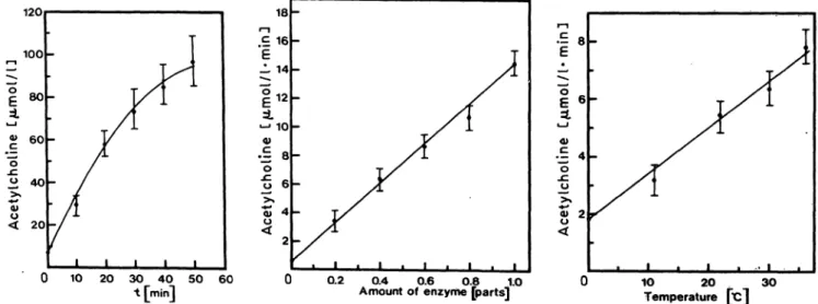
time and temperature under optimal conditions is shown. The typical decrease of the reaction velocity of choline acetyltransferase after 20 min, due to en- zyme inhibition by accumulation of free Coenzyme A (6), is also depicted in figure 1. Measurements with pig brain homogenates were found to be similar to those with partially purified enzymes from human placenta or bovine brain.
When testing choline acetyltransferase activity in pooled fractions of cerebrospinal fluid and serum (10 patients each) we could not detect any formation of radioactive acetylcholine, either in unboiled or boiled samples (3). Tenfold concentrated body fluids also showed no activity in the assay. Our results are thus, at first glance, in contradiction to those of 1. c. (1—3).
These authors report extremely low amounts of ace- tylcholine formed in cerebrospinal fluid. But Aquilo- nius & Eckernas (3) suggested that about 95% of the product measured was due to non-enzymatic conver- sion of the substrate.
Due to these ambiguous results we studied choline acetyltransferase activity in cerebrospinal fluid and serum in more detail. When partially purified enzyme from human placenta or bovine brain was added either to cerebrospinal fluid, serum or phosphate buffer, choline acetyltransferase activity stayed un- changed at 4 °C for 7 days and at 22 °C for 24 hours (data not shown). As choline acetyltransferase activity is usually measured at 37 °C, we incubated samples
120
100
οΕ
60
< 20ο
10 20 30 40 50 t [min]
60
ι-ι ,18
.£ 16 E -L 14
i 2
«-» 10 .Ξ βφ
Ο
Ι
6-Ε 4ο _cυ
s
20.2 0.4 0.6 Ο.8 1.0
Amount of enzyme [parts] 10 20
Temperature 30
Fig. 1. Acetylcholine formation by pig brain homogenate in relation to time, amount of enzyme (parts of enzyme preparation) and temperature as monitored by the Fonnum assay (see Materials and Methods) under optimal conditions The reaction time was 10 min if not otherwise indicated. . .
Mäder and Dickmann: Choline acetyltransferase activity in human cerebrospinal fluid and serum 859
0.50- 0.40- 0.30- 0.20- 0.10-
60 Fig. 2. Stability of choline acetyltransferase
a) Incubation of enzyme from human placenta in phosphate buffer at 0 °C — o—, 22 °C — —, 37 °C — o —.
b) Incubation of enzyme from pig brain — o —, bovine brain — A —, and human placenta — o— in serum at 37 °C.
c) Incubation of enzyme from human placenta in cerebrospinal fluid with dithiothreitol (1 mmol/1) — o— and without dithiothreitol -o— at 37 °C.
at 0 °C, 22 °C, and 37 °C for 1 hour and recorded choline acetyltransferase activity every 15 minutes. A sharp decline of choline acetyltransferase is noticed (fig. 2 a) at 37 °C. This applies particularly to serum and cerebrospinal fluid (data not shown). The decline of enzyme activity is fastest for human placenta fol- lowed by bovine brain and pig brain (fig. 2b). The degree of acetyltransferase inactivation is thus due to the matrix (serum, cerebrospinal fluid or buffer) and to the source of enzyme. When using preheated (70 °C, 30 min) body fluids the decrease of choline acetyltransferase did not alter. Thus, the biological inactivation of acetyltransferase (e.g. by proteases) can be ruled out.
We noticed then that pig brain homogenates prepared by addition of thiol reagents (ß-mercaptoethanol or dithiothreitol) were less or not at all affected in the experiments described above. Thiol reagents are reg- ularly used in choline acetyltransferase purification procedures to stabilize the enzyme (10, 11). When thiol reagents (e. g. l mmol/1 dithiothreitol) are added to serum or cerebrospinal fluid before the enzyme the decrease of activity is completely stopped as shown in figure 2 c. From this result we draw the conclusion that some labile thiol groups of the enzyme (10, 12) will be oxidized chemically in body fluids at 37 °C if not protected sufficiently by addition of a thiol re- agent. This suggestion is in good accord with the fast decline of enzyme activtiy immediately after incuba- tion (fig. 2). Inactivation of choline acetyltransferase in body fluids may be explained in this way. The various degrees of inactivation that occurred with the partually purified enzymes from different species may be explained by different quantities and qualities of inherent protecting factors. It is well known (12, 9)
that choline acetyltransferase becomes more and more labile during purification if thiol reagents are not added.
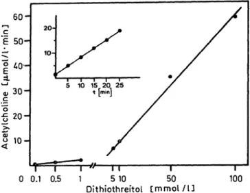
We do not know whether acetyltransferase inherent in body fluids — if existing at all — will be protected from autoxidation. Addition of thiol reagents to body fluids immediately after tapping in order to stabilize inherent choline acetyltransferase seems not advisable because thiol reagents themselves produce choline ace- tyltransferase-like activity in the Fonnum assay as shown in figure 3. This fact has to our knowledge not been described to date in the literature. The choline acetyltransferase-like activity depends linearly on the amount of thiol reagent and the reaction time (fig.
3). As Roskoski et al. (12) suggest, an acetyl-thioen- zyme may be involved as an intermediate in the cho- line acetyltransferase reaction mechanism. It seems that thiol reagents may replace this intermediate by possible forming an acetyl thio-compound. The latter has not been characterized. Acetyltransferase-like ac- tivity was low when 1 mmol/1 or less thiol reagent was used (fig. 3), and therefore it was negligible in the experiments described above where high amounts of enzyme activity were added to the body fluids. But thiol reagents will cause serious problems if traces of inherent choline acetyltransferase activity are to be measured. The minimum amount of dithiothreitol for stabilizing choline acetyltransferase (human placenta) in cerebrospinal fluid during 20 min is 0.1 mmol/1.
Even this amount produces measurable quantities of radioactive acetylcholine (fig. 4).
In the following investigations the body fluids were cooled to 4 °C or frozen at —25 °C immediately after veni or lumbar puncture of the patients. Before testing choline acetyltransferase activity in cerebrospinal
J. Clin. Chem. Gun. Biochem. / Vol. 26, 1988 / No. 12
c 30-
"ο .cjj
>»
Ο)ο
<
20-
0.1
r
"T"0.5
-"—n
Dithiothreitol5 10 50
1
[mmol /U
T"
100
Fig. 3. Non-enzymatic acetylcholine formation in the presence of dithiothreitol in relation to its concentration and the reaction time (insert, 5 mmol/1). In the Fonnum assay (see Materials and Methods) the enzyme sample was replaced by a solution of dithiothreitol.
Fig. 4. Stability of choline acetyltransferase of human placenta in cerebrospinal fluid with various amounts of dithio- threitol added. 1 mmol/1 -Δ-, 0.1 mmol/1 -o-, or 0.01 mmol/1 -o-. Insert: choline acetyltransferase like activity caused by 1 ml of 0.1 mmol/1 dithiothreitol.
fluid, the samples were concentrated tenfold as re- ported by other authors (1, 3), using a Speed Vac apparatus (Savant) which evaporates the solvent un- der vacuum while rotating the samples. All solvents will be concentrated equally. This technique is thought to be mild. But when choline acetyltransferase was added before concentration the recovery of activity was only about 20% of the calculated tenfold aug- mented value, which in turn was only twice that of.
the unconcentrated fluid. Inactivation and denatura-
detect choline acetyltransferase activity seems to be limited.
Cerebrospinal fluid and serum samples from 50 con- secutive patients were studied for choline acetyltrans- ferase activity under conditions just characterized.
Each sample, unconcentrated and tenfold concen- trated, was tested at 22 °C and at 37 °C for 10 and 20 min. None of the samples really showed an enzyme- like reaction. A very faint but significant increase in radioactive acetylcholine could be observed at 37 °C with the concentrated samples (start: 205 ± 41, after 20 min: 248 ± 41 nmol/1 · min, n = 50). This low product formation was never found with serum or with blanks. The values measured were in the range of those found by Aquilonius & Eckernas (3), Johnson
& Domino (1), Rimon et al. (2). To check the possibility of an extremely faint enzymatic activity we therefore performed experiments to compare heated and un^
heated samples of cerebrospinal fluid with those in- hibited with Cibacron Blue. Cibacron Blue is a potent inhibitor of choline acetyltransferase (12). In this ex- periment all samples except blanks show a similar increase in radioactivity (fig. 5). This result indicates a very low non-enzymatic formation of product. Cho- line acetyltransferase activity in cerebrospinal fluid described in the past may fall into this category. As
20 Fig. 5. Acetylcholine formation by cerebrospinal fluid. Blank
(phosphate buffer, pH 7.4) — o—, unboiled — ο—, boiled ^A —. Cibacron Blue inhibited cerebrospinal fluid — Q—, serum —D —. *?
Mäder and Dickmann: Choline acetyltransferase activity in human cerebrospinal fluid and serum 861
Aquilonius & Eckernas (3) have already pointed out, non-enzymatic formation of acetylcholine is possible, e. g. by imidazole. As we have shown, it is also possible by thiol reagents.
In conclusion no measurable amounts of choline ace- tyltransferase activity can be determined in human cerebrospinal fluid or serum with the very sensitive assay of Fonnwn (detection limit, 0.5 — 1.0 / 1 · min acetylcholine formation). There is thus a chal-
lenge to develop new methods based on immuno techniques, which will be capable of detecting traces of choline acetyltransferase molecules, even if the enzyme is inactivated.
Acknowledgement
This work was supported by the Deutsche Forschungsgemein- schaft. (SFB 330).
References
1. Johnson, S. & Domino, E. F. (1971) Clin. Chim. Acta 35, 421 -428.
2. Rimon, R., Puhakka, P., Venäläinen, E. & Mandell, A. J.
(1973) Psychiatria Fennica 19, 265-267.
3. Aquilonius, S. M. & Eckernas, S. A. (1976) J. Neurochem.
27, 317-318.
4. Banik, N. L. & Hogan, E. L. (1983) Cerebrospinal fluid enzymes in neurological disease. In: Neurobiology of cere- brospinal fluid (Wood, J. H., ed.) Vol. 2, Plenum Press New York, p. 211.
5. McCaman, R. E. & Hunt, J. M. (1965) J. Neurochem. 12, 253-259.
6. Schrier, B. K. & Shuster, L. (1967) J. Neurochem. 14, 977- 7. Fonnum, F. (1969) Biochem. J. 775, 465-469.985.
8. Fonnum, F. (1975) J. Neurochem. 24, 407-409.
9. Eckenstein, F., Barde, A. A. & Thoenen, H. (1981) Neu- roscience 6, 993 — 1000.
10. Chao, L. P. (1980) J. Neurosci. Res. 5, 85-115.
11. Peng, J. H., McGeer, P. L., Kimura, H., Sugn, S. C. &
McGeer, E. G. (1980) Neurochem. Res. J, 943-962.
12. Roskoski, R., Lim, C. H. T. & Roskoski, L. M. (1975) Biochemistry 14, 5105-5110.
13. Scopes, R. (1982) Protein Purification. Springer-Verlag New York Heidelberg Berlin.
Dr. M. Mäder
Neurologische Klinik der Universität Robert-Koch-Straße 40
D-3400 Göttingen
J. Clin. Chem. Clin. Biochem. / Vol. 26, 1988 / No. 12