Mating-induced central release of oxytocin:
Implication for social fear extinction.
DISSERTATION ZUR ERLANGUNG DES DOKTORGRADES DER NATURWISSENSCHAFTEN (DR. RER. NAT.) DER FAKULTÄT FÜR
BIOLOGIE UND VORKLINISCHE MEDIZIN DER UNIVERSITÄT REGENSBURG
vorgelegt von Cindy Grossmann
aus Castres, Frankreich
im Jahr 2020
Das Promotionsgesuch wurde eingereicht am:
Die Arbeit wurde angeleitet von: Prof. Dr. rer. nat. Inga D. Neumann
Unterschrift:
Dissertation
Durchgeführt am Institut für Zoologie der Universität Regensburg
Lehrstuhl für Tierphysiologie und Neurobiologie
unter Anleitung von
Prof. Dr. rer. nat. Inga D. Neumann
Contents
4
TABLE OF CONTENTS
Table of contents ___________________________________________________________ 4 Abstract ___________________________________________________________________ 7 Introduction ______________________________________________________________ 10
Emotions ________________________________________________________________ 10
Anxiety and fear __________________________________________________________ 11
Anxiety disorders __________________________________________________________ 11
Modeling anxiety disorders in rodents _________________________________________ 13 4.1. Measuring anxiety in rodents _____________________________________________________ 13 4.2. The Cued Fear Conditioning (CFC) _________________________________________________ 14 4.3. The Social Fear Conditioning (SFC) _________________________________________________ 15 Regulation of anxiety and fear: focus on Oxytocin (OXT) __________________________ 16 5.1. The OXT system ________________________________________________________________ 16 5.2. OXT in anxiety and fear __________________________________________________________ 19 5.3. Brain regions involved in anxiety and fear ___________________________________________ 20 5.3.1. Amygdala _____________________________________________________________________ 20 5.3.2. Lateral Septum ________________________________________________________________ 23 Studying the OXT system: an endogenous model of OXT release in male _____________ 25 6.1. Male sexual behavior in mice: from female encounter to ejaculation _____________________ 26 6.2. Neuronal circuit of male sexual behavior ____________________________________________ 29 6.3. Effect of sexual behavior on anxiety and fear ________________________________________ 31 6.4. Regulation of male sexual behavior by the PVN and OXT _______________________________ 32
Aims of the present thesis ___________________________________________________ 35 Materials and methods _____________________________________________________ 36 Animals__________________________________________________________________ 36
Behavioral techniques ______________________________________________________ 36
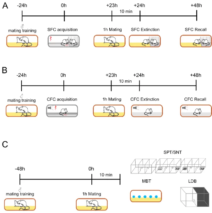
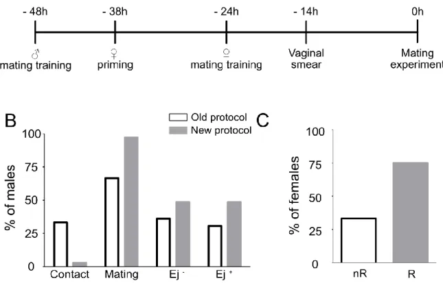
2.1. Mating experiments ____________________________________________________________ 36
2.2. Experimental design of the behavioral experiments ___________________________________ 37
2.3. Social Fear Conditioning paradigm (SFC) ____________________________________________ 39
2.4. Cued-Fear Conditioning (CFC) _____________________________________________________ 40
2.5. Social Preference test (SPT) and Social Novelty test (SNT) ______________________________ 41
2.6. The light/dark box (LDB) _________________________________________________________ 42
Contents
5
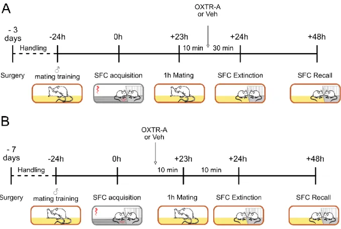
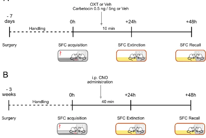
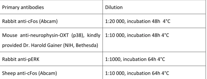
2.7. The marbles burying test ________________________________________________________ 42 Pharmacology and stereotactic techniques _____________________________________ 43 3.1. Experimental design of the pharmacological and chemogenetic experiments ______________ 43 3.2. Stereotactic guide cannulas and microdialysis probe implantations and intracerebral infusion 45 3.3. Intracerebral infusions __________________________________________________________ 45 3.4. Virus microinfusion and chemogenetic experiments __________________________________ 47 3.5. Microdialysis for measuring OXT release ____________________________________________ 47 Histological and microscopy techniques ________________________________________ 48 4.1. Sacrifice and transcardial perfusion ________________________________________________ 48 4.2. Immunohistochemistry __________________________________________________________ 48 4.3. Image Analysis and Cell Quantification _____________________________________________ 51 Statistical analysis _________________________________________________________ 52
Results ___________________________________________________________________ 53
Investigation of the effect of sexual behavior on social fear in male mice: involvement of the OXT system ________________________________________________________________ 53
1.1. Establishment of the priming and mating experiment _________________________________ 53 1.2. Behavioral characterization of the effect of mating on anxiety-like, fear and social behavior __ 54 1.3. Summary of the behavioral experiments ____________________________________________ 63 1.4. Neuronal activation pattern during SFC extinction ____________________________________ 63 1.5. OXT release during mating behavior _______________________________________________ 66 1.6. i.c.v. administration of OXTR-A after mating prior social fear extinction ___________________ 68 1.7. Septal administration of OXTR-A before mating prior social fear extinction ________________ 70 1.8. Differential neuronal activation during sexual behavior and SFC extinction ________________ 73 1.9. Summary _____________________________________________________________________ 75 Involvement of the OXT system within the CeA in social fear extinction ______________ 76 2.1. Pharmacological manipulation of the OXTR into the CeA _______________________________ 77 2.2. Chemogenetic activation of the OXTR neurons into the CeA ____________________________ 83 2.3. Summary _____________________________________________________________________ 85
General discussion _________________________________________________________ 86
Effect of sexual behavior on social fear in male mice: involvement of the OXT system __ 86
1.1. Effect of mating on emotional, social and socio-emotional behavior ______________________ 86
1.2. Neuronal activity during social fear extinction _______________________________________ 88
1.3. OXT release during mating behavior _______________________________________________ 90
1.4. Effects of blockade of OXTR after mating prior social fear extinction _____________________ 91
Contents
6
1.5. LS administration of OXTR-A after mating prior social fear extinction _____________________ 92 1.6. Differential neuronal activation during sexual behavior and SFC extinction ________________ 93 1.7. Conclusion and future studies ____________________________________________________ 94 1.8. Outlook ______________________________________________________________________ 95 Involvement of the OXT system into the CeA in social fear extinction ________________ 97 2.1. Effects of OXT infusion into the CeA on social fear ____________________________________ 97 2.2. Chemogenetic activation of the OXTR neurons into the CeA ____________________________ 99 2.1. Conclusion and future experiments ________________________________________________ 99 2.2. Outlook _____________________________________________________________________ 100
Bibliography _____________________________________________________________ 101
List of Abbreviations _______________________________________________________ 119
List of Figures ____________________________________________________________ 121
List of tables _____________________________________________________________ 122
Acknowledgements _______________________________________________________ 123
Curriculum Vitae __________________________________________________________ 125
Abstract
7
ABSTRACT
Anxiety disorders are one of the most common psychiatric disorders. Among them, social anxiety disorder (SAD), or social phobia, are characterized by persistent fear and avoidance of social situations. Unfortunately, treatments for SAD are rather unspecific as patients are generally treated with anxiolytics and/or antidepressant drugs combined with cognitive behavioral therapy. However, a lot of patients are resistant to these treatments or relapse once the treatment is over. This, combined with the high prevalence of SAD, highlights the emergency to develop new treatment strategies more specific to SAD. However, to find new potential targets for SAD treatments, animal models are needed to understand the underlying mechanisms of SAD. One of the major restrictions of SAD research was the lack of animal models. In 2012, the model of social fear conditioning (SFC) has been developed specifically to study SAD. After conditioning, the conditioned mice (SFC
+) develop social avoidance, a sign of social fear, and the main symptom of SAD. This is easily measurable during the social fear extinction training, mimicking the cognitive behavioral therapy. The social fear extinction training consists of several presentations of unknown conspecifics. At the first presentation, the fearful mice show low investigation time (social avoidance). After several presentations of social stimuli, the investigation time of the mice increases, the sign of social fear extinction.
Oxytocin, well-known for its prosocial properties, is also a puissant anxiolytic. Previous studies found a crucial role of the neuropeptide oxytocin in social fear. Oxytocin was shown to facilitate social fear extinction when infused within the lateral septum (LS). Moreover, lactating females, which are a model for an upregulation of the OXT system, did not show social fear. Moreover, an infusion of an OXT receptor antagonist (OXTR-A) into the LS of lactating mice induced the expression of social fear during social fear extinction. However, lactation is a model for chronic upregulation of the OXT system as the number of OXT fibers and the OXTR binding are increased. In order to study the effect of an acute endogenous release of OXT, mating in male mice is a more adapted model. Indeed, an acute release of OXT within the paraventricular nucleus of the hypothalamus was found in male rats during mating.
During my PhD thesis, my first aim was then to investigate the effect of mating on social fear
extinction in male mice. Male mice were allowed to mate during 1h before to be subjected to
social fear extinction. Interestingly, I found a facilitation of social fear extinction exclusively
Abstract
8 after successful mating (with ejaculation, Ej
+). The mice that did not ejaculate prior to social fear extinction (Ej
-) did not show this facilitation. I then characterized the effect of mating on cued fear, general anxiety, and social preference/novelty to determine if the benefic effect of ejaculation was specific to social fear. Male mice were allowed to mate during 1h before to undergo cued fear extinction, light-dark box, marbles burying test, and social preference/novelty test. No effect of mating without or with ejaculation has been found on cued fear extinction. I found a general anxiolytic effect of mating (Ej
-and Ej
+) on general anxiety. I found no changes in social preference nor social novelty. Ej
+mice showed a facilitation of social fear extinction, which cannot be explained by the general anxiolytic effect of mating, with and without ejaculation, or an increased social motivation, as no specific effect of ejaculation was found in the social preference test. Next, I investigated the mechanisms underlying social fear extinction. For that, I conducted an experiment aiming to measure the level of the protein cFos, product of an immediate-early gene and thereby marker for neuronal activity, to evaluate which brain regions are recruited during the different stages of social fear extinction. Male mice were sacrificed either at the first stimulus (SFC
-and SFC
+mice) or the last stimulus (only SFC
+) of the social fear extinction. I found an increased cFos protein level within the OXT neurons of the PVN and SON, and within the central amygdala (CeA) after the last stimulus of the extinction in SFC
+mice. I also found a decreased level of cFos in SFC
+mice at the first stimulus of the extinction in comparison with SFC
-mice in the ventral part of the LS. To assess the involvement of OXT in mating behavior, I measured the release of OXT within the LS and CeA during mating behavior using microdialysis. I found a specific release of OXT during mating behavior within the LS, but not within the CeA. In the next experiment, I aimed to inhibit the effect of the release of OXT during mating by blocking the OXTR with an antagonist. I first infused i.c.v. an OXTR-A immediately after mating before social fear extinction. Both Ej
-and Ej
+showed an increased social fear expression during extinction. In order to block more specifically the OXTR during mating, I infused an OXTR-A within the LS before mating prior social fear extinction. No effect of the OXTR-A could be found. The OXT release during mating within the LS is then not or not the only reason for the facilitation of the extinction observed after ejaculation.
In the second part of my PhD work, I aimed to investigate the mechanism underlying social
fear extinction. The CeA is well known for its role in fear conditioning and expression. In the
Abstract
9 context of social fear, I previously found an increased cFos expression in the CeA the last social stimuli of social fear extinction in SFC
+mice. This implies an involvement of the CeA in the extinction of social fear. Also, an upregulation of the OXTR binding was found after social fear acquisition in SFC
+mice, suggesting a regulation of the OXT system during social fear acquisition. I then aimed to investigate the involvement of the OXT system within the CeA during social fear extinction. I first infused synthetic OXT within the CeA before social fear extinction. I found a slight facilitation of social fear extinction. In order to confirm this result, I conducted a similar experiment in which I infused a specific OXTR agonist. However, I could not repeat the previously obtained result. In the last experiment, I used chemogenetic to specifically activate the OXTR-positive neurons within the CeA before social fear extinction.
However, I could not find any effect of CeA OXTR-positive neurons stimulation on social fear extinction.
Altogether, I showed that ejaculation during mating specifically facilitates the extinction of social fear. However, I could not identify the circuit directly responsible for this effect yet.
Independently of mating behavior, I could show the recruitment of the CeA in social fear
extinction. More experiments need to be conducted to understand the role of OXT in the CeA
in social fear extinction.
Introduction
10
INTRODUCTION
Emotions
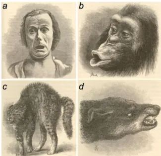
As described by Darwin in “The Expression of the Emotions in Man and Animals” in 1872, emotions can be observed across several species (Figure 1) (Darwin, 1872). However, whether animals experience emotions in the same manner as humans and how to define them is still under debate. Emotions enable the adaptation of the behavioral response towards a specific stimulus. They are essential for survival and they ask the capability to identify an external stimulus, to assess the internal state, and to combine the environmental context and the previous experiences to direct an appropriate behavioral response. Hence, the expression of one emotion is the result of the detection and assessment of the nature of one specific cue combined with the internal body state and the previous experience of the subject. An emotion is expressed by a behavioral, hormonal, and autonomic response (LeDoux, 2000; Anderson and Adolphs, 2014; Barrett and Satpute, 2019). A very prominent section of emotion research is focused on anxiety and fear due to his high relevance for survival.
Figure 1. Expression of emotions observed by Charles Darwin in different species. a, terror in humans. b, disappointment in chimpanzee. c, hostility in cat. d, hostility in dog (Anderson and Adolphs, 2014).
Introduction
11 Anxiety and fear
In the field of emotion, anxiety and fear are probably the most studied as they are highly relevant for survival. The manifestations of anxiety and fear are very similar, however, they are triggered by different stimuli. In 1993, Blanchard et al. attempted to describe the difference between anxiety and fear. Fear is a response to imminent danger, the threat is then physically present and well defined. Anxiety is a response to potential and subjective threats.
Fear could then be defined as a response to an immediate threat while anxiety is a response to a distant or future threat. In rodents, anxiety and fear are manifested by different defensive behaviors depending on the context. When the predator or threat is distant, flight or avoidance can be observed. If no escape is possible, a freezing response will be then observed.
If the predator is getting closer, this passive behavior change in an active defensive threat or attack. Thus, the behavioral response depends on both the threat proximity and the environment. Other defensive behaviors can also be observed like ultrasonic vocalization, defensive burying, or startle response in response to a threat (Blanchard et al., 1993).
Occasional anxiety and fear are beneficial for the survival of an individual, enabling fast responses to possible threats. However, a persistent state of anxiety or a fear response towards non-threatening stimuli can be qualified as pathological. Pathological anxiety and fear are detrimental to the health of an individual, as anxiety and fear are conflicted with other behavior such as feeding, mating, or social interactions among others.
Anxiety disorders
Pathological anxiety and fear, i.e. anxiety disorders, cause personal distress, reduce the quality
of life, impair social functioning, and increase even the risk of suicide. The term “anxiety
disorders” represents a large variety of disorder as general anxiety disorder (chronic
apprehension and anxiety), specific phobia (fear about a specific object/situation), social
anxiety disorder (SAD, fear of social situations), post-traumatic stress disorder (avoidance and
fear of stimuli related with a previous trauma) just to name the most prominent ones
(American Psychological Association (APA), 2013). In Europe, the prevalence to develop any
kind of anxiety disorder is 14.5% according to the European Study of the Epidemiology of
Mental Disorders. Within the broad spectrum of anxiety disorders, specific phobia,
generalized anxiety disorder, and SAD are the more common (Baldwin et al., 2013; Bandelow
Introduction
12 and Michaelis, 2015). Anxiety disorders can be grouped in two kinds of clinical entities: the ones which are characterized by an elevated fear for specific stimuli (e.g. phobias), and the ones which are characterized by an elevated anxious apprehension. However, anxiety and fear are the major symptoms described in the definition of most of the anxiety disorders. In the DSM V, specific phobia is described as a “Marked fear or anxiety about a specific object or situation (e.g. flying, heights, animals receiving an injection, seeing blood). The phobic object or situation almost always provokes immediate fear or anxiety”. Another relevant example is the definition of SAD. In the DSM IV, SAD was described as a “marked and persistent fear of one or more social/performance situations in which the person is exposed to unfamiliar people or possible scrutiny by others”. This definition has changed in the DSM V to include also anxiety: “Marked fear or anxiety about one or more social situations in which the individual is exposed to possible scrutiny by others” (American Psychological Association (APA), 2013; Substance Abuse and Mental Health Services Administration, 2016). Even though they have distinct definitions, anxiety and fear cannot be distinguished at a clinical level as both of them are generally expressed in anxiety disorders. SAD is particularly relevant in the context of my thesis, I will now describe it more in detail.
SAD, also called social phobia, is defined by a persistent fear in certain or all social situations.
The person suffering from SAD fears being judged, humiliated, or rejected. According to the DSM V, a specific form of SAD is the so-called performance anxiety and is defined by fear of giving a speech or performing on stage (American Psychological Association (APA), 2013). The main symptom of SAD is the avoidance of social situations, which is often accompanied by physical symptoms such as sickness, increased heart rate. Moreover, the majority of SAD patients develop other psychiatric disorders such as depression or substance abuse. The 12- months prevalence for SAD is between 0.6-7.9 % in Europe depending on the country (Wittchen and Jacobi, 2005; Wittchen et al., 2011) and 6.8% in the US (Kessler et al., 2005).
The treatments for SAD are non-specifics and have been developed first to treat general
anxiety disorder or depression. These include selective serotonin inhibitors, serotonin-
norepinephrine reuptake inhibitors, and benzodiazepines for the most prescribed. Other
treatments that can be proposed are tricyclic antidepressants, monoamine oxidase inhibitors
or, more unusual treatments like β-adrenergic blockers and anticonvulsants. However, 50% of
relapse has been observed less than 2 months after the end of the treatment (Blanco et al.,
Introduction
13 2013; Williams et al., 2017). That is why pharmacotherapy is often combined with cognitive behavioral therapy, a therapy combining problematic thinking to eliminate negative thoughts and desensitization exposure training, which consists of gradual exposure to anxiety- provoking situations. This combination gives better results and decreases the rate of relapse after treatment (Goldin et al., 2014; Stangier, 2016).
Due to the high prevalence, anxiety disorders are a burden for society. In particular, in the actual context of the COVID-19, the prevalence of anxiety disorders might significantly increase in the post-illness stage (Rogers et al., 2020). Moreover, the prolonged social isolation during the lockdown might increase the probability to develop SAD (Holmes et al., 2020).
Anxiety and fear have been extensively studied in preclinical research, however, the question of how dysregulations in the brain lead to anxiety disorder needs a better understanding on a translational level. Moreover, novel treatments are needed to compensate for the lack of specific treatment for anxiety disorder like SAD. That is why, models were developed in rodents in order to investigate the mechanisms underlying fear and anxiety, and their dysregulations, leading to anxiety disorders.
Modeling anxiety disorders in rodents
To study anxiety disorders in preclinical studies with laboratory animals and understand their underlying mechanisms, animal models were developed. A good animal model has to fulfill 3 criteria. It should first develop the same symptoms as the human disorder, both at a physiological level and at a behavioral level (face validity). Then, the model should be sensible to pharmacological treatments used in patients (predictive validity). And finally, the causality should be the same, meaning that the neurobiological processes involved in anxiety should be the same in the animal model than in humans (construct validity).
4.1. Measuring anxiety in rodents
Anxiety depends on cues or stimuli that cannot be predicted. In this case, the stimulus elicits a state of anxiety over a long period which decays slowly after the removal of the stimulus (De Jongh et al., 2003).
To measure anxiety, a broad spectrum of tests and paradigms were developed. Most of these
tests are based on ethology, as they involved innate anxiety in rodents like exploring new,
Introduction
14 bright, and open environments where they are more vulnerable to predators. These tests are conflictual for the rodents, as they oppose their unconditioned general exploratory drive with their innate anxiety of open spaces. Among others, the elevated-plus maze, the open field, and the light/dark box (LDB) are the most commonly used (Campos et al., 2013). The elevated plus-maze consists of two closed arms and two open arms elevated from the ground. Anxious rodents avoid the open arms (Walf and Frye, 2007). Regarding the open field, this paradigm consists of one big open area. Anxious rodents prefer to stay at the edges rather than investigate the bright and unprotected center of the arena (Gould et al., 2009). In the present thesis, I used the LDB to measure anxiety in mice, this paradigm is composed of one dark, i.e.
“safe” chamber and one bright, i.e. “anxiogenic” chamber. Thus, anxious animals spend more time within the dark chamber than the bright chamber (Griebel et al., 2000; Hascoët and Bourin, 2009; Miller et al., 2011). All these tests were shown to be sensitive to anxiolytic drugs.
However, these tests have the weakness to be sensible to locomotion deficit. Indeed, an animal showing low locomotion can be interpreted as highly anxious while an animal showing high locomotion can be interpreted as non-anxious. Another way to test for anxiety is to use active avoidance tasks, in which rodents try to minimize the threatening stimuli by, for example, burying it. In the shock probe burying (also called defensive burying) paradigm, rats bury an electrified probe after receiving a footshock (Treit et al., 1981; Treit, 1990). This test is mostly used in rats, however, few studies could validate it in mice (Sluyter et al., 1996;
Degroot and Nomikos, 2004). In the marbles burying test, the rodent spontaneously buries non-noxious stimuli such as marbles. This test is more common in mice (Deacon, 2006), however, it was shown that rats also bury non-noxious stimuli (Poling et al., 1981). All these tests are sensitive to anxiolytic drugs.
4.2. The Cued Fear Conditioning (CFC)
Based on associative memory, fear can be induced by a short and discrete cue paired with an aversive stimulus. When the cue is presented, it elicits a state of fear that begins and dissipates quickly once the stimulus is removed. In this context, fear conditioning was extensively used to investigate fear behavior and fear-related circuitries in the brain of rodents.
One of the most famous paradigms is the cued fear conditioning (CFC). It is a type of classical
conditioning, also called respondent or Pavlovian conditioning paradigm. The principle of CFC
is based on the association of two independent stimuli. Before conditioning, one stimulus,
Introduction
15 called unconditioned stimulus (US; a footshock), is able to elicit an unconditioned fear response (e.g.freezing). The other stimulus is neutral (for example a tone), it doesn´t lead to any behavioral response. During the conditioning, the neutral stimulus is presented at the same time as the US. The presence of the US is naturally leading to the expression of the unconditioned fear response. After several US-CS pairings, the CS is presented alone and, if the associative learning happened during the conditioning, it should lead to an equivalent response than the unconditioned response, i.e. freezing, which is then called a conditioned response. A procedure of fear extinction then generally takes place. This procedure aims to induce a new learning, where the animal learns that the CS is not linked with the US anymore.
During extinction, the CS is then presented alone several times. When extinction is successful, the test animal should present a lower level of fear response at the end of the process than initially. The CFC is mostly used to investigate the basis of fear learning and fear extinction. It is also used to investigate anxiety disorders such as general anxiety disorder or post-traumatic disorder. However, this model is not ideal for investigating a disorder like SAD, as it doesn´t induce social avoidance, the main symptom of SAD.
4.3. The Social Fear Conditioning (SFC)
As previously explained, specific treatment options are lacking for SAD. Studying the underlying mechanisms of SAD is then crucial. However, no appropriate model was available to study this disorder. Several models allow to induce social avoidance in rodents as acute or chronic social defeat or foot-shock exposure (Haller and Bakos, 2002; Louvart et al., 2005;
Huhman, 2006). However, these paradigms are unspecific and induce also general anxiety-
and depression-like symptoms. That is why the SFC paradigm was developed in 2012 in our
lab (Toth et al., 2012a). Based on the operant conditioning or instrumental conditioning, this
type of conditioning induces an association between an active behavior and a consequence
that can be either positive (reward) or negative (punishment), allowing either to exacerbate
or to inhibit a specific behavior. The SFC, which was develop in mice, associates the
investigation of a conspecific with a foot shock, leading to a decreased social investigation
(social avoidance). Similarly to the CFC, this conditioning is followed by an extinction training
procedure where several unknown conspecifics are presented. The mouse then has the choice
to investigate the social stimulus or to avoid it. Once the mouse recognizes that the social
investigation is no longer paired with the foot shock, the social fear is gradually extinguished
Introduction
16 as the investigation time of the social stimulus is increased. The extinction training in socially fearful mice mimics the cognitive behavioral therapy in SAD patients. This procedure allows us to generate generalized social fear in mice, without alteration in anxiety- or depressive-like behavior (face validity). After the acquisition, social fear lasts for at least 2 weeks. Moreover, antidepressant or anxiolytic treatments reverse the SFC-induced social fear (predictive validity) (Toth et al., 2012a). The SFC paradigm is nowadays the best option to study the neuronal substrates of SAD. The mechanisms underlying both SAD in humans and social fear in mice are not well understood. However, the hypothesis of the involvement of the neuropeptide oxytocin (OXT) is growing (Marazziti et al., 2015). Indeed, a study found lower plasmatic OXT levels in SAD patients while they were performing a pro-social task in comparison with healthy controls (Hoge et al., 2012). Using SFC, a release of OXT release within the lateral septum (LS) was found in control mice but not in socially fearful mice during social fear extinction (Zoicas et al., 2014). In contrast, two other studies found elevated plasma OXT in SAD patients in comparison to healthy controls (Hoge et al., 2008; Oh et al., 2018).
However, the sampling of OXT was not done during a social task in these last two studies, which might explain the difference in the results. Altogether, both SAD patients and social fear conditioned mice seem to present a dysregulation of the OXT system (construct validity?).
Regulation of anxiety and fear: focus on Oxytocin (OXT) 5.1. The OXT system
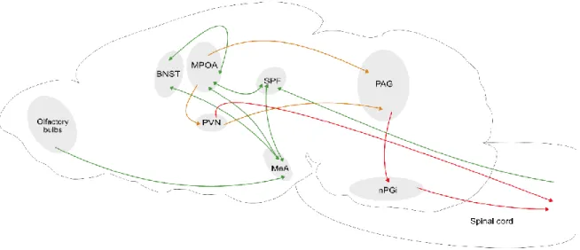
The nonapeptide OXT was discovered in 1909 by Sir Henry H. Dale, and later, its structure was characterized by Du Vigneaud in 1953 (Du Vigneaud et al., 1953). This highly conserved neuropeptide is synthesized within the paraventricular (PVN), supraoptic (SON) and accessory nuclei of the hypothalamus in mammals. The magnocellular neurons of the PVN and SON project to the neurohypophysis, where OXT is released into the bloodstream. OXT is also released by parvocellular neurons of the PVN. This neuronal population is distinct from the magnocellular neurons as they have a smaller size and do not project to the neurohypophysis.
Parvocellular OXT neurons of the PVN project to extrahypothalamic regions including the
brainstem and the spinal cord to regulate autonomic function like pain, analgesia and penile
erection (Wagner and Clemens, 1993; Ackerman et al., 1997; Gerendai et al., 2001; Eliava et
al., 2016). Parvocellular neurons project also to other hypothalamic and extra-hypothalamic
Introduction
17 nuclei (Armstrong et al., 1980; Swanson and Kuypers, 1980; de Vries and Buijs, 1983;
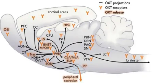
Sofroniew, 1983; Swanson and Sawchenko, 1983; Knobloch et al., 2012). OXT projections from these nuclei are found in numerous brain areas including the septal area, the bed nucleus of the stria terminalis, the nucleus accumbens, the ventral hippocampus, the medial amygdala (MeA), and the central amygdala (CeA)(Neumann and Landgraf, 1989; Knobloch et al., 2012;
Menon et al., 2018; Ferretti et al., 2019) (Figure 2). The central release of OXT within the PVN and SON is due to somato-dendritic release, while the release in the limbic regions is due to axonal release (Ludwig and Leng, 2006). The release of OXT is stimulus- and region-dependent.
Using microdialysis sampling, the intracerebral release of OXT was detected in several brain regions (Neumann, 2007) including the PVN/SON in response to swimming in male rats and maternal aggression in female rats (Wotjak et al., 1998; Bosch et al., 2005; Engelmann et al., 2006; Torner et al., 2017), the septum during suckling in lactating rats (Neumann and Landgraf, 1989), social defeat in male rats (Ebner et al., 2000) and social memory retrieval in male rats (Lukas et al., 2013a), social fear extinction in male and female mice (Zoicas et al., 2014; Menon et al., 2018), the dorsal hippocampus during suckling in lactating rats (Neumann and Landgraf, 1989), the CeA during forced swim test and maternal aggression in rats (Bosch et al., 2005;
Ebner et al., 2005). Optogenetic stimulation of the hypothalamic OXT terminals in rats was
also able to trigger a release of OXT within the CeA (Knobloch et al., 2012; Ferretti et al.,
2019). Studies in other species like sheep allowed to identify other regions where OXT is
released, e.g. the bed nucleus of the stria terminalis (BNST), the medial preoptic area (MPOA),
and the olfactory bulb in parturient ewes, during suckling or, during vaginocervical stimulation
(Kendrick et al., 1988, 1992; Lévy et al., 1995). In humans, a peripheral release of OXT has been
detected in plasma and saliva in response to stress like running (Landgraf et al., 1982; de Jong
et al., 2015). The release of OXT also occurs in response to martial art (Rassovsky et al., 2019)
or during a psychosocial test Trier Social Stress Test) (Pierrehumbert et al., 2010). Except
during social interactions and stress, the intracerebral release of OXT also occurs following
strong physiological stimuli. Well known in females, lactation and birth are two particularly
strong stimuli that induce the release of OXT in the PVN and SON (Neumann et al., 1993). In
males, a central endogenous release of OXT occurs during mating, like in the PVN for example
(Waldherr and Neumann, 2007).
Introduction
18 Endogenously release of OXT expresses its effect usually via the OXT receptor (OXTR). The OXTR is a G protein-coupled transmembrane receptor, which is highly expressed throughout the social brain network and many other brain areas like the CeA, basolateral nuclei of the amygdala (BLA), MeA, nucleus accumbens, BNST, PVN, MPOA, hippocampus, periaqueductal gray, LS, or olfactory bulbs (Brinton et al., 1984; De Kloet et al., 1985; Elands et al., 1988;
Tribollet et al., 1988) (Figure 2). The broad distribution of the OXTR within the brain, as well as the numerous OXT projections, enable OXT to modulate the expression of social behavior, as well as other behaviors like fear and anxiety.
Figure 2. Scheme of OXT projections, sites of OXT release , and OXTR expression within the brain.
AON, anterior olfactory nucleus; OB, olfactory bulb; OT, olfactory tubercle; Nac, nucleus accumbens;
OVLT, organum vasculosum laminae terminalis ; SON, supraoptic nucleus; PVN, paraventricular nucleus; PP, posterior pituitary; PFC, prefrontal cortex; CC, cingulate cortex; MPOA, medial preoptic area; BNST, bed nucleus of the stria terminalis; LS, lateral septum; CPu, caudate putamen; PV, periventricular nucleus of the thalamus; CeA, central amygdala; MeA, medial amygdala; BLA, basolateral amygdala; VTA, ventral tegmental area; LC, locus coeruleus; PBN, parabrachial nucleus;
DRN, dorsal raphe nucleus; PAG, periaqueductal gray; SN, substantia nigra; HPC, hippocampus; HDB, nucleus of the horizontal limb of the diagonal band. Figure from (Grinevich and Neumann, 2020).
After binding to its receptor OXT modulates social behaviors such as social approach (Lukas et al., 2011), social memory and recognition (Engelmann et al., 1998; Lukas et al., 2013a), maternal aggression (Bosch et al., 2005), aggression (Calcagnoli et al., 2015), male and female sexual behavior (Arletti and Bertolini, 1985; Arletti et al., 1985; Waldherr and Neumann, 2007;
Nyuyki et al., 2011) and pair-bonding (Williams et al., 1994; Johnson et al., 2016). Aside from
its pro-social effects, OXT also regulates anxiety and fear, this will be discussed in the next
section.
Introduction
19 5.2. OXT in anxiety and fear
The role of OXT in the regulation of stress and anxiety has been well studied. Stressful situations induce the release of OXT both at the periphery and in the brain. For example, in rats, 10 minutes of forced swim induces the peripheral release of OXT into the bloodstream (Wotjak et al., 1998, 2001; Torner et al., 2017), as well as a central release into the PVN/SON (Wotjak et al., 1998, 2001; Torner et al., 2017) and the CeA (Ebner et al., 2000). Also, acute social defeat induces OXT release into the bloodstream, within the hypothalamus (Engelmann et al., 1999) and the LS (Ebner et al., 2000). In accordance, OXT deficient mice showed increased anxiety-like behavior in the elevated plus-maze, a novel environment, and a platform shaker (Mantella et al., 2003; Amico et al., 2004). In addition, OXTRs are expressed in key brain regions involved in the neurocircuitry of anxiety and fear like the prefrontal cortex, the hippocampus, or the amygdala. Hence, OXTR antagonist (OXTR-A) infused either i.c.v. or into the PVN increases the hypothalamic–pituitary–adrenal axis response during stress in the elevated plus-maze, and the forced swim test in male rats (Neumann et al., 2000). Chronic intracerebral infusion of OXT via minipumps (Windle et al., 1997) or OXT infusion into the CeA (Bale et al., 2001) decreases the stress response and anxiety-like behavior in female rats and mice. Moreover, the administration of OXT within the PVN decreases anxiety-like behavior in rats in the elevated plus-maze and the LDB by activating the mitogen-activated protein kinase cell signaling pathway (Blume et al., 2008). In the prelimbic cortex, OXT infusion induces anxiolysis by recruiting GABAergic neurons to modulate downstream regions like the CeA (Sabihi et al., 2014, 2017). In humans, intranasal administration of OXT was also shown to reduce the stress response (Heinrichs et al., 2003; Kirsch et al., 2005) and to reduce aversion to angry faces (Evans et al., 2010). All in all, endogenous OXT is a puissant anxiolytic in both rodents and humans.
Aside from anxiety, OXT also plays a role in the regulation of fear behavior. In CFC, i.c.v. OXT
infusion before fear extinction impairs the extinction. However, when infused before the
acquisition, OXT does not affect fear acquisition but facilitates fear extinction (Toth et al.,
2012b). After contextual fear memory retrieval, microinfusion of OXT, or specific agonists
within the infralimbic region of the medial prefrontal cortex facilitates extinction (Lahoud and
Maroun, 2013). The OXT infusion within the BLA before fear memory retrieval increases
Introduction
20 freezing and impairs fear extinction. Infusions before fear memory retrieval into the CeA does not affect fear extinction. When infused before conditioning into the BLA, OXT decreases the freezing response and facilitates extinction, while, when infused into the CeA, OXT has no effect. However, agonists of the OXTR show different effects than OXT in this study. When infused before fear retrieval into the BLA, OXTR agonists induce a decrease in fear responses during extinction, and, when infused before conditioning into the CeA, they facilitate fear extinction (Lahoud and Maroun, 2013). In another study, an infusion of OXT before extinction reduces fear expression during the extinction. However, OXT impairs the acquisition of context fear when infused into the BLA before the acquisition, reflected by the low level of freezing during extinction. A local infusion or an optogenetically stimulated release of OXT within the CeA suppresses the expression of contextual fear (Viviani et al., 2011; Knobloch et al., 2012). The different effects of OXT into the amygdala sub-regions on fear extinction are puzzling and need more investigation. Moreover, an acute social defeat before context fear conditioning enhanced fear memory, an effect mediated by the OXTR into the LS (Guzmán et al., 2013). However, OXT infusion in the LS decreases fear expression in contextual fear conditioning after positive social exposure (Guzmán et al., 2014). It seems then that OXT effect depends on the valence of the stimulus presented before extinction. In the context of social fear, OXT is released within the LS during social fear extinction and facilitates social fear extinction in both lactating female and male mice (Zoicas et al., 2014; Menon et al., 2018).
Moreover, social fear alters OXTR binding in several brain regions. An increase in OXTR binding is found after conditioning within the dorsal LS (dLS) and the right CeA (Zoicas et al., 2014). In SAD patients, the intranasal administration of OXT dampens the amygdala hyperreactivity (Labuschagne et al., 2010; Dodhia et al., 2014; Gorka et al., 2015).
5.3. Brain regions involved in anxiety and fear 5.3.1. Amygdala
The case of the patient S.M. suffering from a bilateral destruction of the amygdala, described
in 1994, introduced the relevance of the amygdala for emotions. This patient suffers from
bilateral destruction temporal lobes containing the amygdala. The most remarkable change in
behavior was the lack of fear (Adolphs et al., 1994). Brown and Schäfer in 1988, followed by
Klüver and Bucy in 1937, already described a lack of fear as a consequence of a bilateral
ablation of the temporal lobe in Rhesus monkey, the so-called Klüver-Bucy syndrome.
Introduction
21 The amygdala can be divided into several sub-nuclei: the MeA, the BLA, and the CeA nucleus.
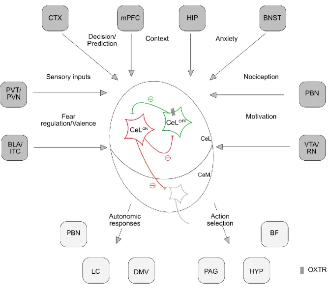
I will here focus on the BLA and CeA as these two regions are participating in the regulation of anxiety and fear. The BLA is mostly constituted of glutamatergic projection neurons and inhibitory interneurons. It receives sensory information from the cortices and the thalamus about the environment but also the hippocampus or the prefrontal cortex. The BLA sends projections to structures like the nucleus accumbens, the BNST, and CeA to then, adapt the behavior in response to a fearful stimulus. Some assume that the CeA plays only the role of a relay between the BLA and the output region, but the CeA also receives direct sensory information and can regulate some behavior via projections to the brainstem independently from the BLA (Pliota et al., 2018). The CeA was strongly investigated concerning its role in defensive behavior and anxiety (LeDoux, 2000). The CeA receives a large variety of inputs including sensory and nociceptive information via projections from the thalamus, the ventral hippocampus, the parabrachial nucleus, or the BLA among others (Figure 3). Thanks to all these connections, the CeA is important for associative learning like conditioning. Even though the CeA is a rather small structure, the microcircuit contained inside is complex. Mainly composed of inhibitory neurons, the CeA can be divided into a medial (CeM) and a lateral (CeL) part (Jolkkonen and Pitkänen, 1998). The CeL is composed of different types of cells, which can be classified depending on their molecular specificity: somatostatin-positive (SOM), corticotropin-releasing factor-positive (CRF) and protein kinase C isoform delta-positive (PKC δ) cells. These cells can be divided also depending on their response to fear stimuli: some cells are activated in response to a conditioned stimulus (CeL
ON) while the others are inhibited (CeL
OFF) (Ciocchi et al., 2010). Interestingly, the CeL
OFFneurons have been identified as the PKC δ-positive cells (Haubensak et al., 2010). All these cells form a micro-inhibitory circuit sending projections to the CeM that modulates fear expression by sending projections to the hypothalamus, the PAG, and the brainstem (Ciocchi et al., 2010; Haubensak et al., 2010; Fadok et al., 2017). The theory of a reciprocal inhibitory circuit would be that the CeL
ON(SOM+) and CeL
OFF(PKC δ+) are connected in a way that one inhibited the other allowing a rapid behavioral change. For example, the activation of the SOM-positive cells induces a freezing response while the activation of the PKC δ-positive cells suppresses freezing (Fadok et al., 2017).
Regarding the CRF-positive cells, they also share reciprocal inhibitory connections with the
SOM-positive cells and promote active defensive behavior.
Introduction
22 The BLA and CeA receive OXTergic projections from the PVN (Knobloch et al., 2012; Ferretti et al., 2019). Both the BLA and the CeA express in abundance the OXTR. Within the CeA, OXTRs are particularly localized in the CeL. Interestingly, the cells expressing the OXTR have been identified as the CeL
OFFor PKC δ-positive cells (Haubensak et al., 2010). Indeed, OXT elicits the activation of approximately 20% of the CeL neurons and inhibits about 50%, leading in fine to the suppression of fear expression (Huber et al., 2005). Moreover, the responses of CeA neurons to OXT show a desensitization of the neurons, as more the neurons are stimulated more the response decreases in brain slices from virgin and lactating rats. This effect is particular to the CeA, as it is not present in the MeA (Terenzi and Ingram, 2005). The action of OXT in the amygdala seems to be time- and region-dependent. In the BLA, an infusion of OXT before conditioning enhances context fear expression during the extinction in rats (Lahoud and Maroun, 2013). However, in another study also in rats, OXT infusion into the BLA impairs acquisition, reflected by a decreased context fear expression (Campbell-Smith et al., 2015).
When infused before context fear extinction into the BLA, OXT and OXT agonists decrease fear expression (Lahoud and Maroun, 2013; Campbell-Smith et al., 2015), this effect can be blocked by an OXTR-A infusion (Campbell-Smith et al., 2015). OXT infused into the CeA before contextual fear conditioning does not affect fear acquisition. However, OXT agonists infusions impair context fear acquisition, reflected by a decreased contextual fear expression during extinction (Lahoud and Maroun, 2013). Campbell-Smith et al. also shown an impairment of contextual fear acquisition by OXT infusion into the CeA (Campbell-Smith et al., 2015). While no effect was found on contextual fear extinction after OXT infusion before extinction (Lahoud and Maroun, 2013), another study showed an increased expression of contextual fear after pre-extinction OXT infusion. This effect could be blocked by OXTR-A, and an OXTR-A infusion alone abolished the expression of fear during extinction (Campbell-Smith et al., 2015).
Optogenetically stimulated release of OXT within the CeA suppresses the expression of
contextual fear (Viviani et al., 2011; Knobloch et al., 2012). Several hypotheses have been
advanced to explain the multiple effects of OXT in the amygdala, which might vary depending
on the protocol, time and doses used. In the context of SFC, OXT is crucial for social fear
extinction. Lactating female mice, a model of high activity of the OXT system, do not show
social fear (Menon et al., 2018). The possible role of OXT within the CeA in SFC has not yet
been studied. However, an increase in OXTR binding was found in the right CeA after
conditioning. This might imply a role of the OXTR in social fear conditioning or expression. In
Introduction
23 humans, intranasal OXT reduces the activation of the amygdala in response to fear in healthy people and SAD patients (Kirsch et al., 2005; Petrovic et al., 2008; Labuschagne et al., 2010;
Dodhia et al., 2014).
Figure 3. Connectivity of the CeA. The CeA receives multiple connections from various brain regions to coordinate an appropriate response face to a threat. BF, Basal forebrain; BLA, Basolateral nucleus of the amygdala; BNST, Bed nucleus of the stria terminalis; CTX, cortices; DMV, Dorsal motor nucleus;
HIP, Hippocampus; HYP, Hypothalmus; ITC, Intercalated cells of the amygdala; LC, Locus coeruleus;
PAG, Periaqueductal grey; PBN, Parabrachial nucleus; mPFC, medial prefrontal cortex; PVN, Paravevntriclar nucleus of the hypothalamus; PVT, Paraventricular nucleus of the thalamus, RN, Raphe nucleus; VTA, Ventral tegmental area. Figure adapted from (Fadok et al., 2018).