Differential effects of central oxytocin on social versus cued fear in male rodents
DISSERTATION ZUR ERLANGUNG DES DOKTORGRADES DER NATURWISSENSCHAFTEN (DR. RER. NAT.) DER FAKULTÄT DER
BIOLOGIE UND VORKLINISCHEN MEDIZIN DER UNIVERSITÄT REGENSBURG
vorgelegt von Iulia Toth aus Temeschburg, Rumänien
im Jahr 2012
Das Promotionsgesuch wurde eingereicht am: 04.10.2012 Die Arbeit wurde angeleitet von: Prof. Dr. Inga D. Neumann Unterschrift
Table of contents
Abstract ………... 1
Chapter 1 General introduction ……….... 4
1.1. Fear and the fear response ………... 5
1.2. Social anxiety disorder ………... 7
1.3. Post-traumatic stress disorder ……….… 8
1.4. Neurocircuitry of SAD and PTSD ………..…... 9
1.5. Treatment of SAD and PTSD ………...……….... 12
1.6. Oxytocin ………...……… 13
1.7. Animal models used to study SAD and PTSD ………... 16
1.7.1. Social defeat ……….... 16
1.7.2. Cued fear conditioning ………....… 17
1.8. Aim of the present thesis ………... 19
1.9. Outline of the present thesis ………... 20
Chapter 2 Social fear conditioning: a novel and specific animal model to study social anxiety disorder ………... 21
Chapter 3 Social fear conditioning as an animal model of social anxiety disorder ………... 46
Chapter 4 Brain oxytocin in social fear conditioning and its extinction: involvement of the lateral septum ………... 69
Chapter 5 Central administration of oxytocin receptor ligands affects cued fear extinction in rats and mice in a time-point-dependent manner ………... 91
Chapter 6 General discussion ………...……….... 110
6.1. Summary of results ………... 111
6.2. Social fear conditioning as an animal model of SAD ……… 113
6.3. Relevance of the social fear conditioning model for future research ……….… 114
6.4. Effects of central OT system manipulation on social and cued fear …………. 117
6.4.1. Effects of exogenous OT on social and cued fear …………..….….. 117
6.4.2. Effects of exogenous OT on learning ……… 118
6.4.3. Effects of endogenous OT on social and cued fear …………..….… 119
6.5. Neurocircuitry of social and cued fear ………..……. 120
6.6. A possible mechanism underlying OT effects on social and cued fear .…..….. 123
6.7. Relevance of the present thesis ……….……... 126
6.8. Perspective for future studies ……….……….... 127
References ……….………... 133
Abbreviations …………..……….... 154
Acknowledgements …..………... 157
Curriculum Vitae ………..………..… 159
Publications ………..… 161
Abstract
Fear is an adaptive emotional response to threatening situations that is crucial for the survival of the individual. In human anxiety disorders, such as social anxiety disorder (SAD) and post- traumatic stress disorder (PTSD), fear is unreasonable and excessive and becomes, therefore, maladaptive. SAD and PTSD are both characterized by debilitating fear and avoidance of fearful situations. These patients show similar dysfunctions in the activity of the brain regions implicated in the fear circuitry and are treated with similar psycho- and pharmacotherapy.
However, the medication for SAD and PTSD is rather unspecific and, despite considerable efforts, their efficacy is unsatisfactory. The neuropeptide oxytocin (OT) has been suggested as a possible therapeutic agent for SAD and PTSD, as it facilitates a wide variety of social behaviors and decreases stress and anxiety in both humans and rodents.
As the fear responses, the fear circuitry, and the behavioral and physiological effects of OT are highly conserved among mammalian species, findings from animal models of SAD and PTSD might provide important information for clinical trials using OT. The cued fear conditioning paradigm has proven to be a powerful model to study the normal and pathological processes involved in fear learning and extinction (the gradual decrease in the fear response as result of repeated exposure to the feared situation), and models some symptoms of PTSD. This paradigm implies exposing animals to a well-defined cue, usually a tone or light, which co-terminates with a foot shock, and results in specific fear of this certain cue. Although several paradigms have been shown to induce social avoidance and fear in addition to other behavioral deficits, there are no appropriate and specific animal models available to study SAD.
Therefore, the three main aims of this thesis were (1) to develop and validate an animal model that mimics the main behavioral symptom of SAD, namely social fear and avoidance, and (2)
to use this paradigm, which I termed “social fear conditioning”, to assess the therapeutic efficacy of OT in reversing social fear. Finally, in order to compare OT effects on social and non-social fear, I aimed (3) to assess the effects of OT in cued fear in an animal model of PTSD, i.e. the cued fear conditioning paradigm.
I could show that specific social fear can be induced in naïve mice by administering electric foot shocks when they approach and actively investigate a con-specific, i.e. a social stimulus.
Social fear was expressed as reduced investigation of unfamiliar social stimuli and aversive responses towards them, such as freezing, stretched approaches, and defensive burying.
Furthermore, the social fear was expressed for at least two weeks and sensitized over time; it was only triggered by social stimuli and did not lead to other behavioral deficits, such as fear of novelty, general anxiety, depression, or impaired locomotion. Moreover, I could demonstrate the predictive validity of the social fear conditioning model by showing that social fear is reversed by acute diazepam and chronic paroxetine treatment, pharmacotherapy currently used in SAD patients.
Having achieved the first aim, I could then show that central infusion of synthetic OT before the extinction procedure, which would be the comparable time-point for psychotherapy in SAD and PTSD patients, reversed social fear, but impaired extinction of cued fear, indicating that OT might represent a promising therapeutic approach for SAD, but not PTSD patients when administered at this time-point. Both the reversing effect on social fear and the impairing effect on cued fear extinction were mediated by the OT receptors (OTR), as central administration of an OT receptor antagonist (OTR-A) prior to OT blocked the observed effects. Furthermore, blockade of OT neurotransmission with OTR-A before the extinction procedure impaired social investigation in unconditioned animals, indicating that the endogenous OT system is needed for naturally-occurring social investigation. In contrast,
blockade of OT neurotransmission at this same time-point did not affect cued fear extinction, indicating that the endogenous OT system is not involved in this process.
Administration of OT before cued fear conditioning, however, facilitated, whereas OTR-A impaired cued fear extinction 24 h later, indicating that an activated endogenous OT system during traumatic events is likely to attenuate formation of fear memories. As both the conditioning and extinction procedure involve learning, these results suggest that exogenous OT impairs learning processes that occur during non-social fearful situations.
In an attempt to identify the neurocircuitry of social fear, I could show that social fear was accompanied by alterations in the brain OT system at the level of the limbic system, namely by an increased OTR binding in the bilateral dorso-lateral septum (DLS), right central amygdala, and right hippocampus (dentate gyrus, cornu ammunis 1). Importantly, these alterations normalized after extinction of social fear, suggesting that OTR expression within these brain regions might be involved in the development and/or neural support of social fear.
Based on these OTR alterations, I could localize the effects of OT on social fear within the DLS by bilateral administration.
Taken together, I could show that OT has a differential effect on social versus cued fear in rodents, at least when administered before the extinction procedure. This result raises attention to the importance of the feared situation when recommending OT for the treatment of SAD and PTSD. Thus, OT represents a promising therapeutic approach for disorders associated with social deficits, such as SAD and possibly PTSD due to social trauma, but caution is needed before recommending OT for the treatment of PTSD due to non-social trauma. Furthermore, I could identify the DLS as a part of the brain network involved in mediating OT effects on social fear.
Chapter 1
General introduction
Fear and anxiety are crucial emotional behaviors that are expressed to some degree in all chordates. Although many definitions exist, most of these agree that fear is a normal emotional response to a real external threat or danger, whereas anxiety is an emotional response to a potential threat or danger (McNaughton and Zangrossi, 2008). From an ethological perspective, both fear and anxiety are highly adaptive responses, whereas in modern psychiatry fear is generally regarded as adaptive, while anxiety is generally regarded as maladaptive. For the purposes of this thesis, the term “fear” will refer to a response to threat in both humans and animals. The term “anxiety” will refer to a pathological condition in humans, and to an unconditioned fear response (see Table 1) in animals.
The behavioral patterns, functions, and mechanisms of fear and anxiety received intense scientific attention, mainly fueled by the desire to understand and ameliorate human anxiety disorders. In this chapter, I will shortly address the behavioral and physiological fear responses, as well as their function and underlying neural circuitry. I will describe the symptoms, current treatment, and some animal models used to study two highly prevalent anxiety disorders, namely social anxiety disorder (SAD) and post-traumatic stress disorder (PTSD). This description will highlight the need for more specific models to assess the etiology and treatment of these disorders in preclinical research and the rationale for using oxytocin (OT) in the treatment of SAD- and PTSD-like symptoms.
1.1. Fear and the fear response
The current belief is that fears can be both innate and learned throughout life. From an evolutionary perspective, innate fears have been selected for across generations as they help an organism to adapt better to the environment and, therefore, survive. Startle responses to loud noises and eye blinking when objects rapidly approach the eye are examples of innate fears. Fear can also be learned by direct experience, by observing the experiences of others, or
by verbal instruction. In humans and monkeys, for example, although fear of snakes is not innate, it can develop by getting attacked, observing a con-specific being attacked, or, at least in humans, by being told that snakes are dangerous.
Activation of the fear system leads to a series of behavioral and physiological responses which are highly conserved among mammals, highlighting their evolutionary importance.
These responses function to remove the organism from the threat, so called escape behaviors, or to prevent the organism from entering or re-entering a dangerous situation, so called avoidant behaviors. The behavioral responses have been characterized in a variety of species and include flight, avoidance, freezing, defensive threat, defensive attack, and risk assessment (Edmunds, 1974; Blanchard et al., 2003). Burying novel, aversive or potentially dangerous objects (Treit et al., 1981), alarm cries (Litvin et al., 2007), and cessation of ongoing behavior (Estes and Skinner, 1941; Brady and Hunt, 1951) have also been described. The physiological responses to threat include activation of the autonomic nervous system (Schneidermann et al., 1974; Cohen and Randall, 1984), release of stress hormones (Mason et al., 1961; Korte et al., 1992), pain suppression (Watkins and Mayer, 1982), and potentiation of somatic reflexes such as startle (Davis, 1986) and eyeblink (Weisz and McInerney, 1990). All these responses are innate and have evolved to increase the chance of survival and to reduce harm to the threatened individual. The freezing response, for example, reduces the likelihood of attack for two reasons. First, if the predator has not yet spotted the prey, it is more difficult to detect when motionless, and second, even if the prey is detected, it is less likely to be attacked if motionless (Fanselow and Lester, 1988). Defensive threat and attack reduce predation by signaling the intention to attack or by actually hurting the predator (Cowlishaw, 1994). Risk assessment enables the individual to assess the characteristics of a threatening stimulus or to determine whether a threat is present or not (Pinel et al., 1989), whereas defensive burying may reduce the risk of accidental contact with potentially harmful stimuli (Pinel et al., 1989).
Fear responses are therefore adaptive, as they protect the individual from life-threatening situations and improve the evolutionary fitness of a species.
In the context of human pathology, however, fear responses become maladaptive as they are inappropriately activated by innocuous stimuli, or are activated in an excessive, recurring, or prolonged way. Excessive and debilitating fear is a prominent symptom in many anxiety disorders, such as generalized anxiety disorder, SAD, specific phobia, obsessive-compulsive disorder, panic disorder, and PTSD. As SAD and PTSD are the most relevant anxiety disorders for my thesis, I will describe them in more detail.
1.2. Social anxiety disorder
SAD has been defined as a "marked and persistent fear of one or more social or performance situations in which the person is exposed to unfamiliar people or possible scrutiny by others”
in the fourth edition of the Diagnostic and Statistical Manual of Mental Disorders (DSM-IV;
American Psychiatric Association, 1994). SAD patients often avoid the feared social situations or else endure them with intense anxiety and distress. Epidemiologically, SAD is the second most common anxiety disorder after specific phobia, with a 12-month and lifetime prevalence of 6.8% and 12.1%, respectively (Kessler et al., 2005a,b), and is more prevalent in women than in men (Schneier et al., 1992; Talepasand and Nokani, 2010). Two subtypes of SAD can currently be diagnosed according to DSM-IV criteria, namely specific and generalized SAD. Specific SAD involves fear and avoidance of a particular social situation and includes performance anxiety (e.g. fear of public speaking, eating or drinking in public), interaction anxiety (fear of social interaction and observation situations), and fear of showing anxiety symptoms (Bögels et al., 2010). Patients with generalized SAD are more impaired as they fear and avoid a wide range of social situations (den Boer, 1997; Kessler et al., 1998;
Ruipérez et al., 2002). The avoidant behavior is often the greatest impairment in SAD and
ranges from subtle safety behavior, such as avoidance of eye contact, to avoidance of all interpersonal contact outside the family. Moreover, the avoidant behavior plays a critical role in the maintenance of SAD and prevents the reversal of fear in social situations (American Psychiatric Association, 1994; Stangier et al., 2006). The majority of SAD patients report at least one other psychiatric disorder, such as agoraphobia (Magee et al., 1996), depression (Schneier et al., 1992; Regier et al., 1998), or substance abuse (Schneier et al., 2010; Regier et al., 1998). However, SAD generally precedes all these disorders, indicating that SAD is a major risk factor for developing additional psychiatric disorders and that the comorbid disorders are secondary to the main SAD symptoms.
1.3. Post-traumatic stress disorder
PTSD is a severe anxiety disorder that can develop after exposure to an event that results in psychological trauma and is characterized by persistent re-experiencing of the trauma through flashbacks or nightmares, avoidance of stimuli associated with the trauma, and hyper-arousal.
Although the consequences of severe trauma have been described since ancient times and several war-related syndromes were reported after the World Wars (Jones, 1995), PTSD has only been officially categorized as an anxiety disorder in DSM-III (American Psychiatric Association, 1980). Epidemiologically, PTSD is the third most common anxiety disorder, with a 12-month and lifetime prevalence of 3.5% and 6.8%, respectively (Kessler et al., 2005a,b), and, like SAD, is more prevalent in women than in men (Kessler et al., 1995;
Breslau et al., 1998). The type of trauma is an important predictor of PTSD, as individuals who experienced assaultive violence, especially rape and physical assault, are more likely to develop PTSD than individuals who experienced other types of trauma, such as natural disasters (Kilpatrick et al., 1989; Rothbaum et al., 1992; Breslau et al., 1998). PTSD patients who experienced assaultive violence often show impaired social interaction and emotional reward in addition to the main symptoms (Olff et al., 2010). Like SAD, PTSD is a highly
comorbid anxiety disorder, with depression, substance abuse, and specific phobia being the most common comorbid diagnoses (Kessler et al., 1995). However, unlike SAD, it is not clear which diagnosis occurred first. Estimates provided by Kessler et al., 1995 suggest that while PTSD often precedes other comorbid diagnoses, it usually succeeds at least one diagnosis.
1.4. Neurocircuitry of SAD and PTSD
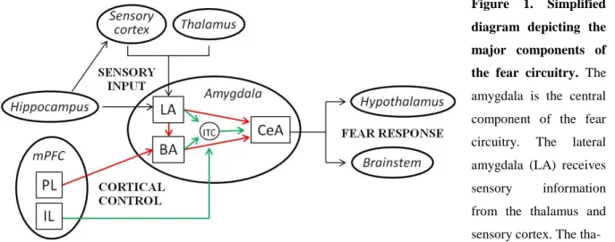
Given that excessive fear and avoidance of fearful situations are key components of both SAD and PTSD, it is not surprising that the neurocircuitry of these disorders show considerable overlap. A series of neuroimaging studies identified the amygdala, medial prefrontal cortex (mPFC), and hippocampus as the most important brain regions mediating learned fear responses in healthy humans (see Figure 1). These same brain regions are often dysfunctional in SAD and PTSD, as demonstrated by symptom provocation and cognitive-emotional studies. Symptom provocation studies are designed to elicit fear responses by public speaking anticipation and/or performance (SAD) or by specifically recalling a traumatic event (PTSD), whereas cognitive-emotional studies involve exposure to emotional stimuli, such as fearful or angry faces, to study generalization of fear responses. These studies indicate that the amygdala, an almond-shaped group of nuclei involved in the perception, learning, and expression of fear (Fanselow and LeDoux, 1999), is hyperactive in both SAD (Tillfors et al., 2001; Lorberbaum et al., 2004; Phan et al., 2006; Guyer et al., 2008) and PTSD (Shin et al., 1997; Liberzon et al., 1999; Rauch et al., 2000; Pissiota et al., 2002) patients, which might account for exaggerated fear responses and persistence of traumatic memories.
The mPFC, a brain region involved in decision making, goal-directed behavior, and working memory (Vertes, 2004), regulates amygdala output and, thereby, the fear response. More specifically, a region in the ventral part of the mPFC (vmPFC), the infralimbic cortex (IL), inhibits amygdala output and fear expression, whereas a region in the dorsal part of the mPFC
(dmPFC), the prelimibic cortex (PL), increases amygdala output and promotes fear expression (Sotres-Bayon and Quirk, 2010). Accordingly, symptom provocation and cognitive-emotional studies indicate that the vmPFC is hypoactive in PTSD patients (Bremner et al., 1999;
Lindauer et al., 2004; Shin et al., 2004a; Yang et al., 2004; Hou et al., 2007) and fails to inhibit the amygdala, whereas the dmPFC shows either normal activity or hyperactivity (Bryant et al., 2005; Felmingham et al., 2009). It is not clear which of these dysfunctions is responsible for the overall outcome in PTSD patients, but a hyperactive amygdala coupled with a hypoactive vmPFC may lead to deficits in fear extinction and emotion regulation in these patients (Liberzon and Sripada, 2008). The involvement of vmPFC in SAD is less clear, with studies showing either hyperactivity (Guyer et al., 2008; Blair et al., 2010), or hypoactivity (Van Ameringen et al., 2004; Evans et al., 2009) of the vmPFC. Additionally, both SAD and PTSD patients show reduced functional connectivity between the amygdala and mPFC during resting-states (Hahn et al., 2011; Sripada et al., 2012), which may contribute to the pathological fear responses observed in these patients.
The hippocampus, a brain region involved in declarative memory (Squire, 1992), processes information about the context, i.e. environment of a fearful situation (Kim et al., 1993). Its involvement in SAD and PTSD is less clear, with studies showing either hypoactivity of this region in SAD (Kilts et al., 2006) and PTSD (Bremner et al., 2003; Shin et al., 2004b) patients, or hyperactivity in SAD (Schneider et al., 1999) and PTSD (Thomaes et al., 2009;
Werner et al., 2009) patients. The differences in brain activity between studies may relate to the type of task employed and the selection of patients, as both SAD and PTSD are highly heterogeneous anxiety disorders.
Preclinical research using SAD- and PTSD-relevant animal models (see section 1.7. for more details) has identified that the same neurocircuity mediates learned fear responses in animals (especially rodents and primates; Figure 1), highlighting again the evolutionary importance of
the fear responses. Although human studies tend to focus on the amygdala, mPFC, and hippocampus, animal studies revealed additional brain regions, such as the striatum, lateral septum (LS), bed nucleus of the stria terminalis (BNST), dorsal raphe nucleus, and periaqueductal gray (Maren, 2001; Calandreau et al., 2007), which are critical either for recognition of a dangerous situation, fear learning, or for appropriate fear expression.
Considering the highly conserved fear circuitry among mammals, these regions are likely to play an important role in humans as well.
Figure 1. Simplified diagram depicting the major components of the fear circuitry. The amygdala is the central component of the fear circuitry. The lateral amygdala (LA) receives sensory information from the thalamus and sensory cortex. The tha- lamo-amygdala pathway sends rapid and crude information about the fear-eliciting stimulus without filtering by conscious control. The cortico-amygdala pathway provides slower, but more detailed sensory information. The LA also receives information about the context of the fear-eliciting stimulus from the hippocampus either directly or indirectly through the perirhinal, entorhinal, and parahippocampal cortices. The LA projects to the central amygdala (CeA) either directly or indirectly through the basal amygdala (BA). The LA and BA also project to the intercalated (ITC) cell masses, a GABAergic neuronal network that inhibits the CeA when activated. The CeA mediates fear responses through projections to the hypothalamus and brainstem. The expression of fear is regulated by the medial prefrontal cortex (mPFC) via projections to the LA, BA, and ITC. A region in the dorsal part of the mPFC, the prelimbic cortex (PL), projects to the BA, which excites the CeA, thereby promoting fear expression, whereas a region in the ventral part of the mPFC, the infralimbic cortex (IL), projects to ITC, which inhibits the CeA, and, thereby, fear expression. Red lines indicate pathways that promote CeA output, green lines indicate pathways that dampen CeA output, and black lines provide input to or output from the amygdala.
1.5. Treatment of SAD and PTSD
The best treatment outcomes in both SAD and PTSD patients are obtained with cognitive- behavioral therapy (Gould et al., 1997; Fedoroff and Taylor, 2001), a psychotherapeutic approach where the maladaptive thoughts that produce and maintain anxiety are identified and replaced with more realistic and positive thoughts, thereby reducing emotional distress. One of the techniques used in cognitive-behavioral therapy is exposure therapy, which leads to a gradual fear extinction, i.e. a decline in the fear response as a result of repeated exposure to the feared situation. This psychotherapy is often combined with a rather unspecific pharmacotherapy, originally designed for generalized anxiety or depression, such as benzodiazepines and antidepressants, with selective serotonin reuptake inhibitors (SSRIs) providing the best response rates in both SAD (Liebowitz et al., 1992; Baldwin et al., 1999;
Van Ameringen et al., 2001) and PTSD (Marshall and Pierce, 2000; Stein et al., 2006, 2009) patients. Probably due to the insufficient understanding of these disorders and the unspecific medication, many SAD and PTSD patients fail to respond to the available treatment options, achieve only partial remission of symptoms, or show a high relapse rate after treatment discontinuation (Blanco et al., 2002; Davidson et al., 2004; Bisson et al., 2007; Brunello et al., 2001; Ipser et al., 2006). Novel therapeutic strategies make use of cognitive enhancers, which are administered either before the psychotherapy session to increase learning, or immediately after the psychotherapy session to facilitate the consolidation of the “safety” memory trained during the session. For example, augmentation of exposure therapy with D-cycloserine, a glutamatergic N-methyl-d-aspartate receptor agonist, was shown to improve some anxiety symptoms in both SAD (Hofmann et al., 2006; Guastella et al., 2008) and PTSD (Heresco- Levy et al., 2002; de Kleine et al., 2012) patients. Brain neuropeptide systems, such as OT, arginine vasopressin (AVP), neuropeptide Y, neuropeptide S (NPS), and substance P (Fendt et al., 2010; Viero et al., 2010; Meyer-Lindenberg et al., 2011; Bowers et al., 2012; Dunlop et
al., 2012), characterized by discrete neuropeptide synthesis and release sites, distinct receptor distribution, and multiple behavioral functions, represent other interesting research candidates with respect to both pathophysiology and treatment of SAD and PTSD. In this thesis, I investigated the effects of OT in two animal models of SAD and PTSD.
1.6. Oxytocin
OT is a nine amino acid neuropeptide that has been recently proposed as a potential therapeutic agent for SAD (Heinrichs et al., 2003; Kirsch et al., 2005; Labuschagne et al., 2010) and PTSD (Olff et al., 2010) due to its pro-social, anxiolytic, and stress-attenuating effects. OT is mainly synthesized in magnocellular neurons of the paraventricular (PVN) and supraoptic (SON) nuclei of the hypothalamus, which project to the posterior pituitary and release OT into the bloodstream (Swaab et al., 1975; Vandesande et al., 1975; Brownstein et al., 1980). OT is also synthesized in parvocellular neurons of the PVN, which project to the brainstem, spinal cord, and the limbic system (Swanson and McKellar, 1979; de Vries and Buijs, 1983; Wagner and Clemens, 1991). Centrally, OT is released within the PVN and SON (Moos et al., 1989; Russell et al., 1992; Neumann et al., 1993; Bosch et al., 2005) from dendrites or perikarya of magnocellular neurons (Ludwig and Leng, 2006), and within limbic regions, such as the septum, hippocampus, and central amygdala (CeA; Landgraf et al., 1988;
Neumann and Landgraf, 1989; Ebner et al., 2000, 2005; Bosch et al., 2005) from axonal or collateral projections of parvocellular and magnocellular neurons.
The effects of OT are mediated via one single type of OT receptor (OTR), which is a polypeptide with 7 transmembrane domains belonging to the G protein-coupled receptor family. OTR are widespread throughout the central nervous system, in regions such as the cortex, the olfactory system, the basal ganglia, limbic areas such as the BNST, septum, hippocampus, CeA, and ventral subiculum, the thalamus, hypothalamus, the brain stem, and
the spinal cord (Brinton et al., 1984; de Kloet et al., 1985; Elands et al., 1988; Tribollet et al., 1988). Therefore, OT can exert its effects in all brain regions implicated in the fear circuitry so far (see section 1.4. and Figure 1), providing an important reason for studying its involvement in SAD and PTSD, especially in fear learning and extinction (see section 1.7.2.
and Table 1).
The reason for the interest in OT in relation with SAD and PTSD includes the fact that OT has been shown to exert pro-social, anxiolytic, and stress-attenuating effects both in rodents and humans. In more detail, OT facilitates a broad variety of social behaviors in rodents, including the onset and maintenance of maternal behavior in lactation (Pedersen and Prange, 1979; Bosch et al., 2005), regulation of male and female sexual behavior (Arletti and Bertolini, 1985; Arletti et al., 1985; Waldherr and Neumann, 2007; Nyuyki et al., 2011), pair bonding in female voles (Williams et al., 1994), social preference (Lukas et al., 2011), and social recognition in male and female rodents (Popik and van Ree, 1991; Ferguson et al., 2000; Choleris et al., 2003; Engelmann et al., 1998; Lukas, Toth et al., in press). In addition to these pro-social effects, both synthetic and endogenous OT exert anxiolytic properties in rodents (McCarthy et al., 1996; Waldherr and Neumann, 2007; Blume et al., 2008; Jurek et al., 2012), and inhibit the activity of the hypothalamic-pituitary-adrenal (HPA) axis (Windle et al., 1997; Neumann et al., 2000).
Comparable effects have also been described in humans, where synthetic OT was shown to exert pro-social effects, such as increased trust and social risk-taking (Kosfeld et al., 2005;
Baumgartner et al., 2008; Theodoridou et al., 2009; Mikolajczak et al., 2010a,b), increased attachment (Buchheim et al., 2009), cooperative behavior (Declerck et al., 2010), emotional and cognitive empathy (Bartz et al., 2010; Hurlemann et al., 2010), improved emotion recognition (Domes et al., 2007a; Savaskan et al., 2008) and memory for positive social information (i.e. happy faces, Di Simplicio et al., 2009; Rimmele et al., 2009; Marsh et al.,
2010). Additionally, OT was shown to reduce the autonomic response to stress (Heinrichs et al., 2003; Kirsch et al., 2005; Kubzansky et al., 2009; Quirin et al., 2011), to decrease aversion to angry faces (Evans et al., 2010), and to increase positive communication during couple conflict (Ditzen et al., 2009). Imaging studies revealed that OT reduces the activity of the amygdala to painful stimulation (Singer et al., 2008), threatening faces and scenes (Kirsch et al., 2005; Domes et al., 2007b; Gamer et al., 2010), and conditioned faces (Petrovic et al., 2008a). Although OT did not reduce anxiety symptoms in SAD patients (Kirsch et al., 2005;
Domes et al., 2007b), it improved speech performance (Guastella et al., 2009) and dampened the hyperactivity of the amygdala to fearful faces (Labuschagne et al., 2010). In autistic patients, which are characterized by marked social deficits, OT was shown to increase social interaction (Andari et al., 2010), emotion recognition (Guastella et al., 2010), comprehension of affective speech (Hollander et al., 2007), and gaze to the eye region (Andari et al., 2010).
More indirect evidence for the anxiolytic and antistress effects of OT in humans comes from nursing mothers who are calmer and less anxious during stressful situations, possibly due to high brain OT activity (Heinrichs et al., 2001; Carter et al., 2001; Slattery and Neumann, 2008).
As fear responses are highly conserved across species, studies on the neural basis of fear in experimental animals made important contribution to understanding the normal processes underlying fear responses. Importantly, the highly conserved fear circuitry combined with the highly conserved effects of OT among mammalian species suggests that findings from experimental animals might be translatable to humans. However, in order to understand the etiology and the underlying neurobiological mechanisms of pathological social and cued fear, which might in turn provide important information for the development of more specific medication for SAD and PTSD, animal models that mimic the main behavioral symptoms of these anxiety disorders are needed.
1.7. Animal models used to study SAD and PTSD
As both SAD and PTSD are complex anxiety disorders with strong psychological components, animal models cannot mimic these disorders in their entire complexity.
However, although human performance anxiety and fear of showing anxiety symptoms (see section 1.2.) cannot be reliably modeled in laboratory animals, several paradigms have been shown to induce severe behavioral deficits in rodents, including social avoidance and fear, and can be used to model human interaction anxiety. These paradigms include social defeat (Huhman, 2006; Yan et al., 2010), conditioned defeat (Hammack et al., 2012), foot-shock exposure (Haller and Bakos, 2002; Louvart et al., 2005), maternal separation (Franklin et al., 2011), and restraint stress (Gehlert et al., 2005; Doremus-Fitzwater et al., 2010). Among the animal models used to study PTSD, cued and context fear conditioning (Sanders et al., 2003), active and passive avoidance (Kovács et al., 1979; Ibragimov, 1990), predator and predator scent exposure (Adamec and Shallow, 1993, Takahashi et al., 2008), and fear potentiated startle (Davis, 1986) have proven to be powerful models to study the neural circuitries and mechanisms involved in fear learning and, perhaps more importantly, in fear extinction. As social defeat and cued fear conditioning are the most relevant models for my thesis, I will describe these two paradigms in more detail. For more details about the other models, please see the following reviews: Davis, 1986; Korte, 2001; Sanders et al., 2003; Henn and Vollmayr, 2005; Takahashi et al., 2008; Kikusui and Mori, 2009; Hammack et al., 2012.
1.7.1. Social defeat
The social defeat paradigm was established by Klaus Miczek (1979) and consists of placing a smaller animal in the home cage of a larger aggressive male, which defends its territory and defeats the experimental animal, forming thereby a typical dominant-subordinate relationship.
This procedure can be performed once, i.e. acute social defeat, or repeatedly using several
dominant males, i.e. chronic social defeat (Huhman, 2006; Yan et al., 2010). Both acute and chronic social defeat result in persistent fear and avoidance of con-specifics, however, the severity of the induced behavioral alterations depends on the type and length of the defeat.
Acute social defeat induces avoidance only of the dominant con-specific that performed the defeat (Lai et al., 2005; Lukas et al., 2011) and is, therefore, less relevant as an animal model of SAD. Chronic social defeat, on the other hand, induces a general avoidance of con- specifics (Avgustinovich et al., 2005; Berton et al., 2006), which makes this model a powerful tool to study the neural circuitries underlying social fear. However, chronic social defeat also induces a large variety of behavioral and physiological alterations, such as increased general anxiety (Keeney and Hogg, 1999; Avgustinovich et al., 2005; Denmark et al., 2010), decreased locomotor activity (Koolhaas et al., 1997; Rygula et al., 2005), depressive-like behavior (Avgustinovich et al., 2005; Rygula et al., 2005; Hollis et al., 2010), anhedonia (Von Frijtag et al., 2000; Rygula et al., 2005), changes in circadian rhythms (Tornatzky and Miczek, 1993; Meerlo et al., 1997), sleep patterns (Kinn et al., 2008), feeding and body weight (Foster et al., 2006), increased heart rate and blood pressure (Fokkema et al., 1986;
Sgoifo et al., 1999; Costoli et al., 2004), increased body temperature (Keeney et al., 2001;
Hayashida et al., 2010), and suppression of immune responses (Stefanski, 1998; Avitsur et al., 2009; Chester et al., 2010). Given that social fear might either represent the major symptom of the anxiety disorder, as seen in SAD, or a comorbid condition to other psychiatric disorders, such as depression and schizophrenia, animal models that induce specific social fear without any confounding behavioral alterations are required to study the etiology of social fear, which might lead to more specific and efficient medications for these patients.
1.7.2. Cued fear conditioning
The cued fear conditioning paradigm was originally developed in humans (Watson and Rayner, 1920) and later translated to animals (Estes and Skinner, 1941). With their famous
experiment on “little Albert”, an eleven-month-old child, Watson and Rayner could demonstrate for the first time that fear responses can be conditioned in humans. They exposed the child to a rat, to which he initially showed no fear response, and made a loud noise by hitting a metal pipe with a hammer, causing Albert to startle and fall forward. After repeatedly pairing the rat with the loud noise, Albert started crying at the sight of the rat and crawled away from it (Watson and Rayner, 1920).
Although fear conditioning studies employing association of innocuous stimuli, such as pictures of faces or landscapes, tones, and words with mild aversive stimuli, such as electric shocks, painful pressure, heat pain, air puffs, and aversive pictures, tones, tastes or odors (Mechias et al., 2010) are largely being used in humans nowadays, most of the indepth knowledge we have about the pathways and neural mechanisms involved in conditioned fear (see section 1.4.) comes from studies in rodents. A typical cued fear conditioning experiment in rodents consists of three experimental phases, namely fear conditioning, fear extinction, and extinction recall (see Table 1 for terminology used in fear conditioning experiments and throughout this thesis). During fear conditioning, rodents are presented with an innocuous stimulus, usually a tone or light (conditioned stimulus, CS), which co-terminates with an aversive stimulus, such as an electrical foot shock (unconditioned stimulus, US). Through repeated CS-US associations (typically 2 - 5), animals learn that the CS predicts the US, and the CS will elicit a fear response even when presented in the absence of the US. The conditioned fear responses elicited by the CS are highly conserved among mammals and are similar to those elicited by natural threats, such as predators (see section 1.1.). During fear extinction, rodents are repeatedly presented with the CS without the US, which leads to a gradual decrease in the fear responses, and is similar to the repeated exposure to the feared situation during cognitive-behavioral therapy in humans (see section 1.5.). There is a growing body of evidence suggesting that fear extinction is different from forgetting and does not
eliminate the original fear memory, but generates new “safety” memory that inhibits the expression of fear in response to the CS (Bouton, 2004; Quirk et al., 2010). During extinction recall, rodents are presented with the CS without the US to determine whether the “safety”
memory trained during fear extinction was consolidated. Although the cued fear conditioning paradigm does not fully model PTSD due to the complexity of the disorder, it allows the study of the neural circuitries and mechanisms involved in fear learning and fear extinction.
Fear conditioning A process by which fear of an innocuous stimulus is learned through repeated associations of this stimulus with a noxious stimulus
Unconditioned stimulus (US) A noxious stimulus (i.e. electric foot shock) that triggers an automatic, unlearned fear response
Unconditioned response (UR) An automatic, unlearned fear response (i.e. freezing, startle) to an US
Conditioned stimulus (CS) An innocuous stimulus (i.e. tone, light, context) that, after fear conditioning, triggers a learned fear response Conditioned response (CR) A learned fear response (i.e. freezing) to a CS
Fear acquisition A process by which the association between the CS and US is encoded; it represents the initial stage of fear conditioning
Fear consolidation A process by which the association between the CS and US is stabilized and maintained after fear acquisition Fear expression A process by which CR are triggered by the CS
Fear extinction A process by which CR gradually decrease as a result of repeated presentations of the CS without the US
Extinction recall A process by which is determined whether CR are triggered by the CS after consolidation of fear extinction
Table 1. Terminology used in fear conditioning experiments
1.8. Aim of the present thesis
Given the lack of animal models that induce general social fear without confounding deficits, the high prevalence and unsatisfactory treatment options for both SAD and PTSD, and the
potential therapeutic role of OT in SAD and PTSD, I aimed to:
1. Develop and validate an animal model that mimics the main behavioral symptom of
SAD, namely social fear and avoidance.
2. Use this novel animal model, i.e. the social fear conditioning paradigm, to verify the therapeutic efficacy of OT in reversing social fear.
3. Verify the therapeutic efficacy of OT in reversing cued fear in an animal model of PTSD, the cued fear conditioning paradigm.
1.9. Outline of the present thesis
Chapter 2 describes a novel and specific animal model to study SAD, the social fear conditioning paradigm, and its effects on social investigation, general anxiety, depressive-like behavior, and locomotion. It also describes the effects of typical SAD medication, i.e. the benzodiazepine diazepam and the antidepressant paroxetine, on the induced social fear.
Chapter 3 describes step by step how to perform the social fear conditioning paradigm. It also describes theoretical and practical considerations for rats and mice, and for the analysis and interpretation of the obtained data.
Chapter 4 describes the central and local effects of synthetic OT on social fear induced by social fear conditioning. It also describes the alterations that occur at the level of the brain OT system after social fear conditioning and social fear extinction.
Chapter 5 describes the central effects of synthetic OT on cued fear induced by cued fear conditioning, and emphasizes the importance of the time-point of OT administration during the conditioning procedure.
Chapter 6 summarizes and discusses the most relevant findings of this thesis.
Chapter 2
Social fear conditioning: a novel and specific animal model to study social anxiety disorder
Author’s contribution:
Toth: designed research, performed research, analyzed data, wrote the manuscript Neumann: designed research, revised the manuscript
Slattery: designed research, revised the manuscript
[adapted from: Toth I, Neumann ID, Slattery DA (2012) Social fear conditioning: a novel and specific animal model to study social anxiety disorder. Neuropsychopharmacology 37(6):1433-1443]
ABSTRACT
SAD is a major health concern with high lifetime prevalence. The current medication is rather unspecific and, despite considerable efforts, its efficacy is still unsatisfactory. However, there are no appropriate and specific animal models available to study the underlying etiology of the disorder. Therefore, we aimed to establish a model of specific social fear in mice and use this social fear conditioning paradigm to assess the therapeutic efficacy of the benzodiazepine diazepam and of the antidepressant paroxetine, treatments currently used for SAD patients.
We show that by administering electric foot shocks (2 - 5, 1 s, 0.7 mA, pulsed current) during the investigation of a con-specific, the investigation of unfamiliar con-specifics was reduced for both the short- and long-term, indicating lasting social fear. The induced fear was specific to social stimuli and did not lead to other behavioral alterations, such as fear of novelty, general anxiety, depression, and impaired locomotion. We show that social fear was dose- dependently reversed by acute diazepam, at doses that were not anxiolytic in a non-social context, such as the elevated plus-maze (EPM). Finally, we show that chronic paroxetine treatment reversed social fear. All in all, we demonstrated robust social fear after exposure to social fear conditioning in mice, which was reversed with both acute benzodiazepine and chronic antidepressant treatment. We propose the social fear conditioning model as an appropriate animal model to identify the underlying etiology of SAD and possible novel treatment approaches.
INTRODUCTION
SAD, often referred to as social phobia, is characterized by persistent fear and avoidance of social situations. Epidemiologically, SAD is the third most common psychiatric disorder, with a 12-month and lifetime prevalence of 6.8% and 12.1%, respectively (Kessler et al., 2005a,b).
For diagnostic purposes, SAD has been divided in two subtypes: specific and generalized SAD. The specific form refers to the fear and avoidance of a particular social situation and includes performance anxiety (e.g. fear of giving a public speech), interaction anxiety (fear of social interaction and observation situations), and fear of showing anxiety symptoms (Bögels et al., 2010). Patients with generalized SAD are more impaired as they fear and avoid a wide range of social situations (den Boer., 1997; Kessler et al., 1998; Ruipérez et al., 2002).
This avoidant behavior has an important role in the maintenance of SAD and prevents the reversal of fear in social situations (American Psychiatric Association, 1994; Stangier et al., 2006).
At present, SAD treatment consists of cognitive-behavioral therapy (Gould et al., 1997; Fedoroff and Taylor, 2001), which leads to gradual fear extinction, i.e. a decline in the fear response as a result of repeated exposure to the feared situation, and is often combined with medication originally designed for depression or generalized anxiety, such as antidepressants, β-blockers, and benzodiazepines. However, a high percentage of SAD patients fail to respond to the available treatment options, or achieve only partial remission of symptoms, with SSRIs providing the best response rates (Liebowitz et al., 1992; Baldwin et al., 1999; Van Ameringen et al., 2001). Given the high prevalence and unsatisfactory treatment options for SAD, a better understanding of the etiology and underlying neurobiological mechanisms of social fear, particularly extinction of social fear, is urgently needed. This, in turn, might provide important information for the development of more specific medication and an improved treatment outcome for SAD patients.
However, there are currently no appropriate animal models available to study the disorder.
Social fear and avoidance is presently induced using a number of paradigms, including the social defeat paradigm, which can be used both acutely and chronically (Huhman, 2006;
Yan et al., 2010), and foot-shock exposure (Haller and Bakos, 2002; Louvart et al., 2005;
Mikics et al., 2008a). However, these paradigms are rather unspecific with respect to the behavioral alterations they induce, as increased general anxiety, depression, and impaired locomotion were found to accompany the social avoidance (Denmark et al., 2010; Hollis et al., 2010).
Therefore, we aimed to establish a novel and specific animal model of SAD using the social fear conditioning paradigm, and use this model to assess the therapeutic efficacy of diazepam and paroxetine, currently used for SAD patients. The SAD-like phenotype was induced in naïve mice by punishing them when investigating an unfamiliar con-specific. Mice were conditioned to associate a shock-induced pain with the investigation of a social stimulus and, therefore, avoid social stimuli. The conditioned social fear is specific to several social stimuli, long-lasting and not accompanied by changes in general anxiety, depressive-like behavior, and locomotion. The social fearful phenotype was dose-dependently reversed by acute diazepam, at a dose that was not anxiolytic in a non-social context, i.e. the EPM. Furthermore, chronic antidepressant treatment also reversed social fear, thus validating the social fear conditioning model.
MATERIALS AND METHODS
Animals
Male CD1 mice (Charles River, Sulzfeld, Germany) weighing 30 - 35 g were individually housed in polycarbonate cages (16 × 22 × 14 cm) for 1 week before experiments started, and
remained isolated throughout. Isolation was shown to increase social motivation (Niesink and Van Ree, 1982) and prevent the attenuation of behavioral effects of stressors observed in group-housed mice (Ruis et al., 1999; Cherng et al., 2010). Mice were transferred to observation cages (30 × 23 × 36 cm) 3 days before experiments started. Age- and weight- matched male CD1 mice were used as social stimuli.
Mice were maintained under standard laboratory conditions (12:12 light/dark cycle, lights on at 06:00, 22°C, 60% humidity, food and water ad libitum). Experiments were performed during the light phase, between 08:00 and 12:00, in accordance with the Guide for the Care and Use of Laboratory Animals of the Government of Oberpfalz and the guidelines of the National Institutes of Health.
Social fear conditioning paradigm
On day 1, mice were conditioned for social fear, whereas on days 2 and 3 or 15 and 16, social investigation was assessed as readout of short- and long-term social fear and fear extinction.
Social fear conditioning was performed with a computerized fear conditioning system (TSE System GmbH, Bad Homburg, Germany). The conditioning chamber consisted of a transparent Perspex box (45 × 22 × 40 cm) enclosed in a wooden chamber to reduce external noise and visual stimulation. The floor consisted of a removable stainless steel grid connected to a shock delivery unit used for manual application of foot shocks. A video camera at the top of the chamber enabled video recording.
Social fear conditioning (day 1). Mice were placed in the conditioning chamber and, after a 30-s adaptation period, an empty wire mesh cage (7 × 7 × 6 cm) was placed as a non-social stimulus near one of the short walls. Mice were allowed to investigate the non-social stimulus for 3 min, before it was replaced by an identical cage containing an unfamiliar male mouse.
Unconditioned (UC) mice were allowed to investigate the social stimulus for 3 min.
Conditioned (C) mice were given a 1-s electric foot shock (0.7 mA, pulsed current) each time they investigated the social stimulus, defined by direct contact with the mouse. Mice received between 2 and 5 foot shocks, with a variable inter-shock interval, depending on when direct social contact was made. The first social contact and, therefore, foot shock occurred within 15 - 30 s. Mice were returned to their home cage when no further social contact was made for 2 min, meaning that conditioned mice spent between 3 and 6 min in the conditioning chamber while the social stimulus was present. The time mice spent investigating the non-social stimulus, as a pre-conditioning measure of non-social anxiety, was analyzed using the JWatcher program (V 1.0, Macquarie University and UCLA).
Social fear extinction (day 2 or 15). To investigate whether conditioned mice displayed social fear and whether this fear could be extinguished, social investigation was assessed in the home cage 1 or 15 days after social fear conditioning for short-term and long-term social fear, respectively. In detail, social fear extinction consisted of exposing the mice to 3 empty cages identical to the cage used during day 1 (non-social stimuli) to assess non-social investigation. Mice were then exposed to 6 unfamiliar male mice enclosed in wire mesh cages (social stimuli) to assess social investigation. Each stimulus was placed near a short wall of the home cage and presented for 3 min, with a 3-min inter-exposure interval. Reduced social investigation and aversive responses toward the social stimuli, such as freezing, stretched approaches, and defensive burying indicated social fear and successful social fear conditioning. As the non-social stimulus elicited a fear response in conditioned mice, an empty cage was placed over night in the home cage to extinguish the fear of the cage.
Extinction recall (day 3 or 16). To investigate whether repeated exposure to social stimuli during social fear extinction leads to a complete reversal of social fear, social investigation was assessed in the home cage 1 day after social fear extinction. Extinction recall consisted of
exposing the mice to 6 unfamiliar social stimuli for 3 min, with a 3-min inter-exposure interval.
Elevated plus-maze
The EPM (Pellow et al., 1985; Lister, 1987) was performed 1 day after social fear conditioning to assess general anxiety-related behavior as previously described (Reber et al., 2007). Briefly, the maze consisted of two open (6 × 30 x 0.2 cm, 110 lx) and two closed (6 × 30 × 16 cm, 25 lx) arms radiating from a central platform (6 × 6 cm) elevated 35 cm above the ground. Mice were placed on the central platform facing a closed arm and allowed to explore the maze for 5 min. The percentage of time spent on the open arms was considered as anxiety-related parameter. The number of entries into the closed arms was considered as indicator of locomotor activity. The experiment was analyzed using the Plus-maze program (version 2.0; E. Fricke).
Forced swim test
The forced swim test (FST; Porsolt et al., 1977) was performed 1 day after social fear conditioning to assess depressive-like behavior. Mice were individually placed into a plexiglass cylinder (13 cm diameter, 25 cm height) filled with 25°C water to a depth of 14 cm for 6 min. Immobility was scored during the last 4 min using the JWatcher program, and was defined as the animal floating in water without swimming and making only movements necessary to maintain its head above the water (Slattery et al., 2005).
Locomotor activity
Home cage locomotion was assessed 1 day after social fear conditioning. Mice were taken out of the cage for 1 min to achieve similar arousal levels in all mice and returned to the home
cage thereafter. Locomotion was measured for 1 h using the Noldus Ethovision XT 5.1 program (Noldus Information Technology, Wageningen, The Netherlands).
Novel object investigation
Novel object and social investigation were assessed in the home cage 1 day after social fear conditioning to differentiate between fear of novelty and social fear. Mice were exposed to three non-social stimuli, three cages containing a white ball (novel object stimuli, size and color matched to the social stimuli), and three unfamiliar social stimuli. Each stimulus was presented for 3 min, with a 3-min inter-exposure interval.
Electric foot-shock exposure
To assess the effects of foot-shock exposure in the absence of a social stimulus on social fear, mice were placed in the empty conditioning chamber and, after a 30-s adaptation period, received five electric foot shocks (1 s, 0.7 mA, pulsed current, i.e. the maximum number received during social fear conditioning), with a 2-min inter-shock interval. Mice were returned to their home cage 2 min after the last foot shock. An empty cage was placed in their home cage over night to allow for comparable behavioral effects with the social fear conditioning experiments. One day later, social investigation was assessed during social fear extinction.
Drugs
Diazepam (Ratiopharm GmbH, Germany) was freshly dissolved in saline and administered intraperitoneally (i.p.) at a volume of 5 ml/kg and doses between 0.5 and 1.25 mg/kg. The highest doses were chosen based on previous studies (Corbett et al., 1993; Dalvi and Rodgers, 1996; Stachowicz et al., 2008). Paroxetine (Bayer Schering, Germany) was administered over 14 days via the drinking water at a dose of 10 mg/kg/ day. The paroxetine dose was chosen
based on previous studies (Da-Rocha et al., 1997; Hascoët et al., 2000a,b; Massé et al., 2005;
Elizalde et al., 2008; Thoeringer et al., 2010).
Experimental design
Effects of social fear conditioning on short- and long-term social fear
Initial experiments were designed to characterize the effects of social fear conditioning on short- and long-term social fear. Therefore, separate groups of mice were subjected to social fear conditioning and social investigation was assessed 1 or 15 days later during social fear extinction (n = 13 per group for short- and n = 9 per group for long-term effects). Extinction recall was measured 1 day later.
Specificity of the induced social fear
To verify the specificity of the induced social fear, separate groups of mice were subjected to general anxiety (EPM; n = 8 per group), depressive-like behavior (FST; n = 8 per group), home cage locomotion (n = 7 per group), or novel object investigation (n = 8 per group) testing 1 day after social fear conditioning. Another group of mice was exposed to electric foot shocks in the absence of the social stimulus, and social investigation was assessed 1 day later during social fear extinction (n = 8 per group).
Reversal of short- and long-term social fear by acute diazepam and chronic paroxetine treatment, respectively
To determine whether the effects of social fear conditioning on short- and long-term social fear could be reversed by medication used for SAD patients, we assessed the effects of diazepam and paroxetine, respectively. Mice (n = 10 per group) were subjected to social fear conditioning. The following day, 30 min before social fear extinction, mice were injected i.p.
either with vehicle (5 ml/kg saline) or diazepam (0.5, 0.75, 1.0, and 1.25 mg/kg). Extinction
recall was measured 1 day later. To determine whether diazepam has anxiolytic effects in a non-social context at doses used to reverse social fear, naïve mice were injected i.p. either with vehicle (5 ml/kg saline) or with diazepam (0.5, 0.75, 1.0, and 1.25 mg/kg) 30 min before EPM testing. In a separate group of mice (n = 8 per group), social investigation was assessed 15 days after social fear conditioning. Paroxetine (10 mg/kg/day) was administered chronically via the drinking water over 14 days, starting 1 day after social fear conditioning to prevent possible confounding effects on fear memory consolidation. Extinction recall was measured 1 day later.
Statistical analysis
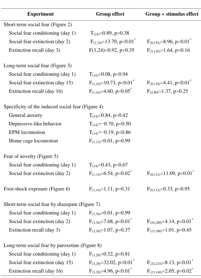
For statistical analysis PASW/SPSS (Version 17) was used. Data were analyzed by Student's t-tests, one-way or two-way analysis of variance (ANOVA) for repeated measures, followed by a Bonferroni post-hoc analysis whenever appropriate. Statistical significance was set at p<0.05. Overall statistics are shown in Table 2.
RESULTS
Effects of social fear conditioning on short- and long-term social fear
Short-term social fear
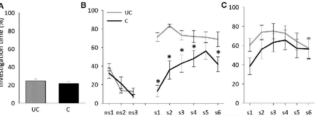
Mice showed similar non-social anxiety during social fear conditioning on day 1 (Figure 2A, Table 2). During social fear extinction on day 2, conditioned mice showed similar non- social investigation, but reduced social investigation (p<0.05; Figure 2B) compared with unconditioned mice, reflecting social fear. No difference between the mice was found during extinction recall on day 3 (Figure 2C).
Experiment Group effect Group × stimulus effect Short-term social fear (Figure 2)
Social fear conditioning (day 1) T(24)=0.89, p=0.38
Social fear extinction (day 2) F(1,24)=13.70, p<0.01* F(8,192)=8.96, p<0.01* Extinction recall (day 3) F(1,24)=0.92, p=0.35 F(5,120)=1.64, p=0.16 Long-term social fear (Figure 3)
Social fear conditioning (day 1) T(16)=0.08, p=0.94
Social fear extinction (day 15) F(1,16)=10.73, p<0.01* F(8,128)=4.41, p<0.01* Extinction recall (day 16) F(1,16)=4.60, p=0.05* F(5,80)=1.37, p=0.25 Specificity of the induced social fear (Figure 4)
General anxiety T(14)=0.84, p=0.42 Depressive-like behavior T(14)=−0.70, p=0.50 EPM locomotion T(14)=−0.19, p=0.86 Home cage locomotion F(1,12)<0.01, p=0.99 Fear of novelty (Figure 5)
Social fear conditioning (day 1) T(14)=0.43, p=0.67
Social fear extinction (day 2) F(1,14)=6.54, p=0.02* F(8,112)=11.09, p<0.01* Foot-shock exposure (Figure 6) F(1,14)=1.11, p=0.31 F(8,112)=0.33, p=0.95 Short-term social fear by diazepam (Figure 7)
Social fear conditioning (day 1) F(3,36)=0.01, p=0.99
Social fear extinction (day 2) F(3,36)=7.68, p<0.01* F(24,288)=4.14, p<0.01* Extinction recall (day 3) F(3,36)=1.07, p=0.37 F(15,180)=1.01, p=0.45 Long-term social fear by paroxetine (Figure 8)
Social fear conditioning (day 1) F(3,28)=0.32, p=0.81
Social fear extinction (day 15) F(3,28)=32.02, p<0.01* F(24,224)=8.13, p<0.01* Extinction recall (day 16) F(3,28)=4.96, p<0.01* F(15,140)=2.05, p=0.02*
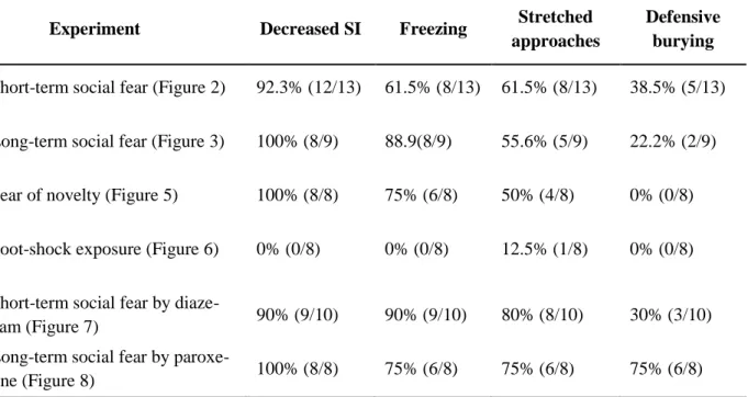
Table 2. Overall effects for the social fear conditioning data. Stimulus effect refers to both non-social and social stimuli during social fear extinction, while during extinction recall it refers to social stimuli only. EPM, elevated plus-maze. Student’s t-tests, one-way or two-way ANOVA for repeated measures followed by Bonferroni post-hoc test; * p<0.05.
Figure 2. Social fear conditioning induces short-term social fear in mice. (A) Investigation of the non-social stimulus (empty cage) by unconditioned (UC) and conditioned (C) mice during social fear conditioning on day 1 (n = 13 per group). (B) Investigation of non-social (ns1-ns3) and social (cages with mice; s1-s6) stimuli during social fear extinction on day 2 (3 min exposure to stimulus, 3 min inter-exposure interval). (C) Investigation of social stimuli (s1-s6) during extinction recall on day 3. Data represent mean percentage of investigation time ± SEM. * p<0.05 vs. UC mice.
Figure 3. Social fear conditioning induces long-term social fear in mice. (A) Investigation of the non-social stimulus (empty cage) by unconditioned (UC) and conditioned (C) mice during social fear conditioning on day 1 (n = 9 per group). (B) Investigation of non-social (ns1-ns3) and social (cages with mice; s1-s6) stimuli during social fear extinction on day 15 (3 min exposure to stimulus, 3 min inter-exposure interval). (C) Investigation of social stimuli (s1-s6) during extinction recall on day 16. Data represent mean percentage of investigation time ± SEM. * p<0.05 vs. UC mice.
Long-term social fear
Mice showed similar non-social anxiety during social fear conditioning on day 1 (Figure 3A, Table 2). During social fear extinction on day 15, conditioned mice showed similar non-
social investigation, but reduced social investigation (p<0.05; Figure 3B) compared with unconditioned mice, reflecting social fear. In contrast to short-term social fear, conditioned mice still showed reduced social investigation during extinction recall on day 16 (p<0.05; Figure 3C).
Specificity of the induced social fear
No effect of social fear conditioning on general anxiety, depressive-like behavior, and home cage locomotion
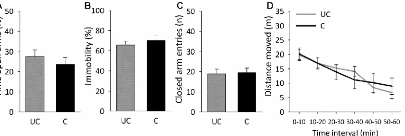
Conditioned mice showed no changes in general anxiety (percentage of time on open arms; Figure 4A, Table 2) or locomotion on the EPM (number of closed arm entries; Figure 4C), in depressive-like behavior in the FST (percentage immobility; Figure 4B), or in home cage locomotion (distance moved; Figure 4D) compared with unconditioned mice 1 day after social fear conditioning.
Figure 4. No effect of social fear conditioning on general anxiety on the elevated plus-maze (EPM) (A), depressive-like behavior in the forced swim test (B), and locomotor activity on the EPM (C) and in the home cage (D). Data represent means ± SEM for n = 7 to 8 mice (separate groups). C, conditioned mice; UC, unconditioned mice.
No effect of social fear conditioning on fear of novelty
Mice showed similar non-social anxiety during social fear conditioning on day 1 (Figure 5A, Table 2). One day after social fear conditioning, conditioned mice showed similar non- social and novel object investigation, but reduced social investigation (p<0.01; Figure 5B) compared with unconditioned mice.
Figure 5. Social fear conditioning does not induce fear of novelty.
(A) Investigation of the non-social stimulus (empty cage) by unconditioned (UC) and conditioned (C) mice during social fear conditioning on day 1 (n = 8 per group). (B) Investigation of non-social stimuli (ns1-ns3), novel object stimuli (cages with objects;
no1-no3), and social stimuli (cages with mice; s1-s6) during novel object investigation on day 2 (3 min exposure to stimulus, 3 min inter-exposure interval). Data represent mean percentage of investigation time ± SEM.
* p<0.05 vs. UC mice.
Figure 6. Exposure to five electric foot shocks in the absence of a social stimulus does not induce social fear. Investigation of non-social (empty cages;
ns1-ns3) and social (cages with mice; s1-s6) stimuli by non-shocked and shocked mice (n = 8 per group) during social fear extinction 1 day after foot-shock exposure (3 min exposure to stimulus, 3 min inter- exposure interval). Data represent mean percentage of investigation time ± SEM.
No effect of foot-shock exposure on social fear
Exposure to foot shocks in the absence of the social stimulus did not alter social investigation 1 day later (Figure 6, Table 2).