Molecular and neuronal correlates of social fear in mice
DISSERTATION ZUR ERLANGUNG DES DOKTORGRADES DER NATURWISSENSCHAFTEN (DR. RER. NAT.) DER FAKULTÄT DER
BIOLOGIE UND VORKLINISCHEN MEDIZIN DER UNIVERSITÄT REGENSBURG
vorgelegt von Rohit Menon aus Kerala, Indien
im Jahr 2017
2
3
Das Promotionsgesuch wurde eingereicht am: 27. Okt 2017
Die Arbeit wurde angeleitet von: Prof. Dr. rer. nat. Inga D. Neumann
Unterschrift:
4
5
Dissertation
Durchgeführt am Institut für Zoologie der Universität Regensburg Unter Anleitung von
Prof. Dr. rer. nat. Inga D. Neumann
6
7
Contents
Abstract ... 11
Introduction ... 15
1.1 Nature of Emotions ... 15
1.2 Anxiety and Fear... 15
1.3 Anxiety disorders ... 16
1.3.1. SAD ... 18
1.3.2. PTSD ... 19
1.4 Treatment of Anxiety disorders ... 21
1.5 Modelling anxiety in rodents ... 22
1.5.1. Measuring general anxiety in rodents ... 23
1.5.2. Cued Fear Conditioning (CFC) ... 23
1.5.3. Social Fear Conditioning (SFC) ... 24
1.6 Neurocircuitry of fear and anxiety ... 25
1.6.1 Amygdala ... 27
1.6.2 Septum ... 28
1.6.3 Hippocampus ... 29
1.6.4 Prefrontal cortex ... 30
1.7. Molecular basis of conditioned fear ... 31
1.7.1. Environment to the neurons: synaptic plasticity and fear acquisition ... 33
1.7.2. From synapse to nucleus: Cell signalling molecules and transcription factors involved in coding of fear ... 34
1.7.3. Changes within the neuronal nucleus: consolidation and formation of long-lasting fear memories ... 36
1.8. Role of neuropeptides in regulation of fear ... 38
1.8.1 Neuropeptide oxytocin and its receptor ... 38
1.8.2 Neuropeptide oxytocin and regulation of general anxiety ... 41
1.8.3 OXT and the regulation of fear ... 42
8
1.9 Epigenetics – the molecular bridge between environment and behaviour ... 44
1.9.1 Histone modifications and regulation of fear ... 45
1.9.2 DNA methylation and regulation for fear and anxiety ... 49
1.10 Aims of the present thesis... 51
1.10.1 Studying the role of endogenous OXT signaling in regulation of social and cued fear in mice ... 51
1.10.2 To delineate the epigenetic mechanisms within the LS the regulate fear in male CD1 mice. ... 52
Materials and Methods ... 53
2.1. Behavioral techniques... 53
2.1.1 Animals ... 53
2.1.2. SFC paradigm ... 53
2.1.3. CFC paradigm ... 54
2.1.4. EPM ... 55
2.1.5. SPT... 56
2.1.6. Hargreaves’ Plantar Test ... 56
2.1.7. Stereotactic implantations ... 56
2.1.8. Intracerebral infusions ... 57
2.3. Scoring of behavior ... 58
2.2. Molecular techniques ... 58
2.2.1. Tissue isolation and mRNA extraction ... 58
2.2.2. Analysis of gene expression using quantitative real-time PCR (qRT-PCR)... 58
2.2.3. Analysis of gene expression using microarray ... 59
2.2.4. cFos immunohistochemistry ... 60
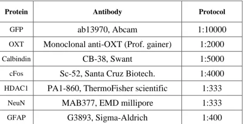
2.2.5. Immunofluorescence ... 60
2.3 Statistical analysis ... 62
Results ... 63
3.1 Septal oxytocin signaling regulates social fear in female mice. ... 63
3.1.1 Summary ... 63
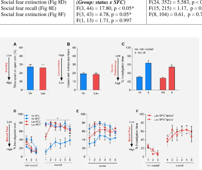
3.1.2 Behavioural characterization of lactating mice: general anxiety, social preference, and social fear conditioning ... 64
3.1.3 Icv pharmacological blockade or activation of the OXTR retrieves SFC-induced social fear in lactating mice or reverses SFC-induced social fear in virgin mice. ... 66
9
3.1.4 Brain regions are differentially activated lactating and virgin mice in response to SFC... 69
3.1.5 Bidirectional manipulation of OXTR-mediated signaling within the LS causes bidirectionally alters SFC-induced fear in lactating and virgin mice ... 72
3.1.6 Lactation prevents CFC-induced cued fear in mice ... 75
3.1.7 Icv pharmacological blockade of OXTR does not affect CFC-induced fear in lactating mice irrespective of the time point of infusion. ... 76
3.1.8 Pain perception remains unaltered in mice during lactation. ... 78
3.1.9 Lactation induces an increase in OXT-positive fibers in the mouse LS ... 79
3.1.10 SFC blocks oxytocin release within the female mouse LS ... 80
3.1.11 Genetic manipulation of the OXTR-expressing neurons alters social fear expression ... 82
3.1.12 Magnocellular OXTergic SON-LS projections are involved in reduced social fear in lactating mice .. 86
3.2 Septal HDAC1 regulates SFC-induced social fear in male mice. ... 89
3.2.1 Summary ... 89
3.2.2 SFC and CFC lead to differential activation of LS and CA2 in male mice. ... 90
3.2.3 Hdac1 mRNA is dynamically regulated within the LS of male mice during SFC. ... 93
3.2.4 Pre-extinction pharmacological inhibition of HDAC1 within the LS of male mice facilitates extinction of SFC-induced social fear. ... 94
3.2.5 Pre-extinction pharmacological inhibition of HDAC1 within the LS of male mice has no effect on CFC- induced fear. ... 96
3.2.6 HDAC1 is ubiquitously expressed in neurons and astrocytes within the adult mouse brain ... 97
3.2.7 Whole genome mRNA expression analysis revealed possible candidates for HDAC1 targets ... 100
Discussion ... 103
4.1 Septal oxytocin signaling regulates social fear in female mice ... 103
4.1.1 Lactating mice exhibit reduced fear expression without confounding alterations in general anxiety, social preference or pain perception ... 103
4.1.2 Neuroanatomical adaptations of the LS-OXT system during lactation ... 105
4.1.3 LS-OXT signaling as a critical regulator of social fear expression in female mice ... 107
4.1.4 Sufficiency of SON-LS OXTergic projections in countering SFC-induced social fear in female mice .. 110
4.1.5 Specificity of LS-OXT signaling in regulation traumatic experiences only in a social context in female mice ... 110
4.1.6 Oxytocin mediates its effects via increasing GABA signaling within the LS ... 112
4.1.7 Outlook ... 113
4.2 Septal HDAC1 regulates social fear in male mice... 115
10
4.2.1 SFC leads to specific activation of the LS... 115
4.2.2 Inhibition of HDAC1 within the LS facilitates the extinction of SFC-induced social fear ... 116
4.2.3 HDAC1 mediated regulation of gene expression ... 117
4.2.4 HDAC1 is ubiquitously expressed across different cell types within the adult mouse brain ... 118
4.2.5 Outlook ... 119
4.3 Future studies ... 121
4.3.1 Mating before extinction reduces SFC-induced social fear in male mice... 121
4.3.2 SFC leads to an increase in the Oxtr mRNA levels within the LS possibly by altering the methylation of specific CpG’s at its promoter. ... 123
References ... 127
Abbreviations ... 147
Acknowledgements ... 151
Curriculum Vitae ... 153
Publications ... 155
11
Abstract
Fear is a basic adaptive emotional response to threatening environmental stimuli. From an evolutionary standpoint, presence and efficient functionality of the neural substrates of fear are imperative for an organism survival. Human anxiety disorders are caused by the impaired functionality of systems within the brain that code for and regulate our responses to fearful and anxiogenic stimuli. Anxiety and fear-based psychopathologies include social anxiety disorder (SAD), generalized anxiety disorder, panic disorders, obsessive-compulsive disorders. SAD is characterized by excessive fear and avoidance of social situations and severely deteriorates the quality of life of the afflicted individual. Treatment for SAD is mainly phenomenological which is mainly caused by the sparse understanding of the neural and molecular underpinnings of this disorder. Another problem is that although these psychopathologies are twice as prevalent in women in comparison to men, most of the current research uses males as primary subjects. To reveal the molecular and neuronal underpinnings of SAD, we have established a model of social fear using a Social Fear Conditioning (SFC) paradigm in male mice which resembles SAD in humans. Using this model we were able to show that local infusion of neuropeptide oxytocin (OXT) which is known for its prosocial and anxiolytic properties into the lateral septum (LS) reverses social fear in male mice. Social fear conditioned (SFC
+) mice showed an increase in OXT receptor (OXTR) binding in the LS which normalized after social fear extinction, while local OXT release in response to social stimuli was found to be blunted in LS of SFC
+mice.
In lieu of these findings, and to address the abovementioned issues I used the SFC paradigm to:
(1) Reveal the role of endogenous OXT system in the regulation of social fear in female mice, and (2) assess the contribution of epigenetic mechanisms in the regulation of social fear memory in male mice.
In order to study the endogenous OXT system in females, I chose the state of lactating mice
which have an activated brain OXT system as a model. SFC
+lactating mice did not show any
SFC-induced fear in comparison to virgin females. This lack of SFC-induced social fear could
12
be reinstated by intracerebroventricular (icv) infusion of OXTR antagonist (OXTR-A).
Conversely, icv infusion of OXT reversed SFC-induced social fear in virgin females. cFos immunohistochemistry revealed increased activation of the LS in SFC
+virgin mice in comparison to the SFC
-controls, and this returned to baseline levels after extinction, whereas LS-activity remained dampened throughout SFC in lactating mice. I also found an increased in the number of OXT-positive fibers within the LS of lactating mice along with increased OXT release in the LS of lactating mice in response to the extinction of social fear. Moreover, calbindin staining of OXTR-Venus mice revealed most of the OXTR-expressing neurons within the LS to be GABAergic interneurons. Corroborating this, local-LS application of the OXTR- A revived, and OXT reversed SFC-induced social fear in lactating and virgin mice respectively implicating LS-OXT system in the reversal of SFC-induced social fear in lactating mice. In line with the pharmacological manipulations, AAV mediated activation of the OXTR-positive neurons within the LS facilitated extinction of social fear whereas constitutive genetic knockdown of OXTR in the mouse brain impaired extinction of social fear. Finally, I was also able to show that specific chemogenetic silencing of magnocellular OXTergic SON afferents to the LS completely blocked social contact in lactating mice.
In the second half of my project, I focused on delineating the epigenetic mechanisms which could underlie the formation of social fear and social fear extinction memory. cFos immunohistochemistry revealed increased activity within the LS of SFC
+male CD1 mice post- acquisition of social fear which reverted to baseline after extinction while such an effect was absent in the case of cued fear conditioning. Following this, I checked for mRNA expression of class I Hdacs and found an increase in Hdac1 in SFC
+mice which again went back to baseline after the extinction of social fear. Pre-extinction pharmacological blockade of HDAC1 within the LS using MS275 led to facilitation of extinction only in the case of social fear.
Finally, I performed a microarray to identify the set of genes which are differentially expressed
in the LS of SFC
+and SFC
-mice. Cross-referencing these genes with the set of putative HDAC1
13
regulated genes led me to a final set of genes which could underlie the HDAC1-mediated regulation of social fear extinction.
Taken together, my data show that molecular mechanisms within the LS are crucial for
regulation of traumatic events associated with a social context in male and female mice. In the
case of female mice, I was able to convincingly show that endogenous OXT-mediated
activation of OXTR-positive GABAergic neurons within the LS is essential for countering
SFC-induced social fear. In the case of males, I was able to show that HDAC1 regulates social
fear extinction memory formation within the LS. Such molecular and neuronal mechanism
probably help define the emotional disposition of an individual and form the neuronal correlates
of social fear in mice. Thus, their better understanding might help us develop better therapeutic
strategies for emotionally crippling psychopathologies such as SAD.
14
15
Introduction
1.1 Nature of Emotions
“Emotions are passions of a short duration which are intimately linked to organic life”
The above-mentioned statement by a Charles Lerourneau (Physiologie des Passions, 1878) states that the link to organic life is a key feature of every emotion. Indeed, it is emotions, whether positive or negative, that make human life meaningful and an implicit assumption made by most studies is that emotions are intrinsic to evolution. In human and non-human primates, emotions have a large cognitive component, which is formed by the ability of these beings to learn and remember the benefits of certain emotions such as love and the negative aspects of others such as fear. A neurobiological approach towards understanding emotions investigates the ability of an organism to perceive emotionally salient cues from the environment, process their valence in accordance with its own survival and then generate an appropriate adaptive behavioural response to cope with the concerned cue. The varied nature of environmental cues warrants development of specific response directed towards each unique cue which quite often leads to one single emotion. Hence, each emotion can be thought of as an agglomeration of several specific behavioural and autonomic responses that manifests as one single, coherent, higher order entity which helps an organism to cope with varied situations.
1.2 Anxiety and Fear
Anxiety and fear are emotions that are often conflated with each other and used interchangeably
in lay terms. Ethologically, both are highly adaptive responses that are very intense and essential
for an organism's healthy survival, as they are the part of their normal emotional repertoire
(McNaughton and Zangrossi, 2008). For the purpose of the present thesis, fear is defined as the
behavioural response to real and clear threatening stimuli, whereas anxiety is defined as the
behavioural response to potential or ambiguous threats. Both these emotions are intense, and
their presence until the real or potential threat is over, seems to be intrinsic for their proper
16
functioning. Anxiety and fear are both coping strategies which could vary depending on the situation at hand. Active coping strategies are deployed in cases where escape is possible, and they are mediated primarily by activation of the sympathetic nervous system leading to hypertension and tachycardia (Cannon 1915, Olds 1956). On the other hand could be passive coping strategies are used in situations where escape is not possible, and it is usually accompanied by autonomic inhibition, i.e. hypotension and bradycardia along with neuroendocrine changes such as activation of the hypothalamo-pituitary-adrenal (HPA) axis (Engel and Schmale 1972). Having said that, the persistence of these emotional response in the absence of a threat is detrimental to other pro-survival behaviours such as mating, food procurement, reproduction, normal social interactions, and self-care amongst others. Such conflict was beautifully demonstrated by Estes and Skinner (1941) in their work wherein rats that were fear conditioned (See section 1.4.1) to a tone, stopped pressing a food supplying lever in the presence of the tone. Such inappropriate over-activation of circuits involved in fear and anxiety leads to anxiety disorders which are extremely debilitating in nature (Gray and McNaughton 1996, Hazen, Stein et al. 1996).
1.3 Anxiety disorders
Anxiety disorders usually result in significant reduction in the quality of the afflicted
individual’s life and have been estimated to have a lifetime prevalence of 30% (Andrade,
Caraveo-Anduaga et al. 2003, dsDemyttenaere, Bruffaerts et al. 2004, Kessler and Wang 2008,
Neumann and Slattery 2016). Maladaptation of anxiety and fear responses leads to various
phobias, panic disorder, obsessive-compulsive disorder, social anxiety disorder (SAD), post-
traumatic stress disorder (PTSD) and general anxiety disorder (GAD) all of which together fall
under the spectrum of anxiety disorders (Neumann and Slattery 2016). SAD is characterized by
intense fear and avoidance of social situations (Turner et al., 1992; Faravelli et al., 2000),
PTSD-afflicted patients suffer from flashbacks of their respective traumatic incident (Nemeroff
17
et al., 2006), and GAD is characterized by chronic apprehension and anxiety, which is not focused on a specific environmental stimulus (Kessler et al., 1994). Albeit the neuronal aberrations which occur alongside anxiety disorders have been studied extensively (Deckersbach, Dougherty et al. 2006, Tovote, Fadok et al. 2015), there are many gaps in our understanding of how much these circuits actually contribute towards generating the states of fear and anxiety and how their dysregulation leads to anxiety disorders. Such lack of understanding forms the most daunting hurdle in the development of effective treatment strategies to counter specific subtypes of anxiety disorders. This situation is only worsened by the fact that most of the current descriptions of anxiety disorders is based on their phenomenology and not their neurobiology (DSM-V, American Psychiatric Association, 2013).
From this point on, the discussions will be limited to SAD and PTSD (with respect to
phenomenology), as obtaining a better understanding of their molecular and neurobiological
underpinning formed the primary framework for this thesis.
18
1.3.1. SAD
In principle, all of us have felt the fear of being judged by others or of making an appearance in front of a group. This stems from a fear that we will end up underperforming, and thus be excluded from the group. For our ancestors belonging to or being included in a group was something that increased their chances of their survival, and hence we have evolved to compete for attractiveness (Gilbert 2001) in a manner which would ideally lead to inclusion. SAD originates from the dysregulation of this normal evolutionary fear of exclusion from the group.
SAD is characterised by intense fear and avoidance of social situations (Kessler et. al., 2005) and is the second most common anxiety disorder with a lifetime prevalence of 12.1% (Alonso, Petukhova et al. 2011, Kessler, Petukhova et al. 2012).
Approximately 60% of patients afflicted with SAD are females, although there seems to be an overrepresentation of men when it comes to seeking treatment (Xu, Schneier et al. 2012). DSM- V has recognised 2 subtypes of SAD, namely generalised SAD and non-generalized SAD. In generalised SAD, patients fear most social situations (Vriends, Becker et al. 2007, Kerns, Comer et al. 2013). It is much more debilitating than non-generalized SAD and could be co- morbid with other anxiety disorders (Stein and Chavira 1998). On the other hand, non- generalized SAD manifests as a fear of specific situations including performance situations such as public speaking or situations with normal social interaction like dating (Vriends, Becker et al. 2007, Bogels, Alden et al. 2010). It is less debilitating in nature, but could still lead to significant reduction in the quality of the patient's life (Hazen, Stein et al. 1996). Studies have
(Photo credit: Shawn Coss)
19
found that people with SAD often (approximately 86.9%) fear more than one social situations like public speaking (89%) being the most commonly feared social situation followed by entering a room occupied by others (63.1%) and meeting strangers (47.3%) (Faravelli, Zucchi et al. 2000, Lecrubier, Wittchen et al. 2000). The symptoms of SAD which include avoidant behaviour is often considered as the biggest hindrance towards extinction and reversal of social fear (Stangier, Esser et al. 2006). SAD usually has an early onset at the age of 5 to 15 and is often comorbid with secondary disorders such as depression (Schneier, Johnson et al. 1992, Stein and Chavira 1998), agoraphobia (Magee, Eaton et al. 1996), or substance abuse (Schneier, Foose et al. 2010, Buckner, Heimberg et al. 2013). Having said that, lack of social contact in SAD patients due to the fear of negative evaluation is the primary symptom and often precedes symptoms of co-morbid conditions like major depressive disorders (Beesdo, Jacobi et al. 2010, Beesdo, Pine et al. 2010).
1.3.2. PTSD
Every organism constantly faces situations which are a threat to its survival. Thus, there needs to be an adaptive neurophysiological system which responds to such a situation and furthermore which encodes that threatening experience to aid survival. These threatening or
“traumatic” experiences often leave a lasting impression leading to flashbacks, nightmare, avoidance behaviour even in the absence of a real threat and hyper-arousal which are the classical behavioural symptoms of PTSD.
The earliest descriptions of traumatic experiences in the aetiology of anxiety disorders can be traced back to Sigmund Freud’s “Theory of Seduction” wherein he stated that during childhood an individual is exposed
(Photo credit: Shawn Coss)
20
to varied types of traumas which could have a distressing effect on an individual’s ego and lead to neurosis. The contemporary definition of PTSD has its origin from the World War I syndrome of ‘shell shock’ which was thought to be due to the actual concussion producing the effect of heavy artillery. Soldiers during this time period were shown to have increased stress response, when exposed to reminders of the wartime period (Southwick et al, 1994). Since then, numerous clinical studies which included not only war veterans, but also people who suffered from industrial accidents, Nazi concentration camps and fire hazards (Kinzie and Goetz 1996, Brady, Pearlstein et al. 2000) amongst others have led to the change in our current understanding of PTSD from its definition as a ‘gross stress reaction’ in DSM-I (American Psychiatric Association, 1952) to an anxiety disorders in DSM-III (American Psychiatric Association, 1980). This has been recently modified, and PTSD is now categorised as trauma- and stressor- related disorder in DSM-V (American Psychiatric Association, 2013). To satisfy this DSM-V criterion, an individual has to be exposed to trauma which involves “exposure to actual or threatened death, serious injury or sexual violence”.
Epidemiologically, PTSD is known to have a lifetime prevalence of 1.3% (Creamer, Burgess et al. 2001), however studies with specific sample sets such as Vietnam war veterans or female rape victims have reported a lifetime prevalence as high as 30% (Andrews, Brewin et al. 2000, Andrews, Brewin et al. 2003, Brewin, Andrews et al. 2003). Studies discussing gender differences in the etiology
y of PTSD find that PTSD is more prevalent in women (10.4%) than in men (5.0%) (Boney-
McCoy and Finkelhor 1996, Perkonigg, Kessler et al. 2000, Tolin and Foa 2006), although
these differences could be caused by inherent gender differences in perception and definition
of trauma (Breslau and Kessler 2001). Just like SAD, PTSD is known to have a very high level
of comorbidity with GAD (53%) followed by specific phobias (50%), depression (37%) and
substance abuse (31%) (Helzer, Robins et al. 1987, Breslau, Davis et al. 1991, Davidson and
21
Foa 1991, Kessler, Sonnega et al. 1995). However, in the case of PTSD, it is not clear as to whether PTSD precedes or is preceded by any of the above-mentioned disorders (Kessler, Sonnega et al. 1995).
1.4 Treatment of Anxiety disorders
Most of the currently used treatment options for anxiety disorders are very non-specific and used to treat not only all the categories of anxiety disorders, but also certain comorbid psychopathologies such as depression. Behavioral and psychological therapy for anxiety disorder includes evidence-based therapies like cognitive-behavioral therapy (CBT), which involves creating a personalized coping strategy for each patient, and exposure-based therapies, which includes exposure to anxiogenic stimuli in a graded and controlled manner for systematic desensitization (Choy, Fyer et al. 2007, Singewald, Schmuckermair et al. 2015, Stangier 2016).
Alternatively, pharmacotherapy is also used to treat anxiety disorders. In this regard, commonly prescribed medication includes selective serotonin reuptake inhibitors (SSRIs), serotonin- norepinephrine reuptake inhibitors (SNRIs) and benzodiazepines, although the use of the later class of anxiolytics has reduced due to its considerable side effects (Bruce, Vasile et al. 2003).
Other drugs, which are seldom used to treat anxiety disorders, includes tricyclic antidepressants
(e.g. imipramine) and monoamine oxidase inhibitors (e.g. phenelzine). Response rates of 50-
55% are commonplace even in the case of the most ideally designed pharmacotherapeutic
regime, and this statistic becomes even more daunting in the face of remission rates, which are
as low as 25-30% (Holmes, Heilig et al. 2003, Stein and Seedat 2004). Thus, pharmacotherapy
is often combined successfully with psychotherapy in order to achieve better remission rates
(Gould et al., 1997; Federoff and Taylor, 2001). Low response and remission rates combined
with a high rate of relapse just go on to signify the need for the development of better and
subtype-specific anxiolytics. However, this endeavour requires a better understanding of the
22
molecular aetiology of these disorders and thus effective modelling of anxiety in animals is essential.
1.5 Modelling anxiety in rodents
When fear and anxiety are viewed from an evolutionary standpoint, it seems logical that the neural and hormonal systems that control behaviour will contain components that are conserved and are likely to have homologous counterparts in other species (McNaughton and Zangrossi, 2008). Indeed, a basic assumption made while developing an animal model is that the neuronal and behavioural responses to human anxiety can be recreated in a rodent by eliciting a threat to its survival. In this regard, the three basic criteria that an animal model needs to fulfil to be deemed useful are the following:
a. Face validity: A behaviour in the animal appears to be analogous to the behaviour in humans.
b. Predictive validity: Refers to the capacity of a model to predict the outcome of a specific manipulation.
c. Construct validity: Refer to the capacity of an animal model to recruit the same neurobiological substrate as its respective disorder in humans.
Even if the above criteria are met, it is almost impossible to develop an animal model that fully
mimics any psychiatric syndrome in its entirety and hence the only criteria that need to be met
by an animal model is that of the purpose for which it was developed. Considering this, it makes
sense to develop models that cater to specific subtypes of anxiety disorders. Behavioural tests
for animals such as the elevated plus-maze (EPM) (Lister 1987), the open field test (OFT)
(Stanford 2007) or the light-dark box (LDB) (Bourin and Hascoet 2003), which utilize the
innate conflictive drive in rodents of exploring novel spaces versus avoiding areas that are open,
at an elevation or brightly illuminated, are often used to measure general innate anxiety. Other
more complicated models based on associative learning like the Pavlovian fear conditioning
23
(Pavlov, 1927) or operant fear conditioning (Skinner, 1938) are often used to study neurobiological mechanisms underlying learned fear. In the following section, we will discuss the EPM and specific fear conditioning paradigms, which were used extensively in my thesis.
1.5.1. Measuring general anxiety in rodents
The EPM is designed for rats and mice (Pellow) and offers the subject a simple choice of exploring open, elevated areas or closed protected areas. This test which was originally developed by Handley and Mithani has been one of the most popular ways for testing anxiety for the last two decades. The EPM consists of two open and two closed arms placed at an elevation. The animal is placed in the closed arm of the plus-maze. Reduction in the novelty of the arm coaxes the animal to explore other parts of the maze and at this point, it is presented with a genuine choice of 2 open arms and 1 closed arm, all with equal novelty. The choice made by the animal at this point indicates the level of anxiety (which is inversely proportional to the time spent exploring the open arms). In the present thesis, EPM was used to measure preconditioning anxiety of lactating, virgin, and male mice.
1.5.2. Cued Fear Conditioning (CFC)
Pavlovian fear conditioning is a process that uses associative learning mechanisms to generate
an adaptive response to environmental stimuli. The CFC paradigm uses this powerful, rapid
and long-lasting effect of Pavlovian fear conditioning to generate a fear response to a non-
threatening cue (light of a particular intensity or sound of a particular frequency). In the CFC
paradigm used by us (described in detail in materials and methods), a neutral stimulus (tone)
called the conditioned stimulus (CS) is paired with an aversive stimulus (foot shock) called as
the unconditioned stimulus (US) and presented to the mice. Through associative learning, the
previously neutral tone will acquire aversive properties and the animal will now exhibit freezing
(called conditioned response) on the presentation of the tone alone. Presentation of the CS
24
during extinction without the US leads to a gradual decline of freezing to a point where the animal is no longer fearful of the CS in a process called fear extinction which is akin to exposure therapy in humans (Myers and Davis 2007). The CFC paradigm generates anxiety-like behaviour as the animal expects a threat (US) on CS-presentation and thus it is a way of studying the neural and molecular substrates that underlie the emotions of anxiety and fear in a general context, making it an apt animal model for anxiety disorders like PTSD and GAD.
1.5.3. Social Fear Conditioning (SFC)
Though CFC is a good model to study anxiety disorders in a general context, the presence of a voluntary social component makes SAD more complicated and, thus, it cannot be satisfactorily modelled using CFC. Situations such as a party or any group activity which involve social contact (positive reinforcement) are heavily rewarding stimuli for humans and, therefore, they are motivated to be in social situations over activities performed in isolation (negative reinforcement). SAD patients, on the other hand, try to avoid all kinds of social contact, when presented with a choice, to avoid punishment. Thus, this legitimate conflict of acceptance versus avoidance, when presented with a social situation needs to be considered while developing an animal model which generates symptoms similar to SAD.
Recently, such a mouse model for SAD was developed by Toth et al, in 2012, which uses the
SFC paradigm (explained in detail in materials and methods) (Toth, Neumann et al. 2012). The
SFC paradigm is based on operant fear conditioning principles, wherein a foot shock
(punishment, consequence) is paired with a social stimulus during the process of fear
acquisition to induce social fear (avoidance of social stimulus, behaviour) in mice. However,
during fear extinction, mice are presented with different social stimuli in their home cage, where
they must make a choice to avoid or approach the respective social stimulus. In this case, the
mouse usually avoids the social stimulus (behaviour) at first, but the realisation of the absence
of a foot shock while making social contact leads to extinction of SFC-induced social fear over
multiple exposures to social stimuli. The SFC paradigm is unique, as it generates social anxiety-
25
like symptoms in mice without other confounding symptoms from co-morbid disorders such as depression or other subtypes of anxiety disorders (Toth, Neumann et al. 2012). Social fear induced by SFC is generalised unlike the other animal models of social avoidance like an acute social defeat (Lukas, Toth et al. 2011, Toth and Neumann 2013, Zoicas, Menon et al. 2016) and lasts for at least 2 weeks (Toth, Neumann et al. 2012). These interesting features of the SFC paradigm put it above the other animal models of social avoidance used in the field of neuroscience to study the molecular psychopathology of SAD.
1.6 Neurocircuitry of fear and anxiety
Conceptually, fear and anxiety are extremely similar emotions, and there is considerable overlap between the neuronal circuits involved in our behavioural response to fearful or anxiogenic stimuli (Davis, Walker et al. 2010, Chen, Wardill et al. 2013, Grupe and Nitschke 2013). Although the anticipatory nature of an anxiogenic stimulus makes it more complicated, development of advanced pharmacogenetics and optogenetic techniques offer much higher spatial and temporal resolution and have helped us functionally characterize individual elements of neuronal circuits and their higher order brain-wide interaction partners (Tye and Deisseroth 2012, Sternson and Roth 2014, Tovote, Fadok et al. 2015).
The basic neurocircuitry involved in our response to fearful and anxiogenic situations includes,
but is not limited to, the amygdala, medial prefrontal cortex (mPFC) and the hippocampus (Hip)
(Tovote, Fadok et al. 2015). Although, specific brain regions have been touted to have specific
functionality in regulation of fear and anxiety, this view is being challenged in the past decade
by studies which implicate novel brain regions such as the bed nucleus of the stria terminals
(BNST) which is considered to be a part of the extended amygdala and the septal nuclei (part
of the septohippocampal system) in the intricate regulation of an entire repertoire of behaviors
from learned fear to innate anxiety.
26
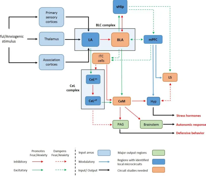
Most of our knowledge about neurocircuitry regulation anxiety comes from the fear conditioning studies and hence from here on forward, we will focus our discussion of circuits involved in processing acquisition and consolidation and extinction conditioned fear. Fig 1 is a representation of neuronal circuits known to be involved in conditioned.
Fig 1. Basic neuronal circuits involved in fear and anxiety. Regions with a major role in fear and anxiety and heavily emphasized in this thesis are in stated in bold letters. LA (lateral amygdala); BLA (basolateral amygdala); BLC (basolateral amygdaloid complex); mPFC (medial prefrontal cortex); LS (lateral septum); vHip (ventral hippocampus); CeL (lateral nucleus of central amygdala); CeM (medial nucleus of central amygdala);
ITC (Intercalated cells); Hyp (hypothalamus); PAG (periaqueductal grey). CeLon and CeLoff cells are described in detail in the section 1.6.1. Fig has been adapted from (Tovote et al., 2015).
27
1.6.1 Amygdala
Various studies in humans, mice and rats have implicated the amygdala, an almond-shaped cluster of nuclei located ventromedially within the temporal lobe of complex vertebrates, as a center for regulation of fear and anxiety (Allman and Brothers 1994, Wolff, Grundemann et al.
2014, Penzo, Robert et al. 2015, Marcinkiewcz, Mazzone et al. 2016, Mendez-Bertolo, Moratti et al. 2016, Zhu, Liu et al. 2016). The amygdala can be divided into two main sub-areas – the basolateral amygdaloid complex (BLC), which is mostly glutamatergic, and the central amygdala (CeA), which is composed mostly of γ-aminobutyric acid (GABA)-ergic neurons. In a pathway often referred to as the “low road” of fear response, any new fearful or anxiogenic stimulus (auditory, visual and somatosensory) generates sensory information within the thalamus and other sensory cortical regions, which is in turn conveyed to and terminates in the lateral amygdala (LA) (a subnuclei of the BLC) (Johansen, Hamanaka et al. 2010, Pessoa and Adolphs 2010, LeDoux 2014). This activates excitatory glutamatergic projections from the LA towards the lateral nucleus of CeA (CeL) which in turn activates the CeL
onneurons, that are characterised by a lack of protein kinase C-delta (PKCδ
-). The CeL
onneuron inhibits the CeL
offneurons, which are characterised by the presence of protein kinase C-delta (PKCδ
+). The CeL
offneurons exercise an inhibitory control over the medial part of the CeA (CeM), which is the main output region of this network and projects towards the periaqueductal grey (PAG), brainstem and hypothalamic regions that regulate downstream defensive behavior such as freezing (Swanson and Petrovich 1998, Tovote, Fadok et al. 2015). Thus, the disinhibition of CeM by a fear-generating stimulus leads to defensive behaviours via a complex pathway involving the different amygdaloid subcircuits (Ciocchi, Herry et al. 2010, Tye, Prakash et al. 2011, Maroun and Wagner 2016). These results are also complemented by studies showing altered GABA receptor levels within the CeA of mice strains bred for high anxiety (Tasan, Bukovac et al.
2011). In addition to the above-mentioned nuclei, a cluster of GABAergic cells that lie at the
interface of CeA and basolateral amygdala (BLA, subnuclei of the BLC) called the intercalated
28
cells (ITC). The ITC cells gate the information flow between BLA and CeA and are thought to be activated by fear extinction procedures (Likhtik, Popa et al. 2008, Tovote, Fadok et al. 2015).
The BLA on its own sends projections to the hippocampus to control anxiogenesis (discussed in detail in the section 1.6.3). The so called “high road” to anxiogenic stimulus involves the cortex (discussed in detail in section 1.6.4).
1.6.2 Septum
The septum or septal nuclei is a subcortical forebrain structure, which is located rostrodorsal to the hypothalamus and in between the lateral ventricles in rodents. It can be divided into two functionally, neurochemically and anatomically distinct nuclei namely the lateral septum (LS) and medial septum (MS). MS sends ascending inputs to the hippocampus which are mostly GABAergic and cholinergic in nature and, along with the hippocampus and diagonal bands of broca, it forms the “septo-hippocampal system”, which has been heavily implicated in operant
LS
Hippocampus Hypothalamus
Prefrontal cortex
Amygdala
Periaqueductal grey
Nucleus accumbens
Locus coeruleus Thalamus
Fig 2. A schematic for the afferent and efferent connections of the lateral septum (LS). Thick arrows indicate strong connections and double sided arrows indicate reciprocal connections (Fig adapted from Sheehan et al, 2004)
29
reward learning (Vega-Flores, Rubio et al. 2014) and spatial memory formation (Durkin 1994).
LS is thought to serve as an essential converging point for cognitive information from the cortex and hippocampus and affective information coming in from amygdala and hypothalamus which it relays to downstream regions to control the behavioural output in response to varied environmental stimuli (Deller, Leranth et al. 1994, Gray and McNaughton 1996). It comprises mostly of GABAergic projection neurons, (Gallagher, Zheng et al. 1995, Sheehan, Chambers et al. 2004) and has been a region of high interest with regards to stress response and aggression.
LS receives strong glutamatergic inputs from the hippocampus and is in turn reciprocally connected (not all) to LS.
cFOS studies have found a negative correlation between aggression and LS activity, and consequently, septal lesions are known to induce a typical “septal rage” phenotype (Potegal, Blau et al. 1981, Goodson, Evans et al. 2005, Lee and Gammie 2009). This is complemented by a gain of function studies showing an increase in LS activity to be correlated with reduced aggression (Wong, Wang et al. 2016). Thus, LS is a key regulation of aggression, which essentially is an enhanced form of social contact. Studies have shown that LS is involved in active stress coping and exerts an inhibitory effect on the HPA axis activity (Herman, Prewitt et al. 1996, Singewald, Rjabokon et al. 2011). These and other studies have led to the prevailing opinion that LS is a region, whose activity could be linked to dampening of anxiety (Sheehan, Chambers et al. 2004). This view has been contested in the past decades by studies which implicate different sub-population of neurons within the LS, for example, the cells expressing corticotropin-releasing factor (CRF) receptor 2 (CRFR2) in the promotion of stress-induced anxiety (Radulovic, Ruhmann et al. 1999, Anthony, Dee et al. 2014).
1.6.3 Hippocampus
The hippocampus (Hip) is a subcortical region located within the medial temporal lobes, which
were traditionally thought to be involved in the processing of declarative memory irrespective
30
of its emotional content. But over the past two decades, studies have shown another side of this interesting brain region, one that is much more plastic and closely linked with emotionality and stress response. Many studies have ascertained a role of the Hip in contextual fear learning and retrieval in rodents (Strekalova, Zorner et al. 2003, Chang and Liang 2017). Reduced hippocampal volumes have been both used as a marker and reported as a consequence of PTSD (Bremner 2002, Gilbertson, Shenton et al. 2002). For example, ventral hip (vHip) lesions have been shown to impair contextual fear conditioning (Kjelstrup, Tuvnes et al. 2002). BLA – vHip connectivity is known to regulate basal anxiety-related behaviour. Optogenetic activation of monosynaptic, glutamatergic BLA projections to the CA1 pyramidal neurons of the ventral Hip (vHip) exerts an anxiogenic effect (Felix-Ortiz, Beyeler et al. 2013). vHip-mPFC synchronicity within the context of anxiety is discussed in section 1.6.4. The septo-hippocampal system, which has been previously mentioned, is known to regulate stress-induced anxiety. Thus, the role of the hippocampus as a regulator of stress-induced anxiety and region which complies context-related information is well documented. Hippocampus also regulates social memory, as shown by a recent study, wherein genetic silencing of the dorsal CA2 pyramidal neurons of the hippocampus impaired social memory in mice (Hitti and Siegelbaum 2014). Considering these results, understanding the role of dorsal Hip (dHip) in coding for a social context during SFC acquisition and retrieval is of importance.
1.6.4 Prefrontal cortex
Higher order brain structures such as the mPFC is key brain structure mediating top-down regulation on brain regions such as the amygdala and thus helping organisms discern safety from danger in a more perceptive response to anxiogenic or fearful stimuli called the “high road” (Pessoa and Adolphs 2010, LeDoux 2014). Theta mPFC input into the BLA is known to provide a safety signal, thereby reducing innate anxiety (Likhtik, Stujenske et al. 2014).
Interestingly, disinhibition of CeM by the lack of functional mPFC – CeA connectivity was
31
hypothesised to be a plausible reason for increased anxiety in humans and monkeys by a recent study (Birn, Shackman et al. 2014). Increase synchronicity in vHip – mPFC connections have also been implicated in anxiety-like behaviour in mice by a study using extracellular in vivo recording especially in the mPFC neurons which encoded an anxiogenic context (Adhikari, Topiwala et al. 2010). Extinction of fear generated by Pavlovian fear conditioning paradigms is known to activate the infralimbic mPFC (IL-mPFC), which sends glutamatergic projection to the BLA activating the GABAergic interneuronal population within the BLA, thus diminishing its excitatory output to the CeA leading to a suppression of the fear response.
1.7. Molecular basis of conditioned fear
As stated before, molecular mechanisms underlying most complex emotions including anxiety and fear are poorly understood. However, the dysregulation of various system implicated in the psychopathology of anxiety disorders have been studied rigorously, and most of the available mechanistic data available to this end come from studies involving fear conditioning paradigms.
Hence, like in the previous section, I will keep the discussion in this part of the introduction
limited to molecular mechanisms relevant to fear conditioning paradigms.
32
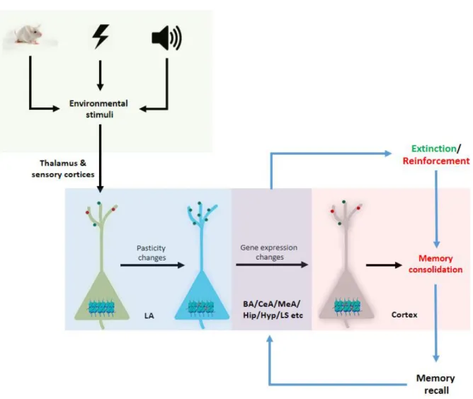
One could confer almost all mechanistic aspects of fear learning and relearning onto changes in synaptic plasticity that affect intracellular signalling cascades, which ultimately affect changes in gene expression via basic transcriptional and epigenetic mechanisms to mediate long-term memory formation. This information is concisely presented in Fig 3.
Here, I will firstly examine the role of mechanisms modulating synaptic plasticity during fear memory formation, which will be followed by an introduction of the role of mechanisms regulating gene expression regulation in consolidation and extinction of conditioned fear.
Fig 3. Schematic representation of the molecular processes involved in fear conditioning, fear memory consolidation and extinction. LA (lateral amygdala); BA (basal amygdala); CeA (central amygdala); MeA (medial amygdala); Hip (hippocampus); Hyp (hypothalamus); LS (lateral septum).
33
1.7.1. Environment to the neurons: synaptic plasticity and fear acquisition
As was shown in Fig 1, most of the sensory information about the CS and US in a fear conditioning paradigm from thalamic and cortical areas converges at the LA and the BLA making them key regions for the acquisition of fear (Quirk, Armony et al. 1997, Lin, Yeh et al.
2003). This idea is complemented by studies, which show that conditioning-induced plasticity
within the LA precedes freezing behaviour and thus seems to be a prerequisite for coding of
fear memory (Repa, Muller et al. 2001). Most of the current evidence suggests that associations
between the CS and US require Hebbian-plasticity (Hebb., 1949) provoked by US-mediated
depolarization within the LA (Johansen, Cain et al. 2011). Most of these plasticity-modulating
environmental stimuli lead to the glutamatergic transmission, which functions by increasing the
intracellular Ca
2+concentration in post-synaptic neurons (Johansen, Cain et al. 2011). A
number of glutamatergic receptors including the post-synaptic N-methyl-d-aspartate receptors
(NMDARs) – both ionotropic and non-ionotropic – are thought to mediate plasticity within the
LA (Rodrigues, Schafe et al. 2001). This, in turn, leads to autophosphorylation of the
Ca
2+/Calmodulin (CaM) dependent protein kinase II (CaMKII), which has been shown to
increase the dendritic spine density and activate fear memory formation via both direct and
indirect mechanisms (Silva 2003). One example of the indirect mechanism could be that
autophosphorylation and thus activation of CaMKII could lead to phosphorylation of the serine
831 residues of α-amino-3-hydroxy-5-methyl-4-isoxazolpropionic acid-type glutamate
receptor (AMPAR) GluA1 subunit which leads to its translocation and insertion into the cell
membrane. Such insertion increases the synaptic strength and is known to aid in short-term
memory formation by activation of the so called ‘silent synapses’ (Malinow and Malenka 2002,
Rumpel, LeDoux et al. 2005, Johansen, Cain et al. 2011). Metabotropic glutamate receptors
(mGluRs), which are also stimulated by such CS- and/or the US- induced glutamate release,
spark acquisition of contextual fear, but are seldom involved in consolidation. Indeed,
activation of the group I mGluR5 within the LA and the BLA is known to aid acquisition of
34
conditioned fear. Such mGluR5 activation within the LA could, in turn, activate the NMDARs and also increase intracellular Ca
2+levels via the inositol 1,4,5-triphosphate pathway and thereby activate the downstream CaMKII leading to further enhancement of fear memory formation (Rodrigues, Bauer et al. 2002, Rudy and Matus-Amat 2009, Johansen, Cain et al.
2011).
Evidence from several pharmacological studies has implicated plasticity changes within the GABAergic interneurons within different amygdalar subnuclei in fear memory modulation (Ehrlich, Humeau et al. 2009) (also see section 1.6.1). Quite a few studies have implicated activation of the GABA-synthesizing interneurons in inhibition of projection neurons within the LA leading to modulation of fear memory formation and expression (Bissiere, Humeau et al. 2003, Tully, Li et al. 2007). Although the exact function of GABAergic interneurons within the LS is not clearly known, few studies have been able to shed light on general GABA function in the context of fear conditioning paradigms. Activation of the GABA
Areceptor was shown to reduce cued fear acquisition, and this is complemented by studies showing a transient reduction in LTP and GAD65 expression following fear conditioning (Muller, Corodimas et al. 1997, Johansen, Cain et al. 2011). Interestingly, pharmacological blockade of the GABA receptor α1 subunit increases cued fear learning. Although the later result is against a popular opinion which is that GABAergic signalling leads to impairment of fear learning, the reason for such results could be because of increase in GABA availability for receptors which lack this subunit (Wiltgen, Godsil et al. 2009).
1.7.2. From synapse to nucleus: Cell signalling molecules and transcription factors involved in coding of fear
Most of the signalling cascades involved in fear memory formation or extinction memory
formation function by increasing intracellular Ca
2+. Such increase leads to a barrage of
molecular changes within the neuron including the activation of CaMKII and CaMKIV which
35
seems to be a prerequisite for strong memory formation following fear conditioning as
mentioned before. Ca
2+also interacts with and activates the Ca
2+/ phospholipid-dependent
protein kinase (PKC) and cAMP-dependent protein kinase (PKA) which eventually leads to
gene expression changes within the nucleus and long-lasting changes in the disposition of a
neuron via LTP. Involvement of PKA and PKC in fear memory formation are supported by
many studies. Pharmacological blockade of PKA with the specific inhibitor RP-cAMPS within
the LA inhibits long-term memory formation, while short-term memories stay intact indicating
that signalling cascades involving PKA are involved in fear memory consolidation (Schafe and
LeDoux 2000). Even studies with transgenic mice expressing an inactive form of PKA within
the hippocampus show reduced LTP and impaired contextual memory formation thus
corroborating the role of PKA in the formation of fear memory. PKMζ, a PKC isoform, was
shown to be upregulated on induction of hippocampal LTP and is necessary and sufficient for
it (Ling, Benardo et al. 2002). CaMKII, PKA and PKC act upon the mitogen-activated kinase
(MAPK) signalling cascade, which ultimately leads to the regulation of gene transcription
leading to fear memory consolidation and long-term memory formation. Activation of MAPK
could lead to subsequent activation of 7 different signalling cascades in vertebrates (Adams and
Sweatt 2002). Synchronous activation of CaMKII, PKA and PKC leads to eventual activation
of extracellular regulated kinase (ERK) 1 and 2 via a pathway involving MAPK/ERK kinase
(MEK) activation. ERK1 and ERK2 translocate to the nucleus and lead to a variety of changes
ultimately leading to changes in gene expression and increase in synaptic strength (Wu,
Deisseroth et al. 2001, Orsini, Yan et al. 2013). Ample data from studies involving
pharmacological and genetic modulation of members of the MAPK signalling cascade have
cemented our understanding of their role in formation of fear memories (English and Sweatt
1996, Brambilla, Gnesutta et al. 1997, Merino and Maren 2006)
36
1.7.3. Changes within the neuronal nucleus: consolidation and formation of long-lasting fear memories
Within the context of fear memories, most signalling cascades converge within the nucleus on the cyclic-AMP (cAMP)/ Ca
2+response element-binding protein (CREB). Indeed, CREB activation by MAPK and PKA signalling within the LA seems to be the key for regulation of fear memory formation and consolidation (Hernandez and Abel 2008). ERK1 activates a downstream kinase RSK within the nucleus and this mediates phosphorylation of CREB at the ser133 residue (Impey, Obrietan et al. 1998, Orsini and Maren 2012, Naqvi, Martin et al. 2014).
Ca
2+dependent activation of CAMKIV also activates CREB. An important difference between
the two modes of CREB activation is that CAMKIV-mediated CREB activation is more of an
activity-dependent rapid activation, whereas ERK/RSK activation exerts a slower and
prolonged effect (Thomas and Huganir 2004). However, the most substantial work in the role
of CREB in fear memory formation comes from the work of Sheena Josselyn’s lab. Over the
last decade, Josselyn and colleagues have shown that neurons expressing an activated form of
CREB are preferentially included in the fear memory trace and that CREB over-activation
within the LA leads to long-lasting fear memories (Han, Kushner et al. 2007). This study also
showed that mice deficient for CREB showed impaired auditory fear conditioning, an effect
that could be completely rescued by transfection of the LA with a herpes simplex virus based
CREB overexpressing vector even though the vector transfected only 15% of LA neurons (Han,
Kushner et al. 2007, Han, Yiu et al. 2008, Yiu, Mercaldo et al. 2014). Once activated CREB
binds to its partner - the CREB binding protein (CBP)/p300 and its target DNA binding site,
i.e. the CREB response element (CRE) leading to activation of more than 100 downstream
target genes including many immediate early genes (IEGs) (Lonze and Ginty 2002). These IEG
can be classified as activity-induced regulatory transcription factors (RTF) which include cFos,
zif268 etc and effector IEGs such as bdnf, arc, homer1a etc (Chaudhuri 1997). Overexpression
of most of these IEGs has been previously correlated with enhanced fear memory formation
37
and strength by studies utilising contextual and auditory fear conditioning paradigms. For example, an elevated cFos expression has been observed in the ventral part of LA after auditory fear conditioning which fit with results from another study that reveals that neurons in this region exhibit increased activity during fear training and extinction (Radulovic, Kammermeier et al. 1998, Ressler, Paschall et al. 2002, Ploski, Park et al. 2010). An interesting gene is Arc/Arg3.1, whose mRNA undergoes nuclear export and is preferentially localised in the dendrites with activated synapses in the rat dentate gyrus in response to LTP (Rodriguez, Davies et al. 2005). Neurotrophic factors such as BDNF, which is also a CREB target and its receptor, the tropomyosin-related kinase B (TrkB) regulate synaptic plasticity. Activity-dependent release of BDNF (pre or post-synaptic) correlates with increased LTP (Schinder and Poo 2000, Messaoudi, Ying et al. 2002, Lessmann, Gottmann et al. 2003). In concert, inhibition of BDNF activity within the LA with either TrkB antagonism or its genetic silencing prevents long-term memory formation (Rattiner, Davis et al. 2004). This is in contrast to a study which shows that ablation of BDNF early during development specifically within the forebrain leads to an enhancement of fear memories.
Once consolidated, fear memories are long-lasting in nature. However, if these associations are
recalled, as it happens during fear extinction, these memories become labile and susceptible to
modifications. During extinction, prolonged or multiple exposures to the CS in absence of the
associated US creates a new memory trace predicting lack of CS aversion and this competes
with the original fear evoking memory trace. On multiple or prolonged exposures to the
unpaired CS, the new memory gets stronger and eventually overshadows the fear memory. In
principle, the neuronal and molecular mechanisms involved in extinction of fear memories are
more or less similar to the ones involved in the formation of the original fear memory, and thus,
they are not discussed here in more detail. However, what I find important to introduce in the
course of my thesis is the modulation of neuropeptide systems and epigenetic mechanisms by
38
cognitive enhancers to aid in the process of extinction with the final aim of developing effective therapeutic strategies for anxiety disorder.
1.8. Role of neuropeptides in regulation of fear
All the factors introduced in the sections above regulate formation, consolidation, and extinction of fear memories in a very intricate and synchronous manner. Although we know a lot about it, fear seems to be more complicated, enigmatic and above these basic mechanisms.
Neuropeptides are defined as ‘small proteinaceous substances produced and released by neurons through a regulated secretory route and act on neural substrates’ and have been implicated in the regulation of fear and anxiety by several studies. They are one of the most diverse class of neuronal signalling molecules, and there are more than 70 genes within the mammalian genome, which code for neuropeptides and their receptors. However, as the focus of my thesis is centred around the oxytocin (OXT) system, I will only elaborate on OXT- mediated regulation of anxiety and fear.
1.8.1 Neuropeptide oxytocin and its receptor The ligand: OXT
OXT is a 9-amino acid long neuropeptide, which was one of the first peptide hormones to be structurally characterised and chemically synthesised (Du Vigneaud, Ressler et al. 1953). It is produced within the hypothalamic paraventricular (PVN), supraoptic (SON) and accessory nuclei by magnocellular and parvocellular neurons, which are distinct in morphology, subnuclear location, projections and the amount on OXT produced (Swanson and Sawchenko 1983, Eliava, Melchior et al. 2016). The parvocellular OXT neurons project mainly to the brainstem and spinal cord where they are involved in the control of feeding
Fig 4. Neuropeptide Oxytocin
39
behaviour, cardiovascular function, nociception and breathing (Swanson and Sawchenko 1983, Mack, Kc et al. 2002, Eliava, Melchior et al. 2016). The magnocellular OXT neurons project to the posterior pituitary leading to the systemic release of OXT via blood in response to a variety of environmental stimuli. Magnocellular neurons also innervate forebrain regions leading to specific control of behaviour (Bargmann and Scharrer 1951, Knobloch, Charlet et al.
2012). Central release of OXT controls a wide range of socio-emotional behaviors such as maternal behavior (Bosch 2011), sexual behavior (Argiolas and Gessa 1991, Waldherr and Neumann 2007), pair bonding (Bales, van Westerhuyzen et al. 2007), social recognition (Oettl, Ravi et al. 2016), social anxiety (Zoicas, Slattery et al. 2014) among others (for review see:
(Landgraf and Neumann 2004)). Local effects of OXT within the SON are more likely due to its dendritic release to attain paracrine effects (Ludwig and Leng 2006).
Effective modulation of these behaviours requires regulation of OXT synthesis and release, which is realised by different genomic and non-genomic mechanisms acting in a fine-tuned way so as to generate appropriate responses to specific environmental and physiological stimuli.
OXT is released both centrally and peripherally in response to various physiological stimuli including stress, suckling during lactation, social interactions, mating etc (Neumann, Russell et al. 1993, Waldherr and Neumann 2007, Zoicas, Slattery et al. 2014). For example, OXT is shown to be released in response to forced swimming in the CeA of male rats, where it presumably supports passive stress-coping (Ebner, Bosch et al. 2005). OXT release is also known to be regulated by other neuropeptides. For example, prolactin is known to inhibit OXT release in virgin rats without interfering with the intracellular signalling cascades and this inhibition is lost during lactation enabling effective milk ejection (Augustine, Ladyman et al.
2017). Another example of such interaction comes from recent unpublished data, which
conclusively show that the anxiolytic effects of the neuropeptide S (NPS) within the PVN are
mediated via the OXT system (Grund et al, unpublished). At the level of DNA, the OXT
promoter contains an oestrogen receptor (ER) response element (ERE) and is activated by
40
interaction with ERβ (Nomura, McKenna et al. 2002, Shughrue, Dellovade et al. 2002). A genomic mechanism which has recently been highlighted in this regard is that of the DNA methylation of the OXT promoter. A recent study has found that low DNA methylation of the OXT promoter (possibly leading to high OXT expression) in human epithelial cells found in saliva samples to be correlated to high sociability and emotional recognition (Haas, Filkowski et al. 2016).
The OXT receptor (OXTR)
As mentioned before, OXTergic neurons have long-range axonal projections which enable OXT release in different far off brain regions. Within these regions, OXT regulates its central effects by acting through the single OXT receptor (OXTR) which is spread throughout the brain (Sofroniew 1980, Knobloch, Charlet et al. 2012, Dolen, Darvishzadeh et al. 2013, Mitre, Marlin et al. 2016, Otero-Garcia, Agustin-Pavon et al. 2016). Thus, the flipside to this story is the controlled regulation of OXTR expression by a variety of mechanisms, which gives an organism additional control over socio-emotional behaviour in response to relevant physiological and environmental cues. The OXTR is a classical class I G-protein-coupled receptor with 7 transmembrane domains and is expressed in a variety of brain regions including MeA, BLA, nucleus accumbens (NAcc), BNST, MPOA, Hyp, Hip, ventral palladium, PAG, striatum, LS, VTA and the olfactory bulb (Grinevich, Desarmenien et al. 2014, Grinevich, Knobloch-Bollmann et al. 2016) (Fig 5). OXTR expression within these regions can be regulated depending upon ligand-availability. For instance, lack of OXT after SFC in male mice is compensated by an increase in the OXTR binding within the LS (Zoicas, Slattery et al. 2014).
Conversely, chronic administration of OXT leads to a downregulation of the OXTR (Peters, Slattery et al. 2014).
At a genomic level, OXTR promoter possesses binding sites for many transcription factors including c-Myc, SP1, nuclear factor kappa B (NFκB), CREB etc (Blanks, Shmygol et al.
2007). OXTR promoter in rodents contain an ERE, and OXTR expression is activated by ERα.
41