DISSERTATION ZUR ELRANGUNG DES DOKTORGRADES DER NATURWISSENSCHAFTEN
(DR. RER. NAT.)
an der Fakultät für Biologie und Vorklinische Medizin der Universität Regensburg
vorgelegt von Melanie Royer aus Burglengenfeld
im Jahr 2020
Septal coding and non-coding RNAs regulated in social fear
─ The role of lncRNA Meg3 in social fear
extinction
Das Promotionsgesuch wurde eingereicht am: 30.04.2020
Die Arbeit wurde angeleitet von: Prof. Dr. Gunter Meister
Unterschrift:
………..
(Melanie Royer)
TABLE OF CONTENTS
Table of Contents
Table of Contents ... III Abstract ... IX Zusammenfassung ... XIII Abbreviations ... XIX
1 Introduction ... 1
1.1 World of RNAs ... 1
1.1.1 Messenger RNAs and the importance of alternative splicing events ... 1
1.1.2 Non-coding RNAs ... 4
1.1.2.1 Long non-coding RNAs ... 5
1.1.2.2 The role of lncRNAs in neuropsychiatric disorders ... 7
1.2 Social anxiety disorders and related animal models ... 10
1.2.1 Social anxiety disorders and treatment options ... 10
1.2.2 Social fear conditioning ... 11
1.2.3 The septum as a key region for social fear ... 12
1.3 Technologies for detecting and manipulating RNAs and chromatin states ... 15
1.3.1 Total RNA-Sequencing ... 15
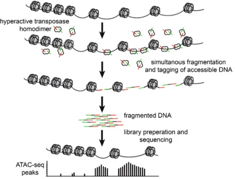
1.3.2 ATAC-Sequencing ... 16
1.3.3 CUT&RUN ... 17
1.3.4 LNA antisense GapmeRs ... 18
1.4 Potential RNA candidates ... 18
1.4.1 Maternally expressed gene 3 ... 18
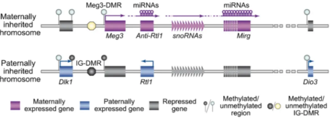
1.4.1.1 Imprinted locus of Meg3 ... 19
1.4.1.2 Knockout models for Meg3 ... 20
1.4.1.3 Function and signalling pathways of Meg3 ... 22
1.4.1.4 The involvement of Meg3 in psychopathologies ... 24
1.4.1.5 PTEN/PI3K/AKT signalling pathway ... 26
1.4.2 Serum and glucocorticoid inducible kinase 1 ... 26
1.5 Aims and outlines of the present study ... 28
2 Materials and Methods ... 31
2.1 Materials ... 31
2.2 Animals and husbandry ... 32
2.3 Behavioural testing ... 32
2.3.1 Social fear conditioning ... 32
2.3.2 Scoring of behaviours ... 34
2.4 Surgical procedures ... 34
2.4.1 Intracerebral microinfusions ... 35
2.4.2 Implantation of microdialysis probes ... 35
2.5 Tissue collection, perfusion and brain slicing ... 36
2.6 Sequencing... 36
2.6.1 Total RNA-Seq ... 36
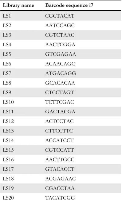
2.6.1.1 Library preparation ... 37
2.6.1.2 Sequencing with HiSeq3000 ... 38
2.6.1.3 Bioinformatical analysis ... 39
2.6.2 FAC-sorting of neuronal nuclei ... 39
2.6.3 ATAC-Seq and data analysis ... 40
2.6.4 CUT&RUN ... 42
2.7 Total RNA and protein extraction from one sample... 44
2.8 Reverse transcription ... 44
2.9 PCR and electrophoretic separation on agarose gel ... 44
2.10 Quantitative real-time PCR ... 45
2.11 RNAscope ... 48
2.12 Protein quantification ... 49
2.13 Western blot analysis ... 49
2.14 Statistical analysis ... 51
2.15 Experimental design ... 52
3 Results ... 55
3.1 Total RNA-Seq ... 55
3.1.1 Sequencing approach I ... 55
3.1.2 Sequencing approach II combined with approach I ... 58
3.2 Meg3 – a lncRNA involved in social fear ... 61
3.2.1 Dynamic regulation of Meg3 expression levels during the SFC paradigm ... 61
3.2.2 Specific regulation of Meg3 in the context of fear extinction learning ... 65
3.2.3 Nuclear localization of Meg3 in neurons of the brain septal region ... 69
3.2.4 Meg3 loss-of-function studies ... 70
3.2.4.1 Establishment of specific LNA antisense GapmeRs for Meg3 variants ... 70
3.2.4.2 Meg3 knockdown effects on social fear extinction ... 72
3.2.4.3 Meg3 knockdown effects on social fear acquisition ... 74
3.2.4.4 Meg3 knockdown effects on long-term extinction memory ... 76
3.2.5 Regulation of the PTEN/PI3K/AKT signalling pathway in context of social extinction ... 78
3.3 Identification of differentially accessible chromatin regions after social fear extinction training ... 82
3.4 CUT&RUN for H3K27me3 modification after social fear extinction ... 83
3.5 Meg3 expression in the HPC in the context of social fear extinction ... 84
3.6 Validation of additional RNA candidates ... 86
3.7 Impairment of social fear extinction by lesions of the LS ... 90
4 Discussion ... 95
4.1 Dynamic regulation of the lncRNA Meg3 within the septum in the context of social fear ... 95
4.2 PI3K/AKT signalling in context of social fear extinction and Meg3 KD ... 100
4.3 Effects of social fear extinction on chromatin accessibility and H3K27me3 histone modifications... 103
4.4 Time-shifted expression of Sgk1 in mice with successful and unsuccessful extinction 104
4.5 Validation of the RNA candidates and confirmation of the RNA-Seq approach ... 105
4.6 The important role of the septum especially for social fear extinction ... 107
4.7 Conclusion and future perspectives ... 108
Appendix ... 113
References ... 117
Curriculum vitae ... 139
Publications ... 141
Acknowledgements ... 145
ABSTRACT
Abstract
Social anxiety disorder (SAD) is characterized by fear and avoidance of social situations and displays the third most common anxiety disorder in society. However, even today, there is only sparse knowledge about the underlying mechanisms of SAD available. So far, applied therapies, which include cognitive-behavioural therapies combined with pharmacological intervention, show indeed partial achievements, but still with a high rate of non-responders and relapse. For the development of specific and more efficient treatment options, a better understanding of especially molecular mechanisms is urgently required. Here, RNA molecules are promising to provide insights on a regulatory as well as subsequently translational level during the state of SAD. In general, coding and non-coding RNAs are dynamically regulated as response to certain stimuli and their dysregulation lead to tremendous effects. Disorders are usually linked to pre- and post-transcriptional alterations and approaching these in context of SAD will help to identify disorder-causing signalling pathways and networks.
Thus, the thesis presented here aimed to characterize RNA profiles that are regulated in context of social fear acquisition and its extinction with a total RNA-Sequencing (RNA-Seq) approach.
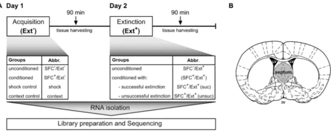
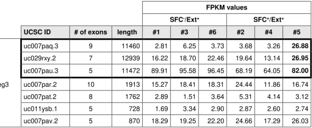
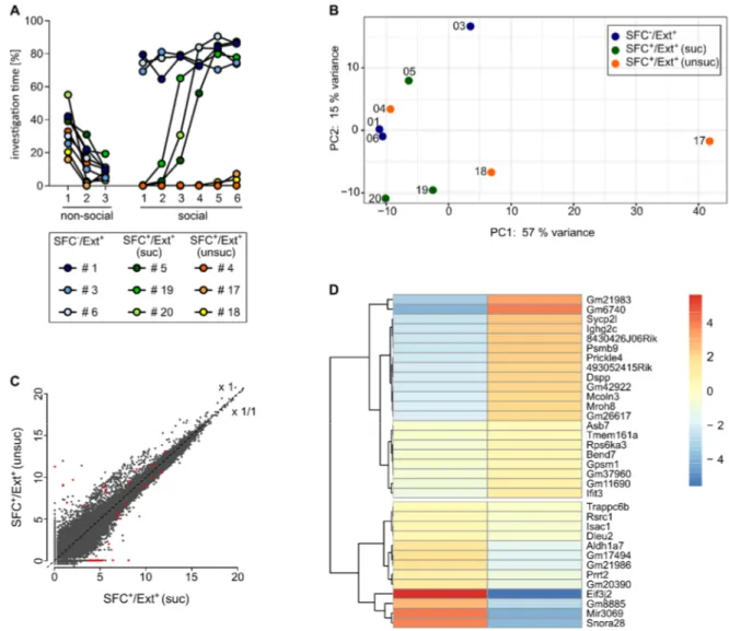
Therefore, I mimicked social fear in male mice by inducing social avoidance – the core symptom of social fear – using the social fear conditioning paradigm (SFC). SFC allows us to induce social fear during social fear acquisition, whereas during social fear extinction training, which is comparable to exposure therapy in humans, animals re-learn to investigate presented conspecifics again. For total RNA-Seq, samples were collected 90 min after the last behavioural assessment during the SFC protocol, with a specific focus on conditioned (SFC+) animals that showed different extinction-success. Here, the septum was chosen as the brain region of interest as it is proven as an important regulator of social fear expression. RNA-Seq revealed many different RNAs regulated on a gene-based and a transcript-based level and after validation of interesting RNA candidates e.g. Sgk1 and Crfr2 among others, with qPCR, I focused mainly on the long non-coding RNA (lncRNA) Meg3, which was regulated in a Meg3 transcript- and extinction success-dependent manner. In more detail, the Meg3 variants containing an alternatively spliced long exon 10 were downregulated in SFC+ animals 24 h after social fear acquisition and levels were restored in case mice could successfully overcome their social fear during the extinction training. In contrast, animals that stayed fearful after the extinction training still displayed reduced Meg3 level even 3 h after extinction training. Control experiments revealed that learning processes, and not social contact per se, are necessary for Meg3 regulation within the septum. Additionally, unique septal Meg3 expression patterns were found compared to hippocampal regions. In vivo Meg3 knockdown within the septum was achieved by the local
application of antisense LNA GapmeRs that induce RNaseH-dependent cleavage of specific Meg3 variants. Meg3 knockdown before acquisition and before extinction training revealed that social fear acquisition was not affected, whereas the extinction and the subsequent memory consolidation seemed to be slightly impaired. Furthermore, I investigated the activation of the PI3K/AKT signalling pathway as it was shown to regulate plasticity and long-term potentiation in neurons, and to be modulated by Meg3. Interestingly, I found increased phosphorylation levels of the regulatory subunit P85 of the PI3K and AKT at Ser473 in SFC+ animals 3 h after unsuccessful extinction training, indicating an activation of this signalling pathway. Activation in successfully extinguishing animals is conceivable to happen at earlier time points and therefore deserves further investigation. In Meg3 knockdown experiments, I observed an activation of PI3K in SFC+ control mice. In contrast, SFC+ knockdown mice showed no increased activation compared to unconditioned (SFC-) knockdown mice, which might be due to high basal PI3K activation in SFC- knockdown mice. Taken together, the presented data provide first, but strong indications on a regulatory link between Meg3 and PI3K/AKT signalling after social fear extinction training. Interestingly, many lncRNAs function as transcription regulators of target genes by altering chromatin structures. To assess changes in chromatin accessibility and histones modifications after social fear extinction on a general level, but also with a direct link to Meg3 for the identification of nuclear Meg3 action sites, I performed ATAC-Seq and CUT&RUN for H3K27me3.
In a second part, I further strengthened the role of the septum in social fear extinction processes.
Based on originally planned microdialysis experiments for monitoring released neurotransmitter during social fear extinction, I observed that the implantation of the microdialysis probe into the lateral septum itself is sufficient to completely impair social fear extinction in male mice.
The unilaterally caused damage still inhibited extinction even if an increased number of social stimuli was presented.
In summary, I was able to generate new data on altered transcriptomics, chromatin accessibility and histone modification H3K27me3 alterations in the context of social fear. Altogether, these data provide a solid background for the further investigation of molecular mechanisms involved in social fear and its extinction. Moreover, I could show for the first time that a lncRNAis dynamically regulated in social fear. Control experiments revealed the importance of learning and memory processes for Meg3 regulation. Moreover, region-specific Meg3 regulation during SFC as well as impaired social fear extinction after septal implantation of a microdialysis probe strengthened the role of the septum in social fear regulation.
ZUSAMMENFASSUNG
Zusammenfassung
Soziale Angst ist durch Angst und Vermeidungsverhalten von sozialen Situationen charakterisiert und stellt die dritthäufigste Angsterkrankung in unserer Gesellschaft dar. Trotz alledem ist auch heute noch wenig über die zugrundeliegenden Mechanismen bekannt. Aktuell zeigen angewandte Therapieansätze, die meist kognitive Verhaltenstherapien mit Medikationen kombinieren, erste Teilerfolge. Jedoch gibt es weiterhin viele Patienten, die nicht darauf ansprechen oder schnell rückfällig werden. Um spezifische und effizientere Behandlungsmöglichkeiten entwickeln zu können, ist vor allem ein besseres Verständnis über die zugrundeliegenden molekularen Mechanismen von Nöten. Hierbei sind RNA-Moleküle besonders vielversprechend, Einblicke in die molekularen Gegebenheiten, sowohl auf regulatorischer als auch auf nachfolgend translationaler Ebene, bei sozialer Angst zu geben.
Generell sind kodierende und nicht-kodierende RNAs als Reaktion auf bestimmte Stimuli sehr dynamisch reguliert und ihre Fehlregulation hat meist schwerwiegende Auswirkungen zur Folge.
Da Krankheiten oftmals mit spezifischen prä- und posttranskriptionellen Modifikationen verbunden sind, kann deren genauere Untersuchung im Kontext von sozialer Angst Aufschluss auf krankheitsrelevante Signalwege und deren Netzwerke geben.
Aus diesem Grund wurde die vorgelegte Doktorarbeit mit dem Ziel verfasst, RNAs, die durch soziale Angstkonditionierung und deren Löschung reguliert werden, mittels RNA- Sequenzierung zu identifizieren und zu charakterisieren. Hierzu habe ich mit Hilfe sozialer Angstkonditionierung soziales Vermeidungsverhalten, die Kernkomponente von sozialer Angst, in männlichen Mäusen induziert. Die soziale Angstkonditionierung ermöglicht die Etablierung von sozialer Angst, wohingegen bei der Angstlöschung, welche vergleichbar zur Konfrontationstherapie beim Menschen ist, die Mäuse lernen, wieder ungestraften Kontakt zu Artgenossen aufzunehmen. Für die RNA-Sequenzierung wurde 90 min nach der letzten Verhaltenstestung der sozialen Angstkonditionierung oder Angstlöschung RNA aus dem Septum der Mäuse isoliert. Hierbei lag ein spezielles Augenmerk auf konditionierten (SFC+) Mäusen, die die Angstlöschung mit unterschiedlichem Erfolg abgeschlossen haben. Das Septum wurde als Zielregion gewählt, da dessen wichtige Funktion für die Regulation von sozialer Angst bereits mehrfach gezeigt wurde. Durch die RNA-Sequenzierung wurden viele RNAs identifiziert, die sowohl auf Gen- als auch auf Transkript-basierter Ebene reguliert waren. Nach anschließender Validierung mittels qPCR von auserwählten RNA-Kandidaten wie beispielsweise Sgk1, Crfr2 und weiteren, habe ich mich hauptsächlich auf die lange nicht- kodierende RNA Meg3 fokussiert, welche transkriptspezifisch und in Abhängigkeit von einer erfolgreichen Angstlöschung reguliert war. Genauer betrachtet waren jene Meg3-Varianten, die
ein langes alternativ gespleißtes Exon 10 enthielten, 24 h nach der Angstkonditionierung in SFC+ Mäusen herunterreguliert. Mäuse, die ihre soziale Angst während des Trainings zur sozialen Angstlöschung überwunden hatten, konnten die Expressionslevel zu unkonditionierten (SFC-) Tieren wieder angleichen. Tiere, die nach dem Training zur Angstlöschung noch immer sozial verängstigt waren, wiesen jedoch sogar noch 3 h nach dem Training niedrige Meg3-Levels auf. Kontrollversuche zeigten, dass Lernprozesse, und nicht soziale Interaktion alleine, für die Regulation von Meg3 nötig sind. Außerdem wurde Meg3 speziell im Septum, und nicht im Hippocampus reguliert vorgefunden. Um in vivo einen Meg3 Knockdown im Septum zu erzielen, wurden antisense LNA GapmeRs, die einen RNase H-abhängigen Abbau von spezifischen Meg3-Varianten induzieren, lokal verabreicht. Meg3 Knockdown-Versuche vor der Angstkonditionierung und vor dem Training zur Angstlöschung zeigten keine Auswirkungen auf die Angstkonditionierung, wohingegen die Angstlöschung und die anschließende Gedächtnisbildung und -festigung etwas beeinträchtigt zu sein schienen. Des Weiteren habe ich die Aktivierung des PI3K/AKT Signalweges untersucht, da diese bewiesenermaßen neuronale Plastizität und Langzeitpotenzierung reguliert und selbst von Meg3 moduliert wird. Interessanterweise konnte ich zeigen, dass die Phosphorylierungslevels von P85, der regulatorischen Untereinheit von PI3K, und AKT Ser473 3 h nach dem Training zur Angstlöschung in Tieren, die die Angst nicht erfolgreich löschen konnten, erhöht waren. Dies lässt auf eine Aktivierung des Signalweges schließen. Es ist denkbar, dass die Aktivierung in Tieren, die ein erfolgreiches Training zur Angstlöschung durchliefen, zu einem früheren Zeitpunkt stattfindet, was weitere Untersuchungen bedarf. In Meg3 Knockdown-Experimenten habe ich eine Aktivierung von PI3K in SFC+ Kontrolltieren beobachtet. Dieser Effekt blieb jedoch in SFC+ Knockdown-Tieren aus, was an den basal erhöhten Aktivierungslevels, die in SFC- Knockdown-Tieren aufzufinden waren, begründet sein könnte. Insgesamt stellen die hier präsentierten Ergebnisse erste, aber bedeutende Hinweise über einen regulatorischen Zusammenhang von Meg3 und dem PI3K/AKT Signalweg nach dem Training zur sozialen Angstlöschung dar, welche noch weiter untersucht werden sollten. Interessanterweise regulieren viele lange nicht-kodierende RNAs die Transkription, indem sie Chromatinstrukturen beeinflussen. Deshalb habe ich ATAC-Seq und CUT&RUN durchgeführt, um Veränderungen auf Chromatinebene und der Histonmodifikation H3K27me3 zu charakterisieren. Hierbei wurden Veränderungen nach der sozialen Angstlöschung im allgemeinem, aber auch mit einem direkten Link zu Meg3 untersucht, um nukleare Interaktionsstellen von Meg3 für Zukunftsexperimente zu identifizieren.
In einem zweiten Teil der Dissertation, habe ich die Rolle des lateralen Septums für Prozesse der sozialen Angstlöschung weiter bestärkt. Basierend auf anfänglich geplanten Mikrodialyse- Experimenten, die die Freisetzung von Neurotransmittern detektieren sollten, musste ich feststellen, dass die alleinige Implantation der Mikrodialyse-Sonde in das LS ausreichend war, die soziale Angstlöschung in männlichen Mäusen stark zu beeinträchtigen. Der unilateral verursachte Gewebsschaden verhinderte die Angstlöschung weiterhin, auch bei einer Erhöhung der Anzahl an präsentierten sozialen Stimuli.
Zusammenfassend konnte ich neue Daten über Veränderungen des Transkriptoms, der Chromatinzugänglichkeit und der Histonmodifikation H3K27me3 im Kontext von sozialer Angst generieren. All dies stellt ein solides Grundgerüst für die weitere Untersuchung von molekularen Mechanismen, die bei sozialer Angst und deren Löschung involviert sind, dar.
Außerdem konnte ich erstmals zeigen, dass eine lange nicht-kodierende RNA dynamisch in sozialer Angst reguliert ist. Kontrollexperimente zeigten, dass Lern- und Gedächtnisprozesse für die Regulation von Meg3 notwendig sind. Zusätzlich bestärkten die regionsspezifische Meg3-Regulation und die eingeschränkte Angstlöschung bei septaler Implantation einer Mikrodialyse-Sonde die Rolle des LS in der Regulation von sozialer Angst.
ABBREVIATIONS
Abbreviations
Abbreviations Meaning
µ micro
µl microliter
µM micromolar
AKT protein kinase B
ANOVA analysis of variance
ATAC-Seq assay for transposase-accessible chromatin with high throughput sequencing
AD Alzheimer disease
AMPAR α-amino-3-hydroxy-5-methyl-4-isoxazolepropionic acid receptor
ASD autism spectrum disorder
BC1 brain cytoplasmic non-coding RNA
bp base pairs
BSA bovine Serum Albumin
cDNA complementary DNA
CRFR2 corticotropin-releasing factor receptor 2 CUT&RUN cleavage under target & release using nuclease
Cwc22 Cwc22 associated protein
DAPI 4',6-Diamidin-2-phenylindol
Dclk3 doublecortin-like kinase 3 DMR differentially methylated region
DNA deoxyribonucleic acid
dNTP deoxyribonucleosid triphosphate
ECL enhanced chemiluminescence
Ext- acquisition
Ext+ extinction (training)
FACS fluorescence-activated cell sorting
FPKM fragments per kilobase million
GABA γ-aminobutyric acid
Gtl2 gene trap locus 2
h hour
(d/v) HPC (dorsal/ventral) hippocampus
HRP horseradish peroxidase
IG-DMR intergenic differentially methylated region
IHC immunohistochemistry
InDA-C insert dependent adaptor cleavage
kb kilobase
KD knockdown
kDa kilodalton
LNA locked nucleic acids
lncRNA long non-coding RNA
LS lateral septum
LTP long-term potentiation
mAB monoclonal antibody
MDM2 murine double minute2; E3 ubiquitin ligase
Meg3 maternally expressed gene 3
miRNA microRNA
mM millimolar
MM mastermix
mRNA messenger RNA; coding RNA
MS medial septum
mTORC (1/2) mammalian target of rapamycin complex (1/2); serine/threonine kinase
n nano
ng nanogram
nm nanometer
nt nucleotide
OFC operant fear conditioning
OFC- unconditioned in context of OFC
OFC+ conditioned in context of OFC
OXT oxytocin
OXTR oxytocin receptor
p phospho
pAB polyclonal antibody
PBS phosphate buffered saline
PCA principal component analysis
PCR polymerase chain reaction
PDK1 phosphoinositide-dependent protein kinase 1
PFA paraformaldehyde
PI3K phosphoinositide-3-kinase
PIP2 phosphatidylinositol-4,5-bisphosphate PIP3 phosphatidylinositol-3,4,5-triphosphate
PolII (RNA) polymerase II
PRC2 polycomb repressive complex 2
qPCR real-time quantitative PCR
REST RE1 silencing transcription factor
RNA ribonucleic acid
RNA-Seq total RNA-sequencing
RT room temperature
SAD social anxiety disorder
sCRFR2 soluble corticotropin-releasing factor receptor 2
SDS sodium dodecyl sulfate
SDS-PAGE sodium dodecyl sulfate polyacrylamide gel electrophoresis
SEM standard error of the mean
Ser serine
SFC social fear conditioning
SFC- unconditioned in context of SFC
SFC+ conditioned in context of SFC
SFC-/Ext+ unconditioned animal that was subjected to extinction training SFC+/Ext+ (suc) conditioned animal showing successful extinction of social fear SFC+/Ext+ (unsuc) conditioned animal showing unsuccessful extinction of social fear SGK1 serum and glucocorticoid inducible kinase 1
TBS Tris-buffered saline
TEMED tetramethylethylendiamin
Thr threonine
Tris tris(hydroxymethyl)-aminomethane
V volt
INTRODUCTION
1 Introduction 1.1 World of RNAs
At the beginning of the 20th century, nucleic acids have already been discovered as the responsible genetic factors, which encode genetic information. In the 1950s, Erwin Chargaff and Rosalind Franklin imaged DNA crystal structures (Franklin & Gosling, 1953a; Franklin &
Gosling, 1953b; Klug, 1968), and the chemistry of DNA, which is composed of deoxyribose, a phosphate group and one of the bases adenine, thymine, guanine or cytosine, was identified (Chargaff, 1950). In parallel, a second class of nucleic acids, containing ribose instead of deoxyribose and uridine instead of thymine, got into the focus of research and is now known as ribose nucleic acid - short RNA. The “Central Dogma of Molecular Biology”, stated by Francis Crick in 1957/58, was considered as a milestone in molecular biology, as it suggested for the first time a direct link of the three macromolecules DNA, RNA and protein (Crick, 1958;
Crick, 1970). With some limitations, this dogma and its basic principles remain valid until today.
Briefly, it states that genetic information within the DNA finally encodes for proteins, which are translated from messenger RNAs (mRNAs), single-stranded molecules transcribed from DNA. Further research and advances in methodology in the last decades identified specific mechanisms and important factors responsible for gene expression. Another milestone in molecular biology was set with completion of the human genome project. Surprisingly, this project revealed that only ~ 2 % of the human genome encodes for proteins (Lander, 2001).
The other ~ 98 % of DNA was considered as “junk DNA” for a long time. However, in the last 20 years, more and more evidence occurred that most of the non-coding “junk DNA” is of highest regulatory significance.
In the following sections, I want to highlight the differences between coding and non-coding RNAs and their important interplay as well as their interaction with DNA and proteins.
1.1.1 Messenger RNAs and the importance of alternative splicing events
mRNAs undergo many processing steps before they can be translated into proteins (Hocine et al., 2010; Montecucco & Biamonti, 2013). RNA polymerase II (PolII) transcribes the primary transcript (pre-mRNA) from DNA. During this transcription process, RNA molecules undergo 5’ capping and specific signal sequences upstream and downstream of the polyadenylation site are recognized by enzyme complexes, which leads the recruitment of poly(A) polymerase that adds up to ~ 250 A residues. The poly(A) tail together with the m7G cap represent important features for the quality control before nuclear export.
In higher eukaryotes, pre-mRNAs are characterized by intronic and exonic sequences. During splicing, the last step of the maturation process, introns are removed by the spliceosome that recognizes boundaries of an intron and an exon (Hocine et al., 2010). By two transesterification reactions, introns are removed and the flanking exons are connected. Here, many transcript variants can be generated depending on the splice site that has been chosen by the spliceosome (Wang et al., 2015a). Weaker splicing signals at splice sites, shorter exons or higher sequence conservation surrounding orthologous exons shift the balance from constitutive to alternative splicing. Whole exon skipping is the most prevalent mechanism for alternative splicing; intron retention and alternative selection of 5’ and 3’ splice sites within exon sequences are also possible. In fact, ~ 95 % of all human genes undergo splicing. In this way, ~ 25,000 human coding genes result in much more transcript variants per gene that often lead to different structures and functions of resulting proteins, thereby increasing the diversity to more than 90,000 different proteins (Wang et al., 2015a). Overall, splicing events are highly tissue- and cell type-specific and strictly regulated in developmental processes (Baralle & Giudice, 2017;
Furlanis et al., 2019).
Splicing events and subcellular localization of RNAs within the central nervous system The brain as highly complex and structured organ is exceedingly affected by splicing events.
Intense research in this field revealed exclusive expression patterns of splice factors within the brain, e. g. NOVA1 that is exclusively expressed in neurons, and PTBP2, which is only expressed in the brain and shown to regulate, amongst others, axonogenesis (Hakimah Ab Hakim et al., 2017; M. Zhang et al., 2019). Splicing events play a crucial role during brain development and neurogenesis including cell-fate decisions, neuronal migration, axon guidance, and synaptogenesis (Su et al., 2018). Moreover, they are associated with psychiatric disorders like Alzheimer disease (AD), schizophrenia and others (Chabot & Shkreta, 2016; Hakimah Ab Hakim et al., 2017; Latorre et al., 2019).
For instance, alternative splicing in the serotonergic system is often linked to psychiatric disorders. This system is composed of the neurotransmitter serotonin that is synthesized by the tryptophan hydroxylase and aromatic amino acid decarboxylase, serotonin transporters, the monoamine oxidases for degradation and seven classes of serotonin receptors. For most of these components, many splice variants are known like 33 splice variants just for the serotonin receptor 2C and three splice variants for the tryptophan hydroxylase. The number of transcript variants and linked differences in dynamics and affinities as well as their tissue-specific
expression already implicate a key role of splicing in regulating the activity of the system and increasing its complexity (Latorre et al., 2019).
Other studies link splicing of certain mRNAs to altered learning capacities and anxiety-like behaviours. Neurexins, e.g., are cell adhesion-proteins on the presynaptic membrane that regulate synapse formation. They are affected by extensive splice events, thereby regulating the specificity of neuronal connectivity. Hence, neurexin 3, containing the alternative splice site 4, modulates plasticity by reducing postsynaptic α-amino-3-hydroxy-5-methyl-4- isoxazolepropionic acid receptor (AMPAR) trafficking and repressing long-term plasticity (Aoto et al., 2013). Besides, inclusion of the splice site 4 in neurexin 1 is required for memory formation and preservation after contextual fear learning within the dentate gyrus (Ding et al., 2017).
Likewise, calcium channels are also important regulators of neuronal connectivity. Cacn1b encodes for the α1-pore forming subunit of the presynaptic Cav2.2 channel, which controls Ca2+
influx that triggers neurotransmitter release. Many known splice variants are cell type-specifically expressed such as the variant including the alternative exon 37a that was recently found to be enriched in Ca2+/calmodulin-dependent protein kinase II excitatory projections neurons (Bunda et al., 2019). The same study found evidence that this alternative variant contributes to transmitter release at cortico-hippocampal synapses, thereby inhibiting exploratory and novelty- induced anxiety-like behaviour.
Another outstanding example, how splicing influences behaviour, is the alternatively spliced corticotropin-releasing factor receptor 2 (Crfr2) mRNA that mediates the anxiogenic effects of chronic oxytocin (OXT) treatment. OXT is generally considered to be anxiolytic, however, some studies observed that chronic treatment exerts fear and anxiety-enhancing effects in rodents and humans (Grillon et al., 2013; Peters et al., 2014; Tabak et al., 2011; Winter et al., in preparation). Winter et al. found that chronic OXT treatment activates the transcription factor MEF2A that induces transcription of the Crfr2 gene by binding to the exon 2. Besides, MEF2A also binds exon 6, promoting alternative splicing and resulting in the soluble form of the CRFR2 (sCRFR2). The shift from the membrane-bound to the soluble form mediates the anxiogenic effects of the OXT treatment as the sCRFR2 levels correlate positively with anxiety-like behaviour. sCRFR2 competes with membrane-bound CRFR2 for ligand binding, hence, it might reduce the usually anxiolytic effects of membrane-bound CRFR2 activation in this context (A. M. Chen et al., 2005; Winter et al., in preparation).
Overall, splicing represents a powerful mechanism to increase diversity and complexity of the RNA pool and the resulting proteins, thereby helping an organism or cell to react to physiological or environmental stimuli. However, the response to stimuli often needs to be
immediate, and therefore, beside de novo transcription and alternative splicing, it is important that RNAs are already stored in certain compartments like so-called cytosolic P-bodies to be released when fast translation is required (Standart & Weil, 2018). Subcellular localization of mRNAs and ribosomes contribute to local synthesis on demand without significant delay. For neurons, dendritic branches and long axons with pre-synaptic compartments exemplify functional domains of cytoplasm that are separated from the cell body by long distances. In this case, several studies showed a high density of ribosomes and mRNAs in dendritic spines close to synapses, which turned out to be important for synaptic plasticity (Willis & Twiss, 2010).
Ribosomes could even be detected in axons of the peripheral nervous system (Jung et al., 2012).
1.1.2 Non-coding RNAs
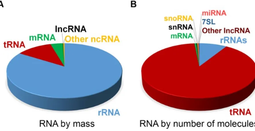
The first types of non-coding RNAs have already been discovered in the 1950s, when transfer and ribosomal RNAs were identified to play an essential role in protein translation. In the last decades, researchers found many additional types of non-coding RNA like small nuclear and nucleolar RNAs, microRNAs (miRNA), PIWI-interacting RNAs, circular RNAs and long non- coding RNAs (lncRNA) that have been mostly well characterized and investigated in the recent past. Non-coding RNAs are transcribed from all over the genome and their abundance within the cell roughly correlates with their level of conservation (Palazzo & Lee, 2015). Comparing the types and classes of RNAs, ribosomal and transfer RNAs represent the most abundant RNA types within a mammalian cell (Figure 1, Palazzo and Lee 2015).
Figure 1 Estimated abundance of different RNA classes in a typical mammalian cell.
Estimated proportions of various RNA classes in a mammalian somatic cell by (A) total mass and by (B) the absolute number of molecules. Non-coding (nc)RNAs shown here include small nuclear (snRNA), small nucleolar (snoRNA) and miRNAs, lncRNAs, 7SL RNA, ribosomal (rRNA) and transfer (tRNA) RNAs (adapted from Palazzo and Lee 2015).
Nevertheless, the role of less abundant non-coding RNA classes should not be neglected, as they often show cell- and tissue-specific expression and function (Quan et al., 2017). Also, their mode of action has been found to be quite varying, including the regulation of chromatin states, recognition and direct binding of various factors and regulatory elements, or the maturation and degradation of target molecules. As many lncRNAs are exclusively expressed in the brain (Derrien et al., 2012) and hold diverse functions due to their sequences and structures, I focus on this class of non-coding RNAs and their role in the regulation of behaviour in the following sections.
1.1.2.1 Long non-coding RNAs
LncRNAs are a fascinating class of RNAs, which are very diverse in their function, expression, localization, and even in their size. The length of lncRNAs varies between 200 nt and up to 100 kb and they are transcribed by RNA PolII. Various methodologies and the comparison of different lncRNA features are necessary to determine the conservation of lncRNAs across species, which might indicate their evolutionary functional importance. Multidimensional aspects like primary sequence conservation as well as the conservation of secondary structures, transcription status and splicing patterns have to be taken into account (Ulitsky, 2016).
Many lncRNAs are similar to mRNAs on the molecular level, as they are also capped, spliced and poly-adenylated. The striking difference is that the open reading frame is usually shorter than 300 nt, which is considered as an indication for the likelihood of non-coding properties.
Therefore, lncRNAs are generally not, or only poorly, translated (Bhat et al., 2016; Cao, 2014;
Frith et al., 2006). As originating from the sense or the antisense strand of the DNA, they can also be transcribed from intronic, intergenic, promoter or 3’ UTR-associated regions (Wu et al.
2013). LncRNA promoters are as evolutionary conserved as the ones of mRNAs (Carninci et al., 2005; Derrien et al., 2012; Guttman et al., 2009). In line with this, expressed lncRNA promoters show similar active histone modifications, like enriched H3K4me3 or H3K27ac, as found for protein-coding promoters. Nevertheless, recent research revealed that there is also a global regulation of lncRNA expression (Zheng et al., 2014). Many promoters are bidirectional, meaning transcripts are produced as sense and antisense RNAs from the same promoter. In this way, sense and antisense transcripts can be simultaneously and coordinately expressed, resulting in similar expression levels (Core et al., 2008; Seila et al., 2008). However, differences in stability and elongation are remarkable, as sense transcripts are enriched for splice sites, whereas antisense transcripts show more polyadenylation signals ensuring the early termination of the
antisense transcript and fully elongation and maturation of the sense transcript (Quinn & Chang, 2016). Most of the lncRNAs belong to the group of these divergent transcripts and are thought to regulate their promoters and corresponding protein-coding genes in cis, meaning they regulate the expression or chromatin states of nearby genes (Guil & Esteller, 2012).
LncRNAs with cis-regulatory properties include lncRNAs from imprinted loci and dosage compensation lncRNAs like Xist (da Rocha et al., 2008; Sahakyan et al., 2018). Xist is transcribed from one of the X-chromosomes of female mammalian cells and subsequently inactivates the X-chromosome from which it is transcribed from (Sahakyan et al., 2018). Moreover, some enhancer RNAs belong to cis-regulatory RNAs as they are transcribed from enhancer regions and positively affect the expression of neighbouring genes (Lai & Shiekhattar, 2014; Li et al., 2016). Trans-acting lncRNAs leave their transcription site and operate at regions far away from their transcription site, even in the cytoplasm and other compartments of the cell (Kopp &
Mendell, 2018).
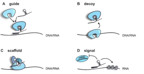
LncRNAs are spatio-temporally regulated, especially within the brain in a cell-type specific manner and with functions in specific subcellular compartments (Cava et al., 2019; Mercer et al., 2008). Depending on their intracellular localization, lncRNAs can fulfil different functions (Figure 2). Due to the length of the RNA molecule, lncRNAs often form secondary structures, thereby providing an interaction and assembly platform for chromatin remodelers and transcription factors. Through sequence complementarity, they guide bound factors or complexes to particular regions of the genome thereby affecting e.g. chromatin remodelling or transcription. On the other hand, lncRNAs are also able to bind components and hence, titrating them away from their original action site. As post-transcriptional regulators, they influence mRNA processing, translation, modification and degradation by the similar mechanisms mentioned above (Wang & Chang, 2012).
Figure 2 Schematic description of the main functions of lncRNAs.
(A) LncRNAs can guide complexes or factors to specific sequences on RNA or DNA molecules. (B) In the same way, factors can be bound by lncRNAs and be titrated away from their action site. (C) Secondary structures provide a platform for protein complex assembly. (D) LncRNA themselves can function as a signal and influence other mechanisms like miRNA-mediated cleavage or protein translation.
On average, lncRNAs have only a slightly shorter half-life than mRNAs. Nevertheless, there is a wide range of variability. Some lncRNAs have a half-life of less than 2 h; others show extreme stability with a half-life longer than 16 h. Intergenic origins or cis-antisense features support stability, whereas nuclear localization is usually linked to a short life span of lncRNAs (Clark et al., 2015). Due to structural similarities to mRNAs and weak open reading frames, a large fraction of lncRNAs is associated with ribosomes and consequently linked to nonsense- mediated decay (Niazi & Valadkhan, 2012; Zeng et al., 2018). Moreover, lncRNA turnover can be regulated by decapping and deadenylation events, miRNA-mediated decay and the binding of RNA binding proteins supporting or preventing degradation (Yoon et al., 2016).
1.1.2.2 The role of lncRNAs in neuropsychiatric disorders
An impressive number of about 40 % of lncRNAs being expressed specifically in the brain indicates a functional role of lncRNA in complex behaviours (Derrien et al., 2012). Their role during brain development is already well established and their involvement in glioma initiation and progression has been reported (Briggs et al., 2015; Nie et al., 2019). Apart from this, increasing evidence reveals lncRNAs as important regulatory players in social behaviour and neuropsychiatric diseases like Autism spectrum disorder (ASD) and schizophrenia.
ASD is a multifactorial and multisymptomal disorder mainly characterized by social and cognitive impairments and repetitive behaviours (Diagnostic and Statistical Manual of Mental Disorders; DSM-5, American Psychiatric Association, 2013). Wang et al. showed that lncRNA levels are strongly altered in blood of ASD patients and that these altered lncRNAs are associated with synaptic vesicle cycling as well as long-term depression and potentiation (Wang et al., 2015b). RNA Sequencing of post mortem frontal and temporal cortex samples from control and ASD subjects identified 60 differentially regulated lncRNAs (Parikshak et al., 2016). In a similar study with human cortical brain samples, Gudenas and colleagues used a computational pipeline combining differential gene expression patterns of ASD tissue in conjunction with gene co-expression networks of tissue-matched control samples to identify differentially expressed lncRNAs. During this analysis, they identified five lncRNAs, which are antisense to ASD risk genes like Rapgef4, Dlx6, Stxbp5, Klc2 and Dmxl2. Furthermore, co-expression network analysis revealed that there is a correlation of regulated lncRNAs and ASD gene sets indicating that the identified lncRNAs are involved in biological processes dysregulated in ASD (Gudenas et al., 2017).Some other lncRNAs could also be linked to schizophrenia, which is characterized by abnormal social behaviour and impaired ability to understand reality. Animal models for schizophrenia were used to successfully identify even potential downstream targets of those lncRNAs. One example for a schizophrenia-linked lncRNA is the brain cytoplasmic non-coding RNA (BC1). BC1 usually forms ribonucleoprotein particles and regulates translation and glutamatergic transmission in neurons (Napoli et al., 2008; Zalfa et al., 2003). BC1 knockout mice revealed deficits in sociability in the three-chamber-test, whereas social memory and social hierarchy were unaffected (Briz et al., 2017). Furthermore, BC1 increases the affinity of two regulatory proteins, FMRP and CYFIP1, in the brains. These proteins are both associated with schizophrenia, ASD and fragile X syndrome (Bardoni et al., 2001; Jansen et al., 2017; Pathania et al., 2014). FMRP, a RNA binding protein, binds the CYFIP1-eIF4E complex in the brain leading to repressed translation of FMRP targeted mRNAs (Napoli et al., 2008).
Another example is the lncRNA Gomafu, which is highly enriched in neurons. It is encoded in a schizophrenia-related locus and also dysregulated in schizophrenia patient brains. There is strong evidence for Gomafu being involved in alternative splicing as it binds QKI and SRSF1, RNA binding proteins that regulate pre-mRNA processing (Barry et al., 2014). In another study, Gomafu could be also linked to the schizophrenia-associated gene Crybb1. Here, Gomafu negatively regulates Crybb1 via the interaction with the polycomb repressive complex 1, thus increasing anxiety-like behaviour in mice (Spadaro et al., 2015).
NEAT1 is one of the most abundant lncRNAs and ubiquitously expressed in the human body, with the lowest expression in the central nervous system (Kukharsky et al., 2019).
Downregulated NEAT1 levels in cerebrocortical regions were linked to schizophrenia and Neat1 knockout mice display an altered socio-behavioural phenotype (Katsel et al., 2019;
Kukharsky et al., 2019). Mice are naturally territorial and exhibit defensive behaviour when unknown conspecifics enter their territory. However, this defensive behaviour towards an intruder was nearly absent in Neat1 knockout mice exposed as residents to the resident-intruder test. In these mice, physical social contact was also decreased, and a general lack of social interest was found in the Social Odor test (Kukharsky et al., 2019).
Contrary to the above-mentioned examples, only a few studies could implicate lncRNAs in the regulation of naturally occurring social behaviours. A recent study by Ma et al. identified the lncRNA AtLAS to regulate social hierarchy by controlling postsynaptic AMPAR trafficking in prefrontal cortical excitatory pyramidal neurons (Ma et al., 2020). They showed that in dominant mice, AtLAS is downregulated in the prefrontal cortex leading to hyperactivity of local neurons and the downregulation of synapsin 2b, which usually inhibits AMPAR insertion into the membrane.
All these above-mentioned examples highlight the regulatory relevance of lncRNA in the brain, especially in complex behaviour and psychiatric disorders. Nevertheless, we are far from a complete list of lncRNAs with specific functions in the central nervous system, their functions and potential interaction partners, or their role in diseases. Still intriguing is the role of lncRNAs in fine-tuning behaviours caused by different social factors or leading to socio-emotional perturbations. Therefore, high-throughput studies should help to provide potential candidates that might be involved in the regulation of socio-emotional behaviours.
1.2 Social anxiety disorders and related animal models 1.2.1 Social anxiety disorders and treatment options
“What will they think about me? They will laugh. I am not pretty and cool enough to be friends with them.
Why do they stare at me? I am not here… I am not here. I do not want to say anything wrong.”
Some of these thoughts come up in one’s mind when a person is worried about how other people think about or judge oneself. A balanced level of these kinds of thoughts usually helps people to work on themselves, perform better and endure particular, social and stressful situations, e.g. entering an occupied room, speaking in public, the first contact with new people at school, work or parties, or starting conversations with unfamiliar people (Ličen et al., 2016).
These events are uncomfortable for many people, although most of the people can finally cope with them. However, if these negative thoughts and feelings become excessive, and if they are omnipresent although there is no concrete reason, stress generated by these social situations is overwhelming and – as consequence – affected people try to avoid such social situation (Morrison & Heimberg, 2013).
People chronically suffering from such states (longer than 6 months) are diagnosed with social anxiety disorder (SAD). SAD, or social phobia, shows an early onset during youth and an estimated lifetime prevalence of 10.7 % in the US, with a higher prevalence in females (Kessler et al., 2012). Next to major depressive disorders and specific phobias, this is the third most common anxiety disorder. According to the current definition (DSM-5), a SAD patient persistently fears one or more social or performance situations, in which he is exposed to unfamiliar people or possible scrutiny by others. A SAD patient fears embarrassment and humiliation. Exposure to such situations evokes anxiety and possibly situationally bound panic attacks. As a consequence, feared situations are avoided and together with the psychological state person’s daily normal routine, occupational functioning and relationships are strongly restricted and quality of life is reduced (DSM-5, American Psychiatric Association, 2013). SAD is often comorbid with depression and other anxiety disorders such as generalized anxiety disorder, agoraphobia or obsessive-compulsive disorders (Fehm et al., 2005, 2008) and many studies found an increased risk for SAD patients to commit suicide (Fehm et al. 2005). This clearly indicates the necessity to find suitable and effective treatment options. To date, a combination of psychological and pharmacological treatment approaches are common.
Psychotherapeutic interventions include mainly cognitive-behavioural exposure therapies and cognitive restructuring, which help SAD patients to understand that the situations are not
harmful but the patient’s thoughts generate anxiety. In addition, at least for some clients, social skills training helps to better cope with social situations (Rodebaugh et al., 2004). Concerning pharmacotherapy, there are no specific chemical compounds available, which target specifically social anxiety. This might be also due to the limited knowledge about underlying molecular mechanisms. So far, serotine-reuptake inhibitors, benzodiazepines, serotonin-norepinephrine- reuptake inhibitors and monoamine-oxidase inhibitors, which are usually applied in context of depression, show positive, symptom-alleviative effects. Nevertheless, only 35-65 % of pharmacologically treated clients respond to medication (Davidson, 2003).
To understand underlying neurobiological mechanisms of SAD, a few fMRI studies were performed to identify brain regions that are differentially activated in patients suffering from SAD. One study investigated neuronal activity patterns in brains of SAD patients, when they were socially excluded during cyber games (Nishiyama et al., 2015). Here they found that social exclusion of socially anxious people led to increased activity of the anterior cingulate cortex, whereas social support through messages during social exclusion increased left dorsal prefrontal cortex activity, thereby positively correlating with social anxiety levels. This indicates that socially anxious individuals perceive social support but still are susceptible to negative evaluation. Another research group used the same cyber game with social exclusion and found that the inferior frontal gyrus plays an important role during re-inclusion (Heeren et al., 2017).
They found that recovery from the negative event “exclusion” is much harder than the exclusion itself. Other studies revealed an amygdala hyperactivation in SAD patients in response to the social threat (Labuschagne et al., 2010; Minkova et al., 2017). Nevertheless, little is known about relevant brain regions involved in the onset and extinction of SAD, which is of importance for exposure therapies and medication.
In summary, it is obvious that the actual state of knowledge and treatment options for SAD are not sufficient. Detailed research for a better understanding of responsible brain regions, circuits and especially of underlying molecular mechanisms is not circumventive in order to help SAD patients and for the development of proper treatment options.
1.2.2 Social fear conditioning
Animal models are consulted to study molecular and underlying mechanisms and develop treatment options for complex psychiatric diseases. The development of animal models is based on the assumption that neuronal and hormonal systems are conserved and that homologous factors exist across various species (Neumann et al. 2011; Greek and Rice 2012). Regarding
social behaviours, there are several models available including models for chronic psychosocial stress, depression, PTSD and others (Langgartner et al., 2015; Neumann et al., 2011).
For studying social fear in rodents, a mouse model of “Social fear conditioning” (SFC) has been developed in 2012. It is based on operant fear conditioning principles and enables researchers to mimic social avoidance – the core symptom of SAD – in adult mice (Toth et al., 2012).
During the social fear acquisition phase, mice associate an aversive event, i.e. a mild electric foot shock, with the contact of an unfamiliar conspecific, inducing a general social avoidance behaviour, which stays manifested up to several weeks (for protocol details see 2.3.1). Social fear extinction training by presenting several unknown conspecifics helps to overcome social fear, and is comparable to human exposure therapies. It results in similar effects, namely that it helps most, but still not all of the mice to extinguish their social fear so that they approach other mice again. The SFC paradigm is a unique model, as it does not induce other common comorbid behavioural changes like depressive-like or general anxiety-like behaviour (Toth et al., 2012), which is often the case in other models like social defeat or subordinate colony housing.
In the last years, pharmacological manipulations revealed that oxytocinergic, neuropeptide S and glutamatergic signalling can influence social fear. Neuropeptide S and OXT intracerebroventricularly infused completely reversed social fear expression during social fear extinction training (Zoicas et al., 2014, 2016), whereas Slattery et al. showed that, unlike non- social fear, blockage of the metabotropic glutamate receptor subtype 5 and activation of subtype 7 lead to impaired social fear extinction (Slattery et al., 2017). These studies provided first important key information, which neuropeptide systems might be involved in the regulation of social fear and its extinction. First hints for brain regions being specifically involved in social fear processes were obtained when Zoicas et al. used oxytocin receptor (OXTR) autoradiography and found differences in OXTR binding over the SFC paradigm especially in the dorsolateral septum (Zoicas et al., 2014).
1.2.3 The septum as a key region for social fear
Findings of Zoicas et al. highlighted the role of the septum in the regulation of social fear. With further experiments, they showed that infusion of OXT directly into the dorsolateral septum abolished social fear expression during social fear extinction training (Zoicas et al., 2014). Based on these findings, further research focused on the lateral septum (LS) in context of social fear.
Menon and colleagues performed manipulations of the OXT system within the LS in virgin mice as well as in lactating mice that have a highly activated endogenous OXT system. Lactating
mice displayed no social fear at all, and blockage of the OXTR in the LS prevented this effect.
The other way around, increasing OXT levels in the LS pharmacologically or genetically of virgin mice reversed SFC-induced social fear (Menon et al., 2018).
Obviously, the LS plays a role in social fear, so what kind of region is it? What makes it so important for social behaviour? The septum is a subcortical forebrain structure, which lies directly between the lateral ventricles in rodents. In humans, the septum pellucidum separates the lateral ventricles as a thin membrane, whereas the septum verum is composed of neuronal somata and runs next to the lateral ventricles. The rodent septum is composed of two main areas: the medial septum (MS) and the lateral septum (LS). The MS sends ascending projections mainly to the hippocampus (HPC), whereas the LS receives descending input from the HPC.
These interconnections form together the septo-hippocampal formation, which is critical for learning and memory processes (Niewiadomska et al., 2009). In contrast to the mainly GABAergic and cholinergic neurons of the MS, the LS receives glutamatergic input from the HPC and sends GABAergic projections to hypothalamic areas and midbrain regions. Newest antero- and retrograde tracing methods confirmed primary projections from the LS also to the nucleus accumbens, Bed nucleus of the stria terminalis, amygdala and parts of the hypothalamus and thalamus (Deng et al., 2019).
With the central location and the high grade of interconnectivity, the LS represents an essential relay station for integrating cognitive information from cortex regions and the HPC with affective information from the amygdala, hypothalamus and bed nucleus. The converged information then is led to regions that directly trigger behavioural responses. Importantly, there is also an intraseptal feedback loop so that the LS inhibits its own activation and the activation of MS by GABAergic collaterals (De France et al., 1975; Stevens et al., 1987). This is in particular important for the septo-hippocampal pathway during learning and memory processes: the MS plays a key role in learning and memory as it stimulates synchronized firing of the HPC.
Reciprocal downregulation of the MS activation can occur via direct GABAergic signalling from the HPC to the MS, or indirectly, via activation of LS, which then inhibits GABAergically the MS (Khakpai et al., 2013).
Functioning as a relay station, the septum is also involved in the regulation of different behaviours. It expresses receptors for various neurotransmitters that are released by input projections from different areas. Therefore, it is not surprising that the septum reacts to pharmacological interventions, which are applied for various psychiatric diseases in humans.
Effective medication for schizophrenia and depression, for example, enhances LS activation