Novel Insights into a microRNA-based Mechanism Underlying Oxytocin-mediated Anxiolysis and
Reversal of Social Fear
DISSERTATION ZUR ERLANGUNG DES DOKTORGRADES DER NATURWISSENSCHAFTEN (DR. RER. NAT.) DER FAKULTÄT FÜR BIOLOGIE UND VORKLINISCHE MEDIZIN DER UNIVERSITÄT REGENSBURG
vorgelegt von Anna Bludau aus Straubing
im Jahr 2019
Das Promotionsgesuch wurde eingereicht am: 25. September 2019
Die Arbeit wurde angeleitet von: Prof. Dr. rer. nat. Inga D. Neumann
Unterschrift: ……….
“Of all base passions, fear is most accursed.”
- William Shakespeare (King Henry IV, First Part)
Table of Contents
Abstract ... 11
Zusammenfassung... 13
Introduction ... 15
1.1 The Social Brain - Implications for Pathologies ... 15
1.2 The Mammalian Oxytocin System ... 15
1.2.1 Brain Oxytocin Receptor Distribution and Intracellular Signaling ... 18
1.2.2 Central Effects of Oxytocin – Focus on Sociability, Fear, and Anxiety ... 19
1.3 Behavioral and Molecular Correlates of Anxiety and Fear ... 21
1.3.1 Anxiety and Fear Responses ... 22
1.3.2 Neurocircuits of Anxiety ... 24
1.3.3 Anatomy, Neurochemistry, and Function of the Paraventricular Nucleus ... 25
1.3.4 Neurocircuits of Fear ... 26
1.3.5 Anatomy, Neurochemistry, and Function of the Septum ... 27
1.4 Anxiety Disorders ... 29
1.4.1 Generalized Anxiety Disorder ... 30
1.4.2 Social Anxiety Disorder... 31
1.4.3 Treatment of Anxiety Disorders ... 32
1.5 Modelling Anxiety and Fear in Rodents ... 33
1.5.1 Evaluation of Anxiety-related Behavior in Rodents ... 34
1.5.2 Evaluation of Fear in Rodents ... 35
1.6 Molecular Changes Underlying Anxiety Disorders and Conditioned Fear ... 36
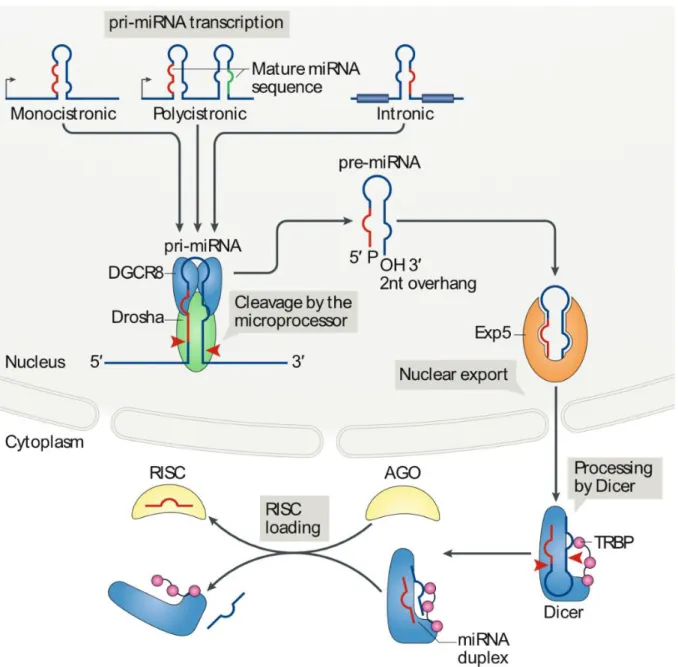
1.7 MicroRNAs – Biogenesis, Function, and Regulation ... 39
1.7.1 Mechanisms of microRNA Biogenesis ... 39
1.7.2 Regulation of microRNA Biogenesis ... 43
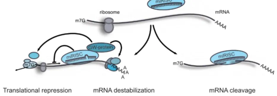
1.7.3 Mechanisms and Regulation of microRNA Function ... 45
1.8 microRNAs in the Central Nervous System ... 47
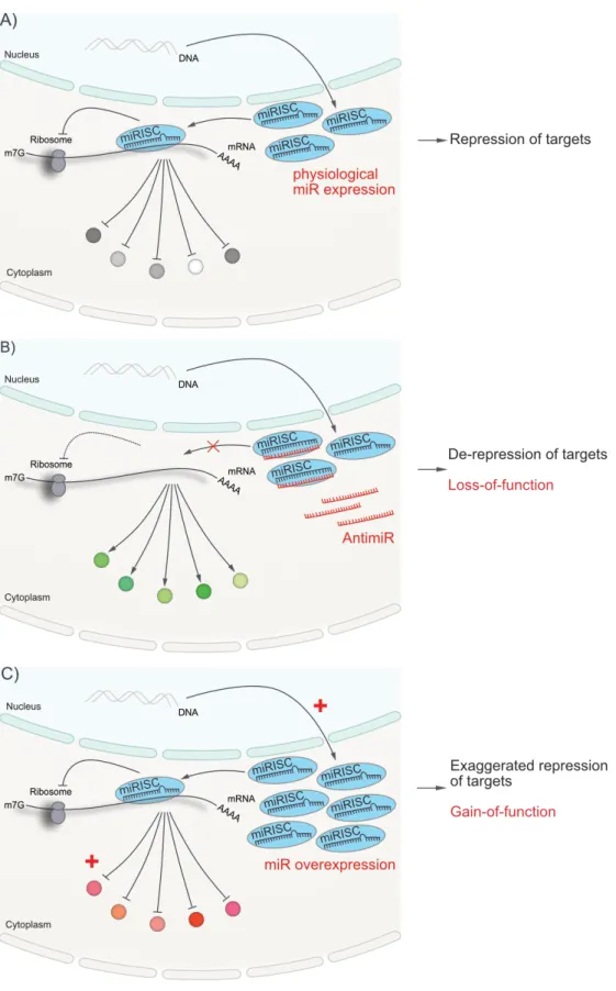
1.8.1 Experimental Manipulation of microRNAs in vivo ... 49
1.8.2 Selected microRNAs Relevant for Anxiety and Fear ... 53
1.8.2.1 Neuronal miR-132/212 ... 53
1.8.2.2 Neuronal miR-124 ... 59
1.8.2.3 Neuronal miR-134 ... 62
1.9 Aims and Outline of the Thesis ... 66
Material and Methods ... 69
2.1 Animals and Animal Husbandry ... 69
2.2 Surgical Procedures ... 69
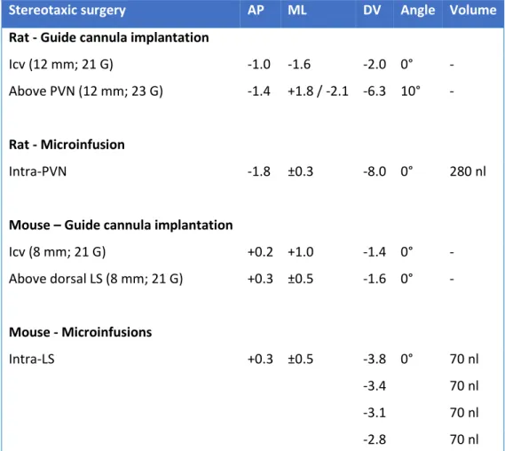
2.2.1 Implantation of Guide Cannulas ... 70
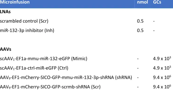
2.2.2 Intracerebral Microinfusion of Locked Nucleic Acids and Adeno-associated Viruses .... 70
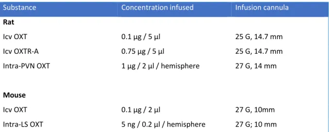
2.3 Drug Infusion in Conscious Animals ... 71
2.4 Verification of Cannula and Probe Placement ... 72
2.5 Extraction of Cerebrospinal Fluid from the Cisterna Cerebromedullaris of Rats ... 72
2.6 Behavioral Tests and Paradigms ... 73
2.6.1 Fear Behavior... 73
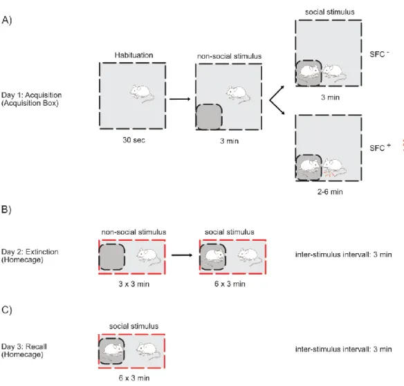
2.6.1.1 Social Fear Conditioning ... 73
2.6.1.2 Cued Fear Conditioning ... 75
2.6.3 Anxiety-related Behavior ... 76
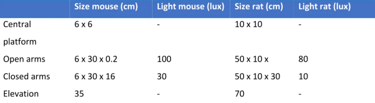
2.6.3.1 Elevated Plus-Maze ... 76
2.6.3.2 Light Dark-Box ... 77
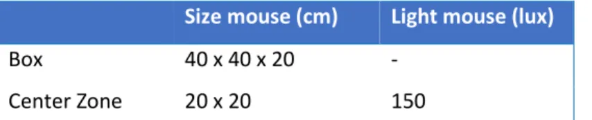
2.6.3.3 Open Field Test and Novel Object Investigation ... 77
2.6.4 Scoring of Behavior... 78
2.7 Molecular Methods ... 78
2.7.1 RNA Isolation from Tissue ... 78
2.7.2 miRNA Isolation from Cerebrospinal Fluid ... 79
2.7.3 Quantitative Real-Time PCR ... 79
2.7.4 Argonaute RNA-Immunoprecipitation-Microarray Analysis ... 80
2.8 Statistical Analysis ... 80
2.9 Experimental Design ... 81
2.9.1 Part I: “Is miR-132 within the PVN Involved in Oxytocin-induced Anxiolysis?” ... 81
2.9.2 Part II: “Does miR-132 Mediate the Oxytocin-induced Reversal of Social Fear?” ... 83
Results ... 87
3.1 Part I: “Is miR-132 within the PVN Involved in Oxytocin-induced Anxiolysis?” ... 87
3.1.1 microRNA Expression within the Rat PVN in Response to central OXT Treatment ... 87
3.1.2 microRNA Expression Alterations within the PVN of Lactating Rats ... 89
3.1.3 Functional Involvement of miR-132 in Anxiety-related Behavior and Cued Fear ... 91
3.2 Part II: “Does miR-132 Mediate the Oxytocin-induced Reversal of Social Fear?” ... 95
3.2.1 Analysis of microRNA Expression Levels in the SFC Paradigm in Mice ... 95
3.2.2 miR-132-3p Inhibition within the Lateral Septum Impairs Extinction of Social Fear ... 99
3.2.3 Septal miR-132 Overexpression Facilitates Extinction of Social Fear... 103
3.2.4 Septal miR-132-3p Inhibition Prevents Oxytocin-mediated Reversal of Social Fear .... 106
3.2.5 Downregulation of miR-132 within OXTR-expressing Neurons Impairs Social Fear Extinction ... 108
3.2.6 miR-132-3p Target Gene Analysis ... 110
Discussion ... 113
4.1 General Discussion ... 113
4.2 Part I: “Is miR-132 within the PVN Involved in Oxytocin-induced Anxiolysis?” ... 116
4.3 Part II: “Does miR-132 Mediate the Oxytocin-induced Reversal of Social Fear?” ... 121
Perspectives and Future Directions ... 129
References ... 133
Abbreviations ... 173
Danksagung ... 177
Curriculum Vitae... 179
List of Publications ... 181
11
Abstract
Everybody knows these situations, which evoke feelings of discomfort: the lonely walk home by night, a big black spider dangling above the bed or giving a speech in front of strangers. Anxiety and fear are natural responses to real or perceived threats and have been conserved throughout evolution. However, excessive fear and anxiety typically manifests as pathological disease state, such as generalized anxiety disorder, posttraumatic stress disorder or social anxiety disorder. To date, anxiety disorders are a high burden for society and ecology, and the available treatment options are limited and elicit numerous adverse side effects.
In the last decade, the anxiolytic and pro-social neuropeptide oxytocin gained focus amongst researchers as novel treatment option for anxiety disorders. Oxytocin binds to a G protein-coupled receptor, thereby activating intracellular signaling pathways, which have not yet been deciphered in detail. In the early 2000´s, non-coding RNAs, especially microRNAs, have been characterized as potent gene-regulatory molecules. microRNAs form regulatory networks to modulate gene expression on a post-transcriptional level. Due to their high regulatory potential and the availability of simple methods to manipulate microRNAs within the central nervous system, they are suggested to be innovative options for the development of new treatment alternatives.
In this thesis, I focused on the functional involvement of microRNAs in intracellular signaling pathways, which are essential for the anxiolytic and social fear-reversing properties of the neuropeptide oxytocin in rodents. Via microRNA expression analysis of the paraventricular nucleus (PVN), I revealed that the transcription of miR-132-3p is induced upon intracerebroventricular oxytocin application in male and female rats, an effect, which is abolished in response to pre- treatment with an oxytocin receptor antagonist. In contrast, chronic activation of the endogenous oxytocin system during lactation did not alter intra-PVN miR-132-3p level, but short-term separation of the mother from its pups increased miR-132-3p transcript levels within the PVN and cerebrospinal fluid of the dams. In a further pilot experiment, I showed that functional inhibition of miR-132-3p via a locked nucleic acid (LNA) prevents the anxiolytic properties of oxytocin applied into the PVN, whereas no explicit effect was seen in cued fear conditioning. In summary, oxytocin has been revealed to induce the transcription of miR-132-3p, which is in turn essential for the anxiolytic properties of the neuropeptide.
Moreover, I showed that septal miR-132-3p is involved in the extinction of social fear in male mice:
Compared to unconditioned mice, conditioned animals had increased septal miR-132-3p levels
after acquisition of social fear. Additionally, functional inhibition of septal miR-132-3p via a LNA
12
impaired extinction, whereas viral overexpression facilitated extinction of social fear. Interestingly, septal LNA-induced miR-132-3p inhibition prevented the oxytocin-induced reversal of social fear and shRNA-mediated downregulation of septal miR-132-3p specifically in oxytocin receptor expressing neurons impaired extinction of social fear. Thereby, miR-132-3p was proven to be essential for the social fear reversing properties of oxytocin. Further analysis of putative septal target messenger RNAs of miR-132-3p via microarray analysis revealed several promising candidates, all of which have not been found to be altered after acquisition and extinction of social fear in mice.
In summary, these experimental results expand our understanding of the mechanisms underlying
the anxiolytic and social fear-reversing properties of the neuropeptide oxytocin, and reveal that
small non-coding ribonucleic acids exert fundamental influence within the central nervous system.
13
Zusammenfassung
Jeder kennt diese eigenartigen Momente, die innerliches Unwohlsein auslösen: der nächtliche Nachhauseweg entlang verlassener, dunkler Gassen, die große schwarze Spinne über dem Bett, oder das Halten einer Rede vor größerem Publikum. Aus evolutionsbiologischer Sicht sind ebendiese Angst- und Furchtreaktionen konservierte, natürliche Handlungsweisen als Antwort auf reale oder gefühlte Bedrohungen. Eine exzessive bzw. unangepasste Ausprägung von Angst- oder Furchtzuständen manifestiert sich allerdings für gewöhnlich in pathologischen Krankheitsbildern. Diese beinhalten unter Anderem generalisierte Angststörungen, posttraumatische Belastungsstörungen und soziale Angststörungen. Heutzutage repräsentieren Angsterkrankungen eine Hauptbelastung der modernen Gesellschaft, vor Allem, da die derzeitigen Behandlungsmethoden stark limitiert und von verheerenden Nebenwirkungen geprägt sind.
In den letzten Jahren ist das Neuropeptid Oxytocin aufgrund seiner potenten angstlösenden und pro- sozialen Wirkung in den Fokus der Erforschung neuer pharmakologischer Behandlungsmöglichkeiten für Angsterkrankungen gerückt. Oxytocin bindet an einen G-Protein-gekoppelten Rezeptor und reguliert dadurch komplexe neuronale Signalwege, die bis heute nicht im Detail entschlüsselt sind. In den frühen 2000er Jahren wurden nicht-kodierende Ribonukleinsäuren, im Speziellen sogenannte microRNAs, als potente Regulatoren diverser intrazellulärer Signalwege gefunden. Diese bilden große regulatorische Netzwerke, mit welchen sie die Expression von Genen auf einem post-transkriptionalen Level beeinflussen. Durch ihr hohes regulatorisches Potential und die Tatsache, im zentralen Nervensystem auf einfachem Wege manipulierbar zu sein, stellen microRNAs innovative Angriffspunkte zur Entwicklung neuer Psychopharmaka dar.
Im Zuge meiner Dissertation untersuchte ich die Fragestellung, welche microRNAs in die neuronalen
Signalwege von Oxytocin involviert sind und dadurch sowohl Angstverhalten, als auch soziale Furcht in
Nagetieren beeinflussen. Mittels Analyse der microRNA Expression konnte ich zeigen, dass die
Transkription einer bestimmten microRNA, nämlich miR-132-3p, im Nucleus paraventricularis (PVN)
der Ratte, durch intracerebroventrikuläre Applikation von Oxytocin geschlechtsunabhängig induziert
wird, wohingegen eine vorangehende Infusion eines Oxytocin Rezeptor Antagonisten diesen Effeckt
verhinderte. Im Gegensatz dazu, hat die langanhaltende Aktivierung des endogenen Oxytocin-Systems
während der Laktation, keine Änderung der miR-132-3p Expression hervorgerufen. Lediglich die
kurzfristige Trennung der Jungtiere von der Mutter, führte sowohl im PVN, als auch in der
Cerebrospinalflüssigkeit der Muttertiere zu einem Anstieg der miR-132-3p Level. In einem Pilotversuch
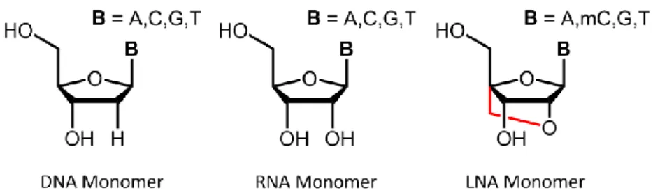
wies ich nach, dass die lokale Applikation (PVN) sogenannter „Locked Nucleic Acids“ (LNAs), welche
14
die Funktion der miR-132-3p verhindern, in männlichen Ratten die angstlösende Wirkung von intracerebral appliziertem Oxytocin unterdrücken, wohingegen sich die Effekte auf konditionierte Furcht als nicht eindeutig herausstellten. Zusammenfassend gesagt, konnte ich also aufzeigen, dass die microRNA miR-132-3p durch das Neuropeptid Oxytocin induziert wird und an der angstlösenden Wirkung des Neuropeptids beteiligt ist.
Wie sich herausstellte, ist dieselbe microRNA im Septum der Maus in die Auslöschung von konditionierter sozialer Furcht involviert: Nach dem Erlernen der sozialen Furcht zeigten sozial konditionierte Mäuse, im Vergleich zu nicht-konditionierten, eine höhere Expression der miR-132-3p.
Ebenso verlangsamte die funktionale Inhibition der miR-132-3p im Septum, mittels LNAs, die Auslöschung von sozialer Furcht, wohingegen eine Virus-induzierte Überexpression derselben, diesen Prozess beschleunigte. Zusätzlich verhinderte die funktionale Inhibition mittels LNAs die Oxytocin- vermittelte Auslöschung von sozialer Furcht. Letztlich führte eine Virus-induzierte Repression der miR- 132-3p Expression, speziell in Oxytocin Rezeptor exprimierenden Neuronen des Septums der Maus, zu einer verschlechterten Auslöschung der sozialen Furcht. Eine weitere Analyse potentieller Ziel-RNAs nach funktionaler Inhibition der miR-132-3p im Maus Septum mittels Microarray-Analyse lieferte einige vielversprechende Kandidaten, deren miR-132-vermittelte Regulation der Transkription im Tiermodell der konditionierten sozialen Furcht allerdings nicht bestätigt werden konnte. Kurz gefasst:
Ich konnte zeigen, dass die microRNA miR-132-3p essentiell in die Oxytocin-vermittelte Auslöschung von konditionierter sozialer Furcht involviert ist.
Diese experimentellen Ergebnisse erweitern das Verständnis jener Mechanismen, welche der
Oxytocin-vermittelten Angstminderung und Auslöschung sozialer Furcht zugrunde liegen, und zeigen,
dass auch kleine nicht-kodierende Ribonukleinsäuren im zentralen Nervensystem einen großen
Einfluss haben können.
15
Introduction
1.1 The Social Brain - Implications for Pathologies
Social neuroscience has developed into a major area of current research in behavioral neurobiology and psychiatry. Studies on the neurobiological basis of social behavior are of particular interest since numerous psychiatric disorders are symptomized by social deficits (Bludau et al., 2019; Fernández et al., 2018). Social behavior requires perception and integration of socially relevant cues through a complex social cognition process, which involves attention, memory, motivation, and emotion. In general, social situations are essential for the survival and propagation of a species. Hence, brain networks, as well as neurobiological and molecular mechanisms underlying social behavior are evolutionary highly conserved across species. The quantitative variation in the synthesis, release, and/or receptor density of crucial molecules accounts for observed inter- and intra-specific variability of the socio-behavioral response (Fernández et al., 2018). Past, recent, and future studies are shedding light onto how dysregulations of specific neuronal circuits and molecular characteristics could lead to psychopathologies, which include social dysfunctions. The activity of involved brain structures and neural circuits is modulated by several neurotransmitter and neuromodulator systems. Here, monoaminercic transmitters as well as neuropeptides, especially the nonapeptide oxytocin (OXT), play a key role.
1.2 The Mammalian Oxytocin System
Sir Henry Dale discovered in 1906 that posterior pituitary extracts stimulate uterus contractions in a cat. Hereupon, he named the found substance OXT from the Greek words “ὀξύς τόκος”, meaning quick birth. Accordingly, the most famous peripheral effect of OXT is the promotion of uterine contractions facilitating and accelerating birth (Fuchs and Poblete, 1970). Even today, intravenous OXT infusions are used in preclinical and clinical obstetrics to speed up the birth process
Social behavior is the behavior displayed when individuals of the same species interact with each other. It includes pair bonding, sexual, maternal, aggressive, and defensive behavior as well as social cognition, which is often constituted by a complex combination of olfactory, auditory, visual, and tactile stimuli.
Monoaminergic
neurotransmitters and neuromodulators include serotonin, dopamine, noradrenaline, and adrenaline.
16
and prevent excessive postpartum hemorrhage via myometrial contraction. Beyond that, OXT maintains neuroendocrine signaling during milk ejection (Jurek and Neumann, 2018). OXT signaling within the mammary gland leads to contraction of myoepithelial cells, which promotes milk ejection, an effect, which only recently has been found to be mediated by alterations in calcium oscillations (Stevenson et al., 2019).
The nonapeptide OXT is synthesized in magnocellular and parvocellular neurons of the paraventricular nucleus (PVN), supraoptic nucleus (only magnocellular neurons; SON), and accessory nucleus of the hypothalamus (Althammer and Grinevich, 2017; Meyer-Lindenberg et al., 2011; Rhodes et al., 1981; Swanson and Sawchenko, 1983).
Magnocellular neurons of the PVN synthesize the two peptide hormones OXT or arginine vasopressin (AVP), whereas parvocellular neurons of the PVN synthesize OXT, AVP, corticotropin-releasing factor (CRF), and thyreotropin-releasing hormone.
Within magnocellular neurons, OXT along with its carrier proteins called neurophysins is stored and transported in large-dense core vesicles (LDCV). Magnocellular OXT neurons are 20-30 μm in diameter and are densely packed with LDCVs (85% of the total neuronal volume) and thus contain substantial amounts of the neuropeptide (Stoop, 2012; Stoop et al., 2015). The LDCVs are axonally transported from the hypothalamus along the neurohypophysial stalk to the respective axon terminals in the neurohypophysis, which form neurohemal contacts, from which OXT is released into the peripheral blood circulation. Within the brain, magnocellular OXT neurons project to forebrain and limbic structures, such as the nucleus accumbens (Dölen et al., 2013), lateral septum (LS) (Menon et al., 2018), and central amygdala (Knobloch et al., 2012). Local release of OXT from these projections into the respective brain regions has been shown to alter social reward or the fear response. Furthermore these magnocellular OXT projections are suggested to modulate maternal aggression as well as aggressive and dominant behavior in males (Bosch et al., 2005; Calcagnoli et al., 2014).
In addition to axonal transport and release of OXT, dendrites have been
Neurohemal contacts aredefined connections between neurons and the blood stream to release neurosecretory
substances into the blood.
The limbic system of the brain includes amygdala, hippocampus, thalamus, hypothalamus, basal ganglia, and cingulate cortex.
The amygdala is well- characerized as center for memory processing, decisiton-making, and emotional responses. It is constituted of several nuclei: Medial nucleus, central nucleus, basal nucleus, and lateral nucleus. Moreover, clusters of intercalated cells also belong to the amygdala.
17
found to be a substantial source of neuropeptide release: Electron microscopic profiles demonstrated high peptide immunoreactivity (Armstrong, 1995) and abundant LDCVs have been shown in somatodendritic structures (Pow and Morris, 1989). This dendritic OXT release (central and peripheral) from magnocellular neurons of the SON differs in its temporal dynamics and does not necessarily follow a linear fashion (Ludwig, 1998; Ludwig and Leng, 2006; Neumann et al., 1993a).
In contrast to magnocellular neurons, parvocellular OXTergic neurons (10-20 μm soma diameter) mainly terminate in the spinal cord and brain stem (Swanson and Sawchenko, 1983), where they modulate autonomic functions, such as cardiovascular reactions (Petersson, 2002), breathing (Mack et al., 2002), erection and copulation (Melis et al., 1986), gastric reflexes (Sabatier et al., 2013), and feeding behavior (Atasoy et al., 2012). Via PVN-SON interconnections, parvocellular OXT neurons are suggested to control and orchestrate the activity of magnocellular OXT neurons within the SON, thereby facilitating analgesia by repression of nociception (Eliava et al., 2016).
Recently, it has been speculated that the brain OXT system consists of at least four neuronal subpopulations, which are distinguishable by the expression of several genetic markers (Romanov et al., 2017), and axonal projections of parvocellular neurons to the forebrain as well as magnocellular neurons to the midbrain have been reported (Althammer and Grinevich, 2017), which further enhances the necessity of a detailed characterization of the brain´s OXTergic projections.
Upon activation of OXTergic neurons, intracellular Ca
2+concentrations are increased, evoking the release of LDCVs (Hökfelt, 1991). The OXT- triggered Ca
2+can originate from extracellular sources, such as influx via N-type voltage gated Ca
2+channels (Fisher and Bourque, 1996) or N- methyl-D-aspartate (NMDA) receptors (Hu and Bourque, 1992), but it is also released from intracellular stores, such as the endoplasmic reticulum (Lambert et al., 1994). The intracellular Ca
2+rise leads to rapid and reversible depolymerization of F-actin to G-actin, and the OXT-filled LDCV is transported to the cell membrane. The subsequent binding of
18
the soluble N-ethylmaleimide-sensitive factor attachment receptor (SNARE) complex leads to fusion of the vesicle and plasma membrane, resulting in release of OXT into the extracellular space (Brown, 2016;
Ludwig et al., 2016).
Once released, OXT has a half-life time of 20 min in the cerebrospinal fluid (CSF) (Ludwig and Leng, 2006) and approximately 1.5 min in the blood (Higuchi et al., 1986). It is mainly degraded in a process that is performed by aminopeptidases (Stoop, 2012). Due to its hydrophilic peptidergic structure, peripheral OXT can not easily cross the blood brain barrier in physiologically-relevant concentrations (Leng and Ludwig, 2016). Only recently, a mechanism by which physiologically- relevant amounts of OXT are transported through the blood brain barrier has been described to involve the vascular receptor for advanced glycation end-products (RAGE) (Yamamoto et al., 2019). This unidirectional transport of OXT from the periphery to the central nervous system (CNS) is suggested to be especially important under conditions of dramatically increased blood OXT concentrations, such as child birth and lactation, which lead to saturation of the peripheral receptor occupancy and thereby promote the transport of the neuropeptide through the blood brain barrier.
Summarized, abovementioned studies reveal that OXT neurons show widespread central projections from the core nuclei of the hypothalamus to distal brain regions, which is essential for the evolution of a fine-tuned interconnected neuro-modulatory network.
1.2.1 Brain Oxytocin Receptor Distribution and Intracellular Signaling
The OXT receptor (OXTR) is a G-protein coupled receptor (GPCR) including a 7-transmembrane domain. High OXTR expression is found in cortical areas, the olfactory system, the limbic system (especially LS, amygdala, subiculum, thalamus, and hypothalamus) (Gimpl and Fahrenholz, 2001; Jurek and Neumann, 2018). As GPCR, it is coupled to a G
qprotein, which activates phospholipase C (PLC). PLC cleaves
The blood brain barrier isa highly semipermeable membrane separating the circulating blood from the extracellular fluid of the central nervous system. It allows passive diffusion and selective transport of molecules, which are essential for neuronal function, such as glucose, water, and amino acids.
GPCRs (also known as 7- transmembrane domain receptors) constitute a large protein family of receptors. They detect extracellular molecules and hence activate intracellular signal transduction pathways.
19
inositol-4,5-bis-phosphate (PIP
2) resulting in 1,2-diacylglycerol (DAG), which activates protein kinase C, and inositol-1,4,5-triphosphate (IP
3) that increases intracellular Ca
2+levels (van den Burg and Neumann, 2011; Jurek and Neumann, 2018).
Recently, it was revealed that the Ca
2+rise elicited by OXTR activation is mainly mediated by transient receptor potential cation channel subfamily V member 2 (TrpV2) channels in a phosphoinositide 3-kinase- dependent manner (van den Burg et al., 2015). Furthermore, this Ca
2+influx from the extracellular space is essential for protein kinase C (PKC), calcium/calmodulin dependent protein kinase I (CaMKI), II, IV, and calcineurin (CaN) cascade activation (Jurek and Neumann, 2018). OXT binding further induces transactivation of the epidermal growth factor receptor (EGFR), subsequent mitogen-activated protein kinase (MAPK) kinase (ERK1/2, ERK5, p38) activation, by which the anxiolytic effect of OXT within the PVN is mediated in male (Blume et al., 2008), as well as female virgin and lactating rats (Jurek et al., 2012). All named signaling cascades converge on the cAMP responsive element binding protein (CREB)-CREB-regulated transcription coactivator (CTRC)/myocyte enhancer factor 2 (MEF-2) transcription factor complex (CREB- CRTC/MEF-2), which results in transcriptional activation of target genes.
Moreover, de novo protein synthesis elicited by OXT is known to be dependent on the eukaryotic elongation factor 2 (eEF2) (Martinetz et al., 2019). However, in-depth information on intracellular signaling cascades and mechanisms is to date still marginal, highlighting that further precise research is required in order to employ OXT as possible treatment option, which is, due to the lack of knowledge, heavily debated.
1.2.2 Central Effects of Oxytocin – Focus on Sociability, Fear, and Anxiety
OXT is released within numerous brain regions in response to reproductive, stressful, and social stimuli (Landgraf and Neumann, 2004; Neumann, 2009; Neumann and Slattery, 2016; Neumann et al.,
MEF2 proteins are a class of transcription factors, which are essential regulators of cellular differentiation and stress response mediation. Four MEF2 isoforms are present in mammals:
MEF2A, MEF2B, MEF2C, MEF2D.
20
1993b; Wotjak et al., 2001; Zoicas et al., 2014). On one hand, synthetic OXT has been found to facilitate sociability and prevent social avoidance in rats and mice (Lukas et al., 2013). On the other hand, optogenetically triggered release of OXT enhances social recognition by modulation of cortical control of early olfactory processing (Oettl et al., 2016), and endogenous OXT is known to regulate pair bonding (Carter et al., 1995;
Cho et al., 1999; Insel and Hulihan, 1995; Shapiro and Insel, 1992), and maternal behavior (Bosch et al., 2004, 2005; Neumann et al., 2000;
Pedersen and Prange, 1979; Pedersen et al., 2006). In the context of this thesis, the effect of central OXT on social memory formation and maintenance is of especial interest. Infusion of OXT into the rat LS or ventral hippocampus have been revealed to improve juvenile recognition in adult males (Ludwig et al., 2013; Popik et al., 1992).
Furthermore, OXT has been found to be crucial for memory formation in a social context, since central OXTR antagonism impairs the maintenance of social memory (Dluzen et al., 2000; Lukas et al., 2013).
In a recently established mouse model of social fear, the so called social fear conditioning (SFC), OXT has been found to crucially regulate social fear expression: Infusion of the neuropeptide into the ventricular system or LS is able to reverse social fear (Zoicas et al., 2014). This data further highlights OXTs pro-social effects (for details on OXT in SFC see section 1.5.2 Evaluation of Fear in Rodents).
In addition to its pro-social properties, central OXT is known to essentially regulate fear- and anxiety-related processes.
Intracerebroventricular (icv) application of OXT affects cued fear conditioning (CFC) in rats and mice in a time-dependent manner (Toth et al., 2012a): In rats, administration of OXT prior to acquisition does not alter the fear conditioning response, but decreases fear expression and facilitates fear extinction. Moreover, OXT infusion prior to extinction of cued fear impaires fear extinction in both, rats and mice.
This effect of OXT is conserved across species and suggested to prevent
the formation of aversive memories during traumatic events. During
lactation, the activity of the brain OXT system is enhanced, an effect
characterized by elevated hypothalamic OXT synthesis (Knobloch et al.,
21
2012), suckling-induced peripheral and central OXT release (Neumann et al., 1993b), increased levels of OXTR expression and binding in various brain regions (Insel, 1986; Meddle et al., 2007), and activation of OXTR-coupled signaling cascades (Jurek et al., 2012; Slattery and Neumann, 2008). During lactation, OXT signaling prevents fear-induced freezing in an odor fear conditioning paradigm in rats (Rickenbacher et al., 2017) and social fear in the SFC paradigm (Menon et al., 2018).
Moreover, studies revealed an anxiolytic phenotype in lactating females (Lonstein, 2005; Neumann, 2001) and in males after mating (Waldherr and Neumann, 2007), which has been suggested to be mediated by OXT. Additionally, local infusions of synthetic OXT into the hypothalamic PVN, central amygdala, or medial prefrontal cortex (PFC) result in acute anxiolysis in male as well as female rodents (Bale et al., 2001; Blume et al., 2008; van den Burg and Neumann, 2011; van den Burg et al., 2015;
Jurek et al., 2012; Martinetz et al., 2019; Neumann, 2008; Neumann et al., 2000; Sabihi et al., 2017).
Abovementioned studies clearly illustrate the significance of the neuropeptide OXT in the regulation of sociability and mood, especially fear- and anxiety-related behavior.
1.3 Behavioral and Molecular Correlates of Anxiety and Fear
Already in 1919, the direct descendant of Darwin, Walter Cannon, highlighted the emergency adaptive functions of anger and fear in terms of promoting fight and flight reactions. This made him the primary investigator of emotional, visceral, and autonomic alterations as responses of anxiety and fear. As emotional behaviors essential for survival, fear and anxiety can be both, innate and adaptive, and are expressed in all vertebrates. Nevertheless, among neuroscientists, there are several perspectives on what differentiates anxiety from fear, resulting in numerous different viewpoints, since the terms of fear and anxiety are often used interchangeably. According to established definitions, fear is commonly specified as an emotional response to a real threat or danger, whereas anxiety is an emotional reaction to a
Freezing responses are commonly observed in prey animals. They reduce the likelihood to be attacked, because it is more complex for the predator to spot the prey when motionless and it is less likely to be attacked by the predator whenever motionless.
Fight and flight reactions are also called hyper- arousal states. They are physiological reactions occurring in response to perceived harmful events, such as attacks or threat to survival.
22
potential, circumstantial, or anticipated threat or danger (McNaughton and Zangrossi, 2008; Tovote et al., 2015). Conceptually, fear and anxiety relate to brain states that are evoked by external or internal stimuli and elicit a specific combination of measurable behavioral, physiological, hormonal, and autonomic responses (Anderson and Adolphs, 2014;
Davis et al., 2010; LeDoux, 2000, 2014). These responses have evolved to enable the organism to survive by adapting to not only beneficial, but also harmful stimuli. Anxiety and fear are highly adaptive and complex responses that are measured through the intensity or persistence of the associated behaviors. Both are coping strategies, which are deployed in dependence of the present situation: (i) Active coping strategies are exerted when escape is feasible. They are primarily mediated by activation of the sympathetic nervous system leading to hypertension and tachycardia (Cannon, 1915; Olds, 1956). (ii) Passive coping strategies are deployed when escape is not possible. They are accompanied by autonomic inhibition, resulting in hypotension and bradycardia (Engel and Schmale, 1972).
1.3.1 Anxiety and Fear Responses
The emotional response to anxiety-eliciting stimuli is highly variable and dynamic, which is predicated on the ambiguity of putative threats.
Anxiety is an adaptive or innate coping mechanism for dangerous situations and is thereby highly associated with emotional as well as cognitive functions, such as learning and memory. It is apparent that anxiety is adaptive in protecting individuals from danger (Scott, 2013):
Anxious avoidance of predators is essential for healthy survival and propagation of species. Anxiety-eliciting stimuli, e.g., open bright spaces, result in species-dependent approach-avoidance behaviors in humans and other land-dwelling species, such as rodents and primates.
These approach-avoidance behaviors are dependent on the goal and the associated motivation (Kenrick and Shiota, 2014). For example, bright open areas induce avoidance behavior in nocturnal rodents, which naturally prefer protected and dark areas, whereas diurnally
Approach-avoidanceconflicts originate if a goal has both, positive and negative characteristics or effects. This makes the goal appealing and unappealing
simultaneously.
23
active humans show diverse behaviors in response to the same environment: approach and avoidance behaviors are present in dependence of the individual´s life experience. Approach-avoidance responses are evolutionary highly conserved and beneficial for the organism in that the unknown provides the possibility of both, opportunity and danger. Another conventional strategy to cope with anxiety, are escape behaviors, which allow withdrawal of the organism from the threat and prevention of re-exposure to the same or similar dangerous situations.
Fear can be both, innate and adaptive (Ramachandran, 1994a). Innate fears (e.g., startle responses to unexpected loud noises) have evolutionary been established across centuries as they assist in the organism´s adaptation to the surrounding environment and thereby mitigate harm and ensure survival. Adaptive or learned fears (e.g., aversive events by getting physically attacked) are achieved by direct and indirect (witnessing) experience or by inter-individual assignment.
Just as anxiogenic stimuli, fear-eliciting stimuli, such as novel objects or situations, provoke mainly avoidance and escape behaviors, as well as prevention of re-exposure, but also approach behaviors to examine the object or situation. Similar to anxiety, behavioral fear responses have been characterized in numerous species and include avoidance (Blanchard et al., 2003; Edmunds, 1974), flight, freezing, defensive threat, defensive attack, risk assessment, burying the threatening object (Treit et al., 1981), alarm cries (Litvin et al., 2007), and cessation of ongoing behavior (Brady and Hunt, 1951; Estes and Skinner, 1941).
In addition to behavioral alterations, states of fear and anxiety evoke physiological responses. These physiological responses to threat include activation of the autonomic nervous system (Cohen and Randall, 1984;
Engel and Schneiderman, 1984), the hypothalamus-pituitary-adrenal (HPA) axis (Graeff and Zangrossi Junior, 2010; Korte et al., 1992; Mason et al., 1961), hyperthermia (Adriaan Bouwknecht et al., 2007), pain suppression (Watkins and Mayer, 1982), and a potentiation of somatic reflexes, such as the startle response (Davis et al., 2010; Ray et al., 2009) and eye blink (Weisz and McInerney, 1990) responses.
The HPA axis is essential for the body´s stress response and represents the interaction between hypothalamus, pituitary, and adrenal glands. Upon stressful stimulation, the hypothalamus releases corticotropin-releasing hormone, which triggerst the anterior pituitary to release
adrenocorticotropic hormone to subsequently stimulate cortisol release from the adrenal glands.
As steroid hormone, cortisol acts as negative feedback regulator of the HPA axis at the level of the hypothalamus and pituitary.
24
The persistence of these behavioral and physiological adaptations in the absence of a real or potential threat leads to detrimental consequences on other pro-survival behaviors, such as self-care and food procurement, but also social interaction and reproduction. Therefore, it is conceivable that inadequate over-activation of mechanisms and circuits involved in anxiety and fear results in debilitating anxiety disorders (see section 1.4 Anxiety Disorders) (Gray and McNaughton, 1996; Hazen et al., 1996).
1.3.2 Neurocircuits of Anxiety
Numerous studies suggest that central mechanisms and neuronal substrates of anxiety and fear in rodents and humans are mediated by at least partially overlapping mechanisms (Davis and Whalen, 2001;
Davis et al., 2010). However, precise brain circuits that underlie anxiety have not been investigated as much.
Anxiety states are mediated on one hand by local microcircuits and on the other hand by long range projections to distal regions (Tovote et al., 2015). Regions, such as the bed nucleus of the stria terminalis (BNST) or the amygdala, which have major roles in anxiety, mediate anxiogenic as well as anxiolytic behavioral effects. Thereby, the functional consequence within anxiety networks is determined by target-specific and/or cell type-specific connections. For example, two paralleled ventral BNST to ventral tegmental area pathways are capable to mediate anxiogenic as well as anxiolytic behavior (Jennings et al., 2013).
Activation of the basolateral amygdala to ventral hippocampus
projections is anxiogenic (Felix-Ortiz et al., 2013), whereas activation of
the basolateral amygdala to central amygdala circuit is anxiolytic (Tye
et al., 2011). Other brain regions that are interconnected with the BNST
and/or amygdala and are involved in modulation of anxiety-related
behavior, include medial PFC, periaqueductal gray, raphe nucleus, locus
coeruleus, LS, and hypothalamus, (Tovote et al., 2015). Importantly, the
hypothalamic PVN became apparent as crucial modulator of anxiety
responses (Blume et al., 2008; van den Burg et al., 2015; Jurek et al.,
25
2012; Martinetz et al., 2019). However, distinct components of the anxiety network, such as cell identity and function, within those local and long-range projections remain to be characterized.
1.3.3 Anatomy, Neurochemistry, and Function of the Paraventricular Nucleus
The hypothalamus is a small, but essential region of the brain, which is formed by numerous nuclei and nervous fibers. The PVN is a bilateral nucleus of the hypothalamus, which is crucially involved in neuroendocrine and behavioral responses to numerous external and internal stimuli. It is located adjacent to the third ventricle and lies within the periventricular zone. The PVN contains two neurosecretory cell types: magnocellular neurons and parvocellular neurons, which have been described in detail in section 1.2 The Mammalian Oxytocin System.
The PVN receives input from other nuclei of the hypothalamus, such as the suprachiasmatic nucleus (SCN) and arcuate nucleus, but also from distal brain regions like periaqueductal gray, parabrachial nucleus, entorhinal cortex, prelimbic cortex, BNST, and amygdala (Hsu et al., 2014). Moreover, it reciprocally interconnects to the contralateral PVN (Jurek and Neumann, 2018). Projections from the PVN reach to the nucleus accumbens, BNST, central and extended amygdala, medial PFC, brainstem, nucleus raphe, and LS (Geerling et al., 2010; Li and Kirouac, 2008; Sofroniew, 1980).
As limbic brain structure, the PVN has been shown to mediate drug relapse (Martin-Fardon and Boutrel, 2012), retrieval of consolidated fear memories (Padilla-Coreano et al., 2012), acute and chronic stress responses (Bhatnagar and Dallman, 1998; Bhatnagar et al., 2002; Hsu et al., 2014), emotional arousal, motivation, and mood, especially anxiety- related behavior (Blume et al., 2008; van den Burg and Neumann, 2011;
van den Burg et al., 2015; Jurek et al., 2012; Martinetz et al., 2019;
Neumann, 2008; Neumann et al., 2000).
The SCN as part of the hypothalamus is seen as the central pacemaker of the circadian rhythm. It is an autonomous nucleus, but also recieves input from the retinothalamic tract to synchronize the day-night-cycle.
26 1.3.4 Neurocircuits of Fear
Most of what we understand about fear originates from studies using Pavlovian fear conditioning, whereby animals learn to predict aversive events. Hence, the following brief disquisition on neurocircuits involved in acquisition, consolidation, and extinction of fear only includes studies based on conditioned fear.
Within the brain, fear states are mediated by long-range projections between brain regions in conjunction with local microcircuits within essential nuclei of this projection network (Tovote et al., 2015). The major center of the fear circuit is located within the amygdala. Briefly, fear expression is elicited by fearful stimuli that activate thalamic centers and cortical regions, such as primary sensory and association cortices. Input from those regions to several nuclei of the amygdala further mediates fear-related neuronal plasticity. Moreover, reciprocal connections between the basal amygdala and the ventral hippocampus as well as the prelimbic cortex modulate this plasticity. The central amygdala projects to hypothalamic, brain stem, and mid brain centers, such as the periaqueductal gray, to modulate neuronal plasticity in order to promote fear behavior and autonomic responses.
Different elements within the same structures mediate extinction of fear: Here the PFC-amygdala pathway is suggested to be most relevant for fear extinction behavior (Muigg et al., 2008, 2009). Bidirectional projections between the infralimbic cortex and basal amygdala or the intercalated cells dampen the fear output from nuclei of the lateral central amygdala to the hypothalamus and periaqueductal grey (Herry et al., 2010; Tovote et al., 2015). Additionally, forebrain to brainstem pathways essentially influence fear extinction, but their identity, connectivity, and specific function remains to be identified. Recently, the group of Valery Grinevich found that magnocellular SON-OXT neurons participate in a fear memory engram, wherein parvocellular OXT neurons from the PVN orchestrate OXT release within distant brain regions, such as the amygdala, in a context-independent manner (Hasan
Pavlovian conditioning isa method that causes a reflex response or behavior by training with repetitive or aversive action. It was invented by the Russian physiologist Ivan Petrovich Pavlov, who conditioned dogs to respond in what seemed to be a predictable manner.
Neuronal plasticity, also known as brain plasticity or neuroplasticity is the capability of neurons to continuously change throughout life to optimize neural networks.
This includes, but is not limited to strengthening and weakening of synapses.
27
et al., 2019). Moreover, magnocellular OXT projections from the SON to the LS are characterized to mediate social fear extinction in female lactating mice, since silencing of these blocks social investigation in those mice (Menon et al., 2018), revealing the LS as crucial component of the social fear brain circuit.
1.3.5 Anatomy, Neurochemistry, and Function of the Septum As abovementioned, the septal region is highly involved in the expression of fear and its extinction. It is a subcortical forebrain structure located between the lateral ventricles and lies rostrodorsal to the hypothalamus. Anatomically, neurochemically, and functionally the septum is divided into two nuclei: the medial septum (MS) and the LS.
The MS receives ascending input from the hypothalamus, ventral tegmental area, substantia nigra, raphe nucleus, locus coeruleus, and hippocampus (Müller and Remy, 2018; Tsanov, 2017). Within the MS, glutamatergic, γ-aminobutyric acid (GABA)-ergic, and cholinergic neurons are highly interconnected and form a local network to synchronize the septal network (Fuhrmann et al., 2015; Hangya et al., 2009; Huh et al., 2010; Manseau et al., 2005; Müller and Remy, 2018).
One major interconnection consists of ascending inputs from the MS as well as the adjacent diagonal band of Broca, which is functionally related to the MS, via the fimbria/fornix fiber bundle into the hippocampus (Khakpai et al., 2013). This septo-hippocampal pathway fine-tunes hippocampal physiology and is indispensable for its behavioral functions. Most septo-hippocampal projections are of cholinergic nature (~65%; (Sun et al., 2014)) and are crucially involved in aversive association learning (Lovett-Barron et al., 2014) and formation of spatial memory (Durkin, 1994; Ikonen et al., 2002).
Glutamatergic neurons account for ~23% of septo-hippocampal projections (Colom et al., 2005) and are essential for processing of environmental and spatial inputs during initiation of movement episodes, general locomotor state, and running speed (Fuhrmann et al., 2015). GABAergic neurons form the minority of septo-hippocampal
28
projections and have been implicated in hippocampal neurogenesis (Van der Borght et al., 2005), operant reward learning (Vega-Flores et al., 2014), and, in interaction with the hippocampal cholinergic system, modulation of anxiety-related behavior (Degroot and Treit, 2003;
Degroot et al., 2001). Hippocampal GABAergic neurons not only receive GABAergic input from the MS, but also project back to the MS (Alonso and Köhler, 1982; Takács et al., 2008; Tóth et al., 1993), forming a reciprocal long-range circuit, which functionally synchronizes remote areas (Caputi et al., 2013). However, the only neuronal subtype classified to modulate social fear extinction are GABAergic neurons, since overexpression of the OXTR in GABAergic neurons of the LS substantially attenuated SFC-elicited social fear in female mice (Menon et al., 2018).
In comparison to the MS, the LS receives descending glutamatergic
input from the hippocampus via the fimbria/fornix bundle (Gallagher et
al., 1995). It is mainly composed of GABAergic neurons, which are
reciprocally interconnected with the hypothalamus and periaqueductal
grey (Sheehan et al., 2004). Moreover, monoaminergic and cholinergic
neurons of the amygdala, BNST, medial PFC, locus coeruleus,
laterodorsal tegmentum, ventral tegmental area, nucleus accumbens,
and entorhinal cortex project to the LS. It is important to note that LS
and MS receive reciprocal projections from each other (Risold and
Swanson, 1997). Thereby, the LS is a crucial region for integrating
cognitive as well as affective functions to directly control appropriate
behavioral responses to particular environmental stimuli. The LS is
essentially involved in various aspects of social behavior, such as social
memory (Engelmann and Landgraf, 1994; Lukas et al., 2013), aggression
(Leroy et al., 2018), and social fear (Menon et al., 2018; Zoicas et al.,
2014), but also anxiety-related behavior (Sheehan et al., 2004) and
avoidance behavior (Troyano-Rodriguez et al., 2019). For example,
social instability stress in male rats results in reduced dendritic spines
within the LS and is suggested to be the underlying cause of reduced
social interaction, impaired social recognition, reduced sexual
performance, and increased aggression (Hodges et al., 2019). Further
29
studies proved that the LS activity negatively correlates with aggressive behavior and consequently, septal lesions are known to induce a “septal rage” phenotype (Goodson et al., 2005; Lee and Gammie, 2009; Potegal et al., 1981; Wong et al., 2016). Thus, the LS is crucial for the regulation of aggression, which in principle is a sophisticated component of social behavior. Most importantly, the LS is a key player of social fear, which will be described within this thesis in section 1.5.2 Evaluation of Fear in Rodents. In addition to the regulation of various cognitive social and emotional behaviors, the LS is involved in the physiological stress response by impacting active stress coping and dampening of the HPA axis activity (Herman et al., 1996; Singewald et al., 2011). Further studies implicate sub-populations of neurons, such as CRF receptor 2 expressing neurons, as promoters of stress-induced anxiety (Anthony et al., 2014; Radulovic et al., 1999). In any case, the involvement of the LS as a main regulatory component of the stress response in undeniable.
1.4 Anxiety Disorders
In the context of human pathology, inappropriate, exaggerated or prolonged activation of anxiety and fear responses by innocuous stimuli becomes detrimental. Debilitating excessive fear is a significant symptom of many anxiety disorders, such as generalized anxiety disorder (GAD), social anxiety disorder (SAD), panic disorder, specific phobia, or obsessive-compulsive disorder (Barton et al., 2014; Craske et al., 2017). Anxiety disorders are associated with immense health care costs and represent a high burden for society and economy. Pursuant to large population-based surveys, they have a constant life-time prevalence of 33.7% throughout the last years (Bandelow and Michaelis, 2015). Additionally, the prevalence in women is approximately twice as high as in men (Angst and Dobler-Mikola, 1985;
Bruce et al., 2005; McLean et al., 2011; Regier et al., 1990), which is discussed to be caused by psychological contributors (e.g., childhood trauma), but also genetic, epigenetic, and neurobiological factors.
Although prospective studies suggest anxiety disorder as chronic
Septal rage, also called sham rage refers to intense dysphoria, hyperexcitability, and anger, which originates from a lesion in the human septum pellucidum.
SAD is suggested to be caused by a composition of genetic, epigenetic, and environmental factors. It is symptomized by a combination of emotional, behavioral, and physical symptoms, as well as a general avoidance of social situations.
30
impairment, prevalence rates decrease throughout age, revealing that an anxiety disorder does not last until old age in most cases (Jacobi et al., 2014). Just as all psychiatric disorders, anxiety disorders show high rates of comorbidity amongst themselves, but also to other psychopathological conditions, such as dysthymia and major depressive disorder (Kessler et al., 2005a). In the following sections, GAD and SAD will be reviewed in detail, as they are the most relevant ones for this thesis.
1.4.1 Generalized Anxiety Disorder
In 1980, GAD appeared for the first time as diagnostic category in the third edition of the Diagnostic and Statistical Manual of Mental Disorders III (DSM-III). The distinctive core symptom of GAD is excessive diffuse worry about a number of life circumstances, which is a cognitive aspect of anxiety. According to DSM-V, GAD patients show symptoms of restlessness, fatigue, irritability, muscle tension, as well as sleep and concentration deficits. Long-term consequences of worry include the inhibition of emotional processing and perpetuation of anxiogenic conditions (Mathews, 1990). Relief of worry is usually only provided short-term by avoidance of the threatening stimuli (Brown, 1997) or intolerance of uncertainty (Bomyea et al., 2015).
The validity of GAD as independent category has been questioned from
DSM-III to DSM-V: On one hand, no clear boundaries between GAD and
personality dimensions, other anxiety-spectrum disorders, and non-
bipolar depression are the major concerns (Crocq, 2017). On the other
hand, epidemiological surveys identified different risk factors for GAD
and depressive disorders, revealing a clear separation between the two
disorders (Kessler et al., 2008). The lifetime prevalence of 9.0% with an
overrepresentation of female patients highlights the need for specific
pharmacological treatment options (Kessler et al., 2012). Of all anxiety
disorders, GAD has the latest median age onset at approximately 31
years (Bandelow and Michaelis, 2015). Moreover, it is comorbid with
other anxiety disorder subtypes and shows particularly high
31
comorbidity with dysthymia and major depression disorder (Kessler et al., 2005a). In the case of major depression disorder, GAD is discussed as a prodromal, residual, or severity marker of a major depressive episode (Kessler et al., 2008). Interestingly, the comorbidity of GAD with other disorders decreases over the duration of GAD itself (Breslau and Davis, 1985). Long-term medication with benzodiazepines worsens the underlying anxiety (Galanter et al., 2014), an effect, which can be reversed by reduction in the treatment dose (Booth, 1995). However, no treatment options with low side effects and relapse rates are known to date, which highlights the necessity of further research.
1.4.2 Social Anxiety Disorder
Although primarily monitoring psychotic patients, the German psychiatrist Emil Kraepelin noted that patients suffering from non- generalized and generalized social phobia experienced “overpowering feelings of aversion […] when they had to establish relations of any kind with other patients”, whereas other patients were “unable to urinate or write a letter in the presence of other people” (Kraepelin E., 1904).
Today, and in a more therapeutically relevant and epidemiological definition, SAD is characterized by intense fear and avoidance of social situations (Kessler et al., 2005b, 2005a), such as meeting strangers or speaking in public. Amongst anxiety disorders, SAD is the second most common with a lifetime prevalence of 12.1% (Alonso et al., 2011;
Kessler et al., 2012). Comparable to the general gender bias of anxiety disorders, 60% of SAD patients are female, although men are overrepresented in the treatment seeking fraction (Xu et al., 2012).
According to the DSM-V, two subtypes of SAD, generalized SAD and non-generalized SAD, are distinguished. Patients suffering from generalized SAD fear most social situations (Kerns et al., 2013; Vriends et al., 2007), whereas in non-generalized SAD patients fear of only a specific social situation manifests (Bögels et al., 2010). Although generalized SAD is much more debilitating in nature than non- generalized SAD, both lead to significant reduction in the quality of the
32
patient´s life (Hazen et al., 1996; Stein and Chavira, 1998). In addition, generalized SAD is usually familial, long-lasting, and shows a lower chance of spontaneous recovery, but carries a higher risk of comorbidity. Common comorbidities include major depression (Schneier et al., 1992; Stein and Chavira, 1998), agoraphobia (Magee et al., 1996), or substance abuse (Buckner et al., 2013; Schneier et al., 2010). Here, SAD symptoms usually appear first, suggesting that SAD may be a crucial risk factor for other psychopathologies (Neumann and Slattery, 2016). SAD has an early onset between age 5 to 15, and its symptom of avoidant behavior is mainly considered the biggest hindrance towards extinction or reversal of social anxiety (Stangier et al., 2006). In healthy humans, memory of negative experience is known to decrease over time (Ritchie et al., 2015). Patients symptomized by high levels of social anxiety tend to show an eroding of positive memories, which leads to a perturbation of their fear of social situations and hinders treatment (Glazier and Alden, 2019). To date, no treatment specifically targeting SAD is present, highlighting the need of further detailed understanding of the underlying mechanisms.
1.4.3 Treatment of Anxiety Disorders
When symptoms are mild, transient, and without associated impairments in social or occupational function, it is not necessary to treat anxiety disorders. However, most patients show marked distress or suffer from severe complications, such as secondary depression, suicidal ideation, or alcohol abuse, which in conclusion make treatment inevitable. All currently available treatment options are rather unspecific and do not primarily treat all categories of anxiety disorders, but also comorbid psychopathologies such as depression. The state-of- the-art therapy consists of a combination of behavioral/psychological and pharmacological treatment to achieve improved remission rates (Fedoroff and Taylor, 2001). Behavioral/psychological therapy includes patient-specific cognitive-behavioral therapy (CBT) (Choy et al., 2007;
Singewald et al., 2015; Stangier, 2016). Conjunctive pharmacotherapy
CBT involves individualcoping strategies and exposure-based therapies including the controlled exposure to anxiogenic stimuli for systemic desensitization.
33
comprises selective serotonin reuptake inhibitors (SSRIs; Escitalopram, Fluoxetine, Paroxetine), serotonin-noradrenalin reuptake inhibitors (SNRIs; e.g., Duloxetine, Venlafaxine), and tricyclic antidepressants (e.g., Clomipramine), but also calcium modulators (e.g., Pregabalin), serotonin receptos 1A agonists (e.g., Buspirone), and reversible monoamine oxidase A inhibitors (e.g., Moclobemide) (Bandelow et al., 2017). All named drugs treat different combinations of indicated symptoms, have variable pre-post effect sizes, and elicit numerous side effects, including sedation, constipation, sexual dysfunction, and a high risk of a toxic overdose (Ravindran and Stein, 2010). Although, plenty treatment options exist, many patients fail to respond, achieve only partial remission of symptoms, or show a high relapse rate after treatment discontinuation (Blanco et al., 2002). The numerous risks, side effects, and low treatment response or high relapse rates of the established anxiolytic drugs, give a strong impetus for future research and the development of new therapeutic strategies for the treatment of anxiety disorders. Numerous of those novel strategies involve a potential use of endogenous or exogenous modulators of glutamate and neuropeptide signaling. Particularly, anxiolytic activities of antagonists of the CRF receptor, glutamate receptor, as well as anxiolysis by neuropeptides such as OXT, AVP, neuropeptide S (NPS), neuropeptide Y, substance P, orexin, galanin, and cholecystokinin are under investigation (Mathew et al., 2008). Moreover, no disease subtype-specific pharmacological treatment option is available. This endeavor requires a more detailed understanding of neuronal and molecular alterations underlying anxiety disorder subtypes. Thus, effective research on anxiety and fear using appropriate animal models is essential.
1.5 Modelling Anxiety and Fear in Rodents
From an evolutionary point of view, neuronal and hormonal systems controlling anxiety and fear behavior contain mechanisms and components, which are highly conserved among species (McNaughton
SSRIs are a group of antidepressant and anxiolytic drugs, which inhibit the uptake of serotonin within the brain.
SNRIs selectively inhibit the uptake of serotonin and norepinephrine and are used as antidepressant and anxiolytic drugs.