Role of the defensin-like protein gene family in plant reproductive isolation and disease resistance in
Arabidopsis thaliana
DISSERTATION ZUR ERLANGUNG DES DOKTORGRADES DER NATURWISSENSCHAFTEN (DR. RER. NAT.) DER FAKULTÄT FÜR
BIOLOGIE UND VORKLINISCHE MEDIZIN DER UNIVERSITÄT REGENSBURG
Vorgelegt von Ajay John Arputharaj
aus
Dubai, U.A.E
im Januar 2017
Das Promotionsgesuch wurde eingereicht am: 7.1.2017
Die Arbeit wurde angeleitet von: Dr. Mariana Mondragón-Palomino Unterschrift:
(Ajay John Arputharaj)
To the Glory of Jesus Christ
i
List of Figures ... vi
List of Tables ... x
Abbreviations ... xi
1. Summary ... 1
2. Zusammenfassung ... 3
3. Introduction ... 5
3.1 Double fertilization ... 5
3.2 The role of cysteine-rich peptides in reproduction ... 7
3.3 Role of DEFLs in species -preferential manner during reproduction ... 8
3.3.1 Arabidopsis species are an ideal experimental model for studying reproductive isolation .... 9
3.4 Role of defensins in immunity ... 10
3.5 Other functional roles of defensins ... 12
3.6 Structure of Defensins ... 13
3.6.1 Defensins under selection pressure... 14
3.7 Fusarium graminearum ... 15
3.8 Aims of the study ... 19
4. Material and Methods ... 20
4.1 Plant materials and growth conditions ... 20
4.1.1 Surface sterilization of Arabidopsis seeds ... 20
4.1.2 Growth conditions of Arabidopsis species ... 20
4.1.3 Arabidopsis thaliana root germination in MS plates ... 20
4.1.4 Pollen grain germination and tube growth ... 21
4.2 Pollination related work ... 21
4.2.1 Emasculation of Arabidopsis species flowers ... 21
4.2.2 Pollination experiments ... 21
4.2.3 Aniline blue staining of the pistils ... 21
4.3 Fusarium graminearum work ... 22
4.3.1 Fusarium graminearum strain ... 22
4.3.2 F. graminearum culturing in potato dextrose agar (PDA) plates ... 22
4.3.3 Preparation of F. graminearum culture for infection ... 22
4.3.4 Inoculation of Arabidopsis species for RNAseq ... 22
4.3.5 Chloral hydrate method for clearing infected tissue ... 23
4.3.6 Wheat germ agglutinin-tetramethylrhodamine (WGA-TMR) staining of the pistil ... 23
4.4 RNAseq related work ... 23
ii
4.4.2 Preparation of cDNA libraries for RNAseq and sequencing ... 24
4.4.3 RNAseq analysis and transcriptomic analysis of differential gene expression ... 24
4.4 Bacterial related work ... 25
4.4.1 Preparation of chemically competent Escherichia coli cells ... 25
4.4.2 Escherichia coli transformation of ligation reaction ... 25
4.4.3. Preparation of competent Agrobacterium cells ... 25
4.4.4 Agrobacterium tumefaciens transformation ... 26
4.4.5 Agrobacterium mediated transformation of Arabidopsis thaliana ... 26
4.5 Molecular biology work ... 27
4.5.1 Isolation of Genomic DNA from plants using CTAB method ... 27
4.5.2 Primer design ... 28
4.5.3 Polymerase chain reaction (PCR) ... 28
4.5.3.1 Amplification of PCR products for cloning ... 28
4.5.3.2 Colony screening from LB plates ... 28
4.5.3.3 Genotyping of transgenic plants ... 29
4.5.4 Digestion of the plasmid ... 29
4.5.5 DNA ligation of digested fragments ... 30
4.5.6 Agarose gel electrophoresis ... 30
4.5.7 Gel Elution... 30
4.5.8 Miniculture of bacterial colony ... 31
4.5.9 Isolation of plasmid DNA ... 31
4.5.10 Cloning strategies ... 31
4.5.10.1 pENTR/D-TOPO cloning reaction ... 31
4.5.10.2 LR reaction ... 32
4.5.10.3 Cloning for subcellular localization ... 32
4.5.10.4 Cloning for promoter analysis. ... 33
4.5.10.5 Cloning of transgenic RNAi lines ... 33
4.6 qPCR related work ... 34
4.6.1 Pistil collection for qPCR analysis ... 34
4.6.1.1 Pollination study ... 34
4.6.1.2 Pollination - infection study ... 34
4.6.1.3 Infection (aging) study ... 34
4.6.2 cDNA synthesis ... 35
4.6.3 qPCR assays ... 35
iii
4.8 Experiments on effect of fungal infection on reproduction ... 37
4.8.1 Developmental studies ... 37
4.8.2 Seed set experiment ... 38
4.8.2.1 Seed set data of infection followed by pollination ... 38
4.8.2.2 Seed set data of pollination followed by infection ... 38
4.8.2.3 Seed set count ... 38
4.9 Microscopy ... 39
5. Results ... 40
5.1 Setting up of experimental conditions for collection of tissue ... 40
5.1.1 Aniline blue staining of pistil ... 40
5.1.2 WGA-TMR to observe fungal hyphae growth ... 42
5.2 Transcriptome analysis and identification of DEFL candidate genes based on their patterns of expression during fungal infection and double fertilization ... 46
5.2.1 Quality of RNAseq ... 46
5.2.2 Transcriptome analysis of A. thaliana data ... 47
5.2.3 DEFL genes expression in transcriptome data ... 49
5.2.4 Plant defensin family (PDF) expression in transcriptome data ... 52
5.2.5 Selection of DEFL genes ... 52
5.2.6 Validation of RNAseq data... 55
5.2.7 Defence related expression ... 57
5.3 Localization of candidate DEFL genes in planta ... 59
5.3.1 DEFL gene expression in female gametophyte ... 60
5.3.2 DEFL gene expression in pollen grains ... 68
5.3.3 DEFL gene expression in roots ... 69
5.4 Testing the effect of mock treatment and pistil age on DEFL transcription during infection with Fusarium graminearum ... 73
5.5. Expression of DEFL candidate gene during double fertilization. ... 78
5.5.1 Quantification of GFP signal ... 78
5.5.2 qPCR assay for pollination ... 79
5.5.3 Comparison of biological replicates used for qPCR... 80
5.6 Examining the effect of Fusarium graminearum in double fertilization ... 82
5.6.1 F. graminearum effect on development of seeds ... 84
5.6.1.1 Seed set development during pollination followed by infection ... 84
5.6.1.2 Seed set development during infection followed by pollination ... 87
5.7 Correlation between patterns of expression of individual candidates ... 91
iv
5.8.1 Endosperm developmental studies during infection ... 95
5.8.1.1 Effect of infection on rate of endosperm development ... 99
5.8.1.2 Ovules at 0h/8h stage are susceptible to F. graminearum infection ... 101
5.8.2 Programmed Cell Death induced in the F. graminearum infected ovule ... 102
6. Discussion ... 104
6.1 Transcriptome analysis of DEFL genes ... 104
6.1.1 Comparison to published transcriptome analyses ... 104
6.1.2 Localization of DEFL gene in reproductive tissue ... 105
6.1.3 DEFLs expression in roots ... 106
6.1.4 Upregulation of CRP in A. thaliana pistil during interspecific crosses ... 107
6.1.5 Importance of DEFL in female gametophyte ... 108
6.2 Coordination of DEFL genes in female gametophyte during double fertilization. ... 109
6.2.1 Central cell role in antipodal degradation ... 109
6.2.2 Central cell interaction with synergid ... 110
6.3 Effects of hemibiotrophic lifestyle of Fusarium graminearum in Arabidopsis pistil ... 112
6.3.1 PDF1.2a-c and PR1 are regulated as defence response towards hemibiotrophic phases of Fusarium graminearum ... 112
6.3.2 – Nectrophic phase of F. graminearum influences the seed development ... 114
6.3.3 F. graminearum initiates programmed cell death in the infected ovule ... 114
6.4 – DEFLs are involved in PTI triggered by Fusarium graminearum infection ... 116
6.4.1 PDF 1.2a activated by mitogen activated protein kinase signalling cascades ... 116
6.4.2 PDF 2.2a is activated by apoplastic peroxidase... 117
6.4.3 Eight DEFL genes are involved as defence response in PTI ... 118
6.4.4 Possible role of DEFLs in proanthocyanidin mediated defence ... 119
6.5 – Bifunctional role of At5g43285 in defence and reproduction ... 120
6.6 Perspectives ... 122
7. Publication ... 124
8. Bibliography ... 125
9. Appendix ... 135
9.1 List of Primers ... 135
9.2 Plasmid for cloning ... 138
9.2.1 Promoter analysis ... 138
9.2.2 Subcellular localization analysis ... 139
9.2.3 RNAi Vector ... 140
v
9.4 Comparison of A. thaliana emasculated pistil with non-emasculated pistil during F.
graminearum infection ... 142
9.5 List of concentration, sample purity (260/230,260/280 ratio) and RIN values for tissue samples used for RNAseq ... 142
9.6 List of differential expression pattern of 72 DEFL genes in the five conditions of our transcriptome data ... 143
9.7 GFP expression of At5g38330, At2g42885, At2g40995 and At3g07005 in the A. thaliana roots ... 146
9.8 qPCR analysis of At5g38330, At2g40995, At5g43285 and At1g60985 to test the effect of age, mock treatment and F. graminearum infection ... 147
9.9 Seed set image ... 148
9.9.1 Seed set image of pollination followed by infection. ... 148
9.9.2 Seed set image of infection followed by pollination. ... 149
9.10 List of pairwise correlation coefficient of relative gene expression using the average CNRQ values during different conditions ... 149
9.11 Prediction of DEFL gene expression based on literature ... 150
9.12 Prediction of DEFLs in root expression based on literature along with the results ... 152
9.13 At5g43285 expression during pollination events ... 153
9.14: List of synthesized cDNA pools for different qPCR analysis. ... 154
9.15 List of differential expression of some genes related to plant immunity ... 155
9.16 List of stuff in CD appendix ... 155
9.17 Log2 fold change of thionins and RALF like peptides in transcriptome data of A. thaliana pistil during foreign pollen ... 155
10. Acknowledgement ... 156
vi List of Figures
Figure 1: Double fertilization in Arabidopsis thaliana ... 6
Figure 2:Schematic diagram showing CRPs involved in communication during plant reproduction ... 8
Figure 3:Zigzag model of plant innate immunity ... 10
Figure 4:Alignment of different DEFL clusters from Arabidopsis. ... 13
Figure 5:Three-dimensional structures of plant defensins MsDef1 and MtDef4. ... 14
Figure 6:Disease life cycle of F. graminearum. ... 16
Figure 7:F.graminearum nfection of Arabidopsis thaliana ... 17
Figure 8:Measurement of GFP signal from the ovules using ImageJ software. ... 37
Figure 9:Aniline blue staining of A. thaliana, A. halleri and A. lyrata pistils 8 hours after pollination. ... 41
Figure 10:Proliferation of Fusarium graminearum after 3 days on pistils and leaves of A. thaliana. .. 42
Figure 11:Proliferation of Fusarium graminearum after 3 days on pistils and leaves of A. halleri. ... 43
Figure 12:Proliferation of Fusarium graminearum after 3 days on pistils and leaves of A. lyrata. ... 43
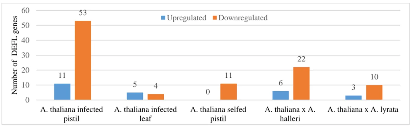
Figure 13:Distribution of the number of DEG in specific conditions. ... 49
Figure 14:Differential expression pattern of DEFL genes at specific conditions... 50
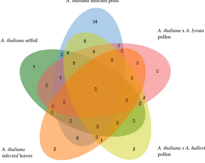
Figure 15:Distribution of differential expressed DEFL genes in A. thaliana during pollination and infection with Fusarium graminearum. ... 51
Figure 16:Positive correlation of log2 fold change between RNAseq and qPCR measurements of 14 candidate DEFL genes. ... 55
Figure 17:Comparison of log2 fold change result of qPCR assay and RNAseq result of 14 candidate DEFL genes validates the RNAseq data... 56
Figure 18:Schematic representation of Arabidopsis thaliana ovule before and after fertilization. ... 60
Figure 19:GFP expression under the regulation of the putative promoter of At5g43285 in the nuclei of synergids. ... 61
Figure 20:GFP expression under the regulation of the putative promoter of At2g20070 in the nucleus of central cell and antipodal cells. ... 61
Figure 21:GFP expression under the regulation of the putative promoter of At5g55132 in central cell nucleus. ... 62
Figure 22:GFP expression under the regulation of the putative promoter of At4g43505 in central cell nucleus. ... 63
Figure 23:GFP expression of At4g30074 under regulation of putative promoter in central cell nucleus before fertilization and in endosperm nuclei after fertilization, and subcellular localization of At4g30074-eGFP protein was found in central cell. ... 64
Figure 24: In non-pollinated ovules, reporter gene GFP indicates the activity in the central cell of the putative promoters of six DEFL genes:At2g40995, At2g42885, At5g38330 and At3g07005 ... 65
vii
putative promoters of six DEFL genes: At2g40995, At2g42885, At5g38330 and At3g07005 ... 66 Figure 26:GFP expression under the regulation of the putative promoter of At4g09153 and At1g60985 in central cell nucleus of an unfertilized ovule and in the endosperm nuclei in 48HAP fertilized ovule. ... 67 Figure 27:At4g11760 recombinant protein labelled with GFP expression in A. thaliana pollen grain. 68 Figure 28:Localization of putative promoter activity indicated by reporter GFP in pollen grains of marker lines of At3g06985, At3g42473 andAt3g65352. ... 69 Figure 29:Representation of root structure ... 70 Figure 30:At4g11760 recombinant protein labelled with GFP expression in A. thaliana root cap and root epidermis. ... 70 Figure 31:At4g30074 recombinant protein labelled with GFP expression in A. thaliana root cap, epidermis and root hair. ... 71 Figure 32:Down regulation of CNRQ values in infected treated samples in comparison to mock treatment and emasculated pistil of At2g42885, At3g07005 and At4g09153 at day 1 and day 3... 74 Figure 33:Data representation for seven candidate genes during 2DAE and 3DAI indicates that 3DAI has downregulation pattern of expression for the seven candidate genes in comparison to 2DAE. ... 75 Figure 34:Similar expression patterns and log2 fold change of candidate genes were observed while comparing the two different F. graminearum strains used during the infection of pistil. ... 75 Figure 35:Log2 fold change of candidate DEFL genes between 4DAE and 3DAI indicates that the age of the pistil has no effect on F. gramainearum infection. ... 76 Figure 36:Comparison of the effects of emasculation, mock treatment and infection in DEFLs gene expression suggest that mock treatment do not significantly differ from the control and
downregulation in DEFLs was due to infection. ... 77 Figure 37:Quantification of GFP signal in the central cell of candidate genes At3g07005, At4g09153 and At2g42885 at different time points after pollination suggests the candidate genes may have a role during fertilization. ... 78 Figure 38:Quantification of GFP signal in antipodal cells and central cell of At2g20070 at different time points after pollination suggests the At2g20070 may have a role in pre-fertilization events. ... 79 Figure 39:Average CNRQ values of candidate genes At3g07005 and At2g20070 during different time points after pollination indicates that At3g07005 and At2g20070 genes have role in early endosperm development. ... 80 Figure 40:Different trends of CNRQ expression were observed while comparing two biological replicates for At3g07005 at different time points after pollination. ... 81 Figure 41:Similar expression trend of CNRQ values were observed while comparing the two replicates for At2g20070 at different time points after pollination. ... 81 Figure 42:Average CNRQ values of candidates At3g07005 and At2gs42885 at either 8HAP or 24 HAP followed by infection or control treatments lasting one day suggests no common trend of
expression between candidate genes. ... 82 Figure 43:Average CNRQ values of candidate DEFLs 8 HAP followed by infection or control
treatments lasting three days indicates that candidate genes are downregulated in mock treatmed and infection treated samples in comparison to control. ... 83
viii
treatments lasting three days suggests that candidate genes have no common pattern of expression in the mock treatment and infection in comparison to control. ... 83 Figure 45:Comparison of seed set data for pollination followed by different treatments indicate that F.
graminearum infection has severe effect on 8HAP pistil in comparison to 24HAP. ... 85 Figure 46:Comparison of seed set data for pollination followed by different treatments indicate that F.
graminearum infection has severe effect on 8HAP pistil in comparison to 24HAP. ... 86 Figure 47:Comparison of infected seed set data at different pollination time points in one days, two days and three days indicates that three days of infection has severe effect in seed set formation
irrespective of pollination time point. ... 87 Figure 48:Comparison of seed set data for different treatment before pollination for one day and three days signifies that infection has drastic effect on seed development. ... 88 Figure 49:Comparison of silique of 3 DAI followed by pollination with two controls. ... 89 Figure 50:Percentage of silique developed during different treatments followed by pollination indicate that the infected pistils followed by pollination had fewer chance in developing into silique. ... 89 Figure 51:Positive correlation coefficient between the levels of relative expression of At2g40995- At1g60985 during pollination followed by infection ... 91 Figure 52:Classification of different ovule stages for endosperm developmental studies using the marker line A. thaliana pAt1g60985:NLS-(3x)eGFP-18. ... 95 Figure 53:Comparison of the endosperm development status of different treatments in different time point indicates that infection and mock treatment cause the cell death of ovules. ... 97 Figure 54:Comparison of the endosperm development status based on day of different treatments indicates mock treatment and infection treated samples had an increase of ovules in degradation stage from one day to two days. ... 98 Figure 55:Comparison of the development stages of ovules during 8HAP after different treatment for one and two days indicates that rate of endosperm development is not effected by F. graminearum infection. ... 100 Figure 56:Comparison of the percentages of ovule in 0h/8h stage to degradation stage in 8HAP
followed by one and two days of different treatment indicates that non-pollinated ovules are more prone to F. graminearum infection. ... 101 Figure 57:Negative correlation between the percentages of ovules at the 0h/8h stage and degradation stage in 8HAP followed by one and two days of different treatments. ... 102 Figure 58:Comparison of AtCEP1-GFP ovules during F. graminearum infection along with control indicates that PCD occurs in the infected ovule. ... 103 Figure 59:Crosstalk between regulatory genes of SA and JA/ET signalling pathway for defence
response along with mycotoxin induced genes in F. graminearum infected pistil which is inferred from infected pistil transcriptome data and the literature. ... 113 Figure 60:Schematic representation of activation of PDF1.2a by MAPK signalling pathway during PTI in Fusarium infected pistils as inferred from the transcriptome and literature. ... 117 Figure 61:Schematic representation of activation of PDF2.2a and other defence mechanism by PRX 33/ 34 during PTI in Fusarium infected pistils as inferred from the transcriptome and literature. ... 118
ix
Figure S1:Comparison of emasculated pistil and non-emasculated pistil during F. graminearum infection ... 142 Figure S2:eGFP expression of At5g38330, At5g42885, At2g40995 and At3g07005 in roots. ... 146 Figure S3:CNRQ values of At5g38330, At2g40995, At5g43285 and At1g60985 during infection along with mock treatment and control at day 1 and day 3. ... 147 Figure S4:Silique comparison of different pollination time point followed by one day of infection or mock treatment. ... 148 Figure S5:Silique comparison of different pollination time point followed by two days of infection or mock treatment. ... 148 Figure S6:Comparison of silique of 1DAI followed by pollination with two controls. ... 149 Figure S7:Expression pattern of At5g43285 gene during different hours after pollination in different studies (A) qPCR study (B) quantification of eGFP signal. ... 153
x List of Tables
Table 1:Tissue samples of three Arabidopsis species taken at different conditions ... 45
Table 2:Characteristics of the transcriptomes sequenced ... 47
Table 3: Differential expression of A. thaliana genes in five condition ... 48
Table 4:Log2 fold change of PDF genes ... 52
Table 5:Log2 fold change of candidate Arabidopsis thaliana DEFL genes ... 54
Table 6:Differential expression of defence related genes in infected pistils of A. thaliana ... 58
Table 7:Candidate DEFLs investigated for expression localization in planta ... 59
Table 8:Summary of candidate DEFL genes expression in plant tissues ... 72
Table 9:Statistical significance of the pairwise comparisons of expression levels of DEFLs in control and infected pistils ... 77
Table 10:Average seed set in two experimental conditions ... 90
Table 11:Pairwise correlation coefficient of relative gene expression using the average CNRQ values during different conditions. ... 92
Table 12:Pairwise correlation coefficient of relative gene expression using the RPKM values ... 93
xi Abbreviations
Abbreviations used in figures are explained in the respective caption.
bp Base pairs
CCG Central cell guidance
cDNA Complementary DNA
CDS Coding sequence
CNRQs Calibrated normalized relative quantities
CLSM Confocal Laser Scanning Microscopy
cm Centimeter(s)
COL-0 Columbia-0
CRP Cysteine-rich peptides
CSα/β Cysteine-stabilized α/β motif
DAE Day after emasculation
DAI Day after infection
DAT Day after mock treatment
DEG Differentially expressed genes
DEFL Defensins and defensin-like proteins
DNA Deoxyribonucleic Acid
DON Deoxynivalenol
eGFP enhanced Green fluorescent signal
ER Endoplasmic reticulum
ETI Effector-triggered immunity
ETS Effector-triggered susceptibility
FER Feronia
FHB Fusarium head blight
g Gramm
GOI Gene of interest
HAP Hours after pollination
hr Hour
JA/ET Jasmonic acid mediated Ethylene signalling
Kg Kilogram
L Liter(s)
LCR Low-molecular-weight Cysteine-Rich Protein
LYK Lysin motif receptor kinase
M Molar
MAP Mitogen activated protein
MAPK MAP kinase
MAPKK MAP kinase kinase
mg milligram
min minutes
mL milliLiter
mM milliMolar
MS Murashige & Skoog
MW Molecular Weight
NB-LRR Nucleotide-Binding Leucine-Rich Repeat
NIV Nivalenol
NLS Nuclear Localization Signal
NP Non-Pollinated pistil
NTC Non-Template Control
PAMPs Pathogen-Associated Molecular Patterns
PBS Phosphate Buffered Saline
xii
PCR Polymerase Chain Reaction
PDA Potato Dextrose Agar
PDF Plant Defensins Family
PG Pollen Grain
PGM Pollen Germination Medium
PRR Pathogen Recognition Receptors
PrsS Papaver rhoeas stigma S-determinant
PrpS Papaver rhoeas pollen S-determinant
PTG Pollen Tube Germination
PTI PAMP-Triggered Immunity
RFP Red Fluorescent Protein
RNA Ribonucleic Acid
RNAi RNA interference
qPCR Quantitative real time Polymerase Chain Reaction
ROS Reactive Oxygen Species
SA Salicylic Acid
SCA Stigma/style Cysteine-rich Adhesin
SCR S-locus Cysteine-Rich
SE Synergid Endosperm
SLR S Locus-Related glycoprotein
SI Self-Incompatibility
SP11 S-locus Protein 11
SRK S-locus Receptor Kinase
T-DNA Transfer-DNA
VPE Vacuolar Processing Enzyme
v/v Volume/volume
WGA-TMR Wheat Germ Agglutinin-TetraMethylRhodamine
WT Wild type
w/v weight /volume
μg Microgram(s)
μL Microliter(s)
μm Micrometer(s)
μM Micromolar
1 1. Summary
In contrast to animals and lower plants, male gametes of angiosperms are immobile and require transportation via the pollen tube cell to reach the female gametophyte, and together complete double fertilization. The path of the pollen tube towards the female gametes is guided by different types of signalling molecules, among them are defensin and defensin-like (DEFL) cysteine-rich peptides. Although A. thaliana has more than 300 genes encoding DEFLs, several of which are involved in plant immunity and cell-to-cell communication during fertilization, the roles of most members of this family are unknown.
The main aim of this project was to systematically identify DEFL genes expressed in A.
thaliana during double fertilization particularly during pollen-tube guidance as well as in response to fungal infection. This was accomplished by analysis of A. thaliana transcriptomes of pistils selfed, treated with A. lyrata or A. halleri pollen or infected with F. graminearum.
Candidate DEFLs exclusively expressed in pistils were selected to carry out a detailed characterization of their expression in planta. The second objective of this project was to gain insight into the molecular basis of Arabidopsis-Fusarium interaction based on the expression patterns of DEFLs. A. thaliana is an appropriate translational model for investigating how DEFLs counteract F. graminearum infection because the immune response of A. thaliana is very well documented.
Analysis of pistil transcriptome data showed that a total of 72 DEFL genes were differentially expressed in A. thaliana pistils. Detailed studies of eGFP localization of 25 DEFL candidate genes, showed 11 of them were expressed before pollination in specific cells of the mature female gametophyte, while four candidates were expressed in mature pollen grains, but not in growing pollen tubes. Post-fertilization, most genes expressed in the central cell of the ovule were expressed in the developing seed endosperm. Key results hinting at the possibility of DEFL involvement in different biological processes are the expression in roots of several candidates detected in the gametophytes and the upregulation of LURE1.1 in infected pistils, suggesting this known pollen tube attractant might also participate in the immune response.
Further statistical analysis of candidate DEFL gene expression data, showed there is a high correlation between the transcription of those expressed in the central cell of the embryo sac and the ones expressed in the synergids, suggesting co-regulated DEFLs play a role in guidance of the pollen tube before fertilization and during polytubey block after fertilization.
2
may be part of the first line of defence to F. graminearum via PAMP-triggered immunity (PTI). Some of these DEFL genes are regulated as secondary messengers of ROS production and also in the downstream process of MAPK signalling cascade. Furthermore, the patterns of differential expression of five DEFL genes (PDF1.2a-c, PDF1.4, PDF1.3), hint they are possibly regulated by the JA/ET defence signalling pathway during the necrotrophic phase of Fusarium infection.
In this context, the detrimental influence of F. graminearum infection in reproduction was investigated through analysis of seed development and seed set. This work suggests unfertilized ovules are more prone to Fusarium infection and its necrotrophic phase has a major detrimental influence in seed development. Furthermore, the reduction in seed set observed during Fusarium infection was caused by programmed cell death (PCD) of unfertilized ovules as documented by observation of ovule micromorphology in marker line AtCEP1-eGFP and analysis of RNAseq data. Specifically, the upregulation of genes encoding proteases involved in PCD in the infected pistil transcriptome suggests a mechanism where necrotrophic Fusarium obtains nutrients by manipulating the immune response of its host.
Our findings suggest that DEFL genes which are specifically expressed in reproductive tissues might play a role in defence and some of them, like LURE1.1 also possess dual function in reproduction. We hypothesize that DEFL genes initially had a role in protecting reproductive tissues and later on some of them acquired additional roles in cell-to-cell communication during pollination. The results of this study are relevant to understand the similarities between the processes of double fertilization and the immune response, identify interesting candidate genes to address the molecular basis of reproductive isolation and to develop strategies to counteract Fusarium head blight, a major crop disease affecting yield and jeopardizing food and feed safety worldwide.
3 2. Zusammenfassung
Im Gegensatz zu Tieren und niederen Pflanzen sind männliche Gameten von Angiospermen unbeweglich und erfordern einen aktiven Transport zu den weiblichen Gameten über die Pollenschlauchzelle.Der Weg des Pollenschlauchs zu den weiblichen Gameten wird von verschiedenen Arten von Signalmolekülen geleitet; Darunter kommt defensinähnliche (DEFL) cysteinreiche Peptiden eine besondere Bedeutung zu. A. thaliana hat mehr als 300 DEFL-Gene, die sowohl an der Pflanzenimmunität als auch an der Zell-Zell-Kommunikation beteiligt sind, jedoch sind die Rollen der meisten DEFL-Gene in A. thaliana weitgehend unbekannt.
In diesem Projekt wurde die Analyse von mehreren Pistil-Transkriptomen zur systematischen Identifizierung von DEFL-Gene herangezogen. Zu diesem Zweck wurde A. thaliana mit sich selbst, gekreuzt und mit den nahestehenden Arten A. lyrata und A. halleri. Um die DEFL Gene zu identifizieren, die während der Abwehrreaktion exprimiert wurden, wurden Infektionsstudien mit F. graminearum durchgeführt. Kandidaten DEFLs, die ausschließlich in Pistillen exprimiert wurden, wurden ausgewählt, um eine detaillierte Charakterisierung ihrer Expression in der Pflanze einschließlich Stempel und Wurzeln durchzuführen. Diese Informationen wurden verwendet, um ihre möglichen Rolle bei der Befruchtung und Infektion zu untersuchen.
Ein Ziel dieses Projekts war es, einen Einblick in die molekulare Basis der Arabidopsis- Fusarium-Wechselwirkung zu gewinnen, basierend auf den Expressionsmustern der DEFLs.Die Ergebnisse dieser Analyse sind relevant für die Entwicklung von Strategien zur Bekämpfung der Fusarium-Kopffäule, einer großen Erntekrankheit, die den Ertrag beeinträchtigt und die Nahrungsmittel- und Futtermittelsicherheit weltweit gefährdet.
Die Transkriptomdaten zeigten, dass insgesamt 72 DEFL Gene differentiell exprimiert wurden. Unter diesen wurde LURE1.1, von infiziertem Pistil differentiell exprimiert, was nahelegt, dass dieses Peptid mehrere Funktionen haben könnte. Detaillierte GFP- Lokalisierungsstudien in 25 Kandidaten zeigten, dass 11 DEFL-Gene vor der Bestäubung in spezifischen Zellen des reifen weiblichen Gametophyten exprimiert wurden, wohingegen vier DEFL-Gene in reifen Pollenkörnern Expression zeigten. Zusätzlich konnte gezeigt werden, dass 6 von 15 DEFLs auch in Wurzeln exprimierten.
Eine weitere statistische Analyse der Kandidaten-DEFL-Genexpressionsdaten zeigte eine hohe Korrelation zwischen der Transkription der ausgeprägten zentralen Zelle und jenen, die in den Synergiden exprimiert wurden, was darauf hindeutet, dass co-regulierte DEFLs eine
4
spielen Polytubey-Block nach der BefruchtungDer Einfluss der F. graminearum-Infektion bei der Reproduktion wurde durch Analyse des Samenkörner je Schote dokumentiert. Hierbei zeigte sich, dass die nekrotrophische Phase von F. graminearum einen großen Einfluss auf die Samenentwicklung hat und dass die unbefruchtete Eizelle anfälliger für eine Fusariuminfektion war, als die umliegenden Zellen. Die Wirkung der Fusarieninfektion auf die unbefruchtete Eizelle wurde auch durch die Entwicklungsstudien unterstützt. Die unbefruchtete Eizelle unterzog PCD während der F. graminearum-Infektion, die durch die PCD-Markerlinie AtCEP1: eGFP beobachtet wurde. Dies wurde durch die Hochregulation der Endopeptidase CEP1 und des α-Vakuol-Verarbeitungsenzyms (αVPE) in den Transkriptomdaten des infizierten Pistils unterstützt. Somit erscheint es naheliegend, dass F.
graminearum Toxine die Eizelle manipulieren, um VPE und andere Proteasen zu produzieren, welche PCD nach sich ziehen, um Nährstoffe während der nekrotrophischen Phase zu erhalten.
Schließlich kann die Analyse des infizierten Pistil-Transkriptoms und der Literaturanalyse zu dem Ergebnis, dass spezifische DEFL-Gene Teil der ersten Verteidigungslinie zu F.
graminearum über PAMP-getriggerte Immunität (PTI) sein können. Einige dieser DEFL-Gene werden als sekundäre Botenstoffe der ROS-Produktion und auch im nachgeschalteten Prozess der MAPK-Signalkaskade reguliert. Darüber hinaus deuten die Muster der differentiellen Expression von fünf DEFL-Genen (PDF1.2a-c, PDF1.4, PDF1.3) darauf hin, dass sie möglicherweise durch den JA / ET-Abwehr-Signalweg während der nekrotrophischen Phase der Fusarium-Infektion reguliert werden.
A. thaliana verwendet DEFLs als einen der Abwehrmechanismen gegen F. graminearum, die in die nährstoffreichen Gewebe der Stempel eindringen. Zusammenfassend bestätigen diese Ergebnisse die Rolle der DEFLs bei der Pflanzenimmunität im Arabidopsis Pistil.
Die gezeigten Ergebnisse deuten darauf hin, dass DEFL-Gene, die spezifisch im Fortpflanzungsgewebe exprimiert werden, eine Rolle bei der Verteidigung spielen können.
5 3. Introduction
3.1 Double fertilization
Double fertilization is the defining feature of angiosperms and was discovered by Nawaschin in 1898 (Nawaschin 1898). Double fertilization involves fusion of two sperm cells with the egg and central cell to form both embryo and endosperm respectively (Berger et al. 2008).
The embryo gives rise to the next plant generation which is nourished by the endosperm during its development (Bleckmann et al. 2014). Signalling events during pollen-pistil interactions are highly orchestrated, which enables plant species to avoid inbreeding and encourages outcrossing. The amount and total mass of seed produced by a given species are closely linked to successful reproduction, and thus we can consider double fertilization as one of the important agronomical traits. Our daily nutrition is highly dependent either directly or indirectly on reproductive success of flowering plants.
In angiosperms such as Arabidopsis thaliana (A. thaliana), immotile sperm cells are transported by the pollen tube through the transmitting tract towards the female gametophytic cells (Dresselhaus et al. 2013). The female gametophyte (embryo sac) consists of seven cells and four cell types: three antipodal cells, two synergid cells, an egg cell and a central cell (Sundaresan et al. 2010).
Sexual reproduction in A. thaliana requires a great deal of coordination between gametic cells of male and female reproductive organs. There is an active crosstalk between the pollen tube and pistil during double fertilization (Dresselhaus et al. 2013). The pollen grains released by the anthers are attached to the papilla cells of the stigma by physical adhesion (Dresselhaus et al. 2013). This adhesion is called pollen capture and the sporopollenin which makes up the exine of the pollen coat plays an important role in that this step takes place in a species- preferential manner (Swanson et al. 2004). The following stage is the pollen-stigma cross- linking where proteins, lipids, and carbohydrates of the stigma and pollen membrane interact for the first time (Swanson et al. 2004). Subsequently, pollen hydration is regulated by plasma-membrane-localized stigmatic proteins along with pollen coat lipids (Dresselhaus et al. 2013). Following germination, pollen tubes penetrate the stigmatic tissues by secreting digestive enzymes and grow through the transmitting tract of style towards the ovule (Swanson et al. 2004). During pollen tube growth, the pollen tube is guided by various chemo-attractants present in the pistil extracellular matrix (Dresselhaus et al. 2013).
Specifically, pollen tube guidance towards the ovule is controlled by two processes known as ovular guidance and micropylar guidance (Takeuchi et al. 2012). Ovular guidance is mediated
6 gametophytic cells (Berger et al. 2008).
The pollen tube is guided by cysteine rich peptide LURE which are secreted by the synergid towards the ovule (Figure 1) (Dresselhaus et al. 2016). The pollen tube enters the embryo sac through the thick synergid cell wall known as filiform apparatus at the micropylar end (Figure 1) (Eckardt 2007). When the pollen tube comes in contact with one of the receptive synergid, the pollen tube ceases to grow and the receptive synergid undergoes cell death. The pollen tube discharges two sperm cells into the cytoplasm of the degenerating synergid in an event known as “pollen tube burst” (Dresselhaus et al. 2016) (Figure 1). The pollen tube burst occurs within 20 seconds after entering the female gametophyte (Drews et al. 2011). After the pollen tube bursts, the two sperm cells move to chalazal region of the degenerated receptive synergid cell within a few seconds (Figure 1). The two sperm cells remain in an immobile state in that region for approximately seven minutes (Figure 1). This is followed by the fusion of one sperm cell with the egg cell to form the embryo and the other sperm cell fuses with central cell to form the endosperm (Hamamura et al. 2012; Dresselhaus et al. 2016).
Figure 1: Double fertilization in Arabidopsis thaliana
(A)The pollen tube is guided by LURE peptides which is secreted by the synergids. (B) The pollen tube enters the ovule through the micropylar opening into a receptive synergid where it bursts. Two sperm cells are released and remain immobile for a few minutes after which one of them fuses with the egg cell to form an embryo, while the other sperm cell fuses with the central cell to form the endosperm.Picture modified from (Sprunck et al.
2015).
A) B)
7
Cysteine-rich peptides (CRP) is a class of small peptides that constitute around 2% of all expressed genes in some plant species (Silverstein et al. 2007). All CRPs have common features such as conserved N-terminal region, C-terminal containing 4–16 cysteine residues, and a size of less than 160 amino acid residues (Marshall et al. 2011). CRPs are categorized by their primary sequence, the position and number of cysteine residues and the location where disulfide bridges form conserved 3D structures (Silverstein et al. 2007). It has been reported that several CRPs play a vital role in pollen-pistil interactions during plant reproduction (Marshall et al. 2011).
Many species of flowering plants have developed a mechanism to prevent self-fertilization during pollen-stigma interaction, which is known as self-incompatibility (SI). Among the first CRPs where a role in reproduction was described are those involved in SI. SI determinants found in pollen and stigma are programmed as a pair in order to control self-non-self-pollen recognition (Swanson et al. 2004). An identical interaction between S-allele ligand–receptor activates SI downstream signalling that results in programmed cell death (PCD) (Thomas et al. 2004). SI is an important mechanism in plants which aids in maintaining genetic diversity by preventing plant species from inbreeding. In Brassicaceae, the male determinant of SI is the so-called S-locus cysteine-rich (SCR)/S-locus protein 11 (SP11), which is a CRP from those first classified as defensins. SCR is found in the pollen coat and contains eight conserved cysteine residues. Binding of SCR/SP11 to the female SI determinant, S-locus receptor kinase (SRK) in the stigma triggers signalling pathways that culminate into pollen rejection through blocking of pollen hydration and inhibition of pollen tube germination (Shiba 2001). In contrast, in Papaver rhoeas, the female SI determinant Papaver rhoeas stigma S-determinant (PrsS) encodes a CRP containing four conserved cysteines (Figure 2) (Wheeler et al. 2009). During the incompatible pollen grain interaction in Papaver rhoeas, papillae cell secretes PrsS which binds to the Papaver rhoeas pollen S-determinant (PrpS) and results in PCD of the pollen tube (Wheeler et al. 2010).
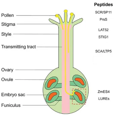
CRPs are also involved in pollen tube growth and guidance. In tomato, pollen-specific secreted protein LAT52 interacts with the pollen receptor LePRK2 for pollen germination (Figure 2) (Zhang et al. 2008). STIG1 a CRP from the stigma and style interacts with LePRK2 to promote pollen tube cell growth in the stigma (Figure 2) (Tang et al. 2004). In the stigma/style of lily, the stigma/style cysteine-rich adhesin (SCA) along with pectin is involved in adhesion of pollen tube and growth through transmitting tract (Figure 2).
8
Figure 2:Schematic diagram showing CRPs involved in communication during plant reproduction
(A) Schematic representation of pollen-tube guidance in Angiosperms. (B) Schematic image representing the CRPs SP11, PrsS, SCA, LAT52 and STIG1 involved during pollen–stigma interactions leading to pollen adhesion, hydration, germination and pollen tube growth. (C) CRPs such as LUREs, ZmEA1 is involved during microphylar guidance prior to double fertilization. Picture modified from (Kanaoka et al. 2015).
LTP5s is secreted from the pollen tube tip in order to maintain the pollen tube polarity (Dresselhaus et al. 2013).CRPs have also been reported to act as short-range micropylar attractants for pollen tubes. Specifically, defensins such as LUREs are secreted by the synergid cells for pollen tube guidance towards ovule (Figure 2) (Takeuchi et al. 2012). After the pollen tube enters the micropyle, Zea mays defensins ZmES4 secreted by synergid ensures pollen tube growth arrest and participates in pollen tube bursting (Amien et al. 2010) (Figure 2). ZmES4 interacts with the pollen tube potassium channel KZM leading to an influx of K+ which results in uptake of water and subsequently leads to pollen tube bursting (Amien et al. 2010).
3.3 Role of DEFLs in species -preferential manner during reproduction
Some of the female gametophyte genes that are involved in the guidance and reception of pollen tube may have species preferential interactions, and thereby contribute to establishing prezygotic reproductive isolation (Escobar-Restrepo et al. 2007; Takeuchi et al. 2012).
Interspecific crosses mostly yield none or a reduced number of seeds due to the failure of
ZmES4
9
phenotype (Escobar-Restrepo et al. 2007). The fer-like phenotypes in some interspecific crosses strongly suggest that species-preferential signal between the pollen tube and ovules mediates pollen tube growth arrest and burst. Prezygotic reproductive isolation takes place when this signal is missing in interspecific crosses.
As mentioned in the previous section, defensins and defensin-like proteins (DEFL) are one of the subgroups of CRPs. DEFLs are involved in pollen-pistil interactions in a species- preferential manner. SCR/SP11 was the first DEFL gene shown to act in species preferential manner during SI during pollen-stigma interaction (Boggs et al. 2009). DEFL peptides such as LUREs and ZmES4 are involved in pollen tube guidance, pollen tube reception and are responsible for the failure of double fertilization events by acting in a species-preferential manner during interspecific crosses (Amien et al. 2010; Takeuchi et al. 2012). The consequences of this preferentiality are that LUREs and ZmES4 peptides secreted by female gametophyte constitute a mechanism of prezygotic reproductive barrier during interspecific crosses. Overcoming this prezygotic barrier would open up possibilities to improve crop productivity. Example of overcoming prezygotic reproductive barrier has been reported in T.
fournieri. A. thaliana LURE peptide was transformed in T. fournieri and T. fournieri ovule was able to recognize A. thaliana pollen (Takeuchi et al. 2012).
3.3.1 Arabidopsis species are an ideal experimental model for studying reproductive isolation
A. thaliana a selfing species and has strong prezygotic reproductive isolation mechanisms triggered when crossed with other species (Grundt et al. 2006). Arabidopsis lyrata (A. lyrata) and Arabidopsis halleri (A. halleri) are self-incompatible species that are related to Arabidopsis thaliana (Clauss et al. 2006).
A. lyrata diverged approximately five million years ago from A. thaliana and is a closely related species to A. thaliana. A. lyrata is a perennial herb with distribution in the northern hemisphere and central Europe in restricted habitats (Schmickl et al. 2010). A. halleri is a heavy metal accumulating species which is distributed in central Europe, eastern Asia and grows on acidic, neutral and oligotrophic soils. A. halleri is mostly studied for its characteristics in tolerance, accumulation of heavy metals, and is an important model of studying phytoremediation (Clauss et al. 2006). A. halleri and A. lyrata are outcrossing diploids with genomes 50% larger than the A. thaliana genome. They are compatible as shown by allopolyploid Arabidopsis kamchatica, a hybrid that originated from the
10
with self-incompatible A. lyrata and A. halleri are ecologically diverged, but occur in geographically overlapping region, making them an ideal plant species for studying the genetic basis of plant reproductive isolation.
In recent years, DEFLs in Arabidopsis species have been shown to mediate the communication between male and female gametophytes in a species preferential manner, this property makes it an ideal gene family for understanding reproductive isolation. For example, the transformation of SCR-complexes along with SRK from S-locus of self-incompatible A.
lyrata to A. thaliana was sufficient to impart SI phenotypes in self-fertile A. thaliana (Boggs et al. 2009).
3.4 Role of defensins in immunity
Although plants have physical barriers to pathogen entry like the cuticle or bark, the size of stomatal pores and alteration of cell walls(Zeng et al. 2010; War et al. 2012), they fundamentally rely on an innate immune system (Dodds et al. 2010). Plant innate immune responses can be represented using a zigzag model (Figure 3).
Figure 3:Zigzag model of plant innate immunity
In phase 1, plants detect PAMPs via PRRs to trigger PTI and this is followed by pathogens delivering effectors that would interfere with PTI and resulting in ETS. In phase 2, effector is recognized either directly or indirectly by an NB-LRR protein, and thereby activating ETI which often cause hypersensitive cell death (HR) and production of defence related gene. Picture was taken from (Incarbone et al. 2013)
In the first phase, pathogen-associated molecular patterns (PAMPs) such as chitin, which is a part of fungal cell wall component, are recognized by pathogen recognition receptors (PRRs), and resulting PAMP-triggered immunity (PTI) stops pathogen growth (Figure 3) (Dodds et al.
2010). In turn, pathogens deploy effectors which interfere with PTI and results in effector- triggered susceptibility (ETS) (Figure 3). In the second phase of innate immunity, effectors are recognized either directly or indirectly by NB-LRR proteins, resulting in effector-triggered
11
those of PTI and specific immune responses for defence (Jones et al. 2006). The plant responds to pathogens with diverse defence strategies such as the expression of defence- related genes, oxidative bursts, increased production of hormones and programmed cell death (Wu et al. 2014; Bigeard et al. 2015). Signalling cascades such as the mitogen-activated protein kinase (MAPK) pathway, are triggered during the defence response. These cascades of protein phosphorylation respond to extracellular biotic stress by activating a wide range of cellular responses (Bigeard et al. 2015).
DEFLs are reported to participate in different biological functions, such as the previously described cell-to-cell communication during fertilization and the immune response which will be described in the following section. Plant defensins with antimicrobial activity are a vital part of the innate immune system of angiosperms (Carvalho et al. 2009). Plant defensins are induced as part of defence response to a broad spectrum of fungal plant pathogens and some bacteria (Carvalho et al. 2009; Penninckx et al. 2003). Lack of antibacterial activity of most plant defensins would possibly be due to the relatively larger infection pressure exhibited by fungal pathogens in comparison to the threat posed by bacteria (Thomma et al. 2002). They also inhibit the in vitro growth of human pathogenic fungi Candida albicans and Saccharomyces cerevisiae (Vriens et al. 2014; Aerts et al. 2011).
Defensins and DEFLs are expressed in all plant tissues reflecting their potential role in the systemic response to fungal infection of vegetative tissues or as constitutive defence barrier, especially in seeds and reproductive organs (Hegedus et al. 2013). Plant defensins are categorized in morphogenic or non-morphogenic according to their effect on the morphology of fungal hyphae. While the inhibition of hyphal elongation by morphogenic defensins results in hyphal hyperbranching, non-morphogenic defensins inhibit hyphal growth without any distortions (Ramamoorthy et al. 2007; Thomma et al. 2002).
Defensins might also counteract the effects of wounding by herbivore insects and parasitic plants. For example, the defensin VrD1 from Vigna radiate seeds inhibits insect α-amylase, leading to indigestibility of plant starch in the insect gut. Defensins which have antifungal activity do not exhibit α-amylase activity and vice versa (Thomma et al. 2002; Carvalho et al.
2009). The sunflower defensin Ha-DEF1 are also involved in the defence against parasitic plant Orobanche cumana, which causes severe yield losses on sunflower (Hegedus et al.
2013).
12
of the mechanisms is by binding to fungal ion channels (Marshall et al. 2011). Blocking of ion channels by defensins leads to inhibition of fungal hyphal tip growth and halts fungal colonization. For example, RsAFP2 defensins which were isolated from radish seeds exhibited antimicrobial activity by affecting K+ and Ca2+ ion transport channels in the fungal membrane (Lacerda et al. 2014). Defensins have developed other mechanism to combat fungal invasion. For example, they have cationic characteristics and interacts with negatively charged plasma membrane components of fungi. During their interaction, defensins alter the fungal membrane by inducing the production of reactive oxygen species (ROS) (Hegedus et al. 2013). For example, NaD1 defensin isolated from Nicotiana alata flowers induces oxidative damage in Candida albicans by hyperproduction of ROS (Hayes et al. 2013).
Transgenic plants expressing defensins have an increased resistance to fungal pathogens. For example, WT1 from wasabi when overexpressed in rice, potato and orchid, resulted in increased resistance against Magnaporthe grisea, Erwinia carotovora and Botrytis cinerea (Kanzaki et al. 2002; Lay et al. 2005; Stotz et al. 2009). Transgenic tomato plants containing defensin Rs-AFP2 decreased the activity of phytopathogenic fungi, including Alternaria solani, F. oxysporum, Phytophthora infestans, and Rhizoctonia solani (Lacerda et al. 2014).
Overexpression of Rs-AFP2 in transgenic rice (Oryza sativa) reduce Magnaporthe oryzae and Rhizoctonia solani infection. These two fungi are the main causative agents for rice blast and sheath blight diseases which leads to rice losses in agriculture (Jha et al. 2010).
Pea defensins enhanced resistance towards blackleg diseases in Brassica napus which is caused by Leptosphaeria maculans (Wang et al. 1999).
3.5 Other functional roles of defensins
Apart from immunity and intercellular communication during fertilization, plant defensins have adopted different roles. For example, AhPDF1.1 from A. halleri has antifungal activity and mediates zinc tolerance (Mith et al. 2015). Plant defensins also play a role in regulating growth and development of tissue. Specifically, MsDef1, MtDef2, RsAFP2 are all capable of inhibiting the growth of plant roots in vitro (Hegedus et al. 2013). The tomato DEF2 is expressed during initial stages of flower development and promotes meiosis. The tomato DEF2 also influences pollen viability and is also involved in the growth of various organs (Stotz et al. 2009). In addition, plant defensins are induced in response to environmental stress. For instance, soybean defensin gene Dhn8 was induced during drought stress (Lay et al. 2005), NeThio1 and NeThio2 from Nicotiana excelsior are induced in response to salt
13
cold induction, and potentially have a role in resistance towards freezing conditions (Gaudet et al. 2003).
3.6 Structure of Defensins
Defensins are structurally conserved in vertebrates and invertebrates including human immune cells. They exist in all plant families, including the Brassicaceae. In the early 1990s, defensins were initially documented to have fewer members in Arabidopsis, the scenario changed over the years with more in-depth studies which enabled in the identification of 324 DEFLs including 15 known defensins (Silverstein et al. 2007).
Most of the defensins genes that have been identified are composed of two exons: the first exon encodes for N-terminal signal peptide, whereas the second exon encodes for the cysteine-rich region that forms a positively charged mature peptide (Figure 4) (Silverstein et al. 2007). Defensins are categorized into two groups based on precursor proteins. In the first group, the precursor protein contains an endoplasmic reticulum (ER) signal peptide sequence and a mature defensin domain (Lay et al. 2014). The protein enters into the secretory pathway without any post-translational modification or subcellular targeting. The second group of defensins comprises of precursor protein with an additional C-terminal prodomain (Lay et al.
2014).C-terminal prodomain functions in subcellular targeting and is removed by proteolytic enzymes while entering through the secretory pathway.
Figure 4:Alignment of different DEFL clusters from Arabidopsis.
Alignment represents four distinct clusters of DEFLs in Arabidopsis. Identical in clusters are shaded black, whereas grey represents similar residues. Signal peptide are box labeled below the alignment. C or G designate conserved Cys are box labelled as C and Gly residues are box labelled with G. CSα/β and γ-core are shown below the alignment. Picture taken from (Silverstein et al. 2005).
14
eight cysteine residues which form intramolecular disulphide bridges (van der Weerden et al.
2013). Intramolecular disulphide bridges are responsible for the structural and thermodynamic stability of the defensins protein. The 3D structure of defensins exhibits a two motif (Figures 4 and 5). The first motif consisting of α -helix connected to triple-stranded, antiparallel β - sheet by three disulfide bonds forming cysteine-stabilized α/β motif (CSα/β) (Figures 4 and 5) (van der Weerden et al. 2013). Defensins have a conserved γ-core motif which consists of two antiparallel β-sheets with loop region. Positively-charged amino acids located at the γ-core motif are important for the antimicrobial activity of defensins (Figures 4 and 5) (Yount et al.
2004).
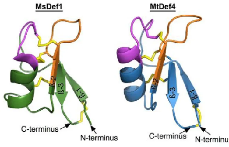
Figure 5:Three-dimensional structures of plant defensins MsDef1 and MtDef4.
MsDef1 and MtDef4 share highly conserved homology sequence. γ-core motif is represented in orange color which carries net positive charge. The CSα/β-core motif is represented in pink color and four disulfide bridges are represented in yellow color which stabilizes the defensins. Picture taken from (Sagaram et al. 2011)
3.6.1 Defensins under selection pressure
Plant defence and reproduction, are two highly conserved processes in the plants which are dependent on the various environment factor and each of the processes have biotic influence.
The pathogen-host and male-female gamete interactions have strong selection pressure on the molecular evolution of genes (Takeuchi et al. 2012). Gene duplication events along with diversifying selection was an important mechanism for plants to evolve in the arms race between microbial attackers and host plants.
Defensins are predicted to exhibit diversifying selection since its primary function is to mediate innate host defence and reproduction (Tesfaye et al. 2013). Defensins have been detected with diversifying selection in ants due to selection pressure caused by microbial pathogens (Viljakainen et al. 2008). The plant defensins tend to show characteristic molecular evolution patterns and selection pressure. These interactions, in particular sexual reproduction,
15 other species (Takeuchi et al. 2012).
Signal peptides of the defensin are conserved, whereas mature peptides of the defensin are possibly subjected to diversifying selection which determines the specific function aspects of these genes (Silverstein et al. 2005). DEFLs occurs both individually and in clusters throughout the A. thaliana genome. The gene duplication followed by successive rounds of diversifying selection might have resulted in 100 subgroups of DEFL with different activities in A. thaliana (Tesfaye et al. 2013; Silverstein et al. 2005).
3.7 Fusarium graminearum
Fusarium graminearum also known by teleomorph stage Gibberella zeae is a soil borne fungi responsible for Fusarium head blight (FHB), a disease from cereal crops which has a dramatic effect on productivity and food safety (Kazan et al. 2012). Between 1990 and 2002, FHB epidemics resulted in a loss of $3 billion of wheat yield and quality in the USA (Schmale et al.
2003).
F. graminearum is a haploid homothallic fungus which has a sexual and an asexual life cycle (Schmale et al. 2003). Both life cycle starts with F. graminearum overwintering on infected crop residues. During the asexual life cycle, F. graminearum produces macroconidia in chlamydospores which enable its survival during unfavorable conditions (Figure 6).
Macrocondia is dispersed to plants by rain-splash, and wind dispersal allowing to resume a new cycle of infection.
During suitable temperature and humidity, the sexual (teleomorph) stage of F. graminearum develops on infested plant debris. They form flask-like fruiting bodies called perithecia on the surface of infested residues (Figure 6). In perithecia, the sexual spores (ascospores) are formed within sacs called asci and forcibly discharged into the air (Figure 6). Ascospores are dispersed to crops by wind and rain. Infection occurs when macroconidia or ascospores land on wheat heads and cause mycelium development in aerial parts of the plant (Paul et al. 2004;
Schmale et al. 2003). Infected seeds might give rise to blighted seedlings if untreated (Figure 6). First symptoms of FHB are diseased spikelets demonstrating premature bleaching. F.
graimearum grows through diseased spikelet and spreads within the head. F. graminearum is also a vascular pathogen which can spread from the rachis of infected flowers to the other parts of plants through vascular bundles of xylem and phloem (Jansen et al. 2005).
16
necrotrophic lifestyle (Ding et al. 2011). The biotrophic lifestyle of F. graminearum is characterized by intercellular hyphae growth and no intracellular hyphae during the initial stages of infection of floral tissues (Brown et al. 2010). This biotrophic phase is followed by a necrotrophic lifestyle driven by nutrients obtained by intracellular colonization and host cell death. F. graminearum also exhibits saprotrophic growth due to its enzymatic ability to degrade crop residues for nutrients (Leplat et al. 2013; Khonga et al. 1988). Thus, F.
graminearum can adapt to different environment conditions.
Figure 6:Disease life cycle of F. graminearum.
The F. graminearum overwinters in infested crop residues. Macroconidia are produced in asexual phase from crop residues and are dispersed by rain. During favourable conditions, perithecia are formed in crop residues in sexual phase. Ascospores are produced from perithecia and dispersed in air. Macroconidia / ascospores infect flower, seeds and stems. Mycotoxins are present in infected seeds. Illustration taken from (Trail 2009).
Analysis of the proteome from F. graminearum during plant colonization revealed several extracellular proteins that facilitate disease establishment and spread in the plant as well as proteins involved in acquiring nutrients (Divon et al. 2007; Paper et al. 2007; Taylor et al.
2008). Some of the proteins secreted by F. graminearum during pathogenesis contained putative secretion signals which might function as effectors to initiate infection (Paper et al.
2007). Along with this, numerous proteins involved in production of reactive oxygen species (ROS) which are linked to pathogenesis are secreted by F. graminearum (Walter et al. 2010), such as cell wall–degrading enzymes (cellulases, hemicellulases, and pectinases) which are