Analysis of components
of the mitochondrial transcription machinery in Arabidopsis thaliana
DISSERTATION
zur Erlangung des akademischen Grades
doctor rerum naturalium (Dr. rer. nat.) im Fach Biologie
eingereicht an der
Mathematisch-Naturwissenschaftlichen Fakultät I der Humboldt-Universität zu Berlin
von
Diplom-Biologin Kristina Kühn geboren am 27.08.1974 in Stollberg
Präsident der Humboldt-Universität zu Berlin in Vertretung Prof. Dr. Hans Jürgen Prömel
Dekan der Mathematisch-Naturwissenschaftlichen Fakultät I Prof. Thomas Buckhout, PhD
Gutachter: 1. Prof. Dr. Thomas Börner 2. Prof. Dr. Wolfgang Hess 3. Prof. Dr. Frank Kempken
Datum der mündlichen Prüfung: 24. Februar 2006
Abstract
In der vorliegenden Arbeit wurde die Transkription mitochondrialer Gene durch die kernkodierten Phagentyp-RNA-Polymerasen RpoTm und RpoTmp der Pflanze Arabidopsis untersucht.
Im Mitochondriengenom von Arabidopsis wurden für 12 Gene Promotoren bestimmt. Diese zeigten verschiedene Sequenzelemente und wichen meist von der für Dikotyle publizierten Konsensussequenz ab. Für die Mehrheit der Gene wurden multiple Promotoren identifiziert. Es wurden weiterhin Promotoren nachgewiesen, welche die Transkription vermutlich nicht funktioneller Sequenzen aktivieren. Architektur, Lokalisation und Nutzung mitochondrialer Promotoren implizieren eine wenig stringente Kontrolle der Transkriptionsinitiation in Arabidopsis-Mitochondrien.
Zur Analyse der Funktionen von RpoTm und RpoTmp wurde ein in vitro-Transkriptions- system entwickelt. Da RpoT-Enzyme möglicherweise Kofaktoren benötigen, wurde in Arabidopsis nach Genen potentieller mitochondrialer Transkriptionsfaktoren gesucht. Als mitochondriales Protein mit Ähnlichkeit zu mtTFB, einem essentiellen Transkriptionsfaktor in Hefemitochondrien, wurde MetA identifiziert. In in vitro-Assays initiierte RpoTm an verschiedenen Promotoren die Transkription, während RpoTmp keine signifikante Promotorspezifität zeigte. Die spezifische Promotornutzung durch RpoTm erforderte superhelikale DNA. Weder RpoTm noch RpoTmp wurde durch MetA stimuliert. Eine mtTFB- ähnliche Funktion von MetA ist daher unwahrscheinlich. Für MetA wurde ausserdem eine engere phylogenetische Beziehung zu nukleären rRNA-Dimethylasen als zu mtTFB ermittelt.
Die hier vorgestellten Studien belegen die Transkription mitochondrialer Gene in Arabidopsis durch RpoTm; für RpoTmp ist eine nicht-redundante Transkriptionsfunktion denkbar. Die Kofaktor-unabhängige Spezifität von RpoTm für verschiedene Promotoren und die wenig stringente Initiationskontrolle in vivo legen nahe, dass eine individuelle Regulation mitochondrialer Gene in Arabidopsis auf Transkriptionsebene nicht erfolgt.
Schlagworte:
Pflanzenmitochondrien Mitochondriengenom Promotor
Transkription
Phagentyp-RNA-Polymerasen
Abstract
Mitochondria depend on a nucleus-encoded transcription machinery to express their genome.
The present study examined the transcription of mitochondrial genes by two nucleus-encoded phage-type RNA polymerases, RpoTm and RpoTmp, in the plant Arabidopsis.
For selected mitochondrial genes in Arabidopsis, transcription initiation sites were determined. Most genes were found to possess multiple promoters. The identified promoters displayed diverse sequence elements and mostly deviated from a nonanucleotide consensus derived previously for dicot mitochondrial promoters. Several promoters were detected that activate transcription of presumably non-functional sequences. Promoter architecture, distribution and utilization suggest a non-stringent control of transcription initiation in Arabidopsis mitochondria.
An in vitro transcription system was set up to elucidate the roles of RpoTm and RpoTmp.
Since RpoT enzymes possibly require auxiliary factors, the Arabidopsis genome was screened for potential cofactors of phage-type RNA polymerases. A mitochondrial protein (MetA) with similarity to mtTFB, an essential transcription factor in yeast mitochondria, was identified. In in vitro transcription studies, RpoTm recognized various promoters whereas RpoTmp displayed no significant promoter specificity. Promoter recognition by RpoTm depended on supercoiled DNA templates. Transcription initiation by RpoTm or RpoTmp was not affected by MetA, indicating that MetA is not functionally equivalent to mtTFB. Besides, MetA was found to be more closely related to non-mitochondrial rRNA dimethylases than to mtTFB.
The present study establishes RpoTm to transcribe mitochondrial genes; RpoTmp may have a non-overlapping transcriptional role in mitochondria. The cofactor-independent promoter specificity of RpoTm and the apparently non-stringent control of transcription initiation in vivo imply that mitochondrial genes in Arabidopsis may not be regulated individually at the transcriptional level.
Keywords:
Plant mitochondria Mitochondrial genome Promoter
Transcription
Phage-type RNA polymerase
Table of Contents
ZUSAMMENFASSUNG 4
SUMMARY 5
I INTRODUCTION 6
I.1 The mitochondrion, an endosymbiont-derived cell organelle 6
I.2 Plant mitochondrial genomes 7
I.3 Transcription in higher plant mitochondria 10
I.3.1 Mitochondrial promoters 10
I.3.2 Mitochondrial T7 bacteriophage-like RNA polymerases 12 I.3.2.1 Plant RpoT genes encoding phage-type transcriptases 12
I.3.2.2 Roles of RpoT enzymes 14
I.3.2.3 Structure of bacteriophage and phage-type RNA polymerases 15
I.3.3 Mitochondrial transcription factors 17
I.3.3.1 Yeast and animal mtTFB 18
I.3.3.2 Yeast and animal mtTFA 20
I.3.3.3 Mitochondrial transcription factors in plants 21 I.3.3.4 Cofactors of phage-type RNA polymerases in plastids 22 I.3.4 Regulation of mitochondrial gene expression at the transcriptional level 23
I.4 Aims of this study 25
II MATERIALS AND METHODS 26
II.1 Growth of Arabidopsis thaliana 26
II.2 Strains and culturing of Escherichia coli 26
II.3 Nucleic acids 26
II.3.1 Isolation of nucleic acids 26
II.3.1.1 Isolation of genomic DNA from Arabidopsis 26
II.3.1.2 Plasmid isolation from E. coli 26
II.3.1.3 Isolation of total RNA and mRNA-enriched RNA from Arabidopsis 26 II.3.2 Determination of nucleic acid concentrations 26
II.3.3 Nucleid acid electrophoreses 26
II.3.3.1 Agarose gel electrophoresis of DNA 26
II.3.3.2 Agarose gel electrophoresis of RNA 27
II.3.3.3 Denaturing polyacrylamide gel eletrophoresis (PAGE) of RNA 27
II.3.3.4 Native PAGE of DNA 27
II.3.4 cDNA synthesis and RT-PCR 28
II.3.5 PCR 28
II.3.6 Cloning and sequencing 28
II.3.6.1 Transformation of E. coli 28
II.3.6.2 Sequencing 28
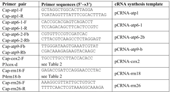
II.3.7 5’-RACE analysis of RNA 29
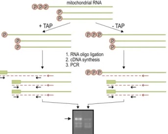
II.3.8 Analysis of in vitro-cappable transcripts 32
II.3.8.1 Preparation of riboprobes 32
II.3.8.2 In vitro capping and RNase protection 32
II.4 Protein analysis 32
II.4.1 Determination of protein concentrations 32
II.4.2 SDS polyacrylamide gel electrophoresis (SDS PAGE) 33
II.4.3 Immunoblotting 33
II.5 Recombinant protein expression 34 II.5.1 Plasmids for the expression of recombinant proteins 34
II.5.2 Protein expression in E. coli 34
II.5.3 Purification of recombinant proteins from E. coli 35
II.5.3.1 Trx-(His)6-tagged RpoTm and RpoTmp 35
II.5.3.2 Proteolytic removal of thioredoxin 36
II.5.3.3 (His)6-tagged MetA and MetB 36
II.6 Electrophoretic mobility shift assay 36
II.6.1 Gel mobility shift probes 36
II.6.2 DNA binding assay 36
II.7 In vitro transcription 37
II.7.1 Template construction 37
II.7.2 In vitro transcription assay 37
II.7.3 5’-end mapping of in vitro-synthesized RNAs 38 II.8 Green fluorescent protein (GFP) import assay 38
II.8.1 GFP targeting constructs 38
II.8.2 Transient expression in tobacco protoplasts and microscopy 38
II.9 Alignments and phylogeny 39
II.10 Material 40
II.11 Providers 40
III RESULTS 41
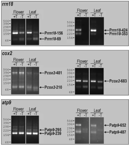
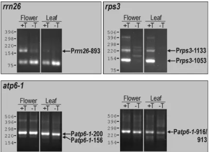
III.1 Analysis of mitochondrial promoters in Arabidopsis thaliana 41 III.1.1 Identification of transcription initiation sites by 5’-RACE 41 III.1.2 Identification of transcription initiation sites by in vitro capping 46 III.1.3 Mitochondrial promoter architecture in Arabidopsis 49 III.1.4 Promoters directing transcription of non-coding sequences 50 III.2 Characterization of a mitochondrial mtTFB-like protein in Arabidopsis 54 III.2.1 Identification of mtTFB-like sequences in the Arabidopsis genome 54 III.2.2 Mitochondrial localization of the mtTFB-like protein MetA 56 III.2.3 Phylogenetic analysis of plant, fungal and animal rRNA dimethylase-like
proteins 57 III.2.4 Non-specific DNA binding by recombinant MetA 59
III.3 Expression of the Arabidopsis phage-type RNA polymerases RpoTm and RpoTmp in E. coli 62 III.4 In vitro transcription studies of Arabidopsis RpoTm and RpoTmp 66 III.4.1 Development of an Arabidopsis in vitro transcription system 66 III.4.2 In vitro transcription from the mitochondrial promoters Patp6-1-200, PtrnM-98
and Prrn26-893 by RpoTm 67
III.4.3 Comparison of the transcriptional performances of RpoTm and RpoTmp 71 III.4.4 Transcription initiation by RpoTm and RpoTmp at non-CRTA promoters 75
IV DISCUSSION 78
IV.1 Multiple promoters as a common feature of mitochondrial genes in
Arabidopsis 78 IV.1.1 Identification of transcription initiation sites 78
IV.1.2 Promoter architecture 79
IV.1.3 Promoter distribution 81
IV.1.4 Non-stringent control of Arabidopsis mtDNA transcription 84
IV.2 Potential mtTFB-like cofactors of phage-type RNA polymerases in
Arabidopsis 85 IV.2.1 A mtTFB-like protein in Arabidopsis mitochondria 85 IV.2.2 Phylogenetic relationship between mtTFB and related rRNA dimethylases 86 IV.2.3 Implications of the function of homologous RNA polymerases in mitochondria
and plastids 88
IV.3 Transcriptional roles of the phage-type RNA polymerases RpoTm and
RpoTmp in Arabidopsis mitochondria 89
IV.3.1 Development of an Arabidopsis in vitro transcription system 90
IV.3.2 Intrinsic promoter specificity of RpoTm 92
IV.3.3 Different transcriptional properties of RpoTm and RpoTmp 95 IV.4 Does a mtTFB homologue function in mitochondrial transcription in plants?
99 IV.5 Transcription initiation in Arabidopsis mitochondria 100
V REFERENCES 102
Abbreviations 114 ANNEX A:Amino acid sequence alignment of methyltransferase-like proteins 115
ANNEX B:Predicted subcellular targeting of plant methyltransferase-like proteins 118 ANNEX C:Threaded structural models of Arabidopsis RpoTm and RpoTmp 119 ANNEX D:Accession numbers of RNA polymerase sequences 120 ANNEX E:T7 phage and phage-type RNA polymerase sequence alignment 121
Curriculum Vitae 123
Publications and Conference Abstracts 124
Danksagung 125
Eidesstattliche Erklärung 126
ZUSAMMENFASSUNG
Im Zellkern kodierte RNA-Polymerasen sind für die Transkription der mitochondrialen Genome eukaryotischer Zellen verantwortlich und übernehmen somit eine zentrale Rolle in der mitochondrialen Genexpression. Für verschiedenen Pflanzenspezies wurden nukleäre T7- phagenähnliche RNA-Polymerasegene (RpoT-Gene) beschrieben, welche vermutlich für katalytische Untereinheiten der mitochondrialen Transkriptionsmaschinerie kodieren. Eine mitochondriale Transkriptionsfunktion von RpoT-Genprodukten in photosynthetisierenden Eukaryoten wurde jedoch bislang nicht nachgewiesen. In der vorliegenden Arbeit wurde die Transkription mitochondrialer Gene durch die mitochondrialen Phagentyp-RNA-Polymerasen RpoTm und RpoTmp der Pflanze Arabidopsis thaliana untersucht.
Im mitochondrialen Genom von Arabidopsis wurden Transkriptionsstartpunkte ausgewählter Gene und Gencluster bestimmt. Erstmals wurde hier für eine dikotyle Pflanze gezeigt, dass mitochondriale Gene häufig multiple Promotoren besitzen. Die identifizierten Promotoren zeigten verschiedene Sequenzelemente und wichen zumeist signifikant von der für Dikotyle publizierten Konsensussequenz ab. Es wurde darüber hinaus die Funktion von Promotoren nachgewiesen, welche die Transkription nichtkodierender und vermutlich nicht funktioneller Sequenzen aktivieren.
Zwischen Promotoraktivitäten in Blüten- und in Blattgewebe wurden keine qualitativen Unterschiede beobachtet. Architektur, Häufigkeit, Lokalisation und Nutzung mitochondrialer Promotoren implizieren eine wenig stringente Kontrolle der Transkriptionsinitiation in Arabidopsis- Mitochondrien.
Die Identifizierung mitochondrialer Promotoren in Arabidopsis ermöglichte die Rekonstitution eines definierten in vitro-Transkriptionssystems zur Analyse der Transkription mitochondrialer Gene durch RpoTm und RpoTmp. In in vitro-Assays mit rekombinanten RNA-Polymerasen initiierte RpoTm an verschiedenen, jedoch nicht allen angebotenen Promotorsequenzen die Transkription, während für RpoTmp keine signifikante Promotorspezifität beobachtet wurde.
Offenbar wird die Spezifität des mitochondrialen Transkriptionsapparates für zahlreiche Promotoren durch RpoTm vermittelt. RpoTm initiierte die in vitro-Transkription an Promotoren auf supercoiled-strukturierten, jedoch nicht auf linearen DNA-Molekülen und unterscheidet sich hierin von in früheren Arbeiten charakterisierten transkriptionsaktiven Extrakten aus Pflanzen- mitochondrien. Dieser Befund impliziert, dass Kofaktoren, deren Funktion bei einer supercoiled- Konformation der DNA nicht essentiell ist, in in vitro-Assays mit mitochondrialen Extrakten und in vivo die Aufschmelzung der DNA unterstützen. Im Genom von Arabidopsis wurde nach Genen potentieller Kofaktoren von Phagentyp-RNA-Polymerasen gesucht. Ein durch den Kernlocus At5g66360 (MetA) kodiertes Protein, dessen mitochondriale Lokalisation hier nachgewiesen wurde, zeigt Ähnlichkeit zu mtTFB, einem essentiellem Kofaktor der T7-ähnlichen RNA-Polymerasen in Hefe- und Säugermitochondrien. In in vitro-Assays wurde die Initiation der RpoTm- und RpoTmp- abhängigen Transkription in keiner Weise durch MetA beeinflusst. Eine mtTFB-ähnliche Funktion von MetA in der mitochondrialen RNA-Synthese ist daher unwahrscheinlich. In Übereinstimmung hiermit wurde für MetA eine engere phylogenetische Beziehung zu nicht-mitochondrialen rRNA- Dimethylasen als zu mtTFB-Proteinen ermittelt.
Die hier vorgestellten Studien belegen die Transkription mitochondrialer Gene in Arabidopsis durch das Enzym RpoTm; für RpoTmp ist eine nicht-redundante mitochondriale Transkriptions- funktion denkbar, welche die Erkennung bekannter Promotoren nicht erfordert. Im Pflanzenreich wird hiermit erstmals der funktionelle Nachweis erbracht, dass ein nukleäres Phagentyp-RNA- Polymerasegen für ein Transkriptionsenzym mit mitochondrialer Promotorspezifität kodiert. Die Kofaktor-unabhängige in vitro-Spezifität von RpoTm für verschiedene Promotorsequenzen und die offenbar nicht stringente Initiationskontrolle in vivo legen nahe, dass eine individuelle Regulation mitochondrialer Gene in Arabidopsis auf Transkriptionsebene nicht erfolgt.
SUMMARY
Mitochondria depend on a nucleus-encoded transcription machinery to express their genome and maintain mitochondrial function. In plants, nuclear T7 bacteriophage-like RpoT genes identified in a variety of species have been suggested to encode catalytic subunits of the mitochondrial transcription apparatus. Still, functional evidence has been lacking for RpoT enzymes being involved in mitochondrial transcription in photosynthetic eukaryotes. The present study examined the transcription of mitochondrial genes by two mitochondrial phage-type RNA polymerases encoded by the RpoTm and RpoTmp genes in the plant Arabidopsis thaliana.
To study cis-elements that are recognized by these enzymes, transcription initiation sites of selected mitochondrial genes and gene clusters in Arabidopsis were determined. Most genes were found to possess multiple promoters, revealing for the first time that promoter multiplicity is a common feature of mitochondrial genes in a dicotyledonous plant. The identified promoters displayed diverse sequence elements and for the most part deviated significantly from a nonanucleotide consensus derived previously for dicot mitochondrial promoters. Several promoters were moreover detected that activate transcription of non-coding and presumably non-functional sequences. No qualitative differences in promoter utilization were observed between leaves and flowers. Promoter architecture, distribution and utilization suggest a non-stringent control of transcription initiation in Arabidopsis mitochondria.
The knowledge of mitochondrial promoters in Arabidopsis allowed a defined in vitro transcription system to be set up in order to elucidate the roles of RpoTm and RpoTmp in mitochondrial transcription. Since RpoT-driven transcription from mitochondrial promoters possibly requires a complementation of RpoT enzymes with as yet unidentified auxiliary factors, the Arabidopsis genome was screened for potential cofactors of phage-type RNA polymerases. A protein encoded by the nuclear locus At5g66360 (MetA) and shown here to be imported into mitochondria displays sequence similarity to mtTFB, an essential cofactor of the mitochondrial T7- like RNA polymerase in yeast and mammals that is related to rRNA methyltransferases. In vitro transcription studies examined the abilities of recombinant RpoTm and RpoTmp to transcribe DNA from mitochondrial promoters, and moreover tested MetA for its potential to modulate the transcriptional performances of RpoTm and RpoTmp. RpoTm recognized a variety of promoters whereas RpoTmp displayed no significant promoter specificity. Sequence specificity of the mitochondrial transcription apparatus thus appears to be conferred by the RpoTm core enzyme for the majority of promoters. RpoTm differed in its transcriptional performance from formerly characterized plant mitochondrial extracts in that it did not specifically initiate transcription at promoters located on linear DNA but required supercoiled templates. This indicates a participation of (an) auxiliary factor(s) in DNA melting in transcription experiments using mitochondrial extracts and in vivo, which is obviated by a supercoiled DNA conformation. Transcription initiation by RpoTm or RpoTmp appeared to be not affected by the presence of MetA in the in vitro assay, indicating that MetA does not have a role equivalent to that of yeast or mammalian mtTFB in mitochondrial RNA synthesis. In line with this, phylogenetic analyses revealed MetA to be more closely related to a group of non-mitochondrial rRNA dimethylases than to fungal or animal mtTFBs.
The data presented here establish RpoTm to transcribe mitochondrial genes. They thus provide the first direct linkage in the plant kingdom between an RNA polymerase activity recognizing mitochondrial promoters and a nuclear gene encoding a mitochondrial phage-type RNA polymerase. Experimental results suggest RpoTmp to have a non-overlapping transcriptional role in mitochondria, which might not involve the recognition of known mitochondrial promoters. The cofactor-independent in vitro specificity of RpoTm for diverse promoter sequences and the apparently non-stringent control of transcription initiation in vivo imply that mitochondrial genes in Arabidopsis may not be regulated individually at the transcriptional level.
I INTRODUCTION
I.1 The mitochondrion, an endosymbiont-derived cell organelle
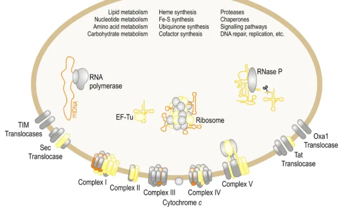
Mitochondria are the compartments of cellular respiration in eukaryotes and accommodate an energy-transducing system that generates ATP by coupling electron transport with oxidative phosphorylation (Saraste, 1999). The respiratory organelle is the descendant of a bacterial endosymbiont (Margulis, 1970; Margulis, 1981) and possesses its own vestigial genome. Analyses of the mitochondrial DNA (mtDNA) trace the evolutionary predecessors of mitochondria to a single ancestor whose closest contemporary relatives are found within the α division of the proteobacteria (Yang, et al., 1985). While the bacterial endosymbiont has long been considered to have been established in a nucleus-containing host cell, studies of unicellular eukaryotes have raised the possibility that the mitochondrion originated at essentially the same time as the nuclear compartment rather than in a subsequent event (Gray, et al., 1999). The majority of the original set of mitochondrial genes was either relocated to the nuclear genome or lost from the cell early in eukaryotic evolution (reviewed in Gray, 1992; Gray, et al., 1999). While animal mtDNA reduction appears to effectively have ceased in the common ancestor of all animals (Boore, 1999), gene transfer from the mitochondrion to the nucleus is an ongoing process in plants (Adams, et al., 2000; Adams, et al., 2002; Adams, et al., 1999). As a consequence of the unidirectional functional gene transfer, components participating in the diverse mitochondrial metabolic pathways and genetic processes largely are encoded in the nucleus and, following synthesis in the cytosol, are imported into the organelle (Herrmann, 2003; Martin and Herrmann, 1998). The protein complexes of the electron transport chain and mitochondrial ribosomes are assembled as mosaics of nucleus- and mitochondrion-encoded components (Figure 1). Correct assembly of these complexes therefore requires the coordinated expression of the mitochondrial and nuclear genomes. In photosynthetic eukaryotes, where the plastid as a second endosymbiont-derived organelle with a residual genome contributes to cellular processes, mitochondria need to coordinate gene function with yet another genetic compartment. The present study investigates as one essential element in organelle gene expression the transcription machinery in plant mitochondria.
Figure 1: Participation of mtDNA-encoded proteins and RNAs in biological processes in mitochondria.
The majority of mitochondrial functions are indicated; only the mitochondrial matrix and inner membrane are shown. Most mitochondrial components are nucleus-encoded (examples shown in grey), and the majority of mitochondrial processes have exclusively nucleus-encoded constituents (listed). Yellow symbols correspond to proteins and RNAs encoded by the mtDNA in some eukaryotes but by the nuclear genome in others. Very few components are specified by the mtDNA in all organisms (orange). Displayed components are involved in electron transport and oxidative phosphorylation (complexes I-V and cytochrome c), protein import and insertion into the inner membrane (TIM), protein export from the matrix and insertion into the inner membrane (Tat, Sec, Oxa1), mtDNA transcription (RNA polymerase), tRNA 5’-end processing (RNase P), protein synthesis (ribosomes and elongation factor EF-Tu). After (Burger, et al., 2003).
I.2 Plant mitochondrial genomes
Plant mitochondrial genomes considerably vary in size but contain a fairly stable number of 50 to 60 genes (Handa, 2003; Kubo, et al., 2000; Notsu, et al., 2002; Sugiyama, et al., 2005; Unseld, et al., 1997). These may be dispersed or organized in gene clusters, and predominantly code for components of the respiratory chain and of the translational apparatus while the machinery that transcribes these genes is encoded in the nucleus. Mitochondrial genome size in land plants ranges from 187 kbp in Marchantia polymorpha to 570 kbp in Zea mays and may even reach up to 2400 kbp in certain Cucurbitaceae (Ward, et al., 1981). In contrast, animal mitochondria contain considerably smaller genomes, yet they do not encode a proportionately smaller number of genes (reviewed in Bullerwell and Gray, 2004; Burger, et al., 2003). For example, the mtDNA of Arabidopsis thaliana (~367 kbp; Figure 2) is 20 times larger than the human mitochondrial genome (~17 kbp) but codes for only approximately
twice the number of proteins and one more RNA than the human mtDNA (Anderson, et al., 1981; Unseld, et al., 1997). 33 proteins, three rRNAs and 20 tRNAs are encoded by the Arabidopsis mtDNA (Duchene and Marechal-Drouard, 2001; Unseld, et al., 1997), whereas the functional plant mitochondrion has been estimated to contain 1000 or more proteins (Millar, et al., 2004).
Figure 2: Distribution of identified genes in the mitochondrial genome of Arabidopsis. (Taken from Dombrowski, et al., 1998).
Mitochondrial genomes of angiosperms display large intergenic regions and have expanded through frequent duplications as well as by incorporating introns and DNA segments from the plastid and the nucleus in addition to sequences of as yet unknown origin (Marienfeld, et al., 1999; Palmer, et al., 2000). Rapid structural evolution of the plant mitochondrial genome, which is accounted for by recombination between the small and large repeats of the mtDNA, is contrasted by slow evolution in sequence (Palmer, 1990).
Recombinational activity of repeated sequences moreover subdivides the mitochondrial genome into several different subgenomic DNA molecules (Andre, et al., 1992; Fauron, et al., 1995; Sugiyama, et al., 2005), the maintenance of which has in particular cases been shown to be under nuclear control (Abdelnoor, et al., 2003; Martinez-Zapater, et al., 1992). Low- frequency intragenic recombination events that are mediated by smaller repeats can result in
chimeric ORFs which, if transcriptionally active, may lead to pollen sterility (reviewed in Hanson and Bentolila, 2004; Linke and Börner, 2005).
Although plant mtDNA sequencing projects have commonly assembled a master circle from the complete genetic information (see Figure 2), the mitochondrial genome is maintained in vivo as not only subgenomic but also largely linear and branched molecules (Backert and Börner, 2000; Bendich, 1993; Oldenburg and Bendich, 1996; Oldenburg and Bendich, 2001; reviewed in Backert, et al., 1997).
DNA-containing fractions prepared from mung bean mitochondria have been characterized as membrane-associated chromatin-like nucleoids (Dai, et al., 2005). Isolated plant mitochondrial nucleoids were found to retain transcriptional activity and the ability to synthesize DNA in vitro, indicating that like in fungi and in mammals, mitochondrial nucleoids in plants are centres of mtDNA maintenance and expression (Dai, et al., 2005; Fey, et al., 1999). While proteins associated with plant mitochondrial nucleoids are still awaiting classification, a high mobility group (HMG) protein designated mtTFA has been identified as a highly abundant protein component of mtDNA-protein complexes in Saccharomyces cerevisiae, Xenopus laevis and humans (Alam, et al., 2003; Antoshechkin and Bogenhagen, 1995; Diffley and Stillman, 1992; Shen and Bogenhagen, 2001). Acting as a nucleoid architectural factor, mtTFA is required for mtDNA maintenance (Diffley and Stillman, 1991;
Kanki, et al., 2004). Besides, mtTFA is an obligatory transcription factor in human mitochondria (see I.3.3.2). A candidate mitochondrial mtTFA-like protein in Arabidopsis has been suggested to be encoded by a sequence positioned in a cluster of nuclear genes that code for mitochondrial proteins involved in DNA and RNA metabolism (Elo, et al., 2003;
Heinhorst, et al., 2004). Computational predictions of subcellular protein targeting are however not in support of this protein being a mitochondrial component (Elo, et al., 2003).
More progress has been made in characterizing nucleoid proteins in the plastid than in plant mitochondria (reviewed in Heinhorst, et al., 2004; Phinney and Thelen, 2005). While the prokaryotic histone-like DNA-binding protein HU is abundant in plastidial nucleoids of red algae, the major DNA-compacting protein in the plastids of plants has been identified as a 70-kDa sulfite reductase (SiR) (Sato, et al., 2001; Sato, et al., 2003). Addition of recombinant maize SiR has been demonstrated to increase the compaction of isolated plastid nucleoids in vitro and to concurrently repress in vitro transcription activity of nucleoids (Sekine, et al., 2002). DNA compaction through SiR is reversible and has been suggested to regulate the transcriptional activity in the chloroplast through changes in nucleoid compaction (Sekine, et
al., 2002). A protein similar in size to SiR was found to compact chloroplast nucleoids and suppress replication in soybean (Cannon, et al., 1999). It thus seems that the major nucleoid proteins of plant plastids and of yeast and animal mitochondria differ fundamentally in their effect on nucleoid activity.
I.3 Transcription in higher plant mitochondria I.3.1 Mitochondrial promoters
Cis-regulatory elements of mitochondrial transcription in metazoa are confined to a discrete mtDNA region known as D-loop, and all genes are transcribed from one or two uni- or bidirectional promoters located in this region (reviewed in Shadel and Clayton, 1993;
Tracy and Stern, 1995). In contrast, multiple promoters are active in yeast mitochondria, which share a sequence motif of nine nucleotides that is sufficient to efficiently promote transcription initiation in vitro (Tracy and Stern, 1995). Plant mitochondria similarly transcribe their genomes from numerous promoters (Tracy and Stern, 1995). Mitochondrial promoters have been analyzed in several plant species through identifying primary 5’ termini of mitochondrial transcripts and aligning sequences surrounding transcription initiation sites, thereby revealing conserved promoter motifs (Figure 3; Fey and Marechal-Drouard, 1999;
Hess and Börner, 1999). Moreover, sequence elements that are relevant for promoter function have been defined in in vitro transcription studies using complex mitochondrial extracts as a source of transcription activity (Binder, et al., 1995; Caoile and Stern, 1997; Dombrowski, et al., 1999; Hoffmann and Binder, 2002; Rapp, et al., 1993; Rapp and Stern, 1992). Sequences of up to 25 nucleotides around the transcription start site, which display the conserved motif YRTA (Y = T or C and R = A or G) immediately upstream of the initiating nucleotide, were found to be required for correct and efficient initiation of transcription in vitro (Hess and Börner, 1999). The majority of higher plant mitochondrial promoters exhibit an A/T-rich sequence element immediately upstream of the promoter core, which has been proven essential for the full function of different dicot and monocot mitochondrial promoters in vitro (Dombrowski, et al., 1999; Rapp, et al., 1993).
In mitochondrial promoters of dicotyledonous plants, the YRTA core motif is embedded in an extended consensus of nine nucleotides, CRTAAGAGA, with the initiating nucleotide at the penultimate position (Figure 3; Binder, et al., 1996). Only a few transcription start sites in dicot mitochondria coincide with sequences lacking a recognizable core motif (Binder, et al., 1994; Brown, et al., 1991). In contrast to mitochondrial promoters of Oenothera berteriana and potato that conform to the nonanucleotide consensus and are recognized by a pea in vitro
transcription system, deviating mitochondrial promoters of both species do not function in the heterologous system, which implies that specific mechanisms are involved in transcription initiation at alternative promoters (Binder, et al., 1995). Moreover, studies on mitochondrial transcription start sites in both dicots and monocots support the idea that the utilization of particular promoters requires a distinct nuclear background (Edqvist and Bergman, 2002;
Newton, et al., 1995).
Figure 3: Conserved sequence elements of plant mitochondrial promoters. Consensus promoter sequences in monocots and dicots are shown essentially as deduced by (Fey and Marechal-Drouard, 1999). The YRTA core motif is written bold, and initiating nucleotides are underlined. Conserved elements of the central promoter domain are highlighted orange; A/T-rich upstream elements are indicated. Upper case nucleotides denote highly conserved positions (frequency of appearance >75%); lower case letters indicate weakly conserved nucleotides (frequency between 50 and 75%). R = A or G; W = A or T; K = T or G; Y = C or T; n = any nucleotide.
Mitochondrial promoters of monocotyledonous plants often deviate in the YRTA core motif and are overall less conserved than dicot mitochondrial promoters (Fey and Marechal- Drouard, 1999; Hess and Börner, 1999). Most promoters comprise a central domain around the transcription initiation site, which contains the YRTA tetranucleotide at varying distance from the initiating nucleotide (Figure 3; Caoile and Stern, 1997; Covello and Gray, 1991;
Rapp, et al., 1993). Among the few promoters displaying no conserved motif is the alternative promoter cpc preceding the cox2 coding sequence in Zea perennis (Newton, et al., 1995).
Mitochondrial genes have been described to display multiple transcription initiation sites in monocots far more frequently than in dicots (Lupold, et al., 1999; Mulligan, et al., 1988). It has been suggested that promoter multiplicity is maintained in order to ensure mitochondrial gene expression despite frequent genome rearrangements (compare I.2; Lupold, et al., 1999);
alternatively, multiple transcription initiation sites may merely be a consequence of a promiscuous mitochondrial transcription machinery (Lupold, et al., 1999).
I.3.2 Mitochondrial T7 bacteriophage-like RNA polymerases I.3.2.1 Plant RpoT genes encoding phage-type transcriptases
Evolution of the mitochondrion was in nearly all organisms accompanied by the loss of genes encoding the bacterial-type RNA polymerase and the acquisition of a different transcription apparatus, the protein components of which are encoded in the nucleus and imported into the organelle (reviewed in Gray and Lang, 1998; Hess and Börner, 1999; Tracy and Stern, 1995). Excepting the brown alga Pylaiella littoralis, none of the mitochondrial genomes of photosynthetic eukaryotes sequenced to date harbours sequence motifs of bacterial-type σ70-dependent promoters. Instead, promoters of diverse architecture have been identified (see I.3.1). Mitochondrial RNA polymerases accordingly differ from enzymes of the bacterial type.
In Saccharomyces cerevisiae, the nuclear RPO41 gene encodes a phage-type RNA polymerase operating as catalytic subunit of the mitochondrial transcription machinery (Greenleaf, et al., 1986; Masters, et al., 1987; see Figure 4). A phage-type enzyme was moreover shown to function as core RNA polymerase in mitochondrial transcription in humans (Falkenberg, et al., 2002; Tiranti, et al., 1997). DNA sequences homologous to mitochondrial phage-type and bacteriophage T3/T7 RNA polymerases have been amplified from a phylogenetically broad range of multicellular and unicellular eukaryotes, suggesting that a phage-type enzyme was recruited to function in mitochondrial transcription at an early stage in the evolution of the mitochondrion (Cermakian, et al., 1996).
Genes encoding T3/T7 phage-like RNA polymerases, which are commonly designated RpoT genes, have been identified in the nuclear genomes of various angiosperms such as Chenopodium album (Weihe, et al., 1997), Arabidopsis (Hedtke, et al., 1997; Hedtke, et al., 2000), maize (Chang, et al., 1999; Young, et al., 1998), wheat (Ikeda and Gray, 1999), Nicotiana tabacum (Hedtke, et al., 2002), Nicotiana sylvestris (Kobayashi, et al., 2002;
Kobayashi, et al., 2001), barley (Emanuel, et al., 2004), and in the moss Physcomitrella patens (Kabeya, et al., 2002; Richter, et al., 2002). Homologous sequences were moreover detected in the gymnosperm Pinus taeda (U. Richter, HU Berlin, personal communication) and in green algae (A. Weihe, HU Berlin, personal communication).
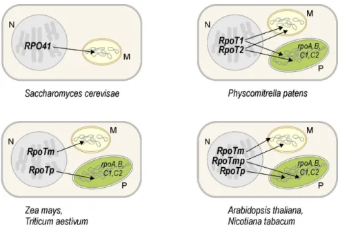
According to in vitro and in vivo import studies, a small family of three RpoT genes in Arabidopsis encodes a mitochondrial RNA polymerase (RpoTm), a plastidial enzyme (RpoTp) as well as a polypeptide imported into both mitochondria and plastids (RpoTmp; see Figure 4) (Hedtke, et al., 1997; Hedtke, et al., 2000; Hedtke, et al., 1999). Comparable
subcellular targeting of RpoT gene products is observed in Nicotiana species (Hedtke, et al., 2002; Kobayashi, et al., 2001). In contrast, monocots have so far been determined to harbour no more than two RpoT genes, of which one codes for a mitochondrial RNA polymerase and the other for a plastidial enzyme (Figure 4; Chang, et al., 1999; Ikeda and Gray, 1999). Dual targeting of RpoT gene products to both mitochondria and plastids being the result of two different translational starts has been reported for the two phage-type RNA polymerases genes RpoT1 and RpoT2 of Physcomitrella (Figure 4; Richter, et al., 2002). For both RpoT1 and RpoT2, translation initiation at the first of two in-frame AUG start codons was found to yield a polypeptide that is targeted to plastids, whereas initiation at the downstream AUG gave rise to a mitochondrial protein (Richter, et al., 2002). In vivo translation initiation at the first AUG and a plastidial localization of both RpoT1 and RpoT2 in Physcomitrella and of RpoTmp in Arabidopsis have recently been questioned by (Kabeya and Sato, 2005) and await further experimental proof.
Figure 4: Nuclear genes encoding organellar phage-type RNA polymerases. Genes in the nucleus (N, grey) of eukaryotic organisms code for T7 phage-like RNA polymerases which, following their synthesis in the cytoplasm, are imported into mitochondria (M, yellow) and plastids (P, green).
The small RpoT gene families found in Physcomitrella and in higher plants appear to be the result of independent gene duplication events dating after the separation of bryophytes from the vascular plant lineage (Richter, et al., 2002). Phylogenetic analyses have shown all higher plant RpoTp enzymes to form a sister clade to the group of RpoTm and RpoTmp enzymes (Richter, et al., 2002). Interestingly, preliminary phylogenetic analyses are indicative
of a closer relationship of monocot RpoTm to dicot RpoTmp than to dicot RpoTm (U.
Richter, HU Berlin, personal communication).
A DNA polymerase or reverse transcriptase has been proposed as predecessor of a single- subunit RNA polymerase that was the common ancestor of phage-encoded and nucleus- encoded phage-type RNA polymerases (Cermakian, et al., 1997). The ancestral single-subunit RNA polymerase gene has moreover been suggested to have originated at essentially the same time as the mitochondriate eukaryotic cell (Cermakian, et al., 1997). The discovery of cryptic prophages related to T3/T7 bacteriophages in several genomes of proteobacteria has inspired a modified scenario: A prophage that encoded among other phage proteins a T3/T7-like RNA polymerase existed in the bacterial endosymbiont being the predecessor of the mitochondrion (Filee and Forterre, 2005). Transfer of the prophage genes to the nucleus may have resulted in the reactivation of these formerly silent genes (Filee and Forterre, 2005).
I.3.2.2 Roles of RpoT enzymes
Distinct functions of RpoTm and RpoTmp in mitochondria and of RpoTp and RpoTmp in plastids of dicots are yet to be assigned. The RpoTm and RpoTmp genes in Arabidopsis have been reported to display overlapping expression patterns in different tissues and at different developmental stages (Emanuel, et al., 2005). Emanuel et al. (2005) therefore suggested RpoTm and RpoTmp to recognize different types of mitochondrial promoters. A contrasting picture of RpoTm and RpoTmp functions has been stimulated by studies of a transgenic Arabidopsis line carrying a T-DNA insertion in the RpoTmp gene (Baba, et al., 2004).
Transgenic plants displayed no apparent alterations compared to wild-type individuals in mitochondrial transcript accumulation. Based predominantly on the observation that in the mutant, the induction of several plastid genes in dark-grown seedlings upon illumination was delayed, Baba et al. (2004) proposed RpoTmp to be the key RNA polymerase transcribing organellar genes during early seedling development and favoured a role of both RpoTm and RpoTp at a later developmental stage.
Only indirect evidence has been provided that the mitochondrial phage-type RNA polymerases encoded by RpoT genes in higher plants have a role in transcription of mitochondrial genes. No sequences that might encode potential mitochondrial RNA polymerases of known enzyme structure have been traced in the fully sequenced Arabidopsis genome besides the previously characterized genes RpoTm and RpoTmp (Hedtke, et al., 1997;
Hedtke, et al., 2000; The Arabidopsis Genome Initiative, 2000). The conservation of functionally critical amino acid positions of the T7 enzyme (McAllister and Raskin, 1993;
Sousa, et al., 1993) in RpoTm and RpoTmp as well as in other plant RpoT enzymes argues for their transcriptional function (Hess and Börner, 1999; see I.3.2.3 and Figure 5). Moreover, recombinant RpoTm and RpoTmp were shown to non-specifically transcribe DNA in vitro (Hedtke, et al., 2000; Kühn, 2001).
The mitochondrial RNA polymerase functions as primase for mtDNA replication in humans and presumably also in yeast (reviewed in Shadel and Clayton, 1997; Tracy and Stern, 1995). A similar role of phage-type RNA polymerases and association of origins of replication with mitochondrial transcription start sites in plants remain to be demonstrated.
I.3.2.3 Structure of bacteriophage and phage-type RNA polymerases
The RNA polymerase of the T7 bacteriophage is a 99-kDa single-polypeptide-chain enzyme that is able to recognize specific promoter sequences, correctly initiate transcription and catalyze transcript elongation (reviewed in Steitz, 2004). Although the phage-type RNA polymerases of eukaryotic organisms most probably require auxiliary proteins to initiate transcription at organellar promoters (see I.3.3), the thoroughly studied T7 enzyme commonly serves as a model for both bacteriophage and eukaryotic phage-type RNA polymerases.
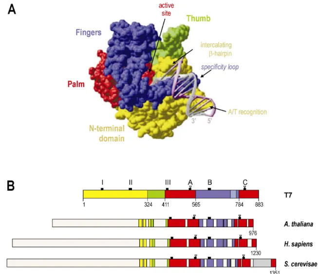
Comparisons between amino acid sequences of phage- and nucleus-encoded enzymes have identified conserved domains comprising identical or conservatively substituted positions (Chang, et al., 1999; Hedtke, 1998). Catalytically relevant structures of the T7 RNA polymerase are formed through amino acids of the C-terminal half of the polypeptide (McAllister and Raskin, 1993; Sousa, et al., 1993). Crystal structures of the T7 RNA polymerase have revealed this portion of the protein to fold into the “fingers”, “palm” and
“thumb” subdomains (Jeruzalmi and Steitz, 1998; Sousa, et al., 1993; see Figure 5) that are typical to members of a superfamily of nucleic acid polymerases also including certain DNA polymerases and reverse transcriptases (reviewed in Sousa, 1996). The similarity of plant organellar RNA polymerases to the T7 enzyme is most apparent for sequence regions corresponding to palm and fingers, which include all essential residues that participate in catalysing RNA synthesis (Figure 5; Hess and Börner, 1999), and references therein). In contrast, elements contributing to promoter recognition by phage RNA polymerases are poorly conserved in plant phage-type transcriptases, possibly reflecting the divergence in architecture between promoters recognized by the two groups of enzymes, as well as different compositions of initiating RNA polymerase complexes (Chang, et al., 1999; Ikeda and Gray, 1999; Jeruzalmi and Steitz, 1998; see I.3.3). Based on structural modelling, Yeast Rpo41 has been suggested to possess two regions that correspond in structure, yet not in sequence, to the
“specificity loop” and intercalating β-hairpin involved in promoter recognition and DNA melting by T7 RNA polymerase (Matsunaga and Jaehning, 2004). In the T7 RNA polymerase-promoter complex, an antiparallel β-hairpin referred to as specificity loop specifically interacts via hydrogen-bonding with bases of the promoter sequence (Cheetham, et al., 1999; Rong, et al., 1998). The intercalating β-hairpin, which is part of the N-terminal domain of the T7 enzyme, facilitates melting of the promoter duplex (Cheetham, et al., 1999).
Rpo41 was recently shown to accurately initiate promoter-specific transcription in vitro from supercoiled and pre-melted DNA templates in the absence of the obligatory yeast mitochondrial transcription factor sc-mtTFB, indicating that promoter specificity determinants indeed reside in the Rpo41 polypeptide rather than in sc-mtTFB (Matsunaga and Jaehning, 2004, see I.3.3.1).
Differences in size between nucleus-encoded phage-type RNA polymerases are primarily accounted for by varying N-terminal extensions. Including transit peptides, T7-like transcriptases of plants are proteins of around 110 kDa, whereas the yeast and human mitochondrial RNA polymerases are 145 and 130 kDa in size (Hess and Börner, 1999), and references therein). The N-terminal extension of yeast Rpo41 and a C-terminal insertion found only in the yeast enzyme are required for stable mtDNA maintenance and represent independent functional domains that may have been acquired through gene fusion events (Lisowsky, et al., 2002; Wang and Shadel, 1999). In heterologous complementation experiments, Arabidopsis RpoTm and various Rpo41/RpoTm chimeras were unable to functionally substitute for Rpo41 in vivo (Lisowsky, et al., 2002).
A number of separate crystal structures have depicted T7 RNA polymerase at different stages from promoter binding to elongation (Cheetham, et al., 1999; Cheetham and Steitz, 1999; Tahirov, et al., 2002; Yin and Steitz, 2002; Yin and Steitz, 2004). Following promoter rcognition, duplex DNA opening and repeated abortive initiation attempts, a major conformational change in the N-terminal domain removes the promoter-binding site and creates a tunnel for the transcript to pass through during the elongation phase in which the enzyme completes the RNA product processively without dissociation until termination (Steitz, 2004). Transcription termination signals recognized by the T7 RNA polymerase have been characterized (He, et al., 1998; Lyakhov, et al., 1998; Macdonald, et al., 1994), whereas no such sequences have been described in organelles. Mitochondrial transcripts in plants are instead considered to be terminated through the action of nucleases that define RNA 3’ ends (Dombrowski, et al., 1997; Perrin, et al., 2004).
Figure 5: Conserved sequence regions of the T7 phage and eukaryotic phage-type RNA polymerases.
(A) Surface representation of the T7 RNA polymerase-promoter complex structure specifying the N-terminal domain (yellow) and the RNA polymerase subdomains “thumb” (green), “palm” (red) and “fingers” (blue; from (Cheetham, et al., 1999). (B) Amino acid sequence organization of T7 RNA polymerase domains (colours as in Figure 5A) and homologous amino acid sequence regions of organellar phage-type RNA polymerases, based on a sequence comparison derived in (Hedtke, 1998). Arabidopsis RpoTm is shown as one member of the group of mitochondrial and plastidial enzymes of land plants which do not greatly differ in their sequence organization and size of the N-terminus. Black squares mark the positions of the motifs T/DxxGR (III), A, B and C (according to (Delarue, et al., 1990) that are important for RNA polymerase function and conserved in all enzymes, and of a T7-specific β-hairpin (I) and a structure involved in recognition of A/T-rich promoter regions (II). Open triangles denote the invariant residues Asp537 and Asp812 acting as ligands to two catalytic Mg2+
ions at the RNA polymerase active site (Woody, et al., 1996). The region corresponding to the specificity loop of the fingers subdomain is depicted in light blue; a yeast-specific C-terminal insertion is shown in grey.
I.3.3 Mitochondrial transcription factors
Unlike the single-subunit RNA polymerases of bacteriophages, mitochondrial phage-type RNA polymerases require auxiliary factors to initiate transcription at promoter sequences. To date, mitochondrial transcription factors have been characterized in S. cerevisiae (e.g.
(Matsunaga and Jaehning, 2004; Schinkel, et al., 1987; Winkley, et al., 1985), X. laevis (Bogenhagen, 1996; Bogenhagen and Insdorf, 1988), Drosophila melanogaster (Matsushima, et al., 2005; Matsushima, et al., 2004), humans (Falkenberg, et al., 2002; Fisher and Clayton,
1988; McCulloch, et al., 2002) and mouse (Gaspari, et al., 2004). While no such proteins have yet been isolated from mitochondria of photosynthetic eukaryotes, a number of studies suggest transcriptional cofactors to function in mitochondrial transcription in plants. An essential role of promoter-specific cofactors in plant mitochondria gained support from analyses of mitochondrial transcription in maize (Young and Lonsdale, 1997). A nucleus- encoded factor has moreover been suggested to be involved in transcription in Z. perennis mitochondria where initiation at the cox2 promoter cpc was found to depend on the presence of the dominant allele of the nuclear MCT gene (Newton, et al., 1995). Attempts to fractionate transcriptionally active mitochondrial extracts resulted in the loss of promoter specificity, indicating that several loosely associated proteins may contribute to promoter recognition (Binder and Brennicke, 2003). Searches for cofactors of mitochondrial phage-type RNA polymerases in higher plants are not unlikely to identify proteins with similarity to mitochondrial transcription factors in yeast and animals, which complement the same type of enzyme (compare I.3.2).
I.3.3.1 Yeast and animal mtTFB
In addition to the core RNA polymerase Rpo41, transcription initiation at mitochondrial promoters in S. cerevisiae requires a single accessory protein of 43 kDa first described as Mtf1 (Figure 6; Lisowsky and Michaelis, 1988; Schinkel, et al., 1987) and also referred to as sc-mtTFB (Shadel and Clayton, 1993). While neither Rpo41 nor sc-mtTFB was able on its own to specifically interact with promoter sequences in gel mobility shift assays (Schinkel, et al., 1988), the non-specifically transcribing core was found to recognize mitochondrial promoters on a linear DNA template when complemented with sc-mtTFB in in vitro transcription experiments (Schinkel, et al., 1987). The latter was therefore considered the specificity factor of Rpo41 (Schinkel, et al., 1987). Functional studies of mutant variants of sc-mtTFB motivated the alternative view that specificity determinants of promoter recognition reside in the core enzyme, and that sc-mtTFB may play a role in unwinding DNA during transcription initiation (Shadel and Clayton, 1995). Intrinsic promoter specificity of the core RNA polymerase was later confirmed not only for yeast Rpo41 (Matsunaga and Jaehning, 2004) but in part also for the human and mouse core enzymes (Gaspari, et al., 2004). On supercoiled or premelted DNA templates, Rpo41 alone was able to accurately initiate transcription in vitro at mitochondrial promoters (Matsunaga and Jaehning, 2004).
While sc-mtTFB was obligatory for specific transcription of linear templates, addition of sc-mtTFB stimulated specific transcription from supercoiled DNA but inhibited escape into
productive elongation from a pre-melted promoter (Matsunaga and Jaehning, 2004). The authors concluded that sc-mtTFB facilitated DNA melting, but not promoter recognition, possibly by inducing and stabilizing structural changes in Rpo41 that enable the enzyme to open the double helix and form an open promoter complex.
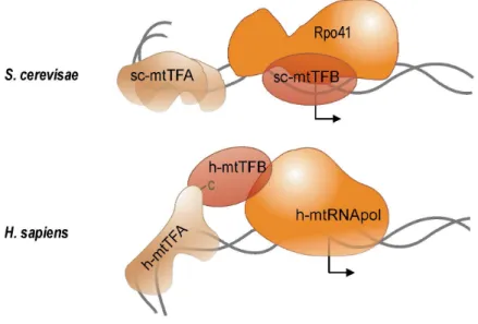
Figure 6: Components of the transcription machineries in yeast and human mitochondria. Yeast transcription initiation model after (Shadel and Clayton, 1993); transcription initiation in human mitochondria as proposed by McCulloch and Shadel (2003). See text for details on mtTFB and mtTFA functions and cofactor interactions with the mitochondrial phage-type RNA polymerases Rpo41 and h-mtRNApol. Cofactor designations adhere to the nomenclature suggested by Shadel and Clayton (1993). mtTFA-induced DNA bending is indicated; bent arrows mark transcriptional starts.
Analyses of the RNA polymerase composition before, during and shortly after transcription initiation indicated that the Rpo41 core and sc-mtTFB form a holoenzyme in solution prior to DNA binding and promoter recognition (Mangus, et al., 1994). Upon binding to the promoter, the RNA polymerase holoenzyme induces a bend in the DNA, which has been shown to enhance promoter activity in vitro (Schinkel, et al., 1988). Following transcription initiation, the factor dissociates from the catalytic subunit and is available for subsequent initiation events (Mangus, et al., 1994). The observation that sc-mtTFB behaved like bacterial sigma factors during transcription initiation stimulated a series of experiments investigating functional and structural similarities between sigma factors and sc-mtTFB (Cliften, et al., 2000; Cliften, et al., 1997; Jang and Jaehning, 1991). However, resolution of the three-dimensional structure of sc-mtTFB later revealed this mitochondrial transcription factor to structurally resemble S-adenosyl-L-methionine (SAM)-dependent rRNA dimethylases such as the Bacillus subtilis rRNA methyltransferase ErmC’ (Schubot, et al., 2001).
Sequence similarity, albeit limited, of mitochondrial transcription factors to rRNA dimethylases became apparent following the identification of two sequences encoding mitochondrial sc-mtTFB-like factors in humans (Falkenberg, et al., 2002; McCulloch, et al., 2002). Homologues to human mtTFB1 and mtTFB2 (referred to as h-mtTFB1 and h-mtTFB2) where subsequently detected in mouse (Rantanen, et al., 2003) and Drosophila (Matsushima, et al., 2005; Matsushima, et al., 2004). Both h-mtTFB1 and h-mtTFB2 were found to form a stable heterodimer with human mitochondrial RNA polymerase and to induce transcription initiation at the human mtDNA promoters LSP and HSP in vitro in the presence of h-mtTFA (Figure 6; Falkenberg, et al., 2002), an additional essential cofactor of human mitochondrial transcription (see I.3.3.2).
Despite poor overall sequence similarity of sc-mtTFB to ErmC’, there is extensive structural agreement between the two proteins around the SAM-binding site, and amino acid residues involved in SAM binding are conserved in sc-mtTFB (Schubot, et al., 2001).
Therefore, Schubot et al. (2001) suggested that sc-mtTFB could bind SAM and methylate the nascent RNA chain in vivo. Alternatively, an RNA-modifying enzyme may have evolved to function solely as transcription factor. While no methyltransferase activity of sc-mtTFB or h-mtTFB2 has been demonstrated so far, h-mtTFB1 has been reported to bind SAM and to substitute for the Escherichia coli rRNA dimethylase KsgA in methylating the 16S rRNA at a conserved stem-loop (McCulloch, et al., 2002; Seidel-Rogol, et al., 2003). The homologous 12S rRNA in human mitochondria was moreover shown to be similarly modified at this site (Seidel-Rogol, et al., 2003). Analyses of transcriptionally competent h-mtTFB1 variants carrying point mutations in conserved methyltransferase motifs indicated that the ability of the protein to function as transcription factor in vitro is independent of SAM binding and methyltransferase activity (McCulloch and Shadel, 2003). The authors therefore concluded h-mtTFB1 to be a bifunctional protein with two separable activities.
I.3.3.2 Yeast and animal mtTFA
In order to accurately and efficiently initiate transcription at the LSP and HSP promoters of the human mtDNA, the heterodimer composed of h-mtTFB and core RNA polymerase depends on the presence of the cofactor h-mtTFA (Falkenberg, et al., 2002; Fisher and Clayton, 1985; Fisher and Clayton, 1988). h-mtTFA is a protein of 25 kDa that comprises two HMG boxes in tandem (Parisi and Clayton, 1991). HMG boxes have been characterized as functional domains of DNA-binding proteins and interact with the minor grove of the double helix primarily at sites of unusual DNA conformation, where they induce dramatic bending
and structural deformation of the DNA (Antoshechkin, et al., 1997; Giese, et al., 1997;
Grosschedl, et al., 1994; Love, et al., 1995). h-mtTFA specifically binds to sequences upstream of the transcription initiation sites, which have been characterized as distal promoter elements (Fisher, et al., 1987; Gaspari, et al., 2004). Specific promoter binding by h-mtTFA as well as exact spacing between distal promoter elements and transcription start sites were shown to be crucial for transcription activation (Dairaghi, et al., 1995).
A h-mtTFA homologue is abundantly found in mitochondria of S. cerevisiae (Diffley and Stillman, 1991). sc-mtTFA is dispensable for transcription initiation at promoters of the yeast mtDNA (Xu and Clayton, 1992) and instead appears to play a major role in structural organization and stable maintenance of the mtDNA (Diffley and Stillman, 1991). Like h-mtTFA, sc-mtTFA is able to specifically associate with regulatory sequences of the mtDNA and also non-specifically bind DNA, and mediates condensation and unwinding as well as bending of the double helix (Diffley and Stillman, 1992; Fisher, et al., 1992). Transcription initiation by the RNA polymerase holoenzyme is slightly stimulated by sc-mtTFA (Parisi, et al., 1993). Presumably, binding of sc-mtTFA to the mtDNA leads to a favourable exposition of cis-regulatory elements to the transcription apparatus (Diffley and Stillman, 1992).
The capacity of h-mtTFA to act as efficient and promoter sequence-specific transcriptional activator has been attributed to a C-terminal extension of the factor and a linker peptide between the two HMG boxes, which are both lacking in sc-mtTFA (Dairaghi, et al., 1995).
Transfer of the C-terminal tail of h-mtTFA onto the yeast factor was shown to turn sc-mtTFA into a transcription factor activating the human mtDNA promoter LSP (Dairaghi, et al., 1995).
Thus, h-mtTFA function appears to have evolved through the acquisition of novel structural domains. The C-terminal tail of h-mtTFA has been proposed to interact with h-mtTFB, thereby positioning the heterodimer composed of h-mtTFB and core RNA polymerase at the transcription initiation site demarcated by a specific h-mtTFA/promoter complex (Figure 6, McCulloch and Shadel, 2003). Corroborating this model, promoter selectivity of the mouse and human transcription machineries has been dissected into binding of mtTFA to distal promoter elements and specificity of the core enzyme for nucleotides proximal to the transcription initiation site (Gaspari, et al., 2004).
I.3.3.3 Mitochondrial transcription factors in plants
To date, no mtTFA or mtTFB homologues have been isolated from plant mitochondria, and the function of such proteins in plant organelles is unclear. Attempts to detect HMG box proteins in maize mitochondrial extracts using a sc-mtTFA antibody did not identify mtTFA
candidates (Andrea T. Descheneau, University of Missouri, Columbia, USA; personal communication). Moreover, application of a protocol that had been successfully employed for the preparation of mtTFA from yeast and human mitochondria failed to purify homologous proteins from pea mitochondria (Däschner, et al., 2001; Hatzack, et al., 1998).
A biochemical approach directed at isolating transcription factors from wheat mitochondria lead to the identification of p63, a 63-kDa protein described to specifically bind to the yeast cox2 promoter (Ikeda and Gray, 1999). Addition of recombinant p63 to a transcriptionally active extract prepared from wheat mitochondria, which per se accurately initiated transcription at the cox2 promoter, appeared to stimulate transcription from this promoter in vitro (Ikeda and Gray, 1999). The authors therefore suggested p63 to play a role in mitochondrial transcription and moreover pointed out a limited amino acid sequence similarity of p63 to sc-mtTFB. However, p63 later emerged to be a member of the large family of organellar PPR proteins and may rather be involved in posttranscriptional processes (Lurin, et al., 2004; Small and Peeters, 2000; J. Gualberto, IBMP CNRS, Strasbourg, France, personal communication).
I.3.3.4 Cofactors of phage-type RNA polymerases in plastids
The homology of nucleus-encoded plastid and mitochondrial RNA polymerases in plants and the presence of yet another phage-type RNA polymerase in both mitochondria and plastids in dicotyledonous plants (Chang, et al., 1999; Hedtke, et al., 1997; Hedtke, et al., 2000; Hedtke, et al., 2002; Ikeda and Gray, 1999), as well as the similarity of promoters that are recognized by these enzymes (Weihe and Börner, 1999) raise the question whether the two organelles harbour similar transcriptional cofactors interacting with these core RNA polymerases. The characterization of auxiliary factors in the plastid may well aid the identification of such proteins in the mitochondrion. CDF2, a DNA-binding factor isolated from spinach chloroplast, has been reported to stimulate transcription of the rrn operon by a nucleus-encoded RNA polymerase activity (Bligny, et al., 2000). However, structural details of CDF2 have hitherto escaped revelation (Bligny, et al., 2000), and no CDF2-like activity has been purified so far from plant mitochondria. Out of six nucleus-encoded sigma factors imported into Arabidopsis plastids, the protein designated Sig2 was found to additionally localize to mitochondria in GFP import assays (Tandara, 2000). Moreover, the maize orthologue Sig2B was determined by immunoblot analyses to co-purify with both mitochondria and plastids (Beardslee, et al., 2002). Yet, available experimental data so far relate Sig2 function to the bacterial-type plastidial RNA polymerase rather than phage-type
enzymes in mitochondria or plastids (Beardslee, et al., 2002; Kanamaru and Tanaka, 2004, and references therein).
A regulatory role has recently been deduced of the plastid-encoded tRNAGlu in plastidial transcription (Hanaoka, et al., 2005). Recombinant RpoTp specifically bound to the tRNA molecule in gel mobility shift experiments (Hanaoka, et al., 2005). Moreover, transcription from the plastidial accD promoter, which is considered to be catalyzed by a phage-type RNA polymerase, was shown to be inhibited by the addition of tRNAGlu to in vitro transcription reactions using proplastid extracts from Arabidopsis as source of transcription activity (Hanaoka, et al., 2005). The authors suggested tRNAGlu to mediate a switch in RNA polymerase utilization from nucleus-encoded RNA polymerases to the plastid-encoded bacterial-type enzyme during chloroplast development.
I.3.4 Regulation of mitochondrial gene expression at the transcriptional level
Coordinated expression of the nuclear and mitochondrial genomes in individual cells and tissues is required for the assembly of functional mitochondria and ensures appropriate metabolic activities of the organelle in response to environmental stimuli. Substantial progress has been made towards understanding nuclear-mitochondrial interaction in yeast (reviewed in Poyton and McEwen, 1996) and illuminating the signalling pathways between the nucleus and the chloroplast (reviewed in Gray, et al., 2003; Leon, et al., 1998; Pfannschmidt and Liere, 2005; Rodermel, 2001). Besides, models have been proposed that describe transcriptional changes occurring in the chloroplast during organelle biogenesis (Cahoon, et al., 2004; Hanaoka, et al., 2005). In higher plants, retrograde signals between the mitochondrion and the nucleus are established (Millar, et al., 2004); yet only a limited number of studies have addressed the question at what levels mitochondrial gene expression may be controlled.
Transcriptional modulation appears to represent a minor means of regulating gene expression in plant mitochondria (Binder and Brennicke, 2003; Mackenzie and McIntosh, 1999), although tissue-specific differences have been observed in transcript levels for particular mitochondrial genes, e.g. in in situ hybridization studies examining various mitochondrial transcripts in maize seedlings (Li, et al., 1996). Similar studies have substantiated a cell-specific regulation of mitochondrial gene expression during sunflower anther development (Smart, et al., 1994). Evidence that such differences are largely the result of posttranscriptional regulation of RNA abundance has been provided by a study that compared transcriptional activities and steady-state RNA levels of mitochondrial genes in
Arabidopsis (Giegé, et al., 2000). While run-on transcription values were found to diverge significantly between genes encoding different subunits of the same protein complex, such differences were less manifest for steady-state RNA levels. Contrary to maize mitochondria where ribosomal RNAs were found to be synthesized at higher rates than other mitochondrial transcripts, thereby accounting for the major contribution of rRNAs to the whole of mitochondrial RNAs (Finnegan and Brown, 1990), the high rRNA levels in Arabidopsis mitochondria were determined to be primarily due to high rRNA stability (Giegé, et al., 2000). In both maize and Arabidopsis, steady-state abundance of protein-coding mRNAs does not correlate with transcriptional processes, emphasizing the importance of posttranscriptional steps in modulating transcript accumulation (Giegé, et al., 2000; Mulligan, et al., 1991).
Posttranscriptional processes have also been suggested to be responsible for an observed elevation of mitochondrial transcript levels induced by impaired chloroplast activity in the barley albostrians mutant (Hedtke, et al., 1999).
The coordinated expression of the mitochondrial and nuclear genome has been investigated comprehensively in a study employing an Arabidopsis cell culture system to modulate mitochondrial biogenesis in response to sugar starvation and refeeding (Giegé, et al., 2005). A comparison of transcript and protein changes during modulation of sugar supply revealed the mitochondrial genome to be insensitive to sugar starvation stress, whereas changes were observed in the expression of nuclear genes encoding mitochondrial components.
Coordination of the expression of mitochondrial and nuclear genes was found to occur at the protein level, possibly during protein-complex assembly (Giegé, et al., 2005).
Limited data support a regulation of mitochondrial gene expression at the transcriptional level. Promoter selection has been reported to be controlled by nucleus-encoded factors in alloplasmic lines of Nicotiana and maize (Edqvist and Bergman, 2002; Newton, et al., 1995);
these factors and their possible roles in regulating mitochondrial function remain to be identified.
I.4 Aims of this study
Although it is generally accepted that RpoT gene products represent catalytic subunits of the mitochondrial transcription machinery in plants, direct evidence is as yet lacking for RpoT enzymes being involved in the transcription of mitochondrial genes in photosynthetic eukaryotes. Attempts to isolate mitochondrial RNA polymerases, including accessory factors, from plants failed to result in the identification of these components.
The present study aims at reconstituting mitochondrial transcription in vitro from recombinant RpoT enzymes, thereby establishing a role of these RNA polymerases in mitochondrial transcription. The nuclear genes RpoTm and RpoTmp encoding phage-type RNA polymerases that are imported into mitochondria have previously been identified in Arabidopsis. Distinct mitochondrial functions of RpoTm and RpoTmp, the latter of which is also imported into plastids, have so far not been specified. Recombinant Arabidopsis RpoTm and RpoTmp will therefore be examined for possible differences in their transcriptional performances in vitro.
Specific transcription initiation by RpoTm or RpoTmp at mitochondrial promoters is likely to require auxiliary factors, which are as yet unknown. Availability of the complete genome sequence of Arabidopsis renders this plant an excellent object of in silico analyses directed at identifying candidate transcription factors, based on their similarity to known essential mitochondrial transcription factors in yeast and animals. The subcellular localization and functional properties of these Arabidopsis proteins will be analyzed and compared to those of related yeast and animal factors. Most importantly, in vitro transcription experiments will attempt to elucidate if these proteins likewise act as auxiliary factors in mitochondrial transcription.
A prerequisite for studying the mitochondrial transcription machinery is the knowledge of mitochondrial promoters. Sequence motifs identified as elements of mitochondrial promoters in various dicotyledonous species are seen upstream of the coding regions of several mitochondrial genes in Arabidopsis; yet experimental evidence is limited to a single promoter. To define cis-elements that direct transcription of the Arabidopsis mitochondrial genome, transcription initiation sites will be mapped and their surrounding sequences, which comprise the promoter, will be analyzed. Experimentally determined promoters will provide a variety of templates for the Arabidopsis in vitro transcription system.