Analysis of the full-length WOX4 promoter activity in Arabidopsis thaliana
Inaugural-Dissertation zur
Erlangung des Doktorgrades
der Mathematisch-Naturwissenschaftlichen Fakultät der Universität zu Köln
Vorgelegt von Satish Kumar Eeda Aus Nandanam (India)
Köln - 2019
2
Berichterstatter: Prof. Dr. Wolfgang Werr
Prof. Dr. Peter Westhoff
Prüfungsvorsitzender: Prof. Dr. Ute Höcker
Tag der mündlichen Prüfung: February 2020
3 Table of Contents
I. SUMMARY... 5
II. ZUSAMMENFASSUNG ... 7
1. INTRODUCTION ... 9
1.1. The Plant vascular system – structure and function ... 9
1.2. Development of the vascular system in Arabidopsis thaliana... 10
1.2.1. Initiation of provascular tissues during the embryogenesis ... 10
1.2.2. Development of the vascular system in post-embryonic tissues ... 12
1.3. Regulation of vascular development ... 18
2. AIM OF THE THESIS ... 22
3. MATERIALS AND METHODS ... 23
3.1. List of chemicals ... 23
3.2. Buffers... 24
3.3. Kits ... 24
3.4. Growth Media ... 24
3.5. Biological materials ... 25
3.5.1. Enzymes ... 25
3.5.2. Primers ... 25
3.5.3. Antibiotics ... 26
3.5.4. Cloning vectors ... 26
3.5.5. Bacterial strains ... 26
3.5.6. Plant material ... 26
3.6. Methods... 27
3.6.1. Phylogenetic Shadowing ... 27
3.6.2. Nucleic acid extraction & purification ... 28
3.6.3. RACE (Rapid amplification of cDNA ends) ... 28
3.6.4. Promoter constructs ... 28
3.6.5. Primer design ... 28
3.6.6. PCR Amplification... 29
3.6.7. Cloning ... 29
4
3.7. Transformation ... 30
3.8. Selection of the transformants ... 31
3.9. Confocal Imaging... 31
3.10. Image Processing ... 31
4. RESULTS ... 32
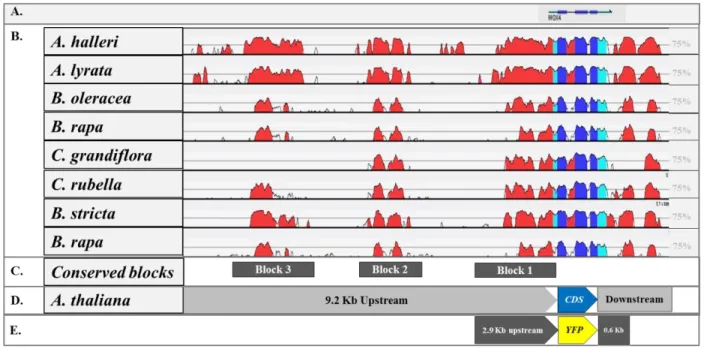
4.1. Phylogenetic shadowing revealed conserved regions within the WOX4 promoter ... 32
4.2. The proximal region of the WOX4 promoter contains a conserved open reading frame ... 35
4.3. The transcriptional start site of the WOX4 is located further upstream ... 36
4.4. The full-length WOX4 promoter construct includes all the conserved regions ... 37
4.5. Expression analysis of the full-length WOX4 promoter-reporters in stem ... 40
4.5.1. WOX4 promoter activity starts early during the shoot development ... 41
4.5.2. WOX4 promoter is active in a circular domain of subcortical cells beneath the IM .. 43
4.5.3. WOX4 promoter activity continues into the subtending nodes of the stem ... 44
4.5.4. WOX4 expression domain continued to exist throughout the developing stem ... 45
4.5.5. The circular WOX4 expression domain persists in the stem during secondary growth ... 47
4.6. Expression analysis of the full-length WOX4 promoter-reporter in the root ... 48
4.7. Full-length WOX4 promoter is active during the leaf development ... 51
5. DISCUSSION ... 55
5.1. The conserved distal elements are essential for WOX4 promoter activity ... 55
5.2. WOX4 promoter activity starts in the shoot apex and continues as a circular domain ... 58
5.3. The circular WOX4 expression domain prepatterns the vasculature ... 60
5.4. WOX4 activity starts in the RAM and continues into the vascular system ... 63
5.5. The WOX4 promoter marks novel expression domains in the leaf ... 64
6. REFERENCES ... 66
III. ACKNOWLEDGMENTS ... 73
IV. ERKLÄRUNG ... 74
V. CURRICULUM VITAE ... 75
5 I. SUMMARY
The understanding of vascular development in plants has been advanced rapidly in the last decades.
Nevertheless, there are still many details to be elucidated about the early stages of vascular differentiation, which requires easily identifiable marker genes. The WUSCHEL-related homeobox 4 (WOX4) is a member of the WOX gene family (Eric van der Graaff, 2009) and it has been demonstrated that WOX4 is involved in cambial stem cell maintenance (Suer et al., 2011).
Existing WOX4 expression analyses in Arabidopsis thaliana were conducted with a short WOX4 promoter-reporter construct, which contains 2.9 Kb of the 5′ flanking region and 0.6 Kb of the 3′
flanking region (Ji et al., 2010; Y Hirakawa et al., 2010; Suer et al., 2011; Shi et al., 2019). These studies describe a cambial cell specific expression pattern of WOX4 promoter in the root, shoot, cotyledons and leaves (Y Hirakawa et al., 2010). In upper part of the inflorescence stem, WOX4 activity was confined only to fascicular cambium, but at the stem-base its activity extends into the interfascicular region and forms a circular expression domain and this pattern was implied to be responsible for the radial outgrowth of the stem (Suer et al., 2011).
However, by detailed sequence analyses in this study we demonstrate that the WOX4 sequence is spatially separated by long intergenic sequences, which contain several distal conserved regions.
Moreover, by comparing phylogenetic shadowing results with published ATAC-seq data (Frerichs et al., 2019) we show the positions of these conserved regions are in open chromatin configurations, suggesting a possible regulatory role of these areas in WOX4 expression pattern.
Hence, we have generated WOX4 promoter-reporter fusions, which contains 9.2 Kb upstream and 1.7 Kb downstream sequences from the WOX4 coding sequence and transferred into A. thaliana with the aim to find a full spectrum of WOX4 activity. Interestingly, the analyses of transgenic plants aligned with previously observed cambium cell specific WOX4 activity but additionally it marked novel WOX4 expression domains in the SAM, RAM, stem and leaves.
In the stem, the full-length WOX4 promoter activity starts in groups of cells of the inflorescence meristem (IM), possibly marking the provascular cells of emerging primordia. Approximately 80- 100 µm beneath the IM, the promoter activity was localized in a circular expression domain of subcortical region that prepatterns the vasculature of young stem. This circular WOX4 expression domain was found to exist throughout the inflorescence stem and intrusions in the circle marks
6 fascicular cambium and interfascicular mark strands. Our findings suggest that the full-length WOX4 promoter was active during the specification of provascular cells in the shoot apex, initiation of fascicular cambium in the young stem without losing competency in the interfascicular mark strands and then continuously active in the cambial ring of the matured stem. Additionally, its activity was also observed in the xylem parenchyma in different growth phases of the inflorescence stem. Similar to the shoot, the WOX4 promoter activity was also found to start in the RAM marking the QC and its adjacent meristematic cells. Then its activity was found to confined to the vascular system of root. In the leaf, the WOX4 promoter was active in the primary, secondary, tertiary and quaternary veins, marking the cambial cells of the complete leaf vascular system. Additionally, it was also active in the xylem parenchyma of leaf vascular bundles and the sub-epidermal cells in adaxial side of the leaf, marking the palisade parenchyma.
Taken together, our study indicates that the inclusion of distal conserved regions of the WOX4 promoter is essential to show the picture of WOX4 expression pattern in different organs of A.
thaliana. Hence, the full-length WOX4 promoter-reporter constructs analysed in this study could further be utilised to elucidate the gene regulatory networks that control vascular development.
However, the complete upstream region is too large to be used as a standard promoter, therefore further promoter dissection studies are needed to identify the cis-regulatory elements, which could then be used for engineering approaches.
7 II. ZUSAMMENFASSUNG
Das molekularbiologische Verständnis der Gefäßentwicklung bei Pflanzen ist in den letzten Jahrzehnten weit vorangeschritten. Nichtsdestotrotz gibt es immernoch Details der frühen Entwicklungsstadien der vaskulären Differenzierung, die noch nicht vollständig beschrieben sind und von einfach detektierbaren Markergenen profitieren würden. WUSCHEL-related homeobox 4 (WOX4) gehört zur WOX Genfamilie (Eric van der Graaff, 2009). Es wurde gezeigt, dass WOX4 an der Stammzell-Aufrechterhaltung im Kambium beteiligt ist (Suer et al., 2011).
Existierende WOX4 Genexpressionsanalysen in Arabidopsis thaliana wurden mit einem vergleichsweise kurzen WOX4 Promoter-Reporter Konstrukt, welches 2.9 Kb der 5′ flankierenden Region und 0.6 Kb der 3′ flankierenden Region umfasst, durchgeführt (Yuki Hirakawa et al., 2010;
Shi et al., 2019). Diese Studien berichten von einer Kambium-spezifischen Expression des WOX4 Promoters in Wurzeln, Spross, Kotelydonen und Blättern (Y Hirakawa et al., 2010). Im oberen Teil der Sprossachse der Infloreszenz beschränkte sich die WOX4 Aktivität auf das faszikuläre Kambium, wohingegen sich die Aktivität in der Basis der Sprossachse auf die interfaszikülare Region ausgeweitete und eine zirkuläre Domäne formte. Dieses Expressionsmuster könnte das radiale Wachstum der Sprossachse einleiten (Suer et al., 2011).
In einer detailierten Sequenzanalyse zeigen wir hier, dass die WOX4 Sequenz räumlich von langen, intergenischen Sequenzen abgegrenzt ist, welche im distalen Bereich konservierte Regionen enthalten. Darüberhinaus zeigen wir durch den Vergleich von phylogenetic shadowing- Ergebnissen mit publizierten ATAC-seq Daten (Frerichs et al., 2019), dass die Positionen der konservierten Regionen in offenen Chromatinbereichen liegen, was uns zu der Hypothese führt, dass sie eine wichtige regulatorische Rolle in der WOX4 Expression spielen könnten. Aus diesem Grund wurden in dieser Studie WOX4 Promoter-Reporter Fusionen generiert, die den 9.2 Kb upstream Bereich und den 1.7 Kb downstream Bereich der WOX4 kodierenden Sequenz umfassen.
Die Promoter-Reporter Konstrukte wurden in A. thaliana transferiert mit dem Ziel das volle Spektrum der WOX4 Aktivität zu erfassen. Interessanterweise stimmten die Analysen der transgenen Pflanzen mit der vorher beschriebenen Kambium-spezifischen WOX4 Aktivität überein, jedoch konnten zusätzlich markierte, neue WOX4 Expressionsdomänen im SAM, RAM, Sprossachse und Blätter identifiziert werden.
8 In der Sprossachse beginnt die Aktivität des WOX4 Volllänge-Promoters in Zellgruppen des Inflorezenzmeristems (IM) was möglicherweise provaskuläre Zellen der sich entwickelnden Primordien markiert. Circa 80-100 µm unterhalb des IMs zeigte sich eine zirkuläre Expressionsdomäne der subcorticalen Region, welche das Muster der Vaskulatur der jungen Sprossachse anzeigt. Diese zirkuläre WOX4 Expressionsdomäne zog sich durch die Sprossachse der gesamten Infloreszenz, wobei Einstülpungen des Kreisens das faszikuläre als auch Kambium markieren.
Unsere Ergebnisse deuten darauf hin, dass der WOX4 Volllänge-Promoter während der Spezifizierung der provaskulären Zellen in der Sprosspitze aktiv ist, während der Initiierung des faszikulären Kambiums in der jungen Sprossachse ohne seine Kompetenz im interfaszikulären Markstrahl zu verlieren aktiv bleibt und seine Aktivität dann auch kontinuierlich im Kambiumring der reifen Sprossachse zu finden ist. Zusätzlich konnte seine Aktivität im Xylemparenchym in verschiedenen Wachstumsphasen der Sprossachse der Infloreszenz beobachtet werden. Wie im Spross zeigte sich die WOX4 Promoteraktivität auch im RAM wo sie das QC und die angrenzenden meristematischen Zellen markierte. Außerdem beschränkte sich die Aktivität auf die Vaskulatur der Wurzel. Im Blatt befand sich die WOX4 Promoteraktivität in den primären, sekundären, tertiären und quartären Venen, wo sie die Kambiumzellen der kompletten Blattvaskulatur markierten. Zusätzlich konnte seine Aktivität in Xylemparenchymzellen der Bündelscheidenzellen und der subepidermalen Zellen der adaxialen Seite des Blattes lokalisiert werden, wo sie das Palisadenparenchym markierte.
Zusammengefasst zeigt unsere Studie, dass der Einbezug der distalen, konservierten Promoterregionen essentiell für die volle Entfaltung der WOX4 Expression in den verschiedenen Pflanzenorganen von A. thaliana ist. Die in dieser Studie analysierten Volllänge WOX4 Promoter- Reporter Konstrukte könnten weiterhin der Erforschung der regulatorischen Netzwerke, welche die Gefäßentwicklung in Pflanzen steuern, dienen. Allerdings eignet sich die vollständige Upstream-Region aufgrund ihrer Größe nicht zum Einsatz als Standardpromoter. Stattdessen wäre es sehr interessant detailierte Promoterstudien an dieser Upstream-Region durchzuführen, um kleinere cis-regulatorische Elemente zu identifizieren, die in Zukunft in biotechnologischen Verfahren an Pflanzen eingesetzt werden können.
9 1. INTRODUCTION
1.1. The Plant vascular system – structure and function
The plant vascular system is a network of specialized conductive tissues that are spread throughout the entire plant body, connecting all the plant organs with each other in an organized manner and it facilitates the transport of water, minerals, and sugars (Lucas et al., 2013). It is composed of two types of differentiated tissues, the xylem and the phloem; that are spatially separated by an undifferentiated tissue, the cambium (Nieminen et al., 2015). The xylem tissue consists of xylem parenchyma, xylem fibers, tracheids and vessel elements and phloem tissue is composed of phloem parenchyma, phloem fibers, sieve tube elements and companion cells (Jouannet et al., 2015).
The primary function of the cambium is to maintain its own stem cell population in the cambial zone, the differentiation of xylem and phloem tissues. The basic function of the xylem tissue is to transport water and mineral nutrients from roots to various parts of the shoot. The phloem tissue transports sugars that are produced in the photosynthetic organs to all other tissues of the plant body (Altamura et al., 2001). The bulk flow of water through the xylem tissue is a passive transport mechanism, which is driven by the osmotic potential difference of the root surface and the tension created in the xylem tissue by transpiration of water through the stomata (Myburg and Sederoff, 2001). As a consequence, the turgor pressure gradient is established in the adjacent phloem tissue that directs the sugar translocation from shoot to root. However, the sugar transport functions via an active transport mechanism that requires direct energy inputs in order to perform the phloem loading and unloading processes (Hölttä et al., 2009; Lucas et al., 2013). Moreover, the plant vascular system plays a major role in long-distance communication, enabling the translocation of phytohormones, RNA molecules, and signal peptides within the shoot and between the shoot and root. For example, in response to water deficit conditions, roots produce the phytohormone abscisic acid, which is then transported to leaves through the xylem, where it reduces the water loss by stomatal closure (Hartung et al., 2002). Another important long-distance communication is that of florigen, a protein encoded by the FLOWERING LOCUS T (FT) gene. It is expressed in the leaves and is then transported to the shoot apical meristem through the phloem, where it promotes the floral transition (Corbesier et al., 2007). Interestingly, CLAVATA3/EMBRYO SURROUNDING REGION peptides (CLE peptides) secreted by roots in response to the plant-
10 microbe interaction are transported to the shoot via the xylem, which then activates a systemic responses that regulate root nodulation (Okamoto et al., 2013; Notaguchi and Okamoto, 2015).
These few examples demonstrate the important role of the plant vascular system in the exchange of signals between plant organs serving either internally as developmental regulators or enable the plant to react quickly to environmental conditions like biotic and abiotic stresses (Lucas et al., 2013). In addition, the vascular system also provides mechanical support to the plant body and in tree species, it contributes to the development of wood through the secondary thickening growth (Ji et al., 2010; Etchells and Turner, 2016).
1.2. Development of the vascular system in Arabidopsis thaliana
The vascular system of Arabidopsis thaliana begins with the specification of vascular initials in the embryo that gives rise to the vascular system of cotyledons, embryonic root and hypocotyl (Figure 1a). After seed germination, the vascular system of the root and shoot develops from the root and shoot apical meristems respectively, and grows into a continuous network by the action of cambium(Turner and Sieburth, 2003). However, the structural arrangement of cambium and vascular tissues varies from organ to organ (Figure 1b). For example, in the root, the vascular tissues are arranged in the diarch symmetry, whereas the stem exhibits the collateral arrangement (Dengler, 2006). Moreover, anatomical differences also exist within the same organ; for example, the anatomy of the inflorescence stem at the apex is generally different from its base (Bowman and Floyd, 2007). In the following sections, the vascular development in Arabidopsis thaliana is described in detail with an emphasis on both embryonic and post-embryonic differences that occur in different organs.
1.2.1. Initiation of provascular tissues during the embryogenesis
During the embryogenesis, the zygote undergoes an asymmetric division that produces a small apical cell with a dense cytoplasm and a larger basal cell with a big vacuole. The apical cell lineage develops into most of the embryo, whereas the basal cell lineage produces the extra-embryonic suspensor (Barton and Poethig, 1993). First, the apical cell goes through two rounds of periclinal divisions to produce the four celled pro-embryo and then an anticlinal division resulting in an octant embryo. The four cells in the upper tier of the octant embryo give rise to the shoot including the shoot meristem, whereas the four cells in the lower-tier give rise to the hypocotyl and
11 hypophysis (Jürgens et al., 1995). Further periclinal divisions occurring in the octant result in an embryo with the specified cell fates, such as protoderm and ground meristem. The elongated cells in lower half of the early-globular embryo are specified as the vascular cells, which is the first sign of vascular tissue development (Jürgens, 1995; Scheres et al., 1995). Further periclinal cell divisions in the globular embryo give rise to the cotyledon primordia and the subsequent divisions in the late heart stage develop the shoot apical meristem in between the developing cotyledons (Boscá et al., 2011). Meanwhile, the simple anticlinal divisions that occurred in the basal cell give rise to the extra-embryonic suspensor. The upper cell of the suspensor develops into the hypophysis which forms the quiescent center of the root meristem and the root cap initials (Armenta-Medina and Gillmor, 2019). Numerous gene expression studies suggested that, although the provascular tissues were specified during the embryogenesis, the actual vascular differentiation marked by certain structural changes including the secondary cell wall formation occurs only after the seed germination (ten Hove et al., 2015).
Figure 1. Schematic representation of the vascular tissue development and its organization in different parts of the Arabidopsis thaliana plant body during embryogenesis (a) and postembryonic development (b) (Ruonala et al., 2017).
Provascular tissues are marked in the orange colour, whereas the xylem and phloem tissues are marked in blue and red colours respectively.
12 1.2.2. Development of the vascular system in post-embryonic tissues
The post-embryonic development of the vascular system in organs such as the hypocotyl and cotyledons emerges from vascular precursors that are prepatterned during embryogenesis whereas the vascular system of shoot organs (leaves, side-shoots, inflorescence stems, flowers and pedicels) and roots originates from the specified vascular precursor cells of the shoot apical meristem and root apical meristem, respectively (Lucas et al., 2013; De Rybel et al., 2015).
1.2.2.1. Vasculature of the root
During early stages of seed germination, the embryonic root or radicle starts protruding from the seed by breaking the seed coat and later develops into the primary root. During root development, three different developmental zones such as meristematic, elongation, and differentiation zones can be observed. These zones are in turn distinguished by the following factors: the external root morphology, the internal root anatomy, the structural and functional differences in the vascular tissues (Turner and Sieburth, 2003).
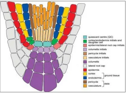
The meristematic zone contains a four-celled quiescent center (QC), which is surrounded by actively dividing meristematic cells (Verbelen et al., 2006). These cells are small in size, packed with cytoplasm, contain small or no vacuoles. They are enclosed by thin cell walls and their nuclei occupy a large area of the cell. The meristematic zone can be further divided into proximal and distal meristematic zones, based on the root anatomy. The proximal meristematic zone contains the stem cell niche formed by a centrally located QC and a layer of meristematic cells that are in direct contact with the QC (Bennett and Scheres, 2010). These meristematic cells are later specified as the mother cells that produce all other root tissues. The cells located below the QC serves as the columella-mother-cells, which differentiate into the columella cells. Similarly, the cells positioned above the QC are specified as the pericycle and vascular initials (Figure 2). The cells that are positioned peripherally to the QC, are specified as endodermis/cortex-mother cells and epidermal/root cap mother cells (Nieminen et al., 2015). These mother cells give rise to different tissues, which forms the distal end of the meristematic zone. The root anatomy at this stage shows the following cell layers - the outer epidermal layer, the middle cortex tissue, a layer of endodermis, and then a single layer of pericycle that encloses the central stele. The central stele is composed of protoxylem tissue at the center, accompanied by two poles of the phloem cells that
13 are located in close proximity to the pericycle, and procambial cells that are positioned between xylem and phloem (Masubelele et al., 2005).
Figure 2. Graphical representation of the organization of different cell types in the root apical meristem (Pérez et al., 2013). The quiescent center (QC) and other meristematic cells of the root apex are shown in differentt colours.
The elongation zone is positioned at an approximately 300-850 µm away from the QC and in this region cell divisions do not occur frequently, but the rapid growth is achieved by cell expansion (Verbelen et al., 2006). Several cytological changes occur in this zone such as enlargement of the vacuole that pushes the nucleus and cytoplasm towards the cell wall (Jing and Strader, 2019). The differentiation zone of the root starts in continuation with the elongation zone. Many anatomical changes occur in this zone with the rapid cell divisions gives rise to various vascular tissue types (De Rybel et al., 2015). Based on the gene expression studies and the differences in cell morphology, the pericycle of the differentiation zone is divided into xylem-pole-pericycle and phloem-pole-pericycle (Parizot et al., 2012). The lateral roots also originate from the specified cells of xylem pole associated pericycle (Jing and Strader, 2019). Inside the central stele of the differentiation root zone, the cambium tissue starts to differentiate into different types of metaxylem tissues such as the xylem vessels, xylem fibers, and the phloem tissues such as sieve elements and companion cells (Ruonala et al., 2017). The root that is positioned between the differentiation zone and the hypocotyl exhibits secondary growth, in which the central part of the root is occupied by the concentric rings of secondary xylem tissue, the cambial cell layer and the secondary phloem tissues (Fukuda and Ohashi-Ito, 2019).
14 1.2.2.2. Vasculature of the Stem
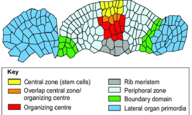
The Arabidopsis stem originates from the shoot apical meristem, which is divided into central zone, peripheral zone, organizing center, and rib zone (Mayer et al., 1998). The central zone contains slowly dividing stem cells that maintain the meristem integrity and also produces daughter cells to the sides and bottom. It is flanked by the peripheral zone, where actively dividing cells develop into lateral organs such as leaves and flowers. The stem cell population of SAM is maintained by the feedback mechanism operated between the central zone and the organizing center (Figure 3). The rib zone located below the organizing center takes part in the development of the vegetative/inflorescence stem (Bowman and Eshed, 2000). It is also believed that the rib zone is further divided into the peripheral-rib zone that produces the cortex and the central-rib zone that produce the pith parenchyma. The boundary cells between the peripheral-rib zone and central- rib zone produce the primary vasculature (Serrano-Mislata and Sablowski, 2018).
However, the continuation of vascular pattern formation in vegetative stem has been seen in two different views: according to the first view, the vasculature of emerging lateral organs are eventually connects to the existing vasculature of the stem, which is supported by the direction of auxin flow from tip of the leaf primordium towards the leaf base (Scarpella, Marcos, Jirí Friml, et al., 2006). In the second view, the existing vasculature of the basal stem extends into the emerging leaf primordia and this view is supported by the fact that the plant growth occurs in an acropetal manner (Turner and Sieburth, 2003). The second hypothesis of the plant vascular development is extensively studied in Arabidopsis during the rosette growth (Kang et al., 2003), in which the authors described the vasculature of the vegetative stem by combining the anatomical features with the expression pattern of provascular marker HOMEOBOX GENE 8 (ATHB8). According to the evidence presented in their report, the vasculature tissue of cotyledons and first four leaves are in direct contact with the central stele of the hypocotyl, suggesting that the primary vasculature develops as a continuation of the embryonic vascular pattern. Whereas, from the fifth leaf onwards the vasculature of each leaf is supplied by a single trace that is derived from two of the eight vascular bundles (or sympodia) of the vegetative stem. It was also found that each leaf vasculature can be traced back to another leaf; for example, the trace to leaf 5 derives from the base of leaf 2 trace (n+3 parastichy), while the traces of leaf number 6, 7 and 8 derives from their n+5 parastichy (Kang et al., 2003). These studies demonstrate that the vascular system of vegetative rosette is
15 composed of approximately eight vascular bundles that form a reticulate pattern by the multiple connections among the different leaves and also with the vascular bundles of the stem (Dengler, 2006).
Figure 3. Schematic representation of the different zones in the shoot apical meristem (Gaillochet and Lohmann, 2015). Different functional zones in the SAM are shown in different colours – Central zone in yellow, organizing center in red, overlapping zone in orange, lateral organs in blue, peripheral zone in the cyan, boundary domain in green and the rib meristem in green colours.
During the vegetative to reproductive transition, the vegetative shoot apical meristem transforms into an inflorescence meristem (IM), which later produces the cauline leaves, flower primordia, lateral branches, nodes and internodes of the inflorescence stem (Lucas et al., 2013). Due to the compressed nature and the difficulties associated with imaging the vegetative stem, the inflorescence stem was used more frequently than the vegetative stem to study stem growth. The structural changes which occur during inflorescence stem growth in Arabidopsis and wood formation in tree species are found to be identical, which has driven researchers to use the Arabidopsis inflorescence stem to study radial outgrowth (Ragni and Greb, 2018).
In general, the development of the inflorescence stem has been divided into primary, intermediary and secondary growth depending on the anatomical variations occurring in the inflorescence stem over the period of plant growth (Tonn and Greb, 2017). The primary growth phase of an inflorescence stem is studied by using either the young inflorescence stem or the matured stem segment located beneath the shoot apical meristem. Both are composed of outer epidermis, middle cortex, vascular bundles, and central pith (Campbell et al., 2016). Similar to the vegetative stem, the young inflorescence stem also possesses approximately eight to ten vascular bundles that are arranged in a circular manner (eustele). Each bundle contains the xylem tissue towards central pith,
16 the phloem tissue towards the cortex and the fascicular cambium positioned in between the xylem and phloem tissues (Figure 1b; Suer et al., 2011). The vascular bundles are separated by an interfascicular region. In the young inflorescence stem, the interfascicular region contains only parenchyma tissue, whereas in the mature stem it has lignified fibers (Altamura et al., 2001). The molecular basis for the existence of a parenchymatic interfascicular region in the young Arabidopsis inflorescence stem is poorly understood so far, because the region is not marked by any well-known vascular or non-vascular markers, therefore it is also not clear whether this region develops from the cambium or cortex or pith.
The inflorescence stem that is located in between the shoot apex and the stem base has been attributed to study of the intermediate growth phase of the stem (Sanchez et al., 2012). The anatomy of the stem undergoing the intermediate growth shows an outer epidermis, the cortex with multiple layers, the enlarged pith, and well-defined fascicular and interfascicular regions. The vascular bundles are highly differentiated and contain various vascular tissue cell types such as the xylem parenchyma, vessel elements, phloem parenchyma, and companion cells, whereas the interfascicular region contains xylem fibers with lignified cell walls. A small portion of the stem which shows the meristematic activity and appears at the junction of fascicular cambium, cortex, and interfascicular regions has been proposed to be the initiation site of stem secondary growth (Suer et al., 2011). Similar to the interfascicular region in the young inflorescence stem, until now no vascular markers are reported to be active in the entire interfascicular region of the stem during the intermediate growth phase. However, one of the cambium associated markers, the WOX4 promoter (2.9 Kb) activity has been shown in a few meristematic cells that are located at the merging point of fascicular and interfascicular regions (Miyashima et al., 2013; Suer et al., 2011).
This expression pattern has been interpreted as the activation of interfascicular cambium that initiates the secondary growth of the inflorescence stem.
The radius of the inflorescence stem progressively becomes larger from the tip to the base which is described as the radial outgrowth or secondary thickening growth (Altamura et al., 2001; Tonn and Greb, 2017). Hence the inflorescence stem-base has been used to describe the anatomical changes that are occurred in the secondary growth phase (Y Hirakawa et al., 2010; Ji et al., 2010;
Barra-Jiménez and Ragni, 2017). In contrast to the anatomy of the apical part of the stem, the base of stem contains vascular tissues that are arranged in concentric rings (Suer et al., 2011; Sanchez
17 et al., 2012). During the secondary growth, fascicular and inter-fascicular cambia merge to form a cambial ring. This cambial ring further produces the secondary xylem tissue towards the center of the stem and the secondary phloem tissue towards the cortex, increasing the number of vascular cell layers of the stem. Together with the subsequent secondary cell wall depositions, secondary growth contributes to the increase in the radius of the stem (Suer et al., 2011; Ragni and Greb, 2018).
Overall, the initiation of meristematic cell divisions at the flank of fascicular cambium that is progressively extending into the interfascicular region to become the circular cambium and their further differentiation into the secondary vascular structures responsible for the radial growth of the stem has been supported by many independent studies (Ji et al., 2010; Suer et al., 2011; Shi et al., 2017; Shi et al., 2019). Interestingly, in all these studies the WOX4 promoter (2.9 Kb) activity has been used to describe the changes that occur in the fascicular and interfascicular regions during the primary, intermediary and secondary phases of Arabidopsis inflorescence stem development.
1.2.2.3. Vasculature of the leaf
Arabidopsis leaf development begins with an initiation of leaf primordia at the periphery of vegetative shoot apical meristem (Kang et al., 2003). The subsequent developmental phases such as the primordium outgrowth, abaxial-adaxial polarization, and leaf blade expansion take place in coordination with the specification and patterning of the leaf vascular system (Kalve et al., 2014).
Early leaf primordia contains L1, L2, and L3 layers, in which the L1 layer is responsible for the development of epidermis, stomata, and trichomes, whereas the L2 and L3 layers produce the ground tissue that is responsible for the development of all internal structures of the leaf including the vascular system (Bowman and Floyd, 2007).
The development of the leaf vascular system starts with the specification of provascular cells at the base of the primordium, where it connects with the stem. Initially, the provascular cells are morphologically indistinguishable from the surrounding cell pool, but the procambial markers like ATHB8 start to express in these cells indicating the prepatterning of the vascular cambium (Nelson and Dengler, 1997; Dengler, 2006). As the leaf primordium expands further, the formation of provascular cells from the ground tissue occurs continuously to form the procambium (Scarpella, Marcos, Jiří Friml, et al., 2006). The procambial cells undergo certain physiological changes to
18 acquire the characteristic thin, slender cellular morphology and these cells are easily distinguishable from the surrounding leaf tissue (Sawchuk et al., 2007). The abaxial-adaxial polarization of the leaf triggers further development in the procambium of the mid-vein (first-order vein) of the leaf vascular system. The procambium tissue located at the center of the vascular bundle differentiates to produce the xylem tissue towards the adaxial side and the phloem tissue towards the abaxial side of the leaf (Rolland-Lagan, 2008).
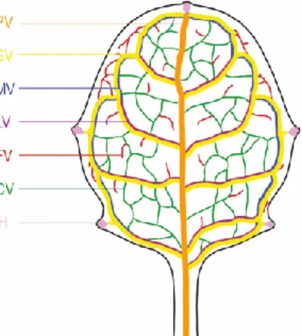
Figure 4. The Arabidopsis leaf shows different vein orders (Biedroń and Banasiak, 2018). The midrib comprised of the first order/primary vein (PV-orange) that extends into the secondary veins forming the loops (SV-Yellow). LV- lateral veins and MV-marginal veins. The secondary veins extend into the tertiary veins (green), which are inter- connected (CV) or free ending (FV).
After the specification of the apical-basal, dorsal-ventral body plan of the leaf, the development is dominated by the leaf blade expansion. In the meantime, second-order veins develop from the mid- vein and merge again with the mid-vein to form secondary vascular loops (Dengler, 2006;
Scarpella et al., 2006b). Then third-order or tertiary veins are formed from the second-order veins.
Small loops are formed either and by making connections with themselves or by looping back into the secondary veins. The reticulate venation of the leaf is completed by the formation of quaternary veins from the tertiary veins (Figure 4). The quaternary veins either merge back to the tertiary veins or end up freely making direct contact with the mesophyll cells (Scarpella, Marcos, Jirí Friml, et al., 2006; Sawchuk et al., 2007).
1.3. Regulation of vascular development
The development and patterning of plant vascular system is controlled by the gene regulatory networks that involve multiple interactions between phytohormones, transcription factors,
19 peptides and microRNAs (Ohtani et al., 2017). Although the phytohormone Auxin is known to play a central role in plant vascular development (Little, 2002; Wenzel et al., 2007), several studies revealed as well a significant involvement of other phytohormones such as cytokinins (Dettmer et al., 2009), and gibberellins (Miyashima et al., 2013). An auxin dependent transcription factor MONOPTEROS or AUXIN RESPONSIVE FACTOR5 (MP/ARF5) plays an important regulatory role in specification and patterning of the vasculature during embryogenesis and post embryonic development and loss of function of the MP leads to severe defects in the vascular development (Hardtke and Berleth, 1998; Berleth et al., 2004). The available evidences suggest that the MP acts as a positive regulator of the PIN-FORMED1 (PIN1) that exports auxin from cells and provides the feedback on the auxin status in the future provascular cells (Wenzel et al., 2007). The direct target of MP is TARGET OF MONOPTEROS 5 (TMO5), which forms a heterodimer with LONESOMEHIGHWAY (LHW) and induces the expression of the LONELYGUY (LOG) to produce the bioactive cytokinins that regulate periclinal cell divisions specifying the provascular cells (De Rybel et al., 2014). Interestingly, MP was also reported to upregulate the expression of ATHB8, which has a crucial role in leaf vascular patterning (Scarpella, Marcos, Jirí Friml, et al., 2006).
Another regulatory pathway that influences several aspects of plant vascular development including the cambial cell divisions, vascular patterning, and differentiation of vascular cell types is the peptide signaling module (Fukuda and Ohashi-Ito, 2019). It is composed of the ligand TRACHEARY ELEMENT DIFFERENTIATION INHIBITORY FACTOR (TDIF) and the receptor PHLOEM INTERCALATED WITH XYLEM (PXY) pair (Etchells and Turner, 2010). The ligand – TDIF peptide is secreted in the phloem tissue, then pairs with the PXY present in the cambium and decides the fate of xylem differentiation (Y Hirakawa et al., 2010). The TDIF-PXY signaling module regulates the vascular development in coordination with the WOX4 (Y Hirakawa et al., 2010; Etchells et al., 2016) which will be described in detail in the following sections.
1.3.1. WOX4 as a regulator of plant vascular system
The WUSCHEL-related homeobox 4 (WOX4) is one of the members of WOX gene family, a plant- specific subclade of homeobox transcription factor superfamily (Ji et al., 2010). In Arabidopsis thaliana, the WOX gene family is comprises of fifteen members, including the founding member, the transcription factor WUSCHEL (WUS) (van der Graaff et al., 2009). Based on the phylogenetic relationship, the WOX gene family is divided into three clades and they are WOX13, WOX9, and
20 WUS clades (Nardmann et al., 2009; Costanzo et al., 2014). The WOX13-clade is comprised of WOX13, 10 and 14 genes, which can be found in vascular and nonvascular plants, including mosses and green algae. The WOX9-clade includes WOX8, 9, 11 and 12 that can be found in vascular plants including the lycophytes. The WUS-clade contains the founding member WUSCHEL and the remaining seven WOX genes from WOX1 to WOX7 (Nardmann and Werr, 2012).
The WOX genes are essential for several plant developmental processes including the embryonic patterning, and the lateral organ development. Some of these WOX genes also show functional similarities such as the maintenance of stem cells. For instance, in the WUS-clade, the founding member WUS regulates the maintenance of stem cells in the shoot apical meristem (SAM) (Mayer et al., 1998). The WUS gene encodes a homeodomain transcription factor in the organizing center and then the protein moves to upper layers of the central zone, where it activates the CLV3 expression. The CLV3, a small extracellular peptide, negatively regulates the WUS expression by activating the CLV1/CLV2 receptor kinase complex. This feedback loop mechanism between CLV3 and WUS is important to maintain the stem cell homeostasis in SAM (Sarkar et al., 2007;
Yadav et al., 2011). Similarly, another WUS-clade member WOX5 exclusively is expressed in the quiescent center (QC) of the root apical meristem (RAM) and regulates the stem cell maintenance of RAM (Sarkar et al., 2007). Here, the CLE40 peptide that is encoded by CLAVATA3/EMBRYO SURROUNDING REGION-related (CLE) family member, regulates the WOX5 expression domain by activating the ARABIDOPSIS CRINKLY4 (ACR4) receptor kinase signaling mechanism (Stahl and Simon, 2009). Moreover, the recent studies indicates that another member of WUS- clade, the WOX4 is preferentially expressed in the cambial cells and promote the proliferation of stem cells in the vascular cambium (Ji et al., 2010; Suer et al., 2011; Shi et al., 2019). The TDIF peptides that are encoded by CLE41 and CLE44 in the phloem regulates the WOX4 expression through the PXY leucine-rich receptor-like kinase (Y Hirakawa et al., 2010). The PXY, similar to WOX4 is expressed in the cambium and required for the normal organization of xylem, phloem and the cambium tissues in the vascular bundles (Etchells and Turner, 2010). Furthermore, the functional WOX4 is necessary for PXY function (Y Hirakawa et al., 2010). Thus, the TDIF-PXY signaling module regulates the vascular development in coordination with the WOX4 (Y Hirakawa et al., 2010; Etchells et al., 2016). The wox4-1 mutant showed a thin stem phenotype, in which the number of vascular tissues were decreased, but the acquisition of fascicular cambium was not much affected (Ji et al., 2010). However, overexpression studies indicated that the WOX4 can
21 promote the cell divisions in both young and matured stems and increase the area of secondary vascular tissue (Ji et al., 2010).
In addition, the WOX3 and WOX4 genes appear to function together during the leaf development.
In Arabidopsis, the WOX4 participates in the development of leaf vascular system (Ji et al., 2010) while the WOX3 play a role in the expansion of the leaf blade by regulating the marginal and plate meristems identity (Matsumoto and Okada, 2001). Whereas, the WOX3 orthologs in maize, the NARROW SHEATH 1 and 2 (NS1 and NS2) regulate both the leaf blade expansion and the number of leaf vascular bundles (Nardmann et al., 2004). Similarly, WOX3 orthologs in rice, the NARROWLEAF 2 & 3 genes also involved in the regulation of both leaf blade expansion and the vascular patterning (Cho et al., 2013). However, it has been shown in rice that AtWOX4 ortholog, OsWOX4 is active in SAM (Ohmori et al., 2013). Interestingly, these studies have shown that OsWOX4 functions in maintaining the stem cell identity in the SAM of rice, replacing the WUS function. Moreover, the OsWOX4 expression was found to extend from the SAM into the early leaf primordia and taking part in the leaf development including the vascular patterning suggesting that the OsWOX4 specifies vascular identity in the SAM in rice (Yasui et al., 2018). In Arabidopsis, the available studies have been shown that WOX4 is expressed in the vasculature of root, shoot, cotyledons and leaves, but its activity was only confined to the cambial cells of all these organs (Y Hirakawa et al., 2010). The WOX4 promoter in Arabidopsis was extensively used to mark the cambial cells during the different developmental phases of the inflorescence stem (Suer et al., 2011). It has been shown that WOX4 promoter was active only in the fascicular cambium of the young stem referring it as the primary growth phase. Then the expression was shown to extend from the fascicular cambium into few cells that are positioned in between the fascicular and interfascicular regions of the stem. This extension was stated as an indication of the initiation of secondary growth. Then the circular WOX4 expression domain was observed in matured stem (Suer et al., 2011). However, the published studies of WOX4 expression pattern (Y Hirakawa et al., 2010; Suer et al., 2011) were based on using 2.9 Kb upstream and 0.6 Kb downstream regions, although long intergenic sequences flank the WOX4 gene.
22 2. AIM OF THE THESIS
The main aim of this thesis work is to find out new information about spatio-temporal expression patterns of the WUSCHEL-related homeobox4 (WOX4) gene in different organs of Arabidopsis thaliana.
Although the published WOX4 promoter has shown cambial specific activity, it contains only 2.9 Kb upstream and 0.6 Kb downstream sequences, which results in weak overall activity. Hence, we analysed the large flanking area, including 9.2 Kb upstream and 1.7 kb downstream from the WOX4 gene sequence to identify the distal conserved elements. Furthermore, full-length WOX4 promoter-reporter constructs, targeting either the endoplasmic reticulum (flWOX4::erGFP and flWOX4::erCERULEAN) or the nucleus (flWOX4::H3-GFP) were generated and transformed into Arabidopsis thaliana. The consequences of addition of all conserved distal elements were studied in detail with respect to the WOX4 expression pattern in the shoot, roots and leaves.
23 3. MATERIALS AND METHODS
3.1. List of chemicals
Name Abbreviation Manufacturer
Acetic acid Roth
Acetone Roth
Agar Roth, Duchefa
Agarose Bio-Budget
Bromophenol blue Merck
Calcium chloride CaCl2 Merck
Chloroform Roth
Dexamethasone Dex Roth
Disodium phosphate Na2HPO4 Roth
Ethanol Roth
Ethidium bromide EtBr Sigma Aldrich
Ethylinediamine tetra acetic acid EDTA Sigma Aldrich
Glacial acetic acid Sigma Aldrich
Glufosinate-ammonium (Basta®) Bayer
Glycerol Sigma Aldrich
Hydrochloric acid HCl Roth
Isoamyl alcohol Roth
Isopropanol Roth
Magnesium chloride MgCl2 Roth
MES salt Sigma Aldrich
Monopotassium phosphate KH2PO4 Sigma Aldrich
Phenol Sigma Aldrich
Phloroglucinol Sigma Aldrich
Potassium chloride KCl Roth
Potassium hydroxide KOH Roth
Propidium iodide PI Sigma Aldrich
Silwet® Sigma Aldrich
Sodium acetate Merck
Sodium chloride NaCl Roth
Sodium dodecyl sulphate SDS Merck
Sodium hydroxide NaOH Merck
Sodium hypochlorite NaClO Roth
Tris base Roth
Tryptone Roth
Yeast-extract Roth
24 3.2. Buffers
TAE Buffer (50X): 242 g Tris base, 57.1 ml Glacial acetic acid and 100 ml of 0.5M EDTA (pH 8.0) per 1L. The 1X working solution is 40 mM Tris-acetate/1 mM EDTA.
TBE Buffer (5X): 54 g Tris base, 27.5 g Boric acid and 20 ml of 0.5M EDTA (pH 8.0) per 1L.
The 0.5X working solution is 45 mM Tris-borate/1 mM EDTA.
PBS Buffer: 25.6 g Disodium Phosphate, 80 g NaCl, 2 g KCl and 2 g Monopotassium phosphate per 1L.
3.3. Kits
Name Purpose Manufacturer
Nucleospin-plasmid Plasmid Purification Macherey-Nagel Nucleospin- Gel and PCR clean-up PCR clean-up & Gel purification Macherey-Nagel
Invitrap spin RNA extraction Startec. molecular
Superscript II Reverse transcriptase cDNA Synthesis Thermo Scientific
SMARTer RACE 5′/3 5′- RACE Clonetech
My-budget 5X Taq PCR master mix Colony PCR Bio-budget Tech
3.4. Growth Media
LB medium: Tryptone – 1 %, Yeast Extract – 0.5 %, NaCl – 1 % , Agar – 15 % , pH: 7.0
SOC-medium: Tryptone – 2 %, Yeast Extract – 0.5 %, NaCl – 1 %, MgCl2 – 10 mM, MgSO4 – 10 mM, KCl – 2.5 mM, Glucose – 20 mM, and Agar – 15 % , pH: 7.0
Infiltration medium: 50 g of Sucrose and 200 µl of Silwet in 1L of distilled water
MS medium: 2.3 g of MS salts with B5 vitamins (Duchefa Biochemie), 10 g Sucrose, 0.5 g MES salt, and 8 g Agar in 1L of distilled water. Total concentration of micro & macro elements including vitamins:
4414.09 mg/l, pH 5.7
25 3.5. Biological materials
3.5.1. Enzymes
Name Purpose Manufacturer
Phusion DNA polymerase DNA amplification Sigma Aldrich Taq DNA polymerase DNA amplification Sigma Aldrich
Restriction endonucleases To cut the DNA specifically New England Biolabs Alkaline Phosphatase,
Calf Intestinal phosphatase (CIP)
Dephosphorylation of 5´ and 3´ ends of DNA
New England Biolabs
Klenow fragment Blunting the DNA ends, Second strand cDNA synthesis
New England Biolabs
T4 DNA ligase Ligation New England Biolabs
3.5.2. Primers
Oligo Name Sequence 5' to 3' Purpose
Up region1-FP_Asc1 GGCGCGCCAGCAAACATACCCACACAAAAG Cloning
Up region1-RP_Sac1 GAGCTCGGGGGTATTTTAAAAAAATCTGATG Cloning
Up region2-FP_Sac1 GAGCTCTCTAAATGCCTTGTCACCAAATC Cloning
Up region2-RP_NcoI CCATGGCTGCTATATGTTAAAACTAGCAAATGC Cloning
Downstream-FP_Apa1 GGGCCCAGTCATGAAGGTGAGG Cloning
Downstream-RP_Asc1 GGCGCGCCTCTTCTCATGGATTCT Cloning
erCerulean-RP_BspHI TCATGAAGACTAATCTTTTTCTCTTTCTCATCTTTTC Cloning
erCerulean-RP_Apa1 GGGCCCTTAGAGTTCGTCGTGCTTGTACAG Cloning
erGFP-FP_BspH1 GTCATGAAGACTAATCTTTTTCTC Cloning
erGFP-RP_Apa1 TTAGAGTTCGTCGTGTTTGTATAG Cloning
H3-FP_Nco1 CCATGGCTCGTACCAAGCAGAC Cloning
H3-RP_Sal1 GTCGACAGCTCGTTCTCCTCTGATTC Cloning
GFP-FP_Sal1 GTCGACAGTAAAGGAGAAGAAC Cloning
GFP-RP_Apa1 GGGCCCTTAGAGTTCGTCGTGTTTG Cloning
WOX4_cDNA_RP1 AAGACACCAGTGGTCGTGAAGC RACE
26
WOX4_cDNA_RP2 GGTTGTTCCTCTTCTGCTTCTGTCTCTCCG RACE
SMARTer-oligoIIA AAGCAGTGGTATCAACGCAGAGTACXXXXX RACE
RACE-UPM-long
CTAATACGACTCACTATAGGGCAAGCAGTGGTATC AACGCAGAGT
RACE
RACE-UPM-short CTAATACGACTCACTATAGGGC RACE
Nested 1_N1 CAATCTGTTGAGCATTAGGAGTACG RACE
Nested 2_N2 CTGTTCTTGAGTCGGGTTCCAC RACE
3.5.3. Antibiotics
Name Working concentration (µg/ml) Manufacturer
Kanamycin 50 Duchefa
Ampicillin / Carbenicillin 100 Duchefa
Gentamycin 25 Duchefa
Rifampicin 50 Duchefa
Hygromycin 100 Duchefa
3.5.4. Cloning vectors
Name Source Resistance Function
pJET 1.2/blunt Thermo Scientific™ Ampicillin Cloning
pBluescript KS (+) (M.A.Alting-Mees and J.M.Short, 1989) Ampicillin Cloning
pGPTV - Kan (Überlacker and Werr, 1996) Kanamycin Binary
pGPTV - Bar (Überlacker and Werr, 1996) Kanamycin, Phosphinothricin
Binary
3.5.5. Bacterial strains
Organism Strain Function
E. coli DH5α (Hanahan, 1983) Cloning
A. tumefaciens GV3101 (pMP90) GentR, RifR (Koncz and Schell, 1986)
Plant transformation
3.5.6. Plant material
All the phenotypic and molecular biology studies were carried out in Arabidopsis thaliana Col-0 as the wild-type control. Same plant background was used to generate the transgenic lines.
27 3.6. Methods
3.6.1. Phylogenetic Shadowing
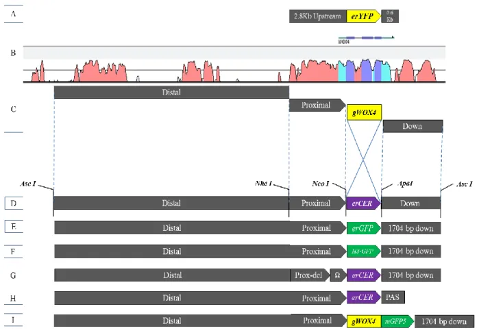
Phylogenetic shadowing was used to identify the conserved sequences in the WOX4 promoter region. First, the WOX4 orthologs were identified by using AtWOX4 coding sequence to BLAST against the genomes of Brassicaceae members’ viz. Arabidopsis thaliana, Arabidopsis lyrata, Arabidopsis halleri, Boechera stricta, Brassica rapa, Brassica oleracea, Capsella rubella, and Capsella grandiflora. Using the locus information of these orthologs, the flanking genes were identified and then, the complete upstream and downstream sequences from the WOX4 coding region were retrieved by using Phytozome 10 and Vector NTI. These sequences were aligned with the mVISTA (http://genome.lbl.gov/vista/mvista/submit.shtml), at the default settings of LAGAN (Brudno et al., 2003) alignment tool. Based on the sequence similarity obtained in vista plots, the conserved elements in the WOX4 promoter are recognized.
Transgenic line Ecotype Selection Purpose Source pWOX4:: YFP Col-0 Phosphinothricin Cambium Thomas Greb pATHB8:: YFP Col-0 Phosphinothricin Pro-cambium E. Scarpella flWOX4::erCER Col-0 Phosphinothricin Full-length WOX4
promoter reporter
Current study
flWOX4::erGFP Col-0 Phosphinothricin Full-length WOX4 promoter reporter
Current study
flWOX4:: H3-GFP Col-0 Phosphinothricin Full-length WOX4 promoter reporter
Current study
flWOX4:gWOX4-GFP Col-0 Phosphinothricin Translational fusion Current study pWOX4-Ω::erCER Col-0 Phosphinothricin 5′-modification in
the WOX4 promoter
Current study
pWOX4::erCER-PAS Col-0 Phosphinothricin 3′-modification in the WOX4 promoter
Current study
28 3.6.2. Nucleic acid extraction & purification
Total genomic DNA was isolated from the leaf tissue according to the protocol described previously (K. Edwards, 1991) and purified by phenol-chloroform-isoamyl alcohol method.
Total RNA was isolated from young seedlings by Invitrap spin plant RNA mini kit, following the manufacturer protocol. An additional DNase treatment was performed and purified by phenol- chloroform-isoamyl alcohol method.
3.6.3. RACE (Rapid amplification of cDNA ends)
SMARTer® RACE 5′/3′ kit was used to identify the 5′ end of WOX4 mRNA. 1µg of total RNA (DNA free) was used to synthesize the cDNA. Then the nested PCRs were performed by using N1, N2 primers that binds in the Exon 2 of WOX4. Subsequently, the RACE-PCR products were cloned into pJET1.2 blunt vector (Thermo Scientific) and sequenced with WOX4 specific primers.
The resulting sequences were aligned with the WOX4 genomic DNA sequence to identify the transcription start site.
3.6.4. Promoter constructs
Following constructs were generated in the current study-
flWOX4::erCER 9183 bp upstream > erCERULEAN > 1704 bp downstream flWOX4::erGFP 9183 bp upstream > erGFP > 1704 bp downstream
pWOX4::erCER:PAS 9183 bp upstream > erCERULEAN> poly-adenylation sequence pWOX4::Ω:erCER 237 bp WOX4 upstream region was replaced by TMV-Ω leader flWOX4::H3-GFP 9183 bp upstream > H3-GFP > 1704 bp downstream
flWOX4:gWOX4-GFP 9183 bp upstream > gWOX4>mGFP5> 1704 bp downstream 3.6.5. Primer design
All primers were designed using the Primer3 software (http://bioinfo.ut.ee/primer3-0.4.0/) and then validated with Sigma-OligoevaluatorTM (https://www.sigmaaldrich.com/technical- documents/articles/biology/oligo-evaluator.html).