Functional Analysis of the SPA Gene Family in Arabidopsis thaliana
Inaugural-Dissertation
zur
Erlangung des Doktorgrades
der Mathematisch-Naturwissenschaftlichen Fakultät der Universität zu Köln
vorgelegt von
Kirsten Fittinghoff aus Velbert
2008
Prüfungsvorsitzender: Prof. Dr. Werr
Tag der mündlichen Prüfung: 02.02.2009
Contents
Contents:
Contents:... I Abbreviations ...IV Zusammenfassung ...VI Abstract...VIII
I. Introduction ... 1
I.1. Plant photoreceptors and light signal transduction ... 1
I.2. The SPA quartet: A family of COP1-interacting proteins with a central role in suppressing photomorphogenesis ... 6
I.3. Functional diversification among Arabidopsis SPA genes ... 8
I.4. Aims of this PhD thesis... 12
II. Results... 13
II.1. SPA1 structure-function analysis... 13
II.1.1. SPA1 N-terminus is not required for SPA1 function in dark- and light- grown seedlings, whereas the coiled-coil domain is essential ... 13
II.1.2. SPA1 N-terminus is required to suppress flowering in short-days ... 16
II.2. SPA transcript analyses... 18
II.2.1. SPA1 mRNA accumulates in blue light... 18
II.2.2. SPA3 and SPA4 but not SPA2 mRNA levels increase by light... 21
II.2.3. SPA mRNA abundance partially correlates with its distinct functions during plant development... 23
II.3. SPA Promoter GUS analyses... 24
II.3.1. SPA1 and SPA2 but not SPA4 promoters are strongly active in the roots of young plants... 24
II.4. Promoter-swap analysis ... 27
II.4.1. Expression of SPA1, SPA2 and SPA4 in spa triple and quadruple mutants phenocopies appropriate mutant phenotypes in seedlings... 27
II.4.2. In darkness cSPA2 and cSPA4 expressed by the SPA1 promoter complement mutant phenotype... 31
II.4.3. SPA2 expressed under the control of SPA1 promoter is not able to rescue mutant phenotypes in FR, whereas SPA4 does partially... 32
II.4.4. SPA1 under control of SPA2 promoter rescue mutant phenotypes in darkness and in FR... 35
II.4.5. Promoter-swap analyses in spa1 single mutants demonstrate that
cSPA1 expressed by SPA4 is partially functional ... 38
II.4.6. GUS analyses verified expression of cGUS-cSPA2-HA under control of SPA1 promoter ... 40
III. Discussion ... 42
III.1. SPA gene transcription is under the control of endogenous and exogenous cues ... 42
III.2. SPA mRNA abundance is regulated by different photoreceptors ... 45
III.3. All SPA proteins act as repressors in darkness – and some also in the light.. ... 46
III.4. The SPA N-terminus has an important function in suppression of flowering .. ... 50
IV. Materials and Methods... 53
IV.1. Materials ... 53
IV.1.1. Chemicals and antibiotics... 53
IV.1.2. Radioactivity, enzymes kits and antibodies ... 53
IV.1.3. Bacterial strains ... 53
IV.1.4. Oligonucleotides ... 54
IV.1.5. Cloning vectors... 57
IV.1.6. Plant lines... 57
IV.2. Methods... 57
IV.2.1. Seed sterilization ... 57
IV.2.2. Plant growth... 58
IV.2.3. Measurement of hypocotyl length... 58
IV.2.4. Measurement of flowering time... 58
IV.2.5. Plasmid DNA preparation of bacteria ... 59
IV.2.6. Genomic DNA preparation... 59
IV.2.7. RNA isolation and Northern analysis ... 59
IV.2.8. Semi-quantitative RT-PCR analysis ... 60
IV.2.9. Protein isolation and immunoblot analysis... 60
IV.2.10. Histochemistry ... 61
IV.2.11. DNA manipulation... 61
IV.2.12. Gateway cloning ... 61
IV.2.13. Plant transformation ... 61
Contents
IV.3. Cloning strategies ... 61
IV.3.1. Promoter-swap constructs... 61
IV.3.2. SPA promoter::GUS constructs ... 64
IV.3.3. Construction of SPA1::SPA1-HA and SPA1 deletion-constructs ... 65
V. Supplement... 68
VI. References ... 73
VII. Danksagung... 80
VIII. Erklärung ... 81
Abbreviations
α-32P-dATP 2’-desoxyadenosin-5’-triphosphate, radioactive labelled at α-P-Atom
35S 35S promoter of Cauliflower Mosaic viruc
B blue light
°C degree Celsius
c continous
µl micro litre
µg micro gram
bp base pair
bHLH basic helix-loop-helix
cDNA complementary DNA
CC coiled-coil structure
Col Columbia; ecotype of Arabidopsis thaliana
cm centimetre
cpm counts per million
WD aspartic acid
D darkness
Da Dalton
DNA desoxyribonucleic acid
FR far-red light
FRc continuous FR
GUS β-Glucuronidase
h hour
HA Influenza hemagglutinin
kb kilo bp
kDA kilo Da
l litre
IR infrared
LD long day
Ler Landsberg errecta; ecotype of Arabidopsis thaliana
M molar; mol/l
mg milligram
mM millimolar
min minute
Abbreveations
mRNA messenger-ribonucleic-acid
NLS nuclear localization signal/sequence
nm nanometre
ORF open reading frame
PCR polymerase chain reaction
Pfr red light absorbing phytochrome conformation
Phy phytochrome
Pr red light absorbing phytochrome conformation
R red light
RLD ecotype of Arabidopsis thaliana
rRNA ribosomal ribonucleic-acid
RNA ribonucleic-acid
RT-PCR reverse-transcription-PCR
s second
SD short day
UTR untranslated region
U unit
UV ultraviolet
WD aspartic acid; tryptophan
WS-0 Wassilewskaja; ecotype of Arabidopsis thaliana
WT wild type
ZT zeitgeber
Nomenclature:
SPA1 gene, locus, wild-type allele
spa1 mutant allele
SPA1 protein
Exception: photoreceptors
PHY gene, locus, wild-type allele
phy mutant allele
PHY apoprotein (without chromophor) phy holoprotein (with chromophor)
Zusammenfassung
Viele Abschnitte im Lebenszyklus von Pflanzen, wie z.B. die Samenkeimung, die frühe Keimlingsentwicklung (De-etiolierung) oder die Induktion der Blütenbildung, werden maßgeblich durch das Sonnenlicht beeinflußt. Dabei spielt der Arabidopsis COP1-SPA Proteinkomplex eine zentrale Rolle, denn er verhindert eine Lichtantwort im Dunkeln. Der COP1-SPA-Komplex ist vor allem in Dunkelheit aktiv und verantwortlich für die Ubiquitin-vermittelte Degradation von positiv wirkenden Faktoren der Lichtsignaltransduktion, wie den Transkriptionsfaktoren HY5, der die Keimlingsentwicklung steuert, oder CONSTANS, der die Blütenbildung fördert. Im Licht wird die Funktion des COP1-SPA Komplexes gedrosselt, eine Aufgabe, die von mehreren Photorezeptoren bewerkstelligt wird. Das COP1 Protein wird in Arabidopsis von einem einzigen Locus kodiert, während die SPA Proteine von einer Genfamilie bestehend aus vier Mitgliedern kodiert werden (SPA1-SPA4). SPA Gene haben überlappende, jedoch auch distinkte Funktionen im Lebenszyklus von Arabidopsis. SPA1 und SPA2 sind hauptverantwortlich für die Unterdrückung der Photomorphogenese im Dunkeln. SPA2 hat keine Funktion bei der lichtgesteuerten Keimlingsentwicklung, die hingegen hauptsächlich von SPA1 und in geringerem Maße auch von SPA3 und SPA4 reguliert wird. SPA1 ist zudem ausreichend, um eine verfrühte Blütenbildung im Kurztag zu verhindern.
Ziel dieser Arbeit war es, die molekularen Grundlagen der unterschiedlichen Funktionen der SPA Gene zu verstehen. Unterschiedliche SPA Gen-Funktionen lassen sich teilweise auf eine unterschiedliche SPA Genexpression zurückführen.
RNA-Blot-Experimente zeigen, dass die mRNA-Mengen von SPA1, SPA3 und SPA4, nicht aber die von S P A 2, positiv durch Licht beeinflußt werden. Mehrere Photorezeptoren wirken dabei zusammen, um die Expression der SPA Gene im Licht unterschiedlicher Wellenlängen zu fördern. SPA-Promotor-Reportergen Analysen zeigen zudem eine zum Teil differentielle Expression der SPA-Gene während der Pflanzenentwicklung. Jedoch kann die unterschiedliche Expression der SPA-Gene nicht alle distinkten Funktionen der SPA Proteine erklären. Promoter-Austausch- Experimente mit den regulatorischen Elementen und cDNAs von SPA1, SPA2 und SPA4 zeigen, dass alle SPA Proteine im Dunkeln wirken können. SPA1 und SPA4 Proteine können außerdem im Licht als Repressoren fungieren, hingegen kann das SPA2-Protein nicht im Licht wirken, selbst wenn die SPA2-cDNA unter der Kontrolle
Zusammenfassung
des licht-induzierten SPA1-Promotors steht. Diese Resultate zeigen, dass Unterschiede in der SPA Proteinsequenz ebenfalls einen Einfluß auf deren Funktionen haben.
Alle SPA Proteine zeigen eine ähnliche Domänen-Anordnung. Im stark konservierten carboxy-terminalen Bereich der SPA Proteine befindet sich eine WD-40 Repeat- Domäne, die ebenso wie die zentrale Coiled-Coil Domäne der SPA Proteine Interaktionen mit anderen Poteinen vermittelt. Die amino-terminale Region (N- Terminus) ist innerhalb der SPA Proteine weniger stark konserviert, zeigt aber in jedem der SPA Proteine eine schwache Ähnlichkeit mit einem Ser/Thr-Kinase-motiv.
Um die Funktion dieser schwach konservierten Region näher zu untersuchen, wurde eine SPA1-Struktur-Funktionsanalyse durchgeführt. Interessanterweise ist ein SPA1 Protein ohne N-Terminus in der Lage, seine Rolle in der Keimlingsentwicklung vollständig auszufüllen. Hingegen ist es unfähig, die verfrühte Induktion der Blütenbildung im Kurztag zu hemmen. Daher zeigen diese Ergebnisse, dass der N- Terminus der SPA Proteine eine essentielle Rolle spielen kann.
Abstract
Ambient light conditions affect development throughout the plant life cycle, including seed germination, seedling development and the induction of flowering. In the model plant Arabidopsis, the COP1-SPA ubiquitin ligase complex plays a central role in suppressing light signaling in darkness. The COP1-SPA complex targets positively acting factors like HY5, a protein necessary for normal seedling development in the light, several photoreceptors and the flowering time regulator CONSTANS for degradation via the 26S proteasome. Therefore, one of the major functions of the light signal transduction pathways is the inactivation of the COP1-SPA complex.
While COP1 is a single copy gene, the SPA proteins are encoded by four different loci (SPA1-SPA4). All SPA proteins have redundant, but also distinct functions in regulating plant development. SPA1 and SPA2 are the key regulators that suppress photomorphogenesis in dark-grown seedlings. Over-stimulation in light-grown seedlings is primarily prevented by SPA1, and to a minor extent, also by SPA3 and SPA4. SPA2, in contrast has only negligible function in the light. SPA1 is sufficient for repressing flowering under non-inductive short-day conditions.
Here, I show that distinct functions of the SPA genes partially correlate with their distinct gene expression patterns. RNA gel blot-analysis revealed that the expression of SPA1, SPA3 and SPA4, but not that of SPA2, is positively influenced by light of different wavelengths. All main photoreceptors contribute to the up-regulation of these SPA transcripts, implying that photoreceptors initiate a negative feedback regulation, which might protect plants from over-stimulation by light. GUS reporter gene experiments show that SPA genes exhibit somewhat distinct tissue-specific expression patterns, which might be important for tissue specific regulation of COP1- SPA targets. However, differences in SPA gene expression cannot account for all distinct SPA gene functions. Promoter-swap experiments with SPA1, SPA2 and SPA4 show that all SPA proteins are potent repressors in dark-grown seedlings.
SPA1 and SPA4 also act as repressor in the light. SPA2, however, can never act as a repressor in the light, not even when it is expressed from the strong light-induced SPA1 promoter. These results show that SPA proteins themselves feature properties that determine characteristic SPA protein functions.
All SPA proteins feature a characteristic domain structure with a C-terminal WD- repeat, a central coiled-coil domain and a less well-conserved N-terminus that
Abstract
includes a kinase-like motif. The WD-repeat domain and the coiled-coil domain are essential for formation of the COP1-SPA complex as well as interactions with various ubiquitination targets. In contrast, the function of the N-terminal domain is unknown.
Aiming to determine the importance of the N-terminal domain of SPA1, I conducted a structure-function analysis. While the N-terminal domain of SPA1 is dispensable for SPA1 function in the seedling stage, this domain is required for SPA1-mediated repression of flowering in non-inductive short-day conditions. These results indicate, that the SPA1 N-terminal domain can full-fill an essential function.
I. Introduction
I.1. Plant photoreceptors and light signal transduction
As sessile organisms, plants need to adopt their growth and development rapidly and optimally to ambient environmental changes. Light is not only the primary source of energy for plants, light is also an important environmental factor that influences many different developmental switches such as seed germination, seedling de-etiolation, shade avoidance, phototropism, stomata and chloroplast movement, circadian rhythm and induction of flowering. Seedling de-etiolation is one of the most drastic light responses. After germination, seedlings growing in the soil, i.e. in darkness, undergo a process called skotomorphogenesis in order to reach the soil surface and start photosynthesis. This is characterized by increased hypocotyl elongation, closed cotyledons and the formation of an apical hook, which protects the shoot apical meristem. On the soil surface seedlings are exposed to light and adopt their morphology for growing in the light. This developmental switch is called photomorphogenesis and is accompanied by inhibition of hypocotyl elongation, expansion of cotyledons and the induction of chlorophyll synthesis. This de-etiolation response in Arabidopsis thaliana seedlings has been used as a model system in forward genetic screens in order to identify photoreceptors and other regulatory factors important for light signaling.
In Arabidopsis, four main classes of photoreceptors are responsible for perceiving light of different intensities, qualities and directions (Briggs and Olney, 2001). Three different types of photoreceptors perceive blue light (B); three cryptochromes (cry1- cry3), two phototropins (phot1-phot2) and members of the zeitlupe gene family (ztl, lkp2, fkf1) (Ahmad and Cashmore, 1993; Lin et al., 1996; Huala et al., 1997; Christie et al., 1998; Mazzella et al., 2001; Kleine et al., 2003). phot1 and phot2 are involved in phototropic plant responses, chloroplast movement and stomatal opening (Briggs and Olney, 2001; Briggs and Christie, 2002; Sakamoto and Briggs, 2002; Ohgishi et al., 2004), whereas the ztl/lkp2/fkf1 photoreceptors regulate light input into the circadian clock and flowering time (Schultz et al., 2001; Imaizumi et al., 2003;
Somers et al., 2004). cry1 is the primary photoreceptor that inhibits hypocotyl elongation in response to high fluence rates of B (Ahmad and Cashmore, 1993; Lin et al., 1996; Mazzella et al., 2001; Kleine et al., 2003). cry2 is important for seedling development under low fluence rates of B and plays an important role in the
Introduction
photoperiodic induction of flowering (Guo et al., 1998; Lin et al., 1998; Mockler et al., 2003).
The fourth class of photoreceptors, the phytochromes (PHYA-PHYE), monitor red light (R) and far-red light (FR) (Sharrock and Quail, 1989; Clack et al., 1994). phyA is the only photoreceptor that can sense FR, but in addition to that phyA can also mediate responses to low fluence rates of R and B (Nagatani et al., 1993; Whitelam et al., 1993). phyB and to a minor extent phyC, phyD and phyE play important roles in R response (Quail, 1997). Phys are known to regulate many different developmental steps such as seed germination, de-etiolation, shade avoidance and regulation of flowering time (Figure 1) (Schepens et al., 2004).
Figure 1: Role of photoreceptors during the plant life cycle.
Specialized classes of photoreceptors monitor light of different wavelengths. Cryptochromes and phototropins perceive B and UVA light. phyB mainly responds to R redundantly with phyA,C,D,E. phyA is the only FR-sensing photoreceptor but can also sense B and R. Photoreceptors modulate adaptive growth and development including seed germination, phototropism, de-etiolation, shade avoidance and induction of flowering.
Although Arabidopsis has evolved this sets of functionally distinct photoreceptors to monitor light, there is also vivid cross-talk between the different photoreceptors and their signaling pathways (Casal, 2000; Devlin and Kay, 2000; Mas et al., 2000;
Mazzella et al., 2001; Yanovsky et al., 2001; Sullivan and Deng, 2003; Usami et al., 2004). In addition, the two major classes of photoreceptors, phys and crys, induce related signaling events. Once activated by light, phyA and cry2 become less stable and are degraded via the 26S proteasome pathway, while phyB and cry1 are stable also in the light (Guo et al., 1999; Hisada et al., 2000). Photo-activated receptors are mainly localized in the nucleus, the place where they initiate further downstream signaling events (Cashmore et al., 1999; Kircher et al., 1999; Kleiner et al., 1999; Yu et al., 2007). These signal cascades lead to a transcriptional reprogramming of the cells, which is coordinated by different classes of transcription factors. But how are these transcription factors regulated by the different photoreceptors? phys and crys follow two distinct strategies that directly and indirectly affect the activity of transcription factors involved in light signaling. First, photo-activated receptors can bind directly to some transcription factors. Phys physically interact with a class of bHLH transcription factors, so-called PHYTOCHROME-INTERACTING-FACTORS (PIFs) and PIF-LIKES (PILs). PIFs and PILs mainly act as repressors of light signaling and phytochromes can phosphorylate PIFs and PILs that are in turn degraded (Al-Sady et al., 2006; Castillon et al., 2007; Al-Sady et al., 2008; Leivar et al., 2008b; Leivar et al., 2008a; Shen et al., 2008). Similarly, photo-activated cry2 was recently shown to interact with the bHLH transcription factor CIB1 to regulate flowering time (Liu et al., 2008).
Several transcription factors with important roles in light signaling do not directly bind to photoreceptors. LONG HYPOCOTYL IN FAR-RED1 (HFR1), another bHLH transcription factor, is a component of phyA and cry1 signaling pathways and does not directly interact with phys (Fairchild et al., 2000; Duek and Fankhauser, 2003).
Also LONG AFTER FAR-RED LIGHT1 (LAF1), a MYB transcription factor, does not directly bind phys but regulates gene expression in response to FR (Ballesteros et al., 2001). HYPOCOTYL 5 (HY5) and HY5 HOMOLOG (HYH), two bZIP-transcription factors, play a more widespread role in mediating light dependent transcriptional activation in seedling development under FR, R, B or UV-B light (Oyama et al., 1997;
Chattopadhyay et al., 1998; Osterlund et al., 2000b; Osterlund et al., 2000a; Ulm et al., 2004). Common to HY5, LAF1 and HFR1 is that their regulation involves light-
Introduction
dependent, post-translational control of protein stability: HY5, LAF1 and HFR1 proteins are low abundant in darkness and accumulate to high levels in the light (Osterlund et al., 2000b; Osterlund et al., 2000a; Seo et al., 2003; Duek et al., 2004;
Jang et al., 2005; Yang et al., 2005b).
Figure 2: COP1 is a central regulator of light signal transduction.
A: Visual phenotypes of dark-grown wild-type and cop1 mutant seedlings (top): In darkness wild-type seedling undergoes normal skotomorphogenesis showing long hypocotyl and closed cotyledons. cop1 mutant seedling undergoes constitutive photomorphogenesis and exhibits the features of a light-grown seedling in darkness. Simplified illustration of molecular mechanism of skotomorphogenesis (bottom):
In darkness, photoreceptors are inactive and cannot suppress negative regulators like COP1. In darkness, COP1 suppresses HY5 function, a transcriptional activator. Mutations in COP1 lead to functional HY5 also in the darkness.
B: Visual phenotypes of light-grown wild-type and cop1 mutant seedlings (top): In light wild-type seedlings exhibit reduced hypocotyls and de-etiolated (green) expanded cotyledons. cop1 mutants show strong constitutive photomorphogenesis. Simplified illustration of molecular mechanism of photomorphogenesis (bottom): In light several photoreceptors suppress COP1 activity. In turn, HY5 protein becomes active and can activate transcription of light-responsive genes.
Photoreceptors promote the stability of these transcription factors indirectly by interfering with the factors that promote their degradation. These factors can be summarized in the group CONSTITUTIVE PHOTOMORPHOGENESIS (COP), DE- ETIOLATED (DET) and FUSCA (FUS) proteins (Chory et al., 1989; Deng et al., 1991). Seedlings with mutations in any of the COP/DET/FUS genes exhibit short hypocotyls and open cotyledons in darkness (constitutive photomorphogenesis). The reason for this is that cop/det/fus mutants exhibit strongly elevated HY5 and HFR1 protein levels also in darkness (Osterlund et al., 2000b; Osterlund et al., 2000a; Seo et al., 2003; Duek et al., 2004; Jang et al., 2005; Yang et al., 2005b).
The most well characterized locus among the COP/DET/FUS genes is COP1 (Deng et al., 1991). COP1 encodes a protein with a carboxy-terminal WD-repeat domain, a coiled-coil domain and an amino-terminal RING motif, which is characteristic for one subclass of E3 ubiquitin ligases (Deng et al., 1992). In fact, COP1 has E3 ubiquitin ligase activity and targets the transcription factors HY5, HFR1 and LAF1 directly for degradation via the 26S proteasome (Osterlund et al., 2000a; Saijo et al., 2003; Seo et al., 2003). However, the molecular mechanism of photoreceptor-mediated inhibition of COP1 activity is not well understood.
The photoreceptors phyA, phyB, cry1 and cry2 can directly bind to the WD-repeat domain of COP1 and these interactions are thought to suppress COP1 activity towards other factors such as HY5 (Wang et al., 2001; Yang et al., 2001; Seo et al., 2004). Interestingly, COP1 seems in turn to be responsible for degradation of the light unstable phyA and probably also cry2 (Shalitin et al., 2002; Seo et al., 2004).
COP1 becomes also inactivated by light-dependent exclusion from the nucleus, a process, which is also initiated by photoreceptor signaling (Von Arnim and Deng, 1994; Von Arnim et al., 1997; Subramanian et al., 2004). However, light does not completely suppress COP1 function, more likely trace amounts of biologically active COP1 remain in the nucleus to prevent over-stimulation by light.
COP1 genetically and physically interacts with other members of the COP/DET/FUS proteins (Schwechheimer and Deng, 2000). COP10, a ubiquitin activating E2 variant, interacts with both COP1 and also components of the COP9 signalosome, a multisubunit, nuclear protein complex involved in cullin-dependent ubiquitin/proteasome pathways (Wei et al., 1994; Yanagawa et al., 2004). COP10 itself forms a stable protein complex (the CDD complex) with DET1 and DDB1 that is thought to be important for COP1 activity (Yanagawa et al., 2004). Also, COP1 forms
Introduction
high molecular weight complexes and interacts with several other proteins that are indispensable for COP1 function. One class of COP1-interacting proteins are the SUPPRESSOR OF PHYTHOCHROME A-105 (SPA) proteins (Hoecker and Quail, 2001; Laubinger and Hoecker, 2003; Saijo et al., 2003; Laubinger et al., 2004; Saijo et al., 2008; Zhu et al., 2008).
Figure 3: COP1 is an E3 ubiquitin ligase with structural similarities to SPA.
The ubiquitin-activating enzyme E1 binds and activates free ubiquitin (U) and transfers it to an ubiquitin-activating enzyme E2. After binding, the E2 ubiquitin-conjugating enzyme associates with COP1, an E3 ubiquitin ligase with a RING motif (typical for one class of E3 ubiquitin ligases). E3 ubiquitin ligases are responsible for substrate recognition. COP1 targets proteins by poly-ubiquitination for degradation via the 26S proteasom. COP1 shows structural similarity to the carboxy-terminal region of SPA including the WD-repeats. Transcription factors like HY5 can bind either the WD-repeat domain of COP1 or SPA1. Both proteins can physically interact through their respective coiled-coil domains (modified from Hoecker, 2005).
I.2. The SPA quartet: A family of COP1-interacting proteins with a central role in suppressing photomorphogenesis
The founding member of the SPA gene family, SPA1, was identified in a mutant screen for genes that suppress the phenotype of a weak phyA mutant allele (Hoecker et al., 1998). spa1 mutants exhibit enhanced photomorphogenic responses in FR, R and B light, but are indistinguishable from wild-type seedlings in complete darkness (Hoecker et al., 1998; Baumgardt et al., 2002; Fittinghoff et al., 2006). The seedling phenotype of spa1 mutants is only detectable in the presence of functional phyA, which led to the conclusion that SPA1 is a repressor of a phyA-specific signaling pathway (Hoecker et al., 1998). SPA1 mRNA levels are strongly upregulated in response to R and FR, a process initiated not only by phyA, but also by phyB (Hoecker et al., 1999). SPA1 encodes a constitutively nuclear-localized protein with three characteristic domains: a carboxy-terminal WD-repeat domain, a central coiled- coil domain and a N-terminal kinase-like domain (Hoecker et al., 1999). Within the
WD-repeat domain, SPA1 exhibit high sequence similarity to the WD-repeat domain of COP1 (Hoecker et al., 1999). The important relationship between COP1 and SPA1 function was corroborated by the observation that spa1 and cop1 mutations genetically interact, and that SPA1 is physically associated with COP1 in planta (Saijo et al., 2003). The interaction between SPA1 and COP1 is mediated by their respective coiled-coil domains and SPA1-binding influences the COP1 E3 ubiquitin ligase activity (Hoecker and Quail, 2001; Saijo et al., 2003; Seo et al., 2003; Saijo et al., 2008). The function of the N-terminal kinase-like domain of SPA1 is unknown and it remains to be elucidated whether the SPA1 protein exhibits kinase activity.
SPA1 is a part of a four-member gene family which includes three more members, SPA1-related 2 (SPA2), SPA1-related 3 (S P A 3), and SPA1-related 4 (SPA4;
(Laubinger and Hoecker, 2003). All SPAs exhibit a similar domain architecture including a kinase-like motif, a coiled-coil domain and WD-repeats (Hoecker et al., 1999; Laubinger and Hoecker, 2003; Laubinger et al., 2004). Highest sequence similarity among all SPAs is found within their WD-repeat domains (Laubinger and Hoecker, 2003). SPA´s amino-termini including are less well conserved (22-27%).
The SPA gene family can be divided into two subgroups. SPA2 is most closely related to SPA1 (Laubinger et al., 2004). SPA1 and SPA2 exhibit almost equal size and show conserved locations of all splice sites. The two members of the other SPA subgroup, SPA3 and SPA4, are highly conserved showing 74% identical amino acids (Laubinger and Hoecker, 2003).
Figure 4: SPAs encode a small protein family that interacts with COP1.
A: All SPA proteins exhibit a carboxy-terminal WD-repeat domain and an amino-terminal kinase -like region. All SPAs feature at least one coiled-coil (CC) domain, which is known to mediate protein interaction or oligomerization. For SPA1 and SPA2 one or two nuclear localization sequences (NLS) are found.
B: All SPAs can form homo- and heterodimers with itself and other SPAs as well as COP1. COP1 can also form homodimers.
Introduction
Reverse genetic approaches were conducted to uncover the role of SPA2, SPA3 and SPA4 in light-regulated plant development. spa3 and spa4 single mutants exhibit, like spa1, enhanced photomorphogenesis in FRc, Rc and Bc but are indistinguishable from wild type in the dark (Laubinger and Hoecker, 2003). Enhanced photomorphogenesis of spa4 mutants, like that of s p a 1, mainly depends on functional phyA, whereas the spa3 mutant phenotype might also depend on other phys (Laubinger and Hoecker, 2003). spa2 single mutants do not show any obvious mutant phenotypes in the light when compared to the wild-type control (Laubinger et al., 2004). Because SPA proteins represent a protein family, it is possible that SPA protein have redundant functions that are partially masked when analyzing only spa single mutants. Indeed, spa1 spa2 spa3 spa4 quadruple mutants undergo constitutive photomorphogenesis in darkness similar to a cop1 mutant (Laubinger et al., 2004). This result indicates that all SPAs act redundantly in suppression of photomorphogenesis in the dark. These results are in agreement with the fact that all SPA proteins directly interact with COP1 and that the spa2 mutant allele genetically interacts with the very weak cop1eid6 mutant allele (Laubinger et al., 2004). Recently, Zhu et al., 2008, showed that SPA proteins and COP1 form heterogeneous complexes in planta, possibly consisting of two COP1 and two SPA proteins. SPA proteins can form homo- as well as heterodimers depending on developmental stage and light regime (Zhu et al., 2008). Furthermore, COP1 complex formation is abolished in the absence of functional SPA proteins and vice versa, indicating that formation of COP1-SPA complexes is an essential step for COP1 and SPA protein function (Zhu et al., 2008).
I.3. Functional diversification among Arabidopsis SPA genes
Important results about the individual SPA gene functions were derived from a variety of spa double and triple mutants (Laubinger et al., 2004; Fittinghoff et al., 2006).
SPA1 and SPA2 are both sufficient to prevent photomorphogenesis in darkness, while SPA3 and SPA4 play a rather minor role in regulating skotomorphogenesis (Laubinger et al., 2004; Fittinghoff et al., 2006). In light-grown seedlings, SPA1 is the main player that suppresses photomorphogenesis (Laubinger et al., 2004; Fittinghoff et al., 2006). SPA3 and SPA4 also contribute to suppression of photomorphogenesis in the light, but the function of these two SPA genes is, when compared to SPA1,
rather dispensable and becomes only obvious when analyzing spa3 spa4 double mutants (Laubinger et al., 2004; Fittinghoff et al., 2006). Interestingly, spa3 spa4 double mutants show reduced adult plant size and the spa quadruple mutant shows dwarfism very similar to cop1 mutants (Laubinger and Hoecker, 2003; Laubinger et al., 2004). A single SPA3 or SPA4 gene is almost sufficient for a normal adult growth, indicating that SPA3 and SPA4 play important roles in controlling adult plant size (Laubinger et al., 2004).
Figure 5: SPAs have redundant and also distinct functions in plant development.
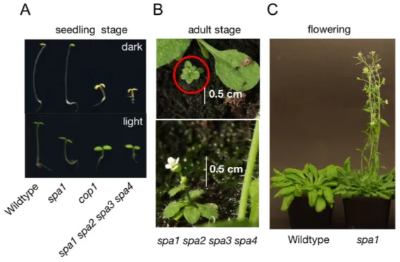
A: Visual phenotypes of wild-type, spa1, cop1 and spa1 spa2 spa3 spa4 mutant seedlings grown in darkness (top) or under Rc (bottom). In darkness, wild-type plants exhibit long hypocotyls and closed cotyledons (skotomorphogenesis), whereas in the light hypocotyl length is reduced and cotyledons are open and green (photomorphogenesis). Mutations in SPA1 result in enhanced photomorphogenesis in the light. cop1 mutants exhibit constitutive photomorphogenesis in light and darkness. spa1 spa2 spa3 s p a 4 mutant seedlings mimic the phenotype of the c o p 1 mutant and show constitutive photomorphogenesis (pictures taken from Hoecker, 2005)
B: Visual phenotypes of a spa1 spa2 spa3 spa4 adult plant which is strongly reduced in size (picture taken from Laubinger et al., 2004).
C: spa1 mutants flower earlier in SD than wild-type plants (picture taken from Laubinger et al., 2006).
Another important, light-regulated step in the plant life cycle is the induction of flowering. Arabidopsis thaliana is a facultative long day (LD) plant that flowers early in long days and late in short days (SD) (Coupland et al., 1998). One major regulator of photoperiodic induction of flowering is CONSTANS (CO) (Putterill et al., 1995). co mutants flower late in LD whereas over-expression of CO leads to an early-flowering phenotype (Putterill et al., 1995; Onouchi et al., 2000). CO encodes a transcription
Introduction
factor, which activates expression of FLOWERING LOCUS T (FT) and its homolog TWIN-SISTER OF FT (TSF) (Koornneef et al., 1991; Yamaguchi et al., 2005). FT protein can move from the leaves to the shoot apical meristem where it interacts with the transcription factor FD to regulate expression of floral genes (Corbesier et al., 2007; Jaeger and Wigge, 2007; Lin et al., 2007a; Mathieu et al., 2007).
Mutations in SPA1 cause an early-flowering phenotype in SD but not in long days (Ishikawa et al., 2006; Laubinger et al., 2006). Additional loss of SPA3 and SPA4 function further enhances the early-flowering phenotype of spa1 mutants (Laubinger et al., 2006). On the other hand, mutants that carry only a functional SPA1 gene flower like wild-type plants indicating that SPA1 alone is sufficient to repress flowering under SD conditions (Ishikawa et al., 2006; Laubinger et al., 2006). spa1 mutants accumulate high levels of FT mRNA while levels of CO are largely unaffected (Ishikawa et al., 2006; Laubinger et al., 2006). spa1 mutants flower only vxx
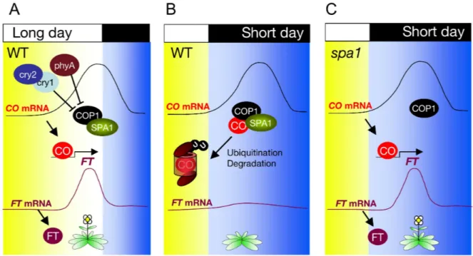
Figure 6: COP1/SPA complexes suppress flowering in SD by destabilization of CO.
A: C O mRNA levels in wild-type plants grown in long days (LD) accumulate in the end of late afternoon and coincide with light. Light stabilizes CO protein that activates FT mRNA transcription, an important inducer of flowering. Possible inactivation of COP1/SPA function could be due to physically interaction with photoreceptors like phys and crys.
B: In wild-type plants grown in short days (SD) the peak of CO mRNA levels occurs during night (darkness). COP1/SPA complex destabilize CO protein levels by targeting CO for degradation via the 26S proteasom. Without CO activation, FT mRNA levels are low abundant and flowering is not induced.
C: In spa1 mutant plants grown SD the peak of C O mRNA levels occurs during night (darkness).
However CO is stable and activates FT transcription, which results in the earlier flowering phenotype of spa mutants in SDs.
early in SD in the presence of functional CO (Ishikawa et al., 2006; Laubinger et al., 2006). CO protein levels are strongly elevated in spa1 and spa1 spa2 spa3 triple mutants (Laubinger et al., 2006; Jang et al., 2008).
In addition, SPA1 interacts with CO in vitro and in planta, raising the possibility that CO is an ubiquitination target of the COP1-SPA complex (Ishikawa et al., 2006;
Laubinger et al., 2006). This is in agreement with the observation that also cop1 mutants flower early in SD, that CO protein accumulates in a cop1 mutant, that COP1 interacts with CO and that COP1 ubiquitinates CO in vitro (Laubinger et al., 2006;
Jang et al., 2008).
Taken together, the COP1-SPA complexes play important roles in many different developmental stages. It seems that the contribution of the individual SPA genes differs in each developmental stage. SPA1 can suppress photomorphogenesis in the dark and the light and also regulates photoperiodic induction of flowering. SPA2 function is limited to dark-grown seedling and it has only very minor functions in later developmental stages that are influenced by light. SPA3 and SPA4 only have minor functions in dark- and light-grown seedlings, but they play important roles in regulating adult plant size.
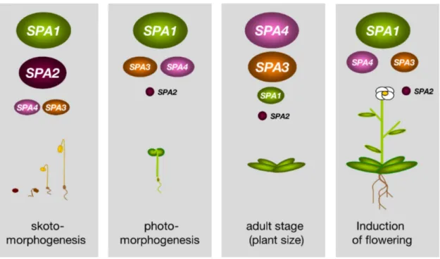
Figure 7: SPA proteins have redundant but also distinct functions in regulating plant development.
SPA1 and SPA2 predominate in suppressing photomorphogenesis in darkness, whereas SPA3 and SPA4 play only minor roles in this developmental stage. SPA1, and to minor extend SPA3 and SPA4, repress photomorphogenesis in the light. SPA3 and SPA4 are the most important SPA genes regulating adult plant size. SPA1 is sufficient for preventing early flowering in non-inductive short days (SD).
Introduction
I.4. Aims of this PhD thesis
Previous genetic analyses have shown that the four SPA genes have partially distinct functions in the control of seedling development in light/darkness, plant size and photoperiodic flowering. This thesis, therefore, aims to identify underlying molecular mechanisms for the functional diversification among SPA genes. Conceptually, differential SPA gene function might be caused by differences in SPA expression levels during development and/or differences among SPA protein sequences. These hypotheses are tested by:
(i) determining SPA transcript levels during development and in light vs. darkness
(ii) analyzing tissue-specificity of SPA expression by examining SPA-promoter::GUS transgenic plants
(ii) conducting promoter/cDNA swaps among SPA genes
The second aim of this thesis addresses a structure-function analysis of SPA1. While spa1 mutant alleles have indicated a functional requirement for the C-terminal WD- repeat domain, little was known about the N-terminal domains of SPA1. Therefore, N- terminal deletion-derivatives of SPA1 are generated and tested for their ability to complement the spa1 mutant phenotype.
II. Results
II.1. SPA1 structure-function analysis
Genetic analysis of diverse multiple spa-mutants showed that SPAs have overlapping but also distinct functions in regulating plant development. Based on their function and sequence similarity, SPA proteins can be divided into two classes. SPA1 and SPA2 proteins are closely related and both important to inhibit photomorphogenesis in dark-grown seedlings (Laubinger et al., 2004). SPA3 and SPA4 proteins share up to 85% identical amino acids and both are mainly involved in regulating growth of adult plants (Laubinger and Hoecker, 2003). All SPA proteins feature a similar protein domain arrangement: High similarity among all SPA proteins is found in their C- terminal regions that include WD-repeats, an important protein domain that is also characteristic for central repressor of light signaling, COP1. For SPA1 and COP1 it was shown that WD-repeats are essential for binding transcription factors like HY5 or HFR1 (Hoecker and Quail, 2001; Saijo et al., 2003; Yang et al., 2005a). All SPAs carry at least one or two predicted coiled-coil domains, which are known to mediate homo- or heterodimerization. Indeed, the predicted coiled-coil regions of SPA proteins are essential for binding COP1 as well as other SPAs (Hoecker and Quail, 2001; Laubinger and Hoecker, 2003; Saijo et al., 2003; Zhu et al., 2008).
While the role of the WD-repeat domain and the central coiled-coil domain of the SPA proteins is well established, the function of the N-terminus is completely unknown.
Although all SPA proteins exhibit similarity with serin-/ threonin- kinases in their N- terminus, it is the most unconserved region within the different SPA proteins. In addition, the N-terminus of SPA1 and SPA2 is much longer than that of SPA3 or SPA4 and carries two putative nuclear localization sequences (NLSs).
II.1.1. SPA1 N-terminus is not required for SPA1 function in dark- and light-grown seedlings, whereas the coiled-coil domain is essential
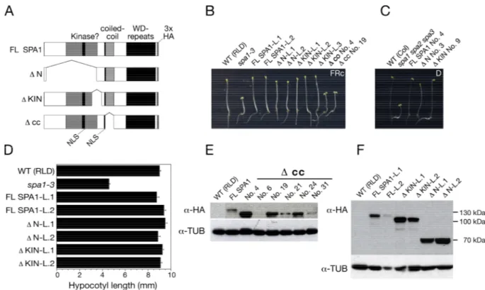
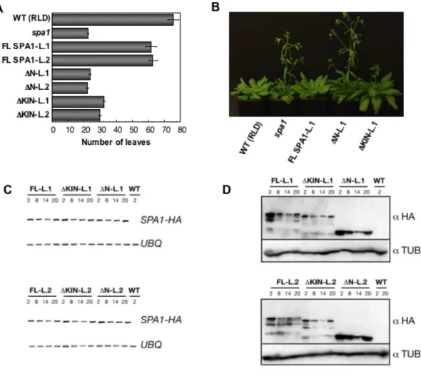
To examine if redundant and non-redundant SPA functions are based on differences in SPA protein structure, especially in the unconserved N-terminal region, it is important to know which structure is relevant for SPA1 function. To test whether the coiled-coil, the kinase-like domain or the whole N-terminus is important for SPA1 function in vivo, SPA1 deletion constructs were generated (Figure 8A): ΔN lacks most
Results
of the SPA1 N-terminus, ΔKIN lacks only a smaller part of the N-terminus, which contains highest sequence homology among the four SPA proteins (Laubinger and Hoecker, 2003). Another deletion-construct produces a SPA1 protein without the predicted coiled-coil domain (Δcc; Hoecker et al., 1999). As a positive control, the SPA1 cDNA coding for the full-length SPA1 protein was used (FL SPA1). The SPA1 deletion-derivates and the full-length cDNA were fused to a sequence encoding a triple influenza hemagglutinin (3xHA). All described constructs (IV.3.3 and Figure 8 A) were placed under the control of SPA1 endogenous 5´ (-2241 base pairs upstream of the SPA1 start codon) and 3´(1026 base pairs downstream of the stop codon) regulatory sequences.
To test which domain is necessary for SPA1 function all deletion-derivates and the full length SPA1 cDNA were transformed into spa1-3 mutant plants. Mutant spa1-3 seedlings show an enhanced de-etiolation in response to FRc with characteristic short hypocotyls and fully opened cotyledons (Hoecker et al., 1998; Figure 8 A).
Therefore, transgenic spa1-3 seedlings were analyzed under fluence rate of FRc.
The vast majority of all investigated transgenic T2 lines carrying FL SPA1, ΔN or ΔKIN deletion-derivates of SPA1 showed segregated seedlings with long hypocotyls and partially closed cotyledons in low FRc, like WT seedlings (Figure 8 B). Thus, expression of FL SPA1 or either its deletion-derivates ΔKIN or ΔN in spa1-3 mutants fully restored the WT phenotype (Figure 8 B). Hence, deletion of SPA1 N-terminus did not affect SPA1 protein function. Because SPA1 is also important for suppression of photomorphogenesis in darkness, SPA1 N-terminal deletion constructs were also transformed into spa1 spa2 spa3 triple mutants that show photomorphogenesis also in complete darkness. SPA1 proteins that lack either the kinase domain or the whole N-terminus fully rescued the phenotype of spa1 spa2 spa3 triple mutants indicating that the N-terminus of SPA1 is dispensable for SPA1 function also in darkness.
To be able to statistically quantify photomorphogenesis in the transgenic lines, complementing lines carrying single insertions were propagated to non-segregating T3 plants that are homozygous for the transgene. Hypocotyl lengths of around 30 seedlings of each T3 line were measured to determine complementation efficiency.
Measurements of two independent transgenic T3 lines for each construct showed that hypocotyls of FR-grown spa1 mutants carrying FL-SPA1, ΔKIN or ΔN deletion- derivates were as tall as those of WT. This results show that the SPA1 C-terminus including the predicted coiled-coil domain and the WD-repeats is sufficient to fully
rescue the spa1-3 mutant phenotype (Figure 8 D). Moreover, these results indicate that the putative NLS located in the SPA1 N-terminus, which is also deleted in ΔN transgenic lines, is not necessary for SPA1 function. If nuclear localization is necessary for SPA1 function these results suggest that the second NLS motif (KKKKASK) is sufficient for SPA1 function.
Figure 8: The N-terminal domain of SPA1 is not required for SPA1 function, whereas the coiled–coil domain is essential.
A: Schematic representation of full-length SPA1 (FL SPA1) and three SPA1 deletion mutants tagged with 3xHA. All constructs are under the control of endogenous SPA1 3´and 5´ regulatory elements.
B, D: Visual phenotypes (B) and hypocotyl lengths (D) of wild-type (WT), spa1-3 and transgenic spa1- 3 seedlings that were transformed with FL SPA1 or SPA1 deletion constructs shown in A. For each construct two independent transgenic lines are shown. For complementing lines in spa1-3 mutant background established non-segregating T3 generation are shown (L1 and L2). Non-complementing lines are shown in segregating T2 generation and presented with numbers (Δcc No.4). Seedlings were grown in 0.3 µmol m-2 s-1 FRc for 3 days. Error bars in D denote one standard error of the mean.
C: Visual phenotypes of dark-grown wild-type (WT), spa1 spa2 spa3 and transgenic spa1 spa2 spa3 seedlings containing FL SPA1, Δ N or Δ KIN deletion derivates, respectively.
E, F: Immunoblot analysis of transgenic spa1-3 seedlings transformed with Δ cc in T2 generation (E), FL SPA1, Δ N or Δ KIN constructs in T3 generation (F). Seedlings were grown for 3 days in 0.3 µmol m-2 s-1 FRc. For immunodetection the membranes were incubated with an α -HA antibody and subsequently rehybridized with an α -tubulin antibody.
On the contrary, spa1-3 seedlings expressing a SPA1 protein lacking the coiled-coil domain (Δcc) showed short hypocotyls and fully expanded cotyledons in FRc, like the spa1 mutant progenitor (Figure 8 B). Out of 39 analyzed transgenic lines none
Results
showed any rescue of the spa1 mutant phenotype. To verify that those non- complementing transgenic lines indeed expressed the Δcc SPA1 deletion-protein, six randomly selected T2 lines were chosen for immunoblot analysis. Five lines showed detectable amounts of expressed Δcc SPA1 protein. Therefore I conclude that the central coiled-coil domain is essential for SPA1 function. Western-blot analysis of complementing T3 lines expressing FL SPA1 showed that the amount of expressed SPA1 differs between the two lines tested. Even weaker expression of FL SPA1 protein was sufficient to rescue the spa1-3 mutant phenotype. The SPA1 deletion- proteins ΔKIN and ΔN were also detectable and in both analyzed T3 lines more abundant than FL SPA1 (Figure 8 F).
These results show that the SPA1 N-terminus including the potential kinase-like domain is not necessary for SPA1-dependent inhibition of photomorphogenesis in dark- or light-grown seedlings. In contrast, the coiled-coil domain is essential for SPA1 function.
II.1.2. SPA1 N-terminus is required to suppress flowering in short-days Apart from suppression of photomorphogenesis in seedlings, SPA1 also plays an important role in the regulation of flowering time. spa1 mutants flower earlier than WT under SD conditions, but not under LD conditions (Laubinger et al. 2006; Ishikawa et al., 2006). Recent studies show that SPA1 and COP1 suppress flowering in SD by destabilizing CO, an important regulator of photoperiodic induction of flowering time (Laubinger et al. 2006; Jang et al.2008).
To investigate whether the SPA1 N-terminal region is important to suppress flowering in short days, 10 to 15 plants of two independent spa1-3 T3 lines each carrying of FL SPA1, ΔN or ΔKIN were grown under SD conditions (eight hours light and 16 hours darkness). To determine flowering time the rosette leaves were counted at the time plants started bolting (Figure 9 A). Transgenic spa1-3 plants expressing FL SPA1 started to flower almost as late as the WT, indicating that FL SPA1 complemented the spa1 phenotype. In contrast, expression of ΔN in spa1-3 mutants was not able to rescue the spa1 mutant flowering time phenotype. These plants flowered as early as spa1-3 mutants in short days. Plants carrying Δ KIN deletion-derivate flowered slightly later than spa1-3 mutants, indicating that the ΔKIN deletion-protein has some residual function.
Figure 9: SPA1 N-terminus is necessary to inhibit photoperiodic induction of flowering in SD.
A: Flowering time in SD of two independent lines of genotypes shown in Figure 8 A.
B: Visual phenotypes of 78 days-old wild type (WT), spa1-3 mutants and spa1-3 mutants transformed with FL SPA1, Δ N or Δ KIN grown in SD.
C: Semi-quantitative RT-PCR of SPA1-HA and UBQ10 transcript in 21 days-old plants grown in SD (8 hours light followed by 16 hours darkness) and harvested at Zeitgeber 2, 8, 14, 20.
D: Immunoblot analysis of 21 days-old wild-type (WT), spa1-3 plants transformed with FL SPA1 (FL), Δ KIN or Δ N. For each construct two independent non-segregating T3 lines were analyzed. All plants were grown in SD and harvested at same Zeitgeber described in C. For immunodetection the membranes were incubated with an α -HA antibody and subsequently re-hybridized with an α -tubulin antibody.
The circadian clock influences flowering time and various genes involved in photoperiodic flowering are regulated in a diurnal or circadian fashion. Also for SPA1 a diurnal and circadian regulation was reported (Harmer et al., 2000; Ishikawa et al., 2006; Laubinger et al., 2006). To investigate whether deletion-derivates show proper diurnal regulation, transcript levels of FL SPA1 and its deletion-derivates ΔN and ΔKIN at were analyzed different time points of the day (Zeitgeber, ZT). On transcriptional levels, no differences in diurnal regulation were observed between the mRNA of FL SPA1 and the mRNA of the deletion-derivates ΔN and ΔKIN (Figure 9).
Results
All lines showed a slight increase of transcript abundance at ZT 2 and ZT 20 (Figure 9 C). On the protein level, FL SPA1 and the truncated SPA1 proteins showed highest protein abundance at ZT 2 and ZT 20. ΔN SPA1 deletion-proteins accumulated to higher levels than FL SPA1 but showed similar diurnal pattern (Figure 9 D).
Taken together, the N-terminus of SPA1 and the sequence including the kinase-like motif are important for SPA1 function in the control of flowering time. RT-PCR and immunoblot-analyses showed that SPA1 deletion-derivates do not exhibit an altered diurnal expression pattern on either transcriptional or protein levels.
II.2. SPA transcript analyses
Results of SPA1 structure-function analysis suggest that only N-terminal sequence diversity among the SPA proteins could not explain their partial distinct functions in light and dark grown seedlings. To investigate whether distinct SPA function are due to differential SPA expression SPA transcript levels were analyzed under various light regimes and developmental stages. Parts of the SPA transcript analyses were conducted during my diploma work and described in my diploma thesis, but are also presented in this work for a complete understanding of SPA transcript regulation.
II.2.1. SPA1 mRNA accumulates in blue light
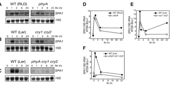
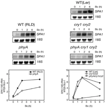
Previous studies showed that SPA1 transcript levels are increased in seedlings transferred from darkness to Rc or FRc (Hoecker et al., 1999). Because spa1 mutants are hypersensitive to Bc, the effect of Bc on SPA1 transcript levels was determined. To this end, RNA was isolated from dark-grown seedlings as well as seedling transferred to Bc and determined SPA1 transcript levels by RNA blot analysis.
After two hours of Bc treatment, SPA1 mRNA accumulated to levels 5- 10-fold higher than in darkness and sustained at high levels after prolonged Bc irradiation (Figure 10 A). SPA1 transcript levels were not influenced within the first 30 minutes after Bc treatment, but accumulated to high levels after 60 minutes. These results indicate that Bc has a similar influence on SPA1 mRNA levels as Rc and FRc (Fittinghoff et al., 2006).
Figure 10: Blue light increases SPA1 mRNA abundance.
A, B: Total RNA gel blot analysis (top) and quantification (bottom) of SPA1 transcript levels in seedlings that were transferred from darkness to 5 µmol m-2 s-1 Bc for 0-24h. Transcript levels were normalized by re-hybridization with an 18SrRNA-specific probe.
Blue light is perceived by the photoreceptors phyA, cry1 and cry2. phot1 and phot2 are also involved in blue light perception but it was shown that they do not play an important role in B regulation of transcripts (Briggs and Christie, 2002; Briggs and Spudich, 2005). To analyze which photoreceptor is responsible for SPA1 mRNA accumulation in Bc, SPA1 transcript levels were analyzed in WT, phyA, cry1 cry2 double and in phyA cry1 cry2 triple mutant seedlings exposed to low or high fluence rates of Bc.
Figure 11: Accumulation of SPA1 mRNA in high B depends on phyA, cry1 and cry2.
RNA-gel-blot analysis (A, B, C) and quantification (D, E, F) of SPA1 transcript levels in phyA, cry1 cry2 and phyA cry1 cry2 mutant seedlings in comparison to wild-type seedlings (WT: Ler, RLD).
Seedlings were transferred from darkness to 5 µmol m-2 s-1 Bc for 0-24 h. Transcript levels were normalized by re-hybridization with an 18SrRNA-specific probe.
Results
SPA1 transcript levels in phyA mutant seedlings exposed to high fluence rates of Bc light (5 µmol m-2 s-1 Bc) were similar to those of WT seedlings (Figure 11 A and D).
SPA1 mRNA accumulation in cry1 cry2 double mutant was somewhat different (Figure 11 B and E). Early accumulation of SPA1 transcript in cry1 cry2 double mutant was weaker than in WT, but still detectable, whereas after two hours of Bc exposure the amount of SPA1 mRNA was strongly reduced when compared to WT (Figure 11 B and E). Only in phyA cry1 cry2 mutant seedlings, Bc induced accumulation of SPA1 mRNA was completely abolished (Figure 11 C and F).
phyA mutants irradiated with lower fluence rates of Bc (0.3 µmol m-2 s-1 Bc) showed reduced amounts of SPA1 transcript whereas cry1 cry2 mutant seedlings did not show any differences in SPA1 mRNA accumulation when compared to WT seedlings (Figure 12). The relevance of cry1 and cry2 for SPA1 transcript accumulation under low fluence rate of Bc became only obvious in the phyA cry1 cry2 mutant, in which SPA1 transcript levels are not responsive to Bc anymore.
Taken together, Bc dependent accumulation of SPA1 transcript depends on functional phyA, cry1 and cry2. More specifically, cry1 and cry2 play predominant roles in high and phyA major functions in low fluence rates of Bc.
Figure 12: phyA, cry1 and cry2 act redundantly in controlling SPA1 mRNA levels in low B.
Total RNA-gel-blot analysis (at the top) and quantification (at the bottom) of SPA1 mRNA from wild- type (RLD/Ler), phyA (RLD), cry1 cry2 (Ler), phyA cry1 cry2 (Ler) mutant seedlings that were transferred from darkness to 0.3 µmol m-2 s-1 Bc for 0-6 hours. Transcript levels were normalized by re- hybridization with an 18SrRNA-specific probe.
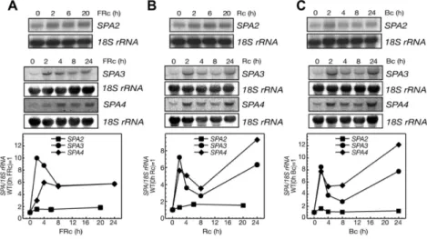
II.2.2. SPA3 and SPA4 but not SPA2 mRNA levels increase by light
For SPA1 transcript levels it was already shown that its transcript levels increase in FRc, Rc and Bc (Hoecker et al., 1999, this study). To further analyze if the partial distinct functions of SPAs in dark- and light-grown seedlings are based on different light-regulated SPA transcript abundance, the transcript levels of SPA2, SPA3 and SPA4 were analyzed under different light conditions. To this end, total RNA from dark-grown seedlings as well as seedling transferred to FRc, Rc or Bc were analyzed and SPA2, SPA3 and SPA4 transcript levels were determined by RNA blot analysis.
Similar to SPA1, the SPA3 and SPA4 transcript levels were strongly increased when dark-grown seedlings were transferred to light. Seedlings exposed to either high intensities of Rc, FRc or Bc, respectively, exhibited a 6- to 10-fold increased in SPA3 or SPA4 mRNA levels when compared to dark-grown seedlings (Figure 13). The increase of SPA3 and SPA4 transcript levels was detectable after two hours of light exposure and stayed at higher levels for all analyzed later time points. In contrast to that, exposure to light did not alter SPA2 transcript abundance (Figure 13).
Taken together, SPA1, SPA3 and SPA4 transcript levels are increased by light, nicely correlating with their function in regulating seedling development in the light.
SPA2 function is mainly restricted to seedling development in darkness, which is in agreement with SPA2 mRNA levels not being influenced by light.
Figure 13: SPA3 and SPA4, but not SPA2, transcript levels increase in light.
A, B, C: Total RNA-gel-blot analysis (top) and quantification (bottom) of SPA2, SPA3 and SPA4 accumulation in 4-day-old dark-grown seedlings (wild type RLD) transferred from darkness to 3 µmol m-2 s-1 FRc (A) 30 µmol m-2 s-1 FRc (B) or 5 µmol m-2 s-1 Bc (C) for 0-24 h. SPA2, SPA3 and SPA4 signals were normalized to 18SrRNA levels after phosphoimager quantification.
Results
To uncover which photoreceptors mediate light dependent accumulation of SPA3 and SPA4 transcript levels, SPA3 and SPA4 transcript levels were determined in WT and in several photoreceptor mutants. After one hour exposure to low intensities of FRc, SPA3 and SPA4 transcript levels were strongly induced in the WT. In phyA mutants, the increase of SPA3 and SPA4 mRNA levels was undetectable (Figure 14). These results are consistent with our knowledge that phyA is the only photoreceptor able to respond to FRc (Casal et al.1997).
Unlike FR, R light signaling depends on functional phyA, phyB, phyC, phyD and phyE whereby phyB plays the predominant role (Reed et al., 1994; Aukerman et al., 1997;
Mathews and Sharrock, 1997). In Rc light, SPA3 and SPA4 mRNA levels showed an early increase after one hour that was only slightly affected in phyA mutants, but completely lost in phyA phyB double mutant (Figure 14 B and D).
Figure 14: Accumulation of SPA3 and SPA4 mRNA in FRc or Rc requires functional phyA or phyB, respectively.
A, B Total RNA gel blot analysis and quantification of SPA3 (A) and SPA4 (B) mRNA levels from wild- type (RLD) and phyA mutant seedlings transferred from darkness to 0.3 µmol m-2 s-1 FRc for 0-2 h.
C, D Quantification of SPA3 (C) and SPA4 (D) transcript accumulation in wild-type (RLD), phyA, phyB, phyA phyB mutant seedlings that were transferred from darkness to 30 µmol m-2 s-1 Rc for 0-24 h. All blots were reprobed by an 18S rRNA–specific probe. SPA3 and SPA4 signals were normalized to 18S rRNA levels after phosphorimager quantification.
In low Bc, the increase of SPA3 and SPA4 transcript levels was not detectable in phyA and phyA cry1 cry2 triple mutant. cry1 cry2 mutants did not show altered regulation of SPA3 and SPA4 transcript abundance, indicating that phyA is the photoreceptor mediating increase of SPA3 and SPA4 transcript levels in low intensities of Bc. In high fluence rates of Bc, lack of phyA had no effect on SPA3 and SPA4 transcript levels. Only in phyA cry1 cry2 triple mutant seedlings B-light dependent increase in SPA3 and SPA4 transcript levels disappeared.
Taken together, SPA3 and SPA4 transcript levels increase in all investigated light qualities and show an expression pattern very similar to that of SPA1. phyA is responsible for increase of SPA levels in FR and low B light, whereas, phyB is mainly involved in the accumulation of SPA3 and SPA4 transcripts in Rc. cry1 and cry2 are mainly responsible for increasing SPA3 and SPA4 mRNA levels in Bc of high intensities.
II.2.3. SPA mRNA abundance partially correlates with its distinct functions during plant development
SPA transcript analysis implies that differences in the regulation of SPA expression might contribute to distinct SPA functions in dark- and light-grown seedlings. To test whether absolute amounts of SPA transcripts correlates with distinct functions in light-, dark-grown seedlings and adult plants, a comparison of SPA transcript abundance were performed. While SPA1 and SPA2 play predominant roles in light and dark-grown seedling, SPA3 and SPA4 mainly regulate vegetative adult plant growth. Therefore, poly(A)+ RNA from seedlings grown for 4 days in darkness or FRc as well as from adult rosettes leaves were isolated and SPA transcript levels were subsequently determined by RNA blot analysis. In order to make band intensities of the different SPA genes comparable, SPA signal were normalized with the respective UBQ10 signals. This normalized ratio was further corrected for differences in SPA probe sizes (see Materials and Methods for details).
In dark-grown seedlings, SPA1, SPA2, SPA3 and SPA4 transcripts were relatively low abundant. However, SPA2 is the most abundant SPA transcript in dark-grown seedlings (Figure 15). In light grown seedlings, SPA1, SPA3 and SPA4 transcripts are more abundant than in dark-grown seedlings while SPA2 transcript levels do not differ between light- and dark-grown seedlings. A direct comparison of SPA mRNA levels revealed that SPA1 and SPA3 are the most abundant SPA transcripts in light
Results
grown seedlings (Figure 15 B). In adult plants, SPA3 and SPA4, but not SPA2 mRNA levels are strongly increased compared to the seedling stage. Levels of SPA1 mRNA in adult plants were almost unchanged when compared to the levels in light-grown seedlings. SPA3 was the most abundant transcript in adult plants. Taken together, comparison of SPA transcript levels showed that SPA2 mRNA levels are largely unaffected among the different developmental stages analyzed. In contrast to that, SPA1, SPA3 and SPA4 mRNA levels are very low in dark-grown seedlings, higher in light-grown seedlings and reach the maximum in adult plants. These expression patterns partially correlate with the known, distinct SPA functions in regulating plant development.
Figure 15: Analysis of SPA1-SPA4 transcript levels during plant development
A: Comparative poly(A)+RNA gel blot analysis of SPA1, SPA2, SPA3 and SPA4 mRNA levels in seedlings grown in darkness or Rc (30 µmol m-2 s-1) for 3 days or in 4 week-old adult plants. SPA mRNAs were detected with SPA-specific probes (for detail see material and methods). For normalization, blots were reprobed with an UBIQUITIN 10 (UBQ10) -specific probe.
B: Quantification of the SPA transcript levels shown in A.
II.3. SPA Promoter GUS analyses
II.3.1. SPA1 and SPA2 but not SPA4 promoters are strongly active in the roots of young plants
While RNA-blot analysis gains important information about SPA mRNA abundance under various light conditions and developmental stages, the weakness of this approach is that especially spatial distribution of SPA transcripts within a tissue cannot be resolved. However, this is of utmost importance because some targets of the COP1-SPA complexes like CO are only localized in specialized cell types. To uncover the spatial distribution of SPA expression, a promoter-reporter-gene analysis
was conducted with the focus on SPA1, SPA2 and one member of the highly redundant SPA3/SPA4 subgroup, SPA4. The reporter gene GUS was transformed under the control of either SPA1-, SPA2- or SPA4- 5´regulatory sequences in wild- type plants (Figure 16A). For each construct approximately 70 transgenic T1 plants were analyzed after selection on kanamycin plates. The reason for this high number of transgenic T1 plants is that not only the promoter but also the insertion site can influence the GUS expression pattern. All following results were found in at least 50%
of all analyzed lines and therefore likely represent the native SPAX promoter activity.
For the analysis of SPA::GUS expression in seedlings, at least 20 to 30 independent T2 lines were analyzed.
Figure 16: Promoter of SPA1 and SPA2 are active in roots of seedlings and young plants.
A: Schematic representation of used constructs. The reporter-gene GUS was expressed under 5`regulatory sequences of SPA1, SPA2 or SPA4.
B: Visual phenotypes of wild-type (WT) and transgenic T2 plants expressing GUS under the control of SPA1, SPA2 or SPA4 promoters described in A.
C: Visual phenotypes of segregating transgenic dark-grown wild-type seedlings expressing GUS under control of SPA1, SPA2, SPA4 promoters in T1 generation. Plants were grown for two weeks on kanamycin plates.
In dark-grown seedlings, SPA1 and SPA2 promoters were predominant active in cotyledons, whereas pSPA4::GUS expression was not detectable in dark-grown seedlings (Figure 16). Young plants expressing GUS under the control of either
Results
SPA1 or SPA2 promoter exhibited GUS staining in roots, hypocotyls and in true leaves (Figure 16). T1 plants carrying the pSPA4::GUS transgene had to be stained two times longer than those carrying SPA1 or SPA2 promoter, which indicates that the SPA4 promoter is less active than those of SPA1 and SPA2.
Figure 17: Promoter of SPA1 and SPA4 are active in vascular bundles of leaves.
Rosette leaves (first two columns), cauline leaves (third column) and inflorescence (fourth column) of 6 week-old transgenic plants expressing GUS under the control of SPA1-, SPA2- or SPA4- promoter. All plants were selected on kanamycin plates, transferred to soil and grown in LD for three weeks. All tissues were stained at 37°C for 8 hours.
Obvious difference in SPA promoter activity was found in the roots. pSPA1::GUS and pSPA2::GUS reporter constructs were strongly expressed in roots indicated by detectable GUS staining after only a view minutes, whereas GUS activity controlled by SPA4 promoter was not or only barely detectable even after several hours of staining (Figure 16). Analysis of older rosette leaves showed that promoters of SPA4 and SPA1 were strongly active in vascular bundles (Figure 17). In contrast, SPA2 promoter conferred strong activity in the leaves, but its expression is not restricted to vascular tissues. SPA genes are also expressed in cauline leaves and stems (Figure 17). All analyzed SPA promoters were active in reproductive tissues. GUS expression was detectable in all flower organs as well as young siliques (Figure 17).
Taken together, the SPA-promoter::GUS analysis demonstrates that the selected SPA1 and SPA2 5´regulatory regions confer to strong expression in roots of young plants, whereas SPA4 promoter show no or only weak detectable activity in roots.