Analysis of trichome pattern formation in Arabidopsis thaliana:
The role of KAKTUS in protein degradation
Inaugural-Dissertation
zur
Erlangung des Doktorgrades
der Mathematisch-Naturwissenschaftlichen Fakultät der Universität zu Köln
vorgelegt von
Elena Galiana Jaime
aus Valencia
2007
Berichterstatter: Prof. Dr. Martin Hülskamp Prof. Dr. Ulf-Ingo Flügge
Prüfungsvorsitzender: Prof. Dr. Wolfgang Werr
Tag der mündlichen Prüfung: 16. April 2007
Acknowledgements
This PhD thesis would not exist without the support of many individuals that have helped me all those years to make my dream of becoming a doctor come true.
My deepest gratitude goes to Professor Dr. HÜLSKAMP for his stimulating advice and constructive criticism during the years of research. I am thankful for the opportunity given to me to develop my own ideas and to work independently.
I extend my gratitude to Professor Dr. FLÜGGE, Professor Dr. WERR and Dr. LUDEWIG who have found time to evaluate this thesis.
I gratefully acknowledge the advice of Dr SCHELLMANN. His knowledge added much to my understanding of the importance of depletion to the trichome pattern formation.
This study could be performed thanks to the assistance of the technicians of the department:
Birgit KERNEBECK, Britta MÜLLER, Irene KLINKHAMMER, Bastian WELTER and Uschi CLAßEN. The personal encouragement and actual help I received from them has made it possible to complete my studies during my time in the university of Cologne.
I am deeply appreciative to many of my colleagues in the university for their understanding and indispensable cooperation. I have learned very much from the long discussions with the members of the Patterning Group: Dr. BOUYER, Dr. PESCH, Rachappa BALKUNDE, Burcu DARTAN, Simona DIGIUNI and Katja WESTER as well as from the members of the Escort Complex Group: Dr. SCHELLMANN, Dr. SPITZER, Aneta SABOVLJEVIK, Mojgan SHAHRIARI and Channa KESHAVAIAH.
I am indebted to Dr. SAEDLER, Dr. PESCH, Dr. SCHELLMANN and Dr. SANTAELLA TENORIO for the critical reading of the whole text and for the valuable contribution to the clearness of the manuscript.
I would like to express my indebtedness to Professor Dr. HERZOG who suggested and stimulated my first steps in the area of trichome development, several years ago, at the university of Grenoble. I realize the great importance of the support of Dr. PERAZZA, Dr.
BONNEVILLE and Dr. VACHON to educate me on what critical scientific reasoning actually means.
In the first three years of my investigation I received financial support from the Graduate School for Molecular Analysis of Developmental Processes for which I express my gratitude.
Several friends have made my life very pleasant during the last four years. Many from them could call themselves experts in the development of trichomes even though they never studied
Acknowledgements
biology, just because they have been curious and patient to listen to me talking about my project. Very often, they made me see aspects from the question that I hadn’t considered.
Beyond any doubt, they deserve to see their names associated to this work. I am also very grateful to those that were there for me any time I needed them. I am talking about Nele DICKMANN, Santiago GÁLVEZ SETTIER, Deborrah GHIRMAI, Delphine GIRAUD, Pari HEIDENREICH, Nermin KOCALAN, Verónica MAURINO, Claudia RAMBKE, Rainer SAEDLER, Mariana SAIGO, Marcella SANTAELLA TENORIO, Enrique TORRES PRIETO and Christina WEINL.
And, last but not least, I was lucky to be working in the best office one can find in the whole universe, thanks to Moola MUTONDO, Silke UHRIG, Valerie MACH, Cho-Chun HUANG and Cordula JÖRGENS, and to the “guests”: Simona DIGIUNI, Philipp THOMAS and Claudia RAMBKE.
This work is dedicated to five persons who are proud of who I am, and have always supported my choices:
Elvira, my mother Ana, my sister Elvira, my grandmother Carmen and Asunción, my grandaunts
Table of contents
Table of contents
Table of contents……….... I Abbreviation list………...………IV Figure index ………...……...V Abstract………..…………...VI Zusammenfassung ………..…VII
A Trichome Morphogenesis... 1
A 1 Introduction ... 2
1.1 General description of the trichome morphogenesis... 2
1.2 The hormone gibberellin is implicated in trichome branching ... 3
1.3 Aim of this work ... 5
A 2 Results ... 7
2.1 Genetic mapping of POLYCHOME (PYM) ... 7
2.1.1 Generation of the mapping population... 7
2.1.2 Creation of markers around PYM... 7
2.1.3 PYM is located between T11A7 and F7D19 ... 7
2.2 Phenotypic characterization of POLYCHOME (PYM) ... 8
2.2.1 Hypocotyl development and stem height ... 9
2.2.2 Trichome development... 11
2.2.3 Flowering ... 12
A 3 Discussion ... 14
3.1 Genetic mapping of the POLYCHOME gene... 14
3.2 Morphological characterization of the polychome mutant and analysis of its dependency on the gibberellins... 15
B Trichome Pattern formation... 18
B 1 Introduction...19
1.1 Trichome initiation in Arabidopsis thaliana... 19
1.2 A model to explain two-dimensional pattern formation ... 19
1.3 Is protein degradation relevant for trichome pattern formation? ... 20
1.4 Elements of the trichome pattern system and their molecular nature ... 21
1.5 Outlook and aim of the work... 23
B 2 Results ... 25
2.1 At4g38600 is the KAKTUS gene... 25
2.2 The kaktus plants carry a mutation in a patterning... 25
2.3 The 35S:YFP:KAKTUS gene is not able to rescue the aberrant trichome patterning phenotype on the kaktus mutant plants ... 26
2.4 KAKTUS interacts in yeast and in Arabidopsis thaliana with proteins implicated in trichome patterning ... 28
2.5 KAKTUS and GLABROUS1 interact genetically in the trichome initiation
pathway. ... 31
2.6 The degradation of GL1 is mediated by the 26S proteasome ... 32
2.7 KAKTUS is implicated in the degradation of GL1... 33
B 3 Discussion ... 35
3.1 KAKTUS plays a role in the trichome patterning ... 35
3.2 KAKTUS is a HECT E3 ligase very likely implicated in the degradation of regulators of trichome initiation... 36
3.3 GL1 is degraded via the 26S proteasome in a KAK-dependent manner ... 37
3.4 The KAKTUS-dependent degradation of GL1 is relevant for trichome pattern formation. ... 38
3.5 Towards a new model to explain trichome pattern formation: the key role of depletion... 39
3.6 Outlook... 41
C 1 Material... 43
1.1 Chemicals, antibiotics ... 43
1.2 Enzymes, primers and kits ... 43
1.3 Cloning vectors ... 43
1.4 Microbiotical strains... 43
1.5 Plant lines ... 44
C 2 Methods... 44
2.1 Plant work...44
2.1.1 Plant growth conditions... 44
2.1.2 Crossing of plants... 44
2.1.3 Plant transformation ... 44
2.1.4 Seed surface sterilisation and subsequent plant treatment ... 45
2.1.5 Selection of transformants... 45
2.1.6 Gibberellin treatment... 45
2.1.7 Uniconazol-P treatment... 45
2.1.8 MG132 (Z-Leu-Leu-Leu-H) treatment ... 46
2.2 Microscopy and cytological methods... 46
2.2.1 Microscopy... 46
2.2.2 Yeast two-hybrid assay ... 46
2.2.3 BiMolecular fluorescent complementation ... 46
2.2.4 Kinetics of protein degradation ... 47
2.3 Molecular-biological methods: ... 48
2.3.1 RNA isolation... 48
2.3.2 Reverse transcription... 48
2.3.3 Semiquantitative RT-PCR... 48
2.3.4 Genomic DNA preparation ... 49
2.3.5 Plasmid DNA preparation from bacteria... 49
2.3.6 DNA-manipulation... 50
Table of contents
2.3.7 Cloning of the KAK cDNA... 50
2.3.8 Primers used for the mapping of PYM... 51
2.4 Biological-chemical methods:... 51
2.4.1 Protein extraction ... 51
2.4.2 Western blotting ... 51
References ... 53
Erklärung. ... 61
Lebenslauf ... 62
Abbreviations list
° degree Celsius
% percent μ micro μm micrometer μM micromolar
aa Amino acid
AD Activation domain AtKLI5 Arabidopsis thaliana KAKTUS LIKE 5
ATP Adenosine triphosphate BAC bacterial artificial chromosome BD Binding domain
bHLH basic helix-loop-helix BiFC Bimolecular fluorescent complementation
bp base pair
CaMV 35S promoter from Cauliflower Mosaic virus C DNA-content of a haploid genome
cDNA complementary DNA CDS coding sequence CFP cyan fluorescent protein
cm centimeters Col Columbia
COP Constitutive morphogenesis CPC CAPRICE
DAPI 4',6-Diamidino-2-phenylindole dCAPs derived cleaved amplified polymorphic sequence
DNA Desoxyribonucleic acid DUB Deubiquitinating enzyme
E3 Enzyme 3
e.g. exempli gratia [Lat.] for example
EGL3 ENHANCER OF GLABRA3
et al. et alterni [Lat.] and others
ETC 1/2 ENHANCER OF TRY CPC1/2 Fig Figure GA gibberelic acid GAI GA INSENSITIVE GAL galactosidase GFP green fluorescent protein
GL1/3 GLABRA1/3 HECT homologous to E6-associated protein carboxyl terminus k kilo
kbp Kilo bp
kDA kilo Dalton KAK KAKTUS Ler Landsberg erecta
LUC Photinus-luciferin 4-monooxygenase LUCIFERASE
mm millimetre mRNA messenger RNA MS Murashige and Shoog
n number p promoter PCR polymerase chain reaction
PYM POLYCHOME RACE-PCR Rapid amplification of cDNA ends
RGA REPRESSOR OF GA1 RNA ribonucleic acid RPM rounds per minute RT-PCR reverse transcription PCR
SSLP simple sequence length polymorphism SPY SPINDLY TTG1/2 TRANSPARENT TESTA GLABRA1/2 TRY TRIPTYCHON UPL1/7 Ubiquitin protein like 1/7
YFP yellow fluorescent protein YFPc/n C/N terminal sub-fragment on the YFP gene
WT wild type
All gene and mutant names are written in italics.
WT genes are written in capital letters.
Proteins are written in non-italic letters.
Figure index
Figure index
Fig. 1: Steps in trichome development
Fig. 2: Phenotype of the kak-like mutants potentially implicated in the gibberellin pathway
Fig. 3: The transduction of the gibberellic acid message Fig. 4: Mapping of POLYCHOME
Fig. 5: Phenotypical characterization of the pym hypocotyl
Fig. 6: Number of trichomes per mm2 in the second pair of leaves Fig. 7: Kinetics of flowering of Ler wild-type and pym mutants Fig. 8: Pattern formation by autocatalysis and long-range inhibition
Fig. 9: Activator-inhibitor model applied to the trichome patterning system Fig. 10: Involvement of KAKTUS in the establishment of trichome patterning
Fig. 11: Interaction of KAK with the proteins implicated in the trichome patterning in yeast two-hybrid assays
Fig. 12: Interaction profile of patterning proteins with KAKTUS in bimolecular fluorescence complementation assays
Fig. 13: Study of the dependence of GL1 degradation on the 26S proteasome Fig. 14: Kinetics of the degradation of GLABROUS1
Fig. 15: Model to explain the role of the KAKTUS-dependent degradation of GL1 in the stabilization of the trichome pattern
Abstract
In this PhD thesis I have studied two different aspects of the cell differentiation: the mechanisms underlying pattern formation and the hormonal regulation of cell morphogenesis.
I concentrated on the development of trichomes, the leaf hairs of Arabidopsis thaliana, as a model system. This cell type is very well suited for the analyses of those processes because it presents a simple two-dimensional spacing pattern on the rosette leaves and develops a predictable and characteristic shape.
The gibberellic acid is implicated in the regulation of cell morphogenesis. In the trichome, this regulation takes place via a SPINDLY-dependent pathway. I have investigated the role of POLYCHOME in this pathway. I have attempted to map it, and I could provide evidence for its implication in the transduction of the GA signal leading to the development of trichome branches in Arabidopsis thaliana.
In the second part of my thesis I examined the part played by protein degradation in the processes that control the formation of a regular trichome spacing pattern on the leaf surface.
My thesis revealed the implication of KAKTUS, which encodes a protein homologous to HECT E3 ligases, in the establishment of a wild-typical patterning. I showed that GL1 is degraded in a KAK-dependent manner through the 26S proteasome. Finally, I could demonstrate the physiological relevance of this regulated depletion for trichome initiation.
These observations are summarised into a model that suggests that the function of KAK in the early events during trichome pattern formation is to stabilize the incipient trichome pattern.
Zusammenfassung
Zusammenfassung
In dieser Doktorarbeit habe ich zwei verschiedene Aspekte der Zelldifferenzierung untersucht: den Mechanismus dem die Musterbildung unterliegt und die hormonelle Regulation der Zellmorphogenese. Ich habe mich auf die Entwicklung von Trichomen, den Blatthaaren von Arabidopsis thaliana, als Modellsystem konzentriert. Dieser Zelltyp ist für die Analyse dieser Prozesse sehr geeignet, da er ein simples zweidimensionales Abstandsmuster präsentiert und eine vorhersehbare und charakteristische Form entwickelt.
Die Gibberellinsäure ist an der Regulation der Zellmorphogenese beteiligt. In den Trichomen findet diese Regulation über den SPINDLY- abhängigen Signalweg statt. Ich habe die Rolle von POLYCHOME in diesem Signalweg untersucht. Ich habe versucht es zu kartieren und ich konnte den Beweis dafür erbringen, dass es an der Weiterleitung des GA-Signals, der zur Entwicklung von verzweigten Trichomen in Arabidopsis thaliana führt, beteiligt ist.
Im zweiten Teil meiner Arbeit habe ich untersucht welche Rolle der Proteinabbau im Prozess der Bildung eines geordneten Trichommusters auf der Blattoberfläche spielt. Meine Arbeit zeigte, dass KAKTUS an der Bildung des wildtypischen Musters beteiligt ist. KAKTUS kodiert ein Protein, das der HECT E3 Ligase homolog ist. Ich konnte zeigen, dass GL1 KAK- abhängig durch das 26S Proteasom abgebaut wird. Schließlich konnte ich die physiologische Relevanz dieser regulierten Abnahme für die Trichominitiation demonstrieren. Diese Beobachtungen werden in einem Modell zusammengefasst, das darauf hinweist, dass die Funktion von KAK in der frühen Trichommusterbildung darin liegt das beginnende Trichommuster zu stabilisieren.
A
Trichome
Morphogenesis
Introduction A
A 1 Introduction
The nature presents an amazing diversity of shapes, ranging from isomorphic bacteria to spermatozoids. The fascinating question of how a cell establishes, regulates and maintains its shape has motivated a wide field of research. Plants represent a good model for the study of those questions, because it is relatively easy to observe some of their cells while they are performing their morphogenesis. In Arabidopsis thaliana, a plant model system, the development of leaf trichomes (Hülskamp et al., 1994; Folkers et al., 1997; Mathur and Hülskamp, 2002), root hairs (Carol and Dolan 2002), pollen tubes (Hepler et al., 2001) and stomata cells (Nadeau and Sack 2003) have been studied in great detail.
The trichomes (also called leaf hairs) of Arabidopsis thaliana are particularly well suited to study cell morphogenesis. Firstly, they are single cells that emerge from the epidermal layer and are therefore easily accessible for observation and experimentation. Secondly, they present a predictable, characteristic and complex stellate shape. Thirdly, it is possible to define genetic distinct steps in trichome development (Hülskamp et al., 1994). Fourthly, trichomes are dispensable for survival of the plant under laboratory conditions.
Figure 1: Steps in trichome development. Scanning electron micrographs of developing wild-type trichomes. (A) Incipient unbranched trichome. (B) Trichome with primary branch point. Note orientation of the branches with respect to the basal-distal leaf axis. (C) Trichome with primary and secondary branch. (D) Mature trichome. (Figure modified from Schwab et al., 2000).
1.1 General description of the trichome morphogenesis
Hülskamp et al. (1994) have described the steps involved in the development of a trichome (Fig1). Trichomes are the first cells that start to differentiate on leaf protoepidermis. Such differentiating cells undergo three rounds of endoreduplication (DNA synthesis without
cytokinesis) that bring the DNA content from 2C to 16C. Thus, the first morphological evidence of a cell entering this pathway is the increase of the nuclear volume. Then, the incipient trichome expands out of the leaf plane and initiates a first branching event. As a consequence, at this stage a plant hair is composed of a stem and two branches. Subsequently, a fourth round of endoreduplication occurs, which brings the DNA content to the one of a mature trichome (32C). The next step of the development of a trichome is the formation of a second branching point on the branch pointing to the distal end of the leaf. Finally, on the last maturation step of the plant hair, incrustations appear on the surface of the cell. The nucleus of a mature trichome has a triangular shape, and is characteristically located under the second branch point.
1.2 The hormone gibberellin is implicated in trichome branching
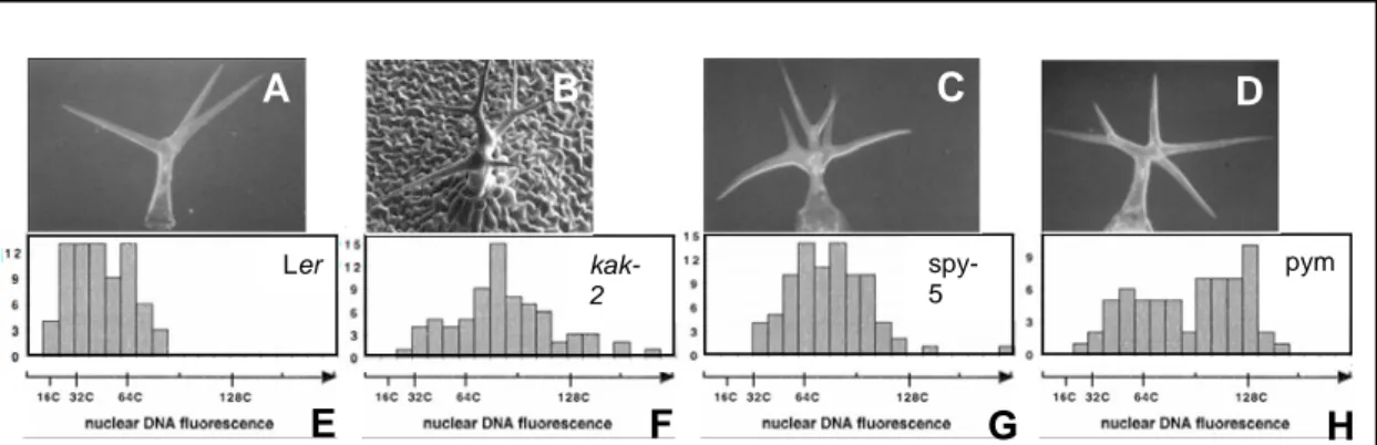
To date, two studies have focused on the relationship between trichome branching and the gibberellins. The link has been first established by the analysis of glabrous1.2, a weak allele of the MYB transcription factor GLABROUS1 (GL1), which develops two-branched trichomes. The promoter of this gene is positively regulated by the gibberellins, indicating that this hormone may regulate trichome branching by up-regulating GL1 (Herman et al., 1989, Esch et al., 1994, Chien and Sussex, 1996; Perazza et al., 1998). More recently, some genes that lead when mutated to an increased DNA content and to the formation of up to eight branches (Fig. 2) have been implicated in the transduction of the gibberellic message. These genes are KAKTUS, SPINDLY and POLYCHOME (Perazza et al., 1999). KAKTUS and SPINDLY are positively implicated in the sensing of the gibberellic acid message: the spy mutant is able to germinate in the absence of the hormone (Jakobsen et al., 1993), whereas an application of gibberellins the kak mutant leads to an abnormal elongation of the hypocotyl (Downes et al., 2003). POLYCHOME seems to act downstream from SPY: the double mutant pym spy displays the same number of trichome branches than the parental lines alone (Perazza et al., 1999). Therefore, PYM may be also involved in the sensing of the GA message.
Since the double mutant kak-2 spy-5 presents no additivity of the overbranching phenotype, it has been suggested that SPINDLY and KAKTUS function on a linear way. Genetic analyses suggest that PYM acts to repress branch formation through a pathway independent of KAK:
the double mutant kak-2 pym is highly overbranched compared to the parental lines. (Perazza et al., 1999).
Introduction A
Ler kak- pym
2
spy- 5
A B C D
E F G H
Figure 2: Phenotype of the kak-like mutants potentially implicated in the gibberellin pathway.
Scanning electron micrographs of mature trichomes (A to D) and DNA fluorescence distributions of trichome nuclei (E to H) (A) Three-branched wild-type trichome (Ler). (B) kak-2 mutant trichome (C) spy-5 mutant trichome (D) pym mutant trichome. DNA fluorescence distributions in wild-type Ler trichome nuclei (E), in kak-2 mutant trichome nuclei (F), in spy-5 mutant trichome nuclei (G) and in pym mutant trichome nuclei (H). (Figures modified from Perazza et al., 1999 and Downes et al., 2003).
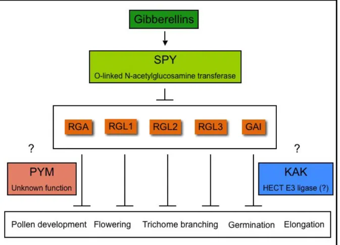
GA signalling operates as a de-repressible system moderated by DELLA-domain proteins, which are transcriptional regulators that repress GA responses (Figure 3). Five DELLA protein genes have been identified in Arabidopsis: GA-insensitive (GAI), Repressor of ga1.3 (RGA), RGA-like1 (RGL1), RGL2 and RGL3, with RGA and GAI being the major repressors during vegetative growth and floral induction (Richards et al., 2001, Olszewski et al., 2002).
The regulatory steps used by the plant to regulate trichome branching through the gibberellins are unclear. Nonetheless, the cloning of SPY and KAK has provided with strong clue about it.
SPY encodes a O-linked N-acetylglucosamine transferase that transfers O-Glc-Nac residues to target proteins (Jakobsen et al., 1996). Based on this, SPY has been hypothesized to modify DELLA proteins in response to the GA (Thomas et al., 2004). One of the target proteins of SPY might be a DELLA protein implicated in the repression of trichome branch formation, and therefore the gibberellins might activate trichome branching by positively regulating SPY, which in turn modifies the DELLA proteins and eventually leads to the activation of trichome branching. KAK encodes for a HECT domain protein and thus may be implicated in the ubiquitin degradatory pathway (Downes et al., 2003, El Refi et al., 2003). Since in response to GA DELLA proteins are rapidly degraded via the ubiquitin proteasome pathway (Sun and Gubbler, 2004), it is reasonable to hypothesize that the role of KAK in trichome branch formation is to degrade activators of this trichome developmental stage in response to the GAs. It appears that PYM is the only gene described to be implicated in the regulation of trichome branching through the GAs that has not been cloned yet. This impeaches to draw a complete picture about this regulatory process.
1.3 Aim of this work
The gibberellic acid is implicated in the regulation of cell morphogenesis. In the trichome, this regulation takes place by the means of a SPINDLY-dependent pathway. To date, no investigation has been carried out to improve our understanding on the mechanisms by which this regulatory cascade leads to the development of branches in the trichome cell. This is partially due to the fact that not all the proteins implicated in the transduction of this hormonal message have been cloned and studied.
Figure 3: The transduction of the gibberellic acid message. The gibberellins regulate developmental processes through the action of SPY. This acetylglucosamine transferase is supposed to transfer O-Glc-Nac residues to the five DELLA proteins existing in Arabidopsis thaliana (represented in orange boxes) in response to the hormone. The DELLA proteins repress the processes indicated in the box, among others. PYM and KAK are implicated in the regulation of this pathway downstream of SPY. Nonetheless, their exact function has not been elucidated yet.
Genetic analysis have shown that PYM plays a role in the hormonal regulation of trichome branching downstream of SPY. This point has not been confirmed by further experimental data. Also, the molecular nature of this gene has not been described. In order to increase the knowledge on the hormonal regulation of cell morphogenesis, it is important to confirm the
Introduction A
implication of POLYCHOME in the gibberellin pathway and to determine its molecular function.
To better understand the role of the gibberellic acid in the context of cell morphogenesis in Arabidopsis thaliana, I focused my thesis work on the study of POLYCHOME, a gene implicated in the trichome branching. I proceeded to a genetic mapping of PYM, as well as to a detailed morphological characterization of the pym mutant and to an observation of the relationship between PYM and the gibberellins.
A 2 Results
2.1 Genetic mapping of POLYCHOME (PYM)
To understand the exact molecular function of the PYM protein, I initiated a genetic mapping of PYM, exploiting positional cloning.
2.1.1 Generation of the mapping population
To generate the mapping population, a Ler pym plant was crossed to a Col-0 wild-type plant and the offspring was screened in the F2 for individuals presenting a pym trichome phenotype.
The mapping was performed with 850 plants generated by two independent crosses.
2.1.2 Creation of markers around PYM
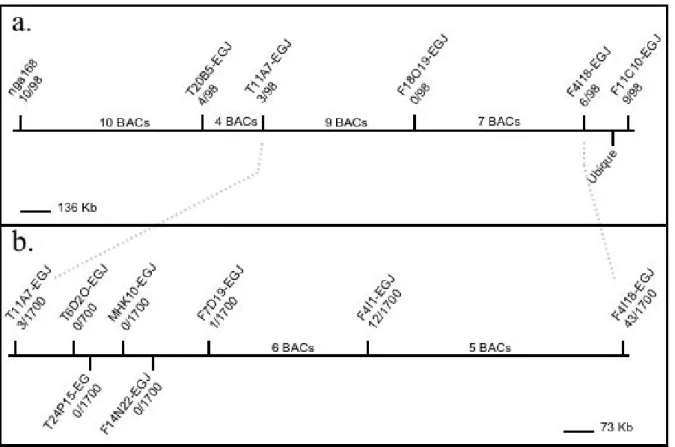
Previous studies have shown that PYM is located at the bottom of chromosome II. pym revealed a linkage to the markers Ubique and nga168 (Perazza et al., 1998). Therefore, I analyzed the mapping population with molecular markers located either in the proximity or between those two. However, the available molecular markers (nga168 and Ubique) for the region of interest on Chromosome II were not sufficient to map the gene. So, I designed new markers based on either simple sequence length polymorphism (SSLP) or derived cleaved amplified polymorphic sequence (dCAPS) techniques. The corresponding data about existing polymorphisms between Col-0 and Ler were obtained from the ‘Monsanto Arabidopsis thaliana polymorphism and Ler sequence collection’.
2.1.3 PYM is located between T11A7 and F7D19
The chromosomal walking was performed in two steps. Firstly, to get an insight into the location of PYM on the second chromosome and to confirm the published localization of this gene, I analyzed 49 plants with 6 SSLP markers located within 37 Bacterial Artificial Chromosomes (BACs). Their analysis showed that no chromatide had recombined between the PYM gene and F18O19-EGJ. Hence, PYM is located between F11A7- EGJ and F4I18-EGJ (Figure 3a). Secondly, to map PYM more precisely, I analyzed the 850 plants comprising the entire mapping population with F11A7- EGJ, F4I18-EGJ and with 6 markers located within
Results A
those two (19 BACs). Three plants and one plant had recombined between pym and T11A7- EGJ and between this gene and F7D19-EGJ, respectively. It was not possible to detect any further recombination in the area between those markers (Figure 3b). To conclude, these data show that the genetic mapping allowed to map PYM between T11A7-EGJ and F7D19-EGJ, but was not sufficient to localize it precisely.
Figure 4: Mapping of POLYCHOME. (a) POLYCHOME is located on the chromosome II between T11A7-EGJ and F4I18-EGJ. (b) POLYCHOME is located on the chromosome II between T11A7-EGJ and F7D19-EGJ. The horizontal lines represent fragments of chromosome II. Each vertical line represents one bacterial artificial chromosome (BAC). The markers used for the mapping are shown on the corresponding BAC. The ratios indicate the number of recombinant events that took place between PYM and the given marker for the tested population.
2.2 Phenotypic characterization of POLYCHOME (PYM)
Genetic analyses indicate that PYM and SPY are part of the same pathway. Since SPY is implicated in the transduction of the gibberellin signal (Jakobsen et al., 1993), it has been suggested that PYM function may be linked to this hormone (Perazza et al., 1999). To test whether this PYM is part of the GA pathway, I observed the behaviour of the corresponding mutant plant during some of the developmental processes controlled by the hormone (hypocotyl and stem elongation, endoreduplication of the hypocotyl epidermal cells, trichome
density and flowering). Subsequently, I analysed the growth of the pym hypocotyl as well as its ability to switch to the reproductive face in the presence of exogenous gibberellic acid.
Finally, I quantified the effect of a drug known to block the biosynthesis of the GAs, the Uniconazol-P, to the trichome branching.
2.2.1 Hypocotyl development and stem height
In Arabidopsis, the hypocotyl appears as the result of a series of apical-basal and radial divisions taking place during the embryonic development followed after germination by a gibberellin-dependent elongation of the cells as well as by endoreduplication (Mayer et al., 1991, Gendreau et al., 1999, Berger et al., 1998a). The requirement of the gibberellic acid for the hypocotyl elongation can be best seen in the GA deficient mutant ga1.3, which is dwarf consistently with the positive role of the gibberellic acid in the elongation of the hypocotyl.
The stem elongation is also under the control of the gibberellins. This organ has a reduced size in the original GA-defective mutants as well as in plants overexpressing genes that encode GA-catabolizing enzymes, the GA2-oxydases (Schomberg et al., 2003) and is abnormally long in the spy mutant plants (Jakobsen et al., 1996).
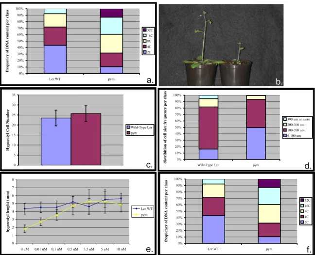
After 8 days on MS media 3% under long day conditions, the size of the pym’s hypocotyl is reduced of 59,6 % compared to the corresponding Ler wild-type (Fig. 5a; pym: 2,25 (+/- 0,55) mm; Ler: 4,21 (+/- 0,74) mm; n= 91). After 25 days growing on soil under green house conditions, the pym stem is almost half as short as the corresponding control (Fig.5b; Ler wild-type: 14,7 (+/- 1,2) cm; pym: 7,8 (+/- 1,3) cm. n=47).
The shortage of the pym’s hypocotyl can be due to a reduction in the number of cells comprising this organ or to a decrease in the cell size. To discriminate between these two hypotheses, I compared the number of cells of both pym and wild-type embryonic hypocotyls, as well as the size of the cells of this organ and of the wild-type one 8 days after germination.
For this assay, the plants were grown on MS media 3% under long day conditions. On the one hand, one hypocotyl cell row of pym is made of 25,7 (+/- 3,86) epidermal cells on average, while the wild-type one comprises 23,5 (+/-3,92) cells (Fig. 5c; n=50). On the other hand, the length of 8-days old wild-type cells fall into four classes: less than 100 μm, 100 to 200 μm, 200 to 300 μm and more than 300 μm (Fig. 5d). The pym mutant presents about 300% more cells smaller than 100 μm than the wild-type (pym: 49,73%; Ler: 16,36%). Also, the number of cells ranging from 100 to 200 μm and from 200 to 300 μm is reduced in the mutant of 33%
and 52,8% respectively (100 to 200 μm: pym: 43,72%, Ler: 65,45%; 200 to 300 μm: pym:
6,01%, Ler: 12,73%). Finally, 5,45% of the wild-type cells are bigger than 300 μm. No pym
Results A
cell belongs to this category. To summarize, the pym hypocotyl comprises the same number of cells than the wild-type one, and the mutant cells in this organ are on average smaller than the wild-type ones: the pym mutant hypocotyl phenotype is due to an elongation defect.
To determine whether this defect is due to a deficiency on hormone gibberellin or to an inability of the mutant to sense the GA message, I have measured the length of this organ in a Ler wild-type and in a pym backgrounds in the presence of increasing concentrations of gibberellic acid (Fig.5e. n>40). The length of the Ler wild-type hypocotyl is the same before and after the application of concentrations of GA ranging from 0,01 μM to 10 μM (0,0 μM:
4,35 +/-0,69 mm; 0,01 μM: 4,53 (+/- 0,67) mm; 0,1 μM: 4,54 (+/- 0,79) mm; 0,5 μM: 5,21(+/- 0,73) mm; 3,5 μM: 4,63 (+/-1,01) mm; 5 μM: 5,51 (+/-0,99) mm; 10 μM: 5,61 (+/- 0,70) mm). The presence of exogenous hormone at a concentration of at least 0,1 μM results in the mutant in a hypocotyl elongation comparable to the one observed for the corresponding wild- type (0 μM: 1,83 (+/- 0,47) mm; 0,01 μM: 2,71(+/- 0,49) mm; 0,1 μM: 3,54 (+/- 0,68) mm;
0,5 μM: 4,72(+/- 1,37) mm; 3,5 μM: 5,2 (+/-1,47) mm; 5 μM: 5,26 (+/-1,47) mm; 10 μM:
4,89 (+/- 0,91) mm). Therefore, an exogenous application of gibberellins rescues the aberrant hypocotyl mutant phenotype.
To characterize the endoreduplication profile of the pym hypocotyl cells, I have determined the ploidy pattern of both pym and wild-type hypocotyls (Fig 5f; n=39). The DNA content of Ler wild-type hypocotyl cells grown for 8 days on MS 3% media can be distributed in 4 classes: 2C (ploidy level corresponding to unreduplicated DNA), 4C (one round of endoreduplication), 8C (two rounds of endoreduplication) and 16C (three rounds of endoreduplication). Respectively, 43,53%, 28,21%, 20,51% and 7,69% of the cells belong to these classes. 10,53%, 21,05%, 28,95% and 26,32% of the mutant nuclei contain 2C, 4C, 8C and 16C respectively. 13,16% of the mutant cells have undergone a fourth round of endoreduplication (32C). Therefore, the pym mutant is affected on the control of the endoreduplication.
0%
10%
20%
30%
40%
50%
60%
70%
80%
90%
100%
Ler WT pym
frequency of DNA content per class
32C 16C 8C 4C 2C
0 5 10 15 20 25 30 35
Hypocotyl Cell Number
Wild-Type Ler pym
0%
10%
20%
30%
40%
50%
60%
70%
80%
90%
100%
Wild-Type Ler pym
distribition of cell size frequency per class
300 um or more 200-300 um 100-200 um 0-100 um
a. b.
0%
10%
20%
30%
40%
50%
60%
70%
80%
90%
100%
Ler WT pym
frequency of DNA content per class
32C 16C 8C 4C 2C
0 1 2 3 4 5 6 7 8
0 uM 0,01 uM 0,1 uM 0,5 uM 3,5 uM 5 uM 10 uM
hypocotyl lenght (mm) Ler WT
pym
f.
d.
e.
c.
Figure 5: Phenotypical characterization of the pym hypocotyl. (a) Hypocotyl length of 8 days old plants. n=91. (b) Flowering Ler wild-type (left) and pym (right) plants grown for 25 days on soil under green house conditions. (c) Hypocotyl cell number of embryonic plants. n=50. (d) Distribution of hypocotyl cell size of 14 days old plants n>150. (e) Dependency of the hypocotyl size of 8 days old plants on GA4+7. n>40. (f) Distribution of hypocotyl cell DNA content of 14 days old plants. n=39.
All the plants except the ones depicted on the figure 3b were grown on MS3% at 22°C.Vertical bars represent the standard deviation.
2.2.2 Trichome development
A mutation in the POLYCHOME gene leads to trichome overbranching (Perazza et al., 1999).
To clarify the relationship between this trichome phenotype of pym and the gibberellins, I have quantified the leaf hairs branches in the presence of different concentrations of Uniconazol-P, an inhibitor of the GA synthesis.
A deficiency in gibberellins caused by an exogenous application of uniconazol-P does not significantly affect the number of pym three-branched trichomes (0 mM: 32,4 (+/- 4,52) %;
10-6 mM: 37,41 (+/-5,66) %; n=30). The same concentration of uniconazol-P is sufficient to
Results A
trigger a significant decrease in the percentage of three-branched trichomes of the Ler wild- type plants (0 mM: 59,52 (+/- 2,62) %; 10-6 mM: 23,5 (+/-3,39) %; n=25).
In addition to playing a role in trichome branching, the gibberellic acid is implicated in trichome initiation (Herman et al., 1989, Chien and Sussex, 1996, Perazza et al., 1998, Payne et al., 2000). To find out whether PYM is implicated in this aspect of the trichome development, I quantified the trichomes in pym compared to the wild-type.
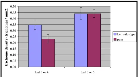
The amount of trichomes per mm2 is significantly reduced in the second pair of leaves of the pym mutant compared to the wild-type (Fig.6; Ler wild-type: 0,35 (+/- 0,04) trichomes/ mm2;
pym: 0,23 (+/-0,04) trichomes/ mm2). This difference does not exist in the third pair of leaves (Ler wild-type: 0,44 (+/- 0,05) trichomes/ mm2; pym: 0,44 (+/-0,03) trichomes/ mm2).
Figure 6: Number of trichomes per mm2 in the second and third pair of leaves. n=20. The bars represent the standard deviation.
0,00 0,05 0,10 0,15 0,20 0,25 0,30 0,35 0,40 0,45 0,50
leaf 3 or 4 leaf 5 or 6
trichome density (trichomes / mm2)
Ler wild-type pym
2.2.3 Flowering
The gibberellic acid is known to play various roles in reproductive development. For example, GA application accelerates flowering, particularly in short days (Landridge 1957, Bagnall 1992). Consistently, mutations that block GA biosynthesis or responsiveness (e.g. sleepy1) cause delayed flowering, whereas mutants with increased GA signalling (e.g. rga, gai and spy) flower early (Olszewski et al., 2002).
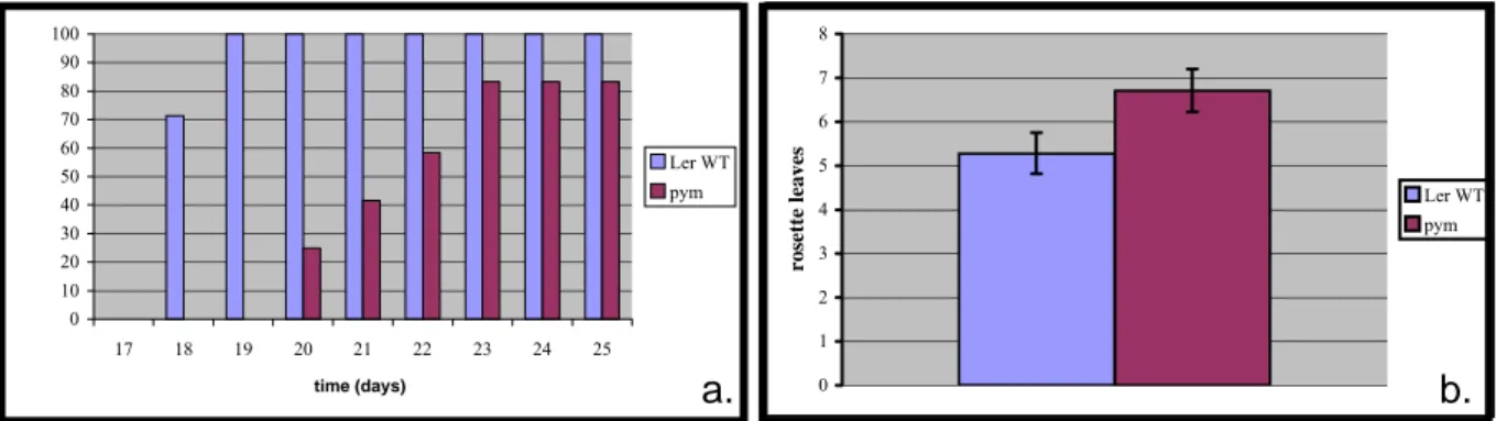
I have compared the kinetics of flowering of pym and Ler wild-type (Fig. 7a, 7b; n=27). The plants were monitored for 25 days and were growing on soil under long day conditions.
0 10 20 30 40 50 60 70 80 90 100
17 18 19 20 21 22 23 24 25
time (days)
Ler WT pym
0 1 2 3 4 5 6 7 8
rosette leaves
Ler WT pym
a. b.
Figure 7: (a) Kinetics of flowering of Ler wild-type and pym plants. The results are shown in percentage of flowering plants in relation to the total population. n=27. (b) The histograms represent the number of rosette leaves of the plants shown in fig 6a when the vegetative meristem switched to a reproductive meristem. The vertical bars indicate the standard deviation.
It appears that after 18 days, 71,4% of the wild-type plants are able to flower, and after 19 days, it is possible to observe a floral bulge in 100% of the population (Fig. 7a). In contrast, after 20 days, 25% of the pym plants have flowered. This percentage increased to 41,6% and to 58,3% after 21 and 22 days, respectively. On the 23rd day, 83,3% of the plants flowered.
No further flowering was observed between the 23rd and the 25th day. On average, Ler produces 5,29 (+/- 0,47) leaves before flowering. The pym mutant produces 6,71 (+/- 0,49) leaves (Fig. 7b). Taken together, those results indicate that pym presents a delay in flowering.
The retard in flowering observed in the pym mutants can be completely rescued by the application of exogenous gibberellins. 100% of both Ler wild-type and pym plants are able to flower after 19 days of growth under GA treatment. When grown without exogenous GAs, only 86,95% Ler wild-type plants and 38,8% pym mutants flowered.
Discussion A
A 3 Discussion
The shaping of the leaf trichomes of Arabidopsis thaliana is under the positive regulation of the gibberellic acid (Chien and Sussex 1996, Perazza et al., 1998). Three genes have been implicated in the hormonal regulation of the trichome cell morphogenesis: SPY, KAK and PYM. Although the cloning SPY and KAK are helping to get a better picture of the mechanisms by which this regulation takes place, this regulatory pathway has been poorly investigated and much is still unknown about its way of action. To perform substantial progress in our understanding of the GA-dependent regulation of cell morphogenesis, it is essential to characterize other members of the pathway.
At the time of the beginning of my theses work, POLYCHOME had been proposed to encode for a protein involved in the GA transduction cascade in the trichome, downstream from SPINDLY (Perazza et al., 1999). To acquire further knowledge on the regulation of cell morphogenesis by the hormone gibberellin, I concentrated my work on the investigation of the POLYCHOME gene. I attempted to map it and I proceeded to a morphological characterization of the mutant, as well as to the observation of the dependency of several pym’s developmental stages on the gibberellins by the application of exogenous gibberellic acid or by the blockage of the hormone biosynthetic pathway.
3.1 Genetic mapping of the POLYCHOME gene
At the beginning of my work, it was known that the PYM gene is located in the south of chromosome II in the genome of Arabidopsis thaliana. It co-segregates with the markers nga168 and Ubique, indicating that PYM and those two markers are in close proximity (Perazza et al., 1999).
To locate PYM, I further fine-mapped the portion between and around nga168 and Ubique.
The PYM gene always co-segregated with the markers located within T11A7-EGJ and F7D19-EGJ. Therefore, my work allowed to locate PYM in the region of 423.140 nucleotides existing between those two markers, but was not sufficient to map PYM precisely.
To overcome this result, one could imagine sequencing all the genes located in this region, to apply a candidate approach or to increase the mapping population. The economical cost of the sequencing is too high to justify the choice of the first possibility. A candidate approach
consists on emitting an educated guess about the molecular nature of the gene of interest, and to screen the area to which the gene has been mapped down looking for candidates to be the one in concern. The only knowledge about PYM is that it may play a role downstream of SPY.
Therefore, it is not possible to attribute a potential molecular nature to this gene. All what can be done is increasing the mapping population and continuing with the chromosomal walking.
Nonetheless, the identification of the pym mutants in the pym Ler x wild-type Col F2 population presented specific problems related to the variability of the occurrence of trichome overbranching on the rosette leaves of Arabidopsis. Indeed, the percentage of 3-branched trichomes on a rosette leaf is not constant; it depends instead on unknown internal and external factors. In addition, the ecotype Columbia presents somewhat more overbranched trichomes than Landsberg erecta (Larkin et al., 1996). Therefore, the offspring of the crossing Ler pym x Col wild-type comprised wild-type and heterozygous plants with a higher overbranching than the Ler wild-type as well as pym mutant plants with a moderate percentage of 4- to 6-branched trichomes. Given the difficulties encountered to obtain a reliable mapping population comprising only pym mutant plants, it was decided not to map PYM further.
There are about 120 genes in the region where PYM is located. From those, 15 are transcription factors, 8 are protein kinases that have not been involved in any specific cascade, 5 play a role in the degradatory pathway, 4 are implicated in protein-protein interactions and 3 are part of the cytoskeleton. All the other genes have no attributed function. None of them have been implicated in the biosynthesis of the GAs or on the transduction of the gibberellin acid message.
3.2 Morphological characterization of the polychome mutant and analysis of its dependency on the gibberellins
The relevance of the PYM protein in the trichome morphogenesis was revealed by the characterization of the corresponding mutant phenotype: the pym mutant trichomes are overbranched and present higher DNA contents than the wild-type. Thus, PYM is essential for the inhibition of the endoreduplication and the branching of the trichomes (Perazza et al., 1999). In addition, my work has shown that the pym mutant plants present pleiotropic alterations: both the hypocotyl and the stem of the mutant are shorter than the ones of the wild-type, the trichome density is reduced, the hypocotyl cells are defective in the regulation
Discussion A
of the endoreduplication and the pym mutant plants flowers late. How to explain those aberrations?
Genetic analyses indicate that PYM functions downstream of SPY in the GA signalling pathway. Since both mutants present the same trichome phenotype, pym may be affected in the same manner than spy: the mutant might transduct constitutively the gibberellin signal. To test this hypothesis, I observed the effect of a blockage of the GA biosynthesis in the trichome morphogenesis. If the transduction of the GA signal is constitutively activated in the pym mutant, the mutant trichomes must be able to branch in the absence of the hormone. In the assay performed for this thesis work, a blockage of the gibberellin biosynthesis did not reduce the branch number in the mutant trichomes, but did in the Ler wild-type ones. This data confirms the function of PYM as a negative regulator of trichome morphogenesis via the GA signalling cascade.
Interestingly, the pym mutant does not present the phenotypic alterations that are characteristic for a constitutive transduction of the gibberellin signal. These mutants typically elongate more, develop more trichomes and flower earlier than the wild-type, which is contrary to the phenotypes observed in pym plants. Moreover, the application of exogenous GA to the pym seedlings resulted in a complete rescue of the hypocotyl length and restored the ability of the mutant to flower, suggesting that the aberrations observed in the mutant are due to a deficiency in GAs. This apparent contradiction can be explained in two ways. It is possible that the phenotypes observed in the pym mutant are the result of mutations in two different genes: in this scenario, a mutation in the PYM gene will affect a protein negatively implicated in the transduction of the gibberellin message specifically in the trichome, and will lead to overbranching. A second side mutation would lead to short stem and hypocotyl as well as to a delay in flowering due to a defect on a gene of the GA biosynthetic pathway. To verify this hypothesis, it is important to perform a genetic analysis of the offspring from a pym x Ler wild-type crossing: the different phenotypes should segregate in an F2 population if they are due to mutations in different genes. In another scenario, all the phenotypes are caused by a single mutation affecting the transduction cascade in the trichomes. This exiting hypothesis is based on the discovery that mutations activating constitutively the transduction of the gibberellin signal lead to decreased levels of bioactive GAs (Xu et al., 1995, Hedden and Kamiya, 1997, Cowling et al., 1998, Silverstone et al., 1998). The role of PYM as a negative regulator of signal transduction seems to be limited in the plant to the control of trichome morphogenesis and to the regulation of endoreduplication in both the trichome and the hypocotyl since all the other aberrant phenotypes that were described during this work
typically correspond to a deficiency on gibberellins. The fact that different proteins in different organs mediate the transduction of the GA signal has already been described, even though the mechanisms implicated in this process have not been understood yet. For instance, DWARF1 of Potato seems to play a specific role in the ability of the second leaf to sense the gibberellin hormone (Ashikari et al., 1999; Fujisawa et al., 1999).
To summarize, PYM is negatively implicated in the transduction of the gibberellin signal leading to the development of trichome branches in Arabidopsis thaliana and very likely also to the control of endoreduplication in the trichome and in the hypocotyl cells. The aberrant phenotypes observed in the pym plants might be due to a second side mutation in the genome of the pym plants but could as well be the result of a deficiency in the GA levels provoked by the up-regulation of the gibberellin signal transduction.
B
Trichome
Pattern formation
B 1 Introduction
1.1 Trichome initiation in Arabidopsis thaliana
The study of the mechanisms underlying pattern formation is a fascinating area of research on developmental biology. How are cells recruited from initially equivalent cells to differentiate and how are they arranged in a well-ordered manner? The development of trichomes, the leaf hairs of Arabidopsis thaliana, is an excellent model system to study pattern formation because they present a simple two-dimensional spacing pattern and are initiated at regular distances to each other (Hülskamp et al., 1994, Marks 1997, Hülskamp et al.,, 1999, Szymanski et al., 2000, Larkin et al., 2003). The resulting pattern must be tightly controlled because the distance between the developing trichomes is at least three to four cells and trichomes adjacent to one another (clusters) are much less frequent as would be expected by a random distribution (Hülskamp et al., 1994, Larkin et al., 1996). Cell lineage is not involved in the decision of becoming a plant hair since trichomes do not derive from systematic cell division patterns (Larkin et al., 1996, Schnittger et al., 1999). Finally, trichomes do not seem to emerge as a response to positional cues, since they are not found associated to any pre-pattern (Hülskamp et al., 1994). It is likely that the trichome pattern is established on the epidermal layer of very young leaves as a result of cell-cell interactions taking place between the trichome precursor and its neighbouring cells (Larkin et al., 1996, Schnittger et al., 1998, Schnittger et al., 1999).
1.2 A model to explain two-dimensional pattern formation
Meinhardt and Gierer have proposed a model to explain biological pattern formation based on the reaction diffusion mechanism studied by Turing in the 50s. Turing demonstrated that two interacting chemicals could generate a spatial concentration pattern if one of the substances diffuses faster than the other (Turing 1952). In the model of Meinhardt and Gierer, thereafter called activator-inhibitor model, the two interacting chemicals described by Turing are an activator and an inhibitor (Meinhardt and Gierer 1974, Koch and Meinhardt 1994, Meinhardt and Gierer 2000). A stable pattern is established by local self-enhancement of a short-ranging activator and an inhibition of this autocatalysis by a long-ranging inhibitor, production of which depends upon the activator (Figure 8a). The activator is engaged in a self-activation
Introduction B
loop, e.g. a slight increase of the activator’s concentration leads to a further increase of this activator. However, this is not sufficient to create a regular pattern because every small fluctuation would lead to an exponential increase on the activator’s concentration. The fast diffusion properties of the inhibitor in this system prevents that the activation takes place in the surrounding tissue and at the same time does not disturb the incipient local increase of the activator. The reaction scheme is shown in figure 8b. A local minute increase of the activating substance (green line) above the concentration range will grow further due to the self- enhancing process while the concomitantly produced inhibition (red line) down-regulates the activation of the surrounding field. A final, stable situation is reached when the local self- enhancement is at equilibrium with the surrounding cloud of inhibition.
a. b.
Figure 8: Pattern formation by autocatalysis and long-range inhibition. (a) The simplest reaction scheme: an activator (blue) catalysis its own production and that of its highly diffusing antagonist, the inhibitor (red) (b) Computer simulation of pattern formation in a chain of cells. Random fluctuations in the ability of the cells to perform the reaction (blue squares) are sufficient to initiate pattern formation. In a field larger than the diffusion range of the inhibitor (red), several activator (green) maxima emerge. Under this condition, the spacing is somewhat irregular and minimum distance is maintained. The initial, an intermediate and the finally stable distributions are shown. (Adapted from Meinhardt and Gierer, 2000)
1.3 Is protein degradation relevant for trichome pattern formation?
The activator-inhibitor model incorporates the existence of a depletion mechanism regulating the concentration on activators and inhibitors (Gierer and Meinhardt 1972, Meinhardt and Gierer 2000). The ability to control the removal of these regulatory elements from the system appears to represent a big advantage for fine-tuning the process, as it may allow rapidly creating and stabilizing incipient concentration maxima. To date, the existence of such a mechanism in the context of trichome pattern formation has not been described. Interestingly, KAKTUS, a gene implicated in trichome morphogenesis, encodes a HECT E3 domain protein and thus may target proteins to degradation via the 26S proteasome (Perazza et al., 1999, El Refi et al., 2003, Downes et al., 2003). Several of the proteins involved in trichome
morphogenesis also play a role in trichome pattern formation (Hülskamp et al., 1994, Larkin et al., 1994, Larkin et al., 1999, Schnittger et al., 1999, Payne et al., 2000, Johnson et al., 2002, Schellmann et al., 2002). Thus, it is reasonable to hypothesize that KAK could play a role in the establishment of trichome formation by promoting the degradation of elements involved in the trichome pattern system.
A protein destined to be degraded by the 26S proteasome enters the pathway via an ATP- dependent conjugation cascade, involving the sequential action of an universal E1 ubiquitin- activating enzyme, one of several E2 ubiquitin-conjugating enzymes and one member of the E3 ubiquitin-ligase family. In the final step, an E3 ligase recruits both the target and an ubiquitinated E2 intermediate and then stimulates ligation of the C-terminal carboxyl group of the ubiquitin to free amino groups in the target (Hershko and Ciechanover 1998, Pickart 2001). A polyubiquitin chain is synthesized by successively adding activated ubiquitin moieties to internal lysine residues on the previously conjugated ubiquitin molecule. In a final step, the 26S proteasome recognizes the chain and degrades the polyubiquitinated substrate.
1.4 Elements of the trichome pattern system and their molecular nature
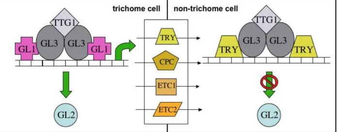
Several genetic screens have allowed the identification of two classes of mutants that show defects in trichome initiation and pattern formation. One class of mutants present few or no trichomes on the leaf surface, and thus the genes affected are positively implicated in trichome initiation. GLABROUS1 (GL1), AtMYB23 (MYB23), GLABROUS2 (GL2), GLABROUS3 (GL3), ENHANCER-OF-GLABROUS3 (EGL3), TRANSPARENT-TESTA- GLABRA1 (TTG1) and TRANSPARENT-TESTA-GLABRA2 (TTG2) belong to this class. In another class of mutants, more or clustered trichomes develop on the leaf: the genes affected encode for inhibitors of trichome formation. The second class of mutants comprises alterations in CAPRICE (CPC), TRYPTICHON (TRY), ENHANCER-OF-TRYPTICHON- AND-CAPRICE1 (ETC1) and ENHANCER-OF-TRYPTICHON-AND-CAPRICE2 (ETC2) (Koornneef 1981 Oppenheimer et al., 1991, Hülskamp et al., 1994, Wada et al., 1997, Johnson et al., 2002, Schellmann et al., 2002, Zhang et al., 2003, Kirik et al., 2004a, Kirik et al., 2004b). GL1 and its homolog MYB23 encode for an R2R3-type MYB transcription factor.
GLABRA2 has been sequenced and shown to have sequence similarity to homeodomain proteins and therefore is likely to coordinate the expression of target genes (Rerie et al., 1994). GLABRA3 and EGL3 are members of the basic helix-loop-helix (bHLH) transcription factor family (Oppenheimer et al., 1991, Payne et al., 2000, Zhang et al., 2003, Kirik et al.,