Understanding the function of the Arabidopsis GLABRA 2 gene in Trichome patterning,
morphogenesis and differentiation
Inaugural-Dissertation
zur
Erlangung des Doktorgrades
der Mathematisch-Naturwissenschaftlichen Fakultät der Universität zu Köln
vorgelegt von
Bhylahalli Purushottam Srinivas
aus Bangalore, Indien
Köln, Mai 2004
Tag der mündlichen Prüfung: 01. Juli 2004
Berichterstatter: Prof. Dr. Martin Hülskamp
Prof. Dr. Klaus Harter
Prüfungsvorsitzender: Prof. Dr. Wolfgang Werr
To
Amma, Appa and Prashanth.
“Reality is merely an illusion, albeit a very persistent one.”
Albert Einstein
Acknowledgements
This work would not have been made possible without all the love, care and support my family has always given me, including when I decided to pursue a Ph.D in a foreign land. No words can convey how much I value their affection and encouragement. My wife Vani has been a source of inspiration for me to complete my thesis in time.
Many thanks are due to my advisor and thesis guide Prof. Martin Hülskamp.
Right from the first day when he kindly offered me a position to work in his lab for my Ph.D to this day he has been supportive, not-so-demanding and gave me complete freedom to think, plan and work as independently as I wanted to. Martin, I sincerely thank you for all this.
No work is successful without having a friendly and understanding atmosphere around you in the work place. Especially so in research, I feel! I am grateful to all my lab mates for having provided me a pleasant atmosphere in the lab during my entire stay. We have had many useful scientific discussions, friendly conversations and many get-togethers and parties…….It will be in my memory for a long time to come. Life in Germany without knowing the German language could have been much more difficult but for the help of some of you. With no hesitation I can single out Herr. Weidner and thank him immensely for patiently listening and helping me with EVERY official form I was required to fill whether it was with the university, Finanzamt, salary department or the Berzirksamt (visa, etc…). And believe me there are far more forms (loads of them) to be filled in Germany than one could have imagined before coming here! Prof.
Schmitz has, with the same kindness and smiling face, helped me whenever I needed his guidance with science, be it with scanning electron microscopy, or vital staining of plants, etc… I am very thankful to you both.
I have had a memorable time cooking Indian cuisine with Birgit and Angi on a few occasions and also got to learn a bit about German food, and not to forget Birgit’s attempts with some Italian cooking! It was wonderful. Jaideep, Neeta and Anshu were a great family. I have spent many days enjoying great north Indian food and watching TV along with them on weekends when they used to either drive me home with them or
invite me regularly. A very productive and successful collaboration with Jaideep on the scientific front, many useful and spontaneous discussions whenever either of us had a new idea and wanted to discuss……all this will be treasured by me. In short I can say the mathur family was in one sense a home away from home for me and I would always be grateful to them for this.
Khadija Chennab, my associate for almost a year was a great person to work with. I enjoyed teaching her things that I knew and she did a good job as a technician helping me initially with some of my projects. I thank her for systematically making hundreds of DNA and bacterial glycerol stocks and perfectly labeling them all. I remember her whenever I see them and rightly so I can feel proud of her job!
The 3+ years I have spent in the Institute of Botany means the list of acknowledgements can go on and on. There are many people within our group as well as from outside whom I would like to thank for all the help and kindness they have offered me during my stay here. It’s been a memorable time having met and made many new friends. Let me sincerely thank them all together without mentioning everyone’s names. The memories are in my heart. And, I wish them all the very best.
Contents
1. Introduction: Arabidopsis trichomes as a model... 3
2. The Arabidopsis GLABRA 2 gene functions in trichome patterning, morphogenesis and differentiation ……… 11
2.1 Summary ………... 12
2.2 Introduction ……… 13
2.3 Results……… 15
2.3.1 Increased trichome cell specifications on gl2 leaves ……….… 15
2.3.2 Ubiquitous expression of GL2 inhibits trichome initiations ………… 17
2.3.3 Genetic interaction of GL2 with TRY ……….…… 19
2.3.4 Lateral inhibition appears to be compromised in gl2 ………….…… 23
2.3.5 TRY/CPC may possibly mediate the inhibitory function of GL2……. 26
2.3.6 Analysis of gl2 double mutants………. 28
2.3.7 Sub-cellular localization of the GL2 protein in Arabidopsis…………. 31
2.3.8 Regulation of the GL2 promoter ……… 33
2.3.9 Ectopic expression of GL2 in different mutant backgrounds ……… 34
2.3.10 Differentiation defects in gl2 trichomes ……… 35
2.3.11 gl2 mutant trichomes appear to be divided ……… 39
2.3.12 GL2 positively regulates trichome branching ……… 43
2.4 Discussion ……….. 46
3. Understanding the mechanism of trichome patterning ………. 54
3.1 Summary ……… 55
3.2 Results ………. 55
3.2.1 Sub-cellular localization and inter-cellular movement of transcription factors ……… 55
3.2.2 Mechanism of Trichome cell selection ……… 56
3.2.3 Expression of TRY or CPC by GL2 promoter inhibits trichome initiation ……….……… 60
3.3 Discussion ……….……..… 60
4. A novel method to simultaneously analyze multiple gene expression patterns in vivo ……….………. 64
4.1 Summary ……… … 65
4.2 Introduction ……… … 65
4.3 Results ……… … 68
4.4 Discussion ……… … 72
5. Materials and Methods ….………. 75
6. References ……… 80 7. Appendix
LIST OF FIGURES
Number Page
Fig.1. Phenotype of trichomes on wt and gl2 mutant leaves ………16
Fig.2. Counting trichome specifications / initiations in wt and gl2 using a molecular marker ………18
Fig.3. Ectopic over-expression of GL2 in wt background and RT-PCR analysis……..18
Fig.4. Analysis of regulation of TRY expression in wt and gl2 leaves……… 20
Fig.5. Temporal expression pattern analysis of TRY in wt and gl2 mutant leaves… 21
Fig.6. Analysis of GL2::GFP-ER expression in 35S::TRY plants……… 21
Fig.7. Ineffective lateral inhibition on gl2 leaves ……… 24
Fig.8. Ubiquitous expression of GL2 in try cpc double mutant……… 27
Fig.9. Analysis of trichome initiation in gl2 double mutants ………29
Fig.10. Sub-cellular localization of EYFP-GL2 protein in epidermal cells…………...32
Fig.11. Regulation of GL2 expression by other patterning genes……… 32
Fig.12. Development of a gl2 trichome followed by time lapse microscopy………….36
Fig.13.Defective differentiation features of gl2 trichomes……….36
Fig.14.Loss of trichome cell identity in gl2 mutants………..38
Fig.15.Trichomes on gl2 leaves frequently appear to be divided……… 38
Fig.16.Expression of trichome specific markers in the apparently divided trichomes on gl2 leaves………40
Fig.17.Expression analysis of GL2::GFP-ER and TRY::GFP-ER in young and older gl2 leaves suggesting some trichomes divide……… 42
Fig.18.Analyzing the strength and temporal pattern of expression of TRY and GL1 promoters in trichomes……….. 44
Fig.19. A cartoon model illustrating the development and fate of trichomes on gl2 leaves………. 48 Fig.20. A genetic model for trichome patterning and differentiation……… 53 Fig.21.Transient assay in Arabidopsis epidermal cells demonstrating inter-cellular movement of TRY and CPC……….. 57 Fig.22.Analysis of the early trichome marker, GL2:GFP-ER, in young leaves to
understand the process of trichome cell selection………. 58 Fig.23. Expression of TRY or CPC under the GL2 promoter in wt plants results in inhibition of trichome initiation………. 63 Fig.24. A genetic model to explain trichome cell selection in wt leaves………...63 Fig.25. A cartoon illustration of how MEPI works………69 Fig.26. Simultaneous analysis of three reporter genes in Onion and Arabidopsis…… 71 epidermal cells
Zusammenfassung
Die Blatthaare von Arabidopsis thaliana (Trichome) sind die mit am besten geeigneten Zelltypen zur funktionellen Untersuchung von Musterbildungsprozessen und Zelldifferenzierungsvorgängen. In dieser Arbeit wurde die Funktion des GLABRA2 Gens in diesen beiden Prozessen näher untersucht. Die Befunde können wie folgt zusammengefasst werden:
- es konnte mit Hilfe von Trichom-Markerlinien gezeigt werden, dass in glabra2 Mutanten während der Blattinitiation mehr Epidermiszellen als in Wildtyp ein Trichomschicksal wählen. Während der Blattentwicklung scheinen viele dieser Zellen wieder zu de-differenzieren, da in älteren Blättern deutlich weniger Trichome als in Wildtyp gefunden werden.
- GLABRA2 hat eine Funktion in der Trichom-Differenzierung und Morphogenese.
Viele Trichome beginnen sich wie in Wildtyp zu entwickeln, stoppen dann jedoch ihre Entwicklung und nehmen dann ein Epidermiszellschicksal an. Trichome, die sich weiter entwickeln, zeigen oft eine reduzierte Verzweigungsanzahl.
- GLABRA2 und das Musterbildungsgen TRIPTYCHON regulieren sich gegenseitig in einer positiven Rückkopplungsschleife.
- Mit Hilfe von Trichom-Markerlinien konnte gezeigt werden, dass einzelne Trichome aus einer Gruppe von kompetenten Zellen ausgewählt werden und das dabei die Entscheidung, ob die Zelle weiter mitotische Zyklen durchläuft oder mit Endoreduplikation beginnt, wichtig ist.
- Experimente bei denen TRIPTYCHON und CAPRICE, zwei Musterbildungsgene, die an der lateralen Inhibition beteiligt sind, als GFP-Fusionen transient exprimiert wurden, zeigen, dass beide Proteine in benachbarte Zellen wandern können.
- Es wurde eine neue Methode entwickelt (MEPI) die es ermöglicht, die Expression mehrerer Gene simultan in vivo zu untersuchen. Zusätzlich zu GFP-Varianten mit verschiedenen Farbspektren werden bei dieser Methode GFP-Fusionen verwendet, die spezifische Kompartimente der Zelle markieren.
Summary of the Thesis
Arabidopsis trichomes (leaf hairs) are one of the best studied plant model cell types with respect to understanding the molecular mechanisms underlying the process of cell patterning and differentiation. Many mutants which exhibit altered trichome patterning and differentiation have been analyzed and insights into the molecular nature of interactions among the genes involved have been obtained. The main focus of this thesis has been to understand the role of the GLABRA 2 (GL2) gene in trichome cell patterning, morphogenesis and differentiation. The main findings can be summarized as follows:
i. In the absence of GL2 function more protodermal cells than wild-type get specified as trichome precursors but most of them exit the trichome differentiation pathway at different stages of their development resulting in lesser number of trichomes on mature gl2 mutant leaves. The role of GL2 in patterning was established by analyzing various gl2 double mutants as well as by ubiquitous expression of GL2 which showed that it is required both for trichome specification and differentiation. The development and fate of trichomes on gl2 leaves was carefully analyzed and it is concluded that in the absence of GL2 function trichomes lose their identity and likely adopt the default epidermal differentiation pathway of pavement cells. GL2 was also found to regulate trichome branching positively. A positive feedback loop between TRIPTYCHON (TRY) and GL2 was discovered and is hypothesized to be important in the final steps of trichome pattern resolution. In a nutshell, GL2 was found to be involved in trichome patterning, branching and differentiation. All the results have been incorporated in a model discussed at the end of chapter 2.
ii. Analysis of an early trichome marker in young wild-type leaves showed that trichome patterning is a de novo process, meaning only a few cells get selected to become trichomes from a pool of apparently equivalent cells. Based on the above analysis it is hypothesized that in addition to the expression of specific transcription factors the final resolution of the trichome pattern is likely an outcome of competition between endoreplication and mitosis modes of cell cycle.
iii. Using transient assays it was found that TRY and CPC gene products exhibit ability for intercellular movement, an essential property for them to act as inhibitors during patterning.
iv. A novel method to simultaneously analyze multiple gene expression patterns in vivo (MEPI) has been proposed which is based on targeting many fluorescent reporter genes
(GFP variants) to distinct intercellular structures / organelles in the same specimen. A proof-of-concept has been demonstrated by simultaneously analyzing three different reporter genes in Arabidopsis epidermal cells using a transient assay.
Introduction
Arabidopsis trichomes as a model system to understand cell
patterning and differentiation
1
Plants, like other multicellular eukaryotes, develop from a single celled zygote that ultimately gives rise to the many specialized cell types of the adult organism. Cell patterning is when cells are guided to their appropriate differentiated fate at the correct time and place in the developing organism. Understanding the mechanisms underlying cell patterning, cell fate specification and cell differentiation has long been the goal of developmental biology. The plant Arabidopsis thaliana has been successfully used as a model system to address such questions in plant biology.
1.1 The Plant epidermis
The plant epidermis is the outermost cell layer of a plant. It contains many specialized cell types which function primarily in protecting the plant from various external threats.
The presence of a layer of waxy cuticle and different cell types serve in defending the plant against various pathogens and herbivores, dehydration, UV damage and other factors. The different specialized cell types found on the epidermis include the trichomes, stomatal guard cells, root hair cells, various secretory glands and nectaries and epidermal pavement cells among others.
The plant epidermis is an excellent tissue for studying cell patterning. The epidermis of the root, hypocotyls and the leaf consist of only a few cell types and is easily accessible for observation. The root and the hypocotyl consist of only two cell types: the root hair and the non-root hair cells in the root epidermis and the stomatal cells and the non stomatal cells on the hypocotyl epidermis. The cells are arranged in files of alternating types in both the tissues and are thought to arise due to a position dependent mechanism. However, the epidermis on the adaxial surface of the leaf consists of three different cell types. The trichomes, which are large branched hair like cells, stomatal guard cells, which help in gas exchange of the plant with its surrounding atmosphere and the epidermal pavement cells which are mostly jigsaw puzzle shaped cells covering the entire leaf.
1.2 Trichomes: An excellent model cell type to study cell patterning, cell fate specification, differentiation and cell cycle
Trichomes, also called as plant ‘leaf hairs’, are present on the aerial surfaces of most plants, ranging from ferns to angiosperms. Trichomes come in various shapes and forms, from single celled to multicellular, and include both glandular secretory hairs
and nonglandular hairs. They have been thought to function in providing the plants with resistance to insect herbivores, reducing water loss by excess transpiration, increasing freeze tolerance and protecting plants from UV light. One of the most thoroughly studied plant cell differentiation pathways is the development of Arabidopsis trichomes.
Trichomes on Arabidopsis are large single polyploidy cells that protrude from the epidermis of aerial organs that include rosette leaves, cauline leaves, sepals and stems.
They are surrounded by a ring of 8-10 specialized accessory cells, also called as socket cells (Fig.1; page 16) visible at very low magnifications and are accessible for manipulations. Under laboratory conditions they are completely dispensable to the plants and thus numerous mutants affected in various steps of trichome fate specification, morphogenesis and differentiation are available for genetic analysis which makes them an unparalleled model cell type for scientific investigation in the above areas.
1.3 Development of trichomes and their spatial distribution pattern
Trichome development in wild-type (wt) Arabidopsis begins near the distal tips of leaves when they are approximately 100 µm long, and proceeds basipetally (Larkin et al., 1996). Trichomes are found adjacent to one another much less frequently than would be expected by chance, suggesting that an active mechanism exists to govern trichome spacing (Hulskamp et al., 1994).
Trichomes are the first differentiated cell type formed on the developing leaf epidermis.
Trichome progenitor cells are specified in young leaf primordia in a field of morphologically similar, undifferentiated dividing epidermal cells. The first steps of trichome differentiation start with an enlargement of the nucleus and an increase in cell size. The growing trichome cell extends out of the leaf surface, elongates and eventually initiates two branching events. Secondary branching occurs in a plane perpendicular to the primary branch plane and is followed by elaboration of the secondary cell wall thickening which eventually results in a mature trichome decorated with cell surface papillae. The single nucleus of a wild-type trichome continues to replicate its genomic DNA during differentiation, reaching an average nuclear DNA levels of 20C – 32C (Hulskamp et al., 1994; Melaragno et al., 1993), a process known as endoreplication or endoreduplication. Endoreplication is a common variant of cell cycle where mitosis and cytokinesis are suppressed, but cycles of DNA replication
continue. Trichome patterning and differentiation continue in the growth zone at the base of the leaf during further leaf expansion.
1.4 Genetic dissection of the various steps of trichome development
A powerful tool to address various questions regarding the regulatory mechanisms underlying the different steps of trichome development is genetic analysis using various mutants defective in each of those steps. More than 36 genes have been identified so far in several mutagenesis screens that affect different aspects of trichome development.
The genes can be grouped into categories which affect a particular step of trichome development (Hulskamp et al., 1999). The trichome initiation step includes the genes GL1, TTG1, GLl3, EGL3, TRY, CPC, ETC1 and ETC2 which when mutated show either increased or decreased trichome initiations compared to wt. The other categories include local outgrowth (GL2), extension growth (CRK, DIS1, DIS2, GRL, KLK, SPI and WRM), endoreplication and primary branching (GL3, STI), secondary branching (AN) and maturation step (UDT, TBR, CHA, CDO and RTS).
1.5 Molecular dissection of Trichome patterning
The first step of trichome development is the selection of a single epidermal cell as a trichome initial, the step of trichome specification. Several mutants affecting this step have been identified and molecularly characterized (Hulskamp et al., 1994; Larkin et al., 2003). They are broadly classified as positive and negative regulators of trichome initiation. The mutation in the positive factors like GLABRA1 (GL1), TRANSPARENT TESTA GLABRA1 (TTG1) and GLABRA3 (GL3) leads to either a reduction or complete absence of trichomes on leaves. Recently a close homolog of GL3, THE ENHANCER OF GLABRA3 (EGL3), has been identified which may function redundantly with GL3 during trichome patterning (Bernhardt et al., 2003). Whereas gl1 and ttg1 mutants display completely glabrous leaves, gl3 mutants show a reduction in trichome number.
But the gl3 egl3 double mutant is completely glabrous supporting the redundancy idea.
GL1 encodes an R2-R3 MYB transcription factor with two repeats of MYB DNA- binding domain. Mutation in gl1 only specifically affects trichome initiations with the leaves being completely glabrous. TTG1 on the other hand encodes for a WD-40 protein which is thought to mediate protein-protein interactions and a mutation in this gene has pleiotropic effect showing reduced anthocyanin pigmentation, absence of seed
coat mucilage, and an increase in the number of root hairs (Walker et al., 1999) along with fully glabrous leaves. The third important gene in this process is GL3. gl3 mutants, along with their reduced trichome number phenotype, also show smaller and less branched trichomes. Their nuclear DNA content is also reduced. GL3 encodes a basic helix-loop-helix (bHLH) protein closely related to the maize R gene (Payne et al., 2000). Recently one report has suggested that GLABRA2 (GL2) could also be involved in the step of trichome initiation by showing that an additional copy of the GL2 gene expressed under its own promoter increased the number of trichome initiations as well as trichome clusters / nests (Ohashi et al., 2002). GL2 encodes a homeo domain transcription factor of the HD-Zip IV class (Rerie et al., 1994).
The TRIPTYCHON (TRY) and CAPRICE (CPC) genes encode for single repeat MYB protein with no apparent transcriptional activation domain. Mutations in try lead to formation of adjacent trichomes at a much higher frequency than in wt. It has been shown to act non-cell autonomously in inhibiting cells neighboring trichomes from acquiring trichome fate. cpc mutants on the other hand show an increased number of trichome initiations. That both TRY and CPC act redundantly in lateral inhibition was suggested by the phenotype of the double mutant (try cpc) where huge nests of trichomes containing sometimes upto 30 were observed (Wada et al., 2002; Schellmann et al., 2002). Recently two close homologs of TRY and CPC have been identified.
ENHANCER of TRIPTYCHON and CAPRICE 1 (ETC1) and ETC2 also seem to act redundantly along with TRY and CPC during trichome patterning (Viktor Kirik, 2004.
personal comm.).
Protein interaction studies using the yeast two hybrid method has shown that GL3 interacts with itself, GL1 and TTG1 by its different domains. Similarly EGL3 has also been shown to interact with GL1 and TTG1 suggesting that a homo or hetero-dimer of GL3/EGL3 along with GL1 and TTG1 bound to them acts as a complex during trichome patterning. TRY has been shown to compete with GL1 to bind to the same site on GL3 and thus form an alternative complex consisting of TRY, GL3/EGL3 and TTG1.
1.6 Current model of the mechanism of trichome patterning
The position of trichomes on leaves does not seem to correlate with the position of underlying cell types nor is a cell lineage dependent stereotyped cell division
mechanism involved in the generation of the their distribution pattern (Larkin et al., 1996; Schnittger et al., 1999). It was therefore postulated to be generated de novo.
According to this concept, trichome precursors are selected from initially equivalent cells by a competition mechanism and that incipient trichome cells inhibit their neighboring cells from acquiring the trichome fate. A mathematical model proposed by Gierer and Meinhardt (Meinhardt and Gierer 2000; Gierer and Meinhardt 1972) has been used to explain the generation of such a de novo pattern starting from a pool of initially equivalent cells. In brief, the model proposes that an ‘Activator’ positively regulates the production of an ‘Inhibitor’ which in turn represses the production of the activator (Fig.1a). The activator also leads to more production of itself because of a positive feedback loop. The inhibitor is proposed to have a higher diffusion rate than the activator. With these properties embedded in the system, one can start with a scenario where the concentration of both the activator and the inhibitor are more or less the same across a field of cells. Due to stochastic fluctuations the activator concentration may slightly increase over that of the inhibitor in one of the cells. This small change is sufficient to be amplified by the positive feedback loop of the activator combined with the ability of the inhibitor to diffuse faster to neighboring cells, finally resulting in this cell having a much higher concentration of the activator than any of its neighbors, resulting in its specification as a trichome (Fig.1b). Thus, a spacing pattern evolves from an initially equipotential field of cells over time.
Currently it is speculated that trichome patterning is in principle based on this model. The positive patterning genes GL1, TTG1 and GL3/EGL3 represent the ‘active complex’ which functions to activate the immediate downstream target gene GL2 leading to trichome fate specification(Larkin et al., 2003). They are assumed to locally activate their own expression and that of TRY and CPC. The inhibitors counteract their activity by a competition mechanism by forming the ‘inactive complex’ as described above and thus inhibiting trichome specification. Cell-cell interactions are likely to be mediated by the movement of TRY and CPC through the plasmodesmata (Fig.1c). This is supported by the finding that in the root system CPC can move from the cells in which it is expressed into neighbouring cells (Wada et al., 2002). However it remains to be seen whether these two proteins move between cells in the context of trichome patterning. Many other aspects of the meinhardt model also need to be tested, though
the protein interaction studies have already given some insights into how these genes possibly interact during trichome patterning.
c) The meinhardt model as applied to trichome patterning: Initially, all epidermal cells are equivalent (protodermal cells) expressing the activators GL1, GL3/EGL3 and TTG1 and begin to communicate with each other via TRY/CPC that are believed to move from cell to cell (top diagram). A bias in the balance of the activators concentration is postulated to increase the activity of the activators in one cell. The increased levels of the activator leads to trichome cell fate determination and causes increased levels of the inhibitor which in turn laterally suppress the neighbouring cells (bottom diagram). (Fig.1: modified from Srinivas BP and Hulskamp M, 2004)
Fig.1: Current model for trichome patterning
a) The regulatory relationship between the ‘Activator’ and the
‘Inhibitor’ in the Meinhardt model to create a spacing pattern. The Activator activates the production of itself and the inhibitor while the inhibitor inhibits the production of the activator.
b) A diagrammatic illustration of how a pattern can arise with time, according to the meinhardt model, starting from an equipotential field of activator (black line) and inhibitor (dotted line) concentrations. Random fluctuations in the activator /
inhibitor concentrations are sufficient to kick start the system.
The Arabidopsis GLABRA 2 gene functions in trichome cell patterning, morphogenesis and differentiation
2
2.1 Summary
This chapter presents and discusses results of a detailed analysis on two aspects of trichome development: a) The role of the GLABRA 2 gene in trichome patterning and b) The development and fate of trichomes on gl2 mutant leaves. A model incorporating all the results is presented at the end of the chapter. The main findings were:
a. Double mutant analysis of gl2 with cpc, try and gl3 revealed that GL2 positively regulates trichome initiation. GL2 was found to positively regulate TRY expression. In the absence of GL2 function more cells seem to enter the trichome pathway on gl2 leaves, likely due to ineffective lateral inhibition, but cannot proceed further in the pathway to initiate the morphogenetic program. Ubiquitous expression of GL2 in wild- type leaves strongly inhibits trichome initiation but has a mild effect in try cpc double mutant leaves suggesting that its inhibitory function could be mediated by TRY / CPC or their homologs. An unexpected positive feedback loop between TRY and GL2 was discovered which may have interesting consequences during trichome patterning.
b. Cells which have entered the trichome differentiation pathway lose their way during development in the absence of GL2 function. They start developing as wt trichomes and express different trichome specific markers but they either abort or exit the differentiation program at different points and appear to enter the default epidermal differentiation pathway of pavement cells. They eventually end up showing many features of pavement cells and the results suggest that they also re-enter mitosis implying de-differentiation. GL2 was also found to positively regulate trichome branching.
2.2 Introduction
gl2 mutants show defects in the differentiation of various epidermal cell types in Arabidopsis(Rerie et al., 1994; Masucci et al., 1996). Wild-type (wt) Arabidopsis roots and hypocotyls have alternate files of root hair / non-root hair and stomatal / non- stomatal cells respectively. In gl2 mutants it has been documented that the non-root hair cells (also called atrichoblasts) develop as root hair cells and some of the non-stomatal cells in the hypocotyl differentiate as stomatal cells. Whereas wt seeds are covered with a coat of mucilage, it is absent around gl2 seeds. GL2 expression pattern in arabidospsis has been well studied. Its expression starts early during embryogenesis where its position dependent expression pattern is established and maintained throughout the remainder of embryogenesis (Costa and Dolan, 2003; Lin and Schiefelbein, 2001). In roots and hypocotyls GL2 is preferentially expressed in non root hair and non-stomatal cell files respectively in a position dependent manner. The cells expressing GL2 directly lie over one underlying cortical cell suggesting that a position dependent mechanism controls GL2 expression (Hung et al., 1998). It has also been shown that CPC plays an important role in this mechanism by repressing GL2 expression in hair cell / stomatal cell files (Lee and Schiefelbein, 2002).
Wild-type (wt) leaves contain trichomes which grow out the leaf surface and show a typical 3 branched morphology. Loss-of-function gl2 mutants produce trichomes that expand aberrantly along the plane of leaf surface (Rerie et al., 1994) or in weaker alleles with reduced branching thereby resembling the combined phenotype of several other differentiation mutants. This phenotype suggests that GL2 acts downstream of the patterning genes after trichome cell selection to activate those genes specifically required for trichome specific differentiation. The GLABRA2 (GL2) gene encodes a putative transcription factor of the Homeo-domain leucine zipper (HD-Zip-IV) family.
It has been hypothesized that GL2 is downstream target of the patterning genes as its expression is dependent on GL1, TTG1 and GL3 (Szymanski and Marks, 1998). In leaves GL2 is found to be expressed in trichomes at all stages of its development including its earliest stage of specification (when it is morphological similar to the neighboring epidermal cells). Studies on Gl2 expression in young leaves showed that it is expressed both in trichomes as well as to a lower level in the cells surrounding it. But
as the trichome develops GL2 expression increases steadily in it and concomitantly ceases to be expressed in other epidermal cells.
The gl2 trichome phenotype combined with promoter regulation studies was used to conclude that GL2 is the essential downstream target of the patterning genes and it initiates the differentiation process by activating other genes. The idea received support by the finding that in roots GL2 directly binds and regulates the activity of the phospholipase Dζ gene, which is possibly involved in signal transduction, thereby acting as an intermediary between the cell patterning and morphogenesis steps (Ohashi et al., 2003). Only one report so far suggested that GL2 quantitatively regulates trichome initiation and spacing using two experiments to support the claim. An additional copy of the GL2 gene under its own promoter was expressed in wt background (entopic expression), which lead to increased trichome initiations as well as trichome clusters. Also, gl2 heterozygotes were reported to have reduced number of trichomes on leaves, 8.1 ± 2.1 as against in wt leaves which had 6.2 ± 1.7. This difference in trichome numbers between gl2 heterozygotes and wt, however, does not appear to be very significant (Ohashi et al., 2002).
Hence it was aimed in this study to investigate whether GL2 is indeed involved in trichome patterning. If so, how does it participate in the process? With which other trichome patterning genes does it interact and how? Also, a thorough analysis of the mutant itself was carried out to understand its role in trichome differentiation, the process which was initially identified to be the main defect in the mutant.
2.3 Results
Understanding the function of GL2 in trichome patterning
2.3.1 Increased trichome cell specifications on gl2 leaves
The first pair of gl2 leaves look superficially glabrous but later leaves show unbranched spike trichomes towards the leaf margin. A closer look however shows that trichomes are present on the leaf blade but fail to expand out of the leaf surface like wt (Fig.1).
Reports so far in literature indicate that gl2 leaves do not show any apparent trichome patterning (initiation) defect but show only trichome morphogenesis /differentiation defects resulting in abnormally expanded trichome cells. However, lately a report by Ohashi et al, suggested that GL2 quantitatively regulates trichome initiation in a positive way ( ). As it is not easy to detect and score the mutant trichomes using normal light microscopy Ohashi et al used gl2 heterozygotes and wt plant lines expressing an additional copy of the GL2 gene (driven by its own promoter; called entopic expression) to study the effect of GL2 on trichome patterning and concluded that GL2 positively regulates trichome initiations.
As gl2 mutants are defective in trichome differentiation it appeared to be a good idea to score for the number of trichome initiations using a trichome molecular marker, GL2::GFP-ER, rather than relying on trichome morphology. In wt plants this marker is expressed in trichomes at all developmental stages irrespective of cell morphology and size (including in trichomes even before they have enlarged or expanded out of the leaf surface) and has thus been considered as an early marker for trichome cell fate (Szymanski and Marks, 1998). It has also been shown that GFP accumulation in plants carrying this construct accurately reflects the transcription pattern of the GL2 gene (Lin and Schiefelbein, 2001). In young leaf primordia the marker is strongly expressed in very early trichome initials and to a lesser extent in the neighboring cells. During further development the intensity of the marker progressively increases in the trichome cell and concomitantly ceases in neighboring cells (Szymanski DB and Marks MD, 1998).
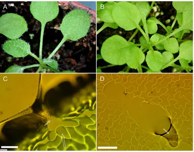
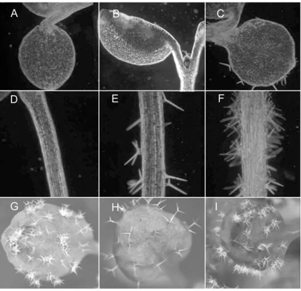
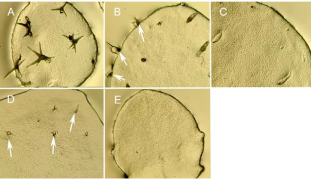
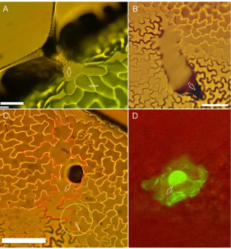
Figure 1: gl2 plants look superficially glabrous.
View of a wt (A) and gl2 mutant (B) plant. WT leaves show many trichomes distributed on their surface while gl2 leaves appear superficially glabrous. However, leaf epidermal imprints on agarose show that laterally expanded trichomes are present on gl2 (D) with a small peak projecting out (arrow). Wild-type trichomes (C) grow out the leaf surface, normally have three extended branches, and at maturity show surface papillae (thin arrow). They are also surrounded by a ring of socket cells (thick arrow). Scale bar: C and D = 50 µm.
Both WT and gl2 mutant plants carrying this reporter construct were analyzed and the number of trichome specifications on the first pair of young leaves (of an average length of 400 µm) was counted. In this analysis all cells that evidently exhibited a highly increased GFP fluorescence level relative to their neighboring cells were considered as trichomes (Fig.2). On gl2 leaves trichome number was increased by about 30% compared to wt (Fig.2, Table.1). This result contradicted the expectations from published reports.
Therefore, to see the extent to which these cells, specified as trichomes at the marker level, proceed in the trichome developmental pathway, epidermal surface imprints of older leaves (first pair), of an average length of 1.5 mm, were made using agarose (see materials and methods section) and observed under a microscope. Young leaf primordia of the stage used in the above marker analysis experiment could not be used because of the limitations of this imprint technique. The number of morphologically identifiable trichomes was counted. Surprisingly, the number of trichomes was less in gl2 when compared to wt-col leaves (Table.1). In summary the above two results suggest that in gl2 mutants many cells get specified and enter the trichome pathway but only some among them proceed further in their development initiating cell morphogenesis steps.
This implies that GL2 positively functions to initiate trichome development but does it negatively regulate the first step of trichome cell selection?
2.3.2 Ubiquitous expression of GL2 inhibits trichome initiations The mutant analysis described above suggests that GL2 may act as a negative regulator during early trichome patterning, though its ‘positive’ function is needed after specification to push the cells entering the trichome pathway to completely undergo morphogenesis and differentiate as trichomes. One possibility to test this would be to ubiquitously express the GL2 gene which should result in a glabrous phenotype similar as observed with TRY or CPC, both of which are inhibitors of trichome initiation. This has been attempted previously. Ubiquitous expression of GL2 cDNA under the control of the CaMV 35S promoter has been reported to be toxic to plants leading to scarcely viable phenotypes (Ohashi et al., 2002). In a wild-type background it was also observed that surviving plants show a gl2-like phenotype and it was concluded that ectopic GL2 expression interferes with endogenous GL2 function thereby effecting normal trichome cell morphogenesis. In order to avoid this toxic affect of ectopic GL2 expression during
Molecular marker criteria
Morphological criteria
WT-Col 22.28 ± 3.4 (n=38) 28 ± 4.6 (n=12) gl2 28.94 ± 3.0 (n=36) 19.33 ± 3.8 (n=15)
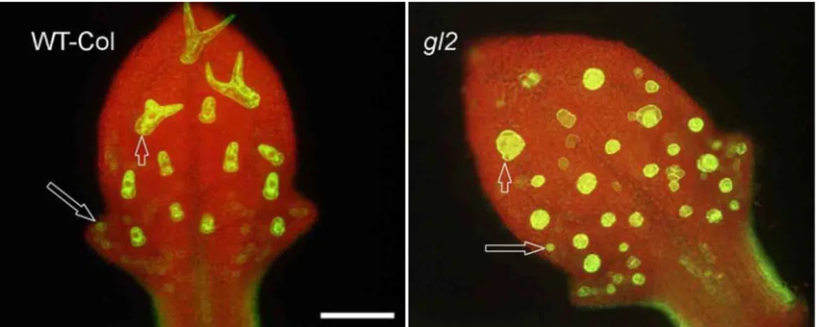
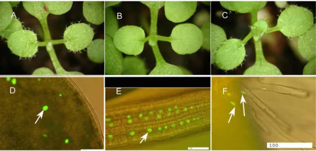
Figure 2: Number of trichome specifications / initiations in wt and gl2
First pair of leaves of WT-col (left) and gl2 plants expressing an early trichome molecular marker (GL2:GFP-ER) were analyzed to count and compare the number of trichome cell specifications. Big arrows point to the earliest cell which has been specified as a trichome and small arrows point to developing larger trichomes. Scale bar = 100 µm
Table 1: Number of trichome specifications / initiations counted using two different methods (trichome molecular marker and morphological criteria) on first pair of gl2 and wt leaves. Young leaves of about 400 µm were used to count GFP marked trichome cells, whereas larger leaves of 1.5 mm were used for the other method (due to limitations of the imprint technique) and hence should not be directly compared.
Figure 3: Ectopic over-expression of GL2 using the constitutively expressed CaMV 35S promoter in wild type (Ler) plants leads to inhibition of trichome initiation (above). Control wt plants after heat shock no trichome inhibition phenotype.
Bottom: RT-PCR analysis of the GL2 gene. Wt-ler (1), 35S:GL2/ler (2), try cpc (3) and 35S:GL2/try cpc (4) (see text for explanation). Elongation factor 1 (EF1) is used as the internal control transcript.
Note the increase in GL2 transcript level in lane 2 when compared to lane 1.
seed germination a modified version of a published recombinase mediated transcriptional induction system (Hoff et al., 2001) was used. The system is designed such that a heat shock induces a recombination event that generates an active 35S::GL2 arrangement on the chromosome (see materials and methods). After heat shock, trichome initiation was compared between wild-type plants and plants carrying the construct system. Wild-type plants showed no patterning defects. Heat shock treated plants containing the construct system showed extreme variability in the phenotype of different siblings of the same line or even between different leaves of the same plant.
Although this variability prevents a statistical analysis, a qualitative description revealed surprising results. Six independent lines were analyzed; 3 lines showed inhibition in trichome initiation after heat shock treatments, with reduced number of trichomes per leaf compared to wt. Lines # 1 and 2 had strong trichome inhibition phenotype with line # 2 showing the most severe phenotype. Most plants were completely glabrous (Fig.3). Leaf epidermal imprints on agarose of these glabrous plants were analyzed to see if gl2 like laterally expanded trichomes are found. The leaves were completely glabrous and had no resemblance to gl2 phenotype where mutant laterally expanded trichomes can still be found. The inducible system showed leakiness, as reported in the original publication, such that also uninduced plants showed trichome inhibition phenotypes to varying degrees (data not shown). Genomic DNA PCR analysis of the lines which showed the trichome inhibition phenotype (both induced as well as uninduced) confirmed that the recombination event had occurred in these lines as expected. In addition, RT-PCR analysis of the lines showing the glabrous phenotype indicated that the level of GL2 transcript was increased as compared to corresponding WT control plants (Fig.3). These results indicated that ubiquitous GL2 expression inhibits trichome initiation. The seeds of the two lines showing strong inhibition phenotype (line # 1,2) did not germinate in the next generation confirming published reports that ectopic over-expression of GL2 during embryogenesis is lethal.
2.3.3 Genetic interaction of GL2 with TRY a) GL2 positively regulates TRY expression:
The finding that GL2 appears to suppress trichome initiations during trichome patterning raises the question of how GL2 function is linked to that of those factors (TRY and CPC) already known to have this function. Hence, it was sought to find if
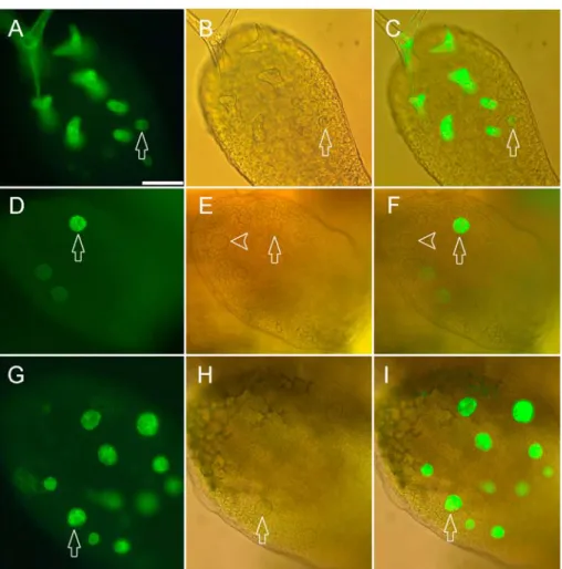
Figure 4: GL2 positively regulates TRYexpression.
TRY gene expression pattern in very young leaves as revealed by the TRY:GFP-ER construct.
A,D,G) Fluorescence of the GFP marker. B,E,H) DIC-light micrograph from the same leaves as A,D,G. C,F,I) Overlay of (A,B), (D,E) and (G,H) respectively. (A-C) TRY expression in wild type. TRY expression is seen as early as incipient trichomes can be recognized by morphological criteria (arrow). (C-E) TRY expression in gl2 mutant leaf expressing TRY only in some (arrow) but not in other (arrow head) trichome initials. (c) AtMYB23 expression in a gl2 mutant leaf. Note this gene is expressed in all trichome initials. Scale bar: 50 µm.
Figure 5: Temporal expression changes of TRY in gl2 mutants and wild type
TRY gene expression pattern in very young leaves as revealed by the TRY:GFP-ER construct. The expression is compared at three different stages of leaf development. A-C) Wild type leaves. D-F) gl2 mutant leaves. A,D) TRY expression in the youngest leaf stage. B,E) TRY expression in an intermediate leaf stage. C,F) TRY expression in a mature leaf. Note that mature trichome cells at the apex of a fully expanded wild type leaf still express the TRY gene while at the same stage there is no detectable TRY expression in gl2 leaves. Scale bars: A,D = 40µm; B,E = 80µm; C,F = 200µm
Figure 6: TRY is a positive regulator of GL2.
GL2:GFP-ER reporter construct expression pattern in wild-type and 35S:TRY plants. A,C,E: wt plants. B,D,E:
35S:TRY plants. A) Wild type cotyledon. No expression of GL2 is seen. B) 35S:TRY cotyledon. Strong expression of GL2 is found in all epidermal cells. C) Epidermal cells of the wild-type hypocotyl. Cells of alternating files express GL2 (arrows). D) Epidermal cells of the 35S:TRY hypocotyl. Note, all cell files express GL2. E) wt young leaf. GL2 expression limited to trichomes and some epidermal cells. F) 35S:TRY young leaf primordium. Trichomes are absent but most epidermal cells express GL2. Scale bars: A,B = 80 µm; C,D = 40 µm; E,F = 50 µm
any regulatory interaction exists between GL2 and TRY. The TRY gene has been shown to act as a negative regulator of trichome development (Schnittger et al., 1999) and is thought to be important in mediating lateral inhibition during trichome patterning (Schellmann et al., 2002). The latter is suggested by the finding that try mutants exhibitnests of 2 or 3 trichomes instead of single separate trichomes as in wild type.
TRY expression was studied in gl2 mutant plants at different stages of leaf development using the TRY::GFP marker line. It has been shown before, that the promoter used in this construct corresponds to the expression pattern observed in in situ hybridizations and is sufficient for rescuing the try mutant phenotype (Schellmann et al., 2002). In very young wild-type leaves TRY is expressed consistently at high levels in all morphologically distinct and recognizable trichome initials (Fig.4 A,B,C). By contrast, in gl2 mutants TRY expression is very variable. Frequently young trichome cells with a typical wt morphology and which have clearly expanded out of the leaf surface show much reduced expression levels or do not express any detectable TRY at all (Fig. 4 D,E,F). Two scenarios can explain these findings. Due to the differentiation defects in some gl2 trichomes the expression of all trichome specific genes could be generally reduced or alternatively, the observed regulation of TRY reflects a specific regulation of TRY by GL2. To distinguish between these two possibilities, we analyzed the expression of another trichome specific gene, AtMYB23 (Kirik et al., 2001). AtMYB23 is expressed in all morphologically distinct trichome initials both in WT as well as in gl2 leaves (Fig.4G,H,I).
The variability of TRY expression intensity suggests that the temporal expression of TRY might be generally effected. We therefore compared its temporal expression pattern in gl2 and wild type leaves. A comparison of different leaf stages revealed that TRY expression is almost absent in more mature leaves of gl2 when wild type plants still show high expression levels (Fig.5). This indicates that GL2 is required not only to initiate TRY expression in all trichome initials but also to maintain it.
b) GL2 is ectopically activated by ubiquitous expression of TRY but not CPC To test if GL2 expression is regulated by TRY, the GL2::GFP-ER marker was analyzed in a 35S::TRY background. At least 10 independent transgenic lines which were analyzed showed the same result. We focused on the analysis of cells in which GL2 is
files of the hypocotyls not overlying a cleft of the underlying cells (Fig.6C) ( ). All epidermal cells of the cotyledons (Fig.6B) and all hypocotyl cell files (Fig.6D) exhibited high levels of GL2 in a 35S::TRY background. In wt, GL2 expression is mostly restricted to trichomes and some epidermal cells at the basal portion of the leaf (Fig.6E). In 35S::TRY plants GL2 was found to be ectopically expressed in most epidermal leaf cells of young leaf primordia (Fig. 6F), though its expression was relatively very weak as leaves developed, with the exception of cells at the margin and towards the apex where high levels of GL2 expression was still seen (data not shown).
Thus, ectopic expression of TRY causes ectopic expression of GL2 indicating that TRY positively regulates GL2 expression. The findings suggest the existence of a positive feedback loop of TRY and GL2. It remains, however, to be determined whether this regulatory relationship is relevant in the context of trichome patterning and if it is dependent on developmental stages. An interesting exception was observed in the roots where only cells in the inner most tissue and not in the epidermis or cortex showed ectopic GL2 expression (data not shown).
As TRY and CPC are highly homologous it has been postulated that they may act partially redundantly during epidermal cell patterning in Arabidopsis. It has been previously shown that 35S::CPC inhibits GL2 expression in roots (Lee and Schiefelbein, 2002). GL2 expression was checked in a 35S::CPC line (which has been previously published and was a kind gift from Takuji Wada, Japan) (Wada et al., 2002).
No expression of GL2 was detected in the hypocotyls/roots/leaves or cotyledons of these plants (data not shown) confirming that CPC represses GL2 expression and suggesting a functional difference between TRY and CPC with respect to the regulation of GL2.
2.3.4 Lateral inhibition during trichome patterning appears to be compromised in gl2
It has been proposed that lateral inhibition is an integral component of the mechanism resulting in the trichome spacing pattern. TRY and CPC are implicated to function in this pathway by inhibiting the cells around a trichome from acquiring a trichome fate.
The finding that GL2 could be involved in early trichome patterning combined with the fact that TRY expression is controlled by GL2 lead us to study patterning during early stages of trichome development as recognized with the trichome molecular marker,
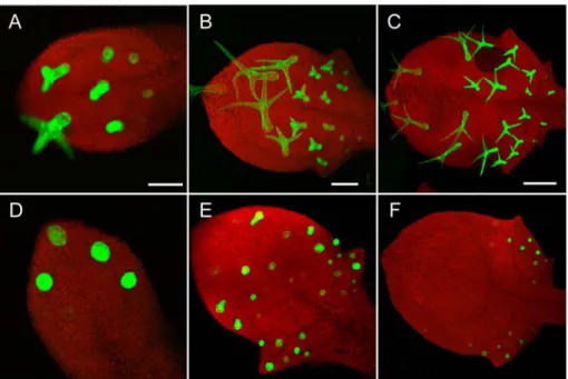
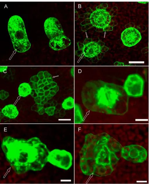
Figure 7: Ineffective lateral inhibition on gl2 leaves
Expression of an early trichome marker (pGL2:GFP-ER ) in wt and gl2 leaves. Epidermal cells surrounding wt (A) trichomes (arrow) express the trichome marker at a very low level or do not express at all suggesting effective lateral inhibition from the trichome. However, in gl2 (B) epidermal cells (thin arrows) surrounding some trichomes (thick arrow) still express the trichome marker relatively at higher levels implying compromise in lateral inhibition (The microscopic settings used were the same for wt and gl2 leaves). Also, occasionally large groups of cells expressing the marker are found in the immediate neighborhood of trichome in gl2 (C). Towards the base of the gl2 leaves trichome clusters resembling try mutants are seen (D, E). However unlike try clusters, the gl2 clusters always have one large dominant trichome (thick arrow) and 1-3 smaller cells surrounding it which start to express the trichome marker as strong as the dominant cell. At exactly the same positions on the leaves similar clusters are found in which the dominant cell is composed of many individual cells (F). Note that D,E and F are 3D reconstructions of many confocal sections (and same magnification) showing the top view of the dominant cell which is clearly bulged out of the leaf surface. In C the dominant cell is clearly one large cell. But in F it is composed of many individual cells with clear cell walls and nuclei. Scale bars: A,B = 20 µm; C = 16 µm; D,E,F = 8 µm.
GL2::GFP-ER. At stages in which epidermal cells around a wild-type trichome initial (Fig.7A) show either very little no marker expression anymore, in gl2 mutants epidermal cells immediately surrounding some trichomes of similar stage were found expressing the marker at relatively high levels (Fig. 7B). Also, in the immediate neighborhood of some trichomes large groups of cells very strongly expressing the marker were found (Fig.7C).This suggests that weaker lateral inhibition in gl2 leaves could lead to such a phenotype. This ineffective inhibition of cells from getting specified as trichomes may consequently lead to more number of trichome specifications in gl2 when compared to wt leaves. This is reminiscent of the cpc mutant phenotype where the number of trichome initiations is increased compared to wt.
The observation that expression of TRY in all trichome initials depends on GL2 led us to speculate that small clusters of trichomes (2 - 3) as seen on try mutants could also be seen on young gl2 leaves. Indeed, trichome clusters reminiscent of those observed in try mutants were observed in gl2 mutants with an average frequency of around 5 - 10%.
Typically, one to two adjacent cells next to a large trichome start to express GL2 at high levels when compared to the other epidermal cells in the neighborhood where no marker expression is found anymore (Fig.7 D, E). Though this phenotype is reminiscent of the try phenotype, the difference lies in the fact that whereas in the try clusters the trichomes are equally large and developed, the try-like cluster seen on gl2 has one dominant large trichome cell surrounded by one – two much smaller cells. At about the same frequency and position on the leaves of similar age try-like clusters of the same size as mentioned before were found but the difference was that the dominant cell was not one single cell any longer but composed of many individual cells (Fig.7F) with clear cell walls and large nuclei (By DAPI staining it was found that they are large nuclei and not vacuoles). 3D reconstructions of many individual confocal sections of all these try-like trichome clusters showed that they are bulged out of the leaf surface considerably. Fig.7D and Fig.7F are two representative pictures of these two classes of try-like clusters. Whereas the dominant cell in Fig.7D is clearly one large cell, the one in Fig.7F is not. The above observations pose the obvious question of whether the trichomes which are seriously compromised in lateral inhibition result in try-like clusters (Fig.7D) and possibly divide later (Fig.7F). The size of the dominant cell, their frequency of occurrence and position on the leaves in both the cases all are suggestive (but not proofs) that it may be the case. It also needs to be investigated in more detail
to see if the large group of cells found in the neighborhood of trichomes as seen in Fig.7C are a later consequence of the event seen in Fig.7F or completely unrelated to it.
Epidermal imprints of gl2 leaves were analyzed carefully to detect any morphologically identifiable try-like clusters seen by the marker study. None were detected. This again points to the fact that in the absence of GL2 function, new trichome specifications seen at the marker level do not proceed further in the pathway by initiating morphogenesis processes. Is the ineffective lateral inhibition on gl2 leaves an indirect effect related to the differentiation status of trichomes? Or is GL2 directly involved in the process of lateral inhibition during trichome patterning in wt?
2.3.5 TRY and / or CPC may possibly mediate the inhibitory function of GL2 during patterning
The finding that GL2 positively regulates TRY expression in trichomes and gl2 mutants show defective lateral inhibition raises the possibility that TRY and/or CPC may be important to mediate the inhibitory effect of GL2. In order to test this, heat shock inducible GL2 expression in the try cpc double mutant background was used. The try cpc double mutant shows large clusters of trichomes containing between 2-30 trichomes in each cluster (Fig.8G) (Schellmann et al., 2002). If TRY and/or CPC would mediate the inhibitory effect of GL2 one would expect that the try cpc double mutant would be insensitive to ubiquitous GL2 expression.
The try cpc double mutant plants were transformed with the same heat inducible GL2 construct as earlier (see “ubiquitous GL2 expression inhibits trichome initiation in wt leaves” paragraph) and 6 resulting transgenic lines were analyzed. After heat shock four lines showed new phenotypes. In all four lines ectopic trichomes on cotyledons and hypocotyls was increased compared to try cpc control plants after heat shock (Fig.8 C, F). Line # 6 showed a strong effect on reduction of trichome cluster size and trichome numbers on leaves when compared to try cpc control after heat shock. Most leaves on line #6 plants had single isolated trichomes in the middle of the leaf and small clusters of 2-4 trichomes on the edges (Fig.8 H). Lines # 3 and 5 produced very high numbers of ectopic trichomes on cotyledons and hypocotyls (more than line # 6) but did not show any discernible difference in their leaf trichome phenotypes (Fig.8 I). Line # 4 had an intermediate leaf trichome phenotype with respect to trichome number and cluster size reduction. Two observations are interesting. No completely glabrous leaves
Figure 8: Ubiquitous expression of GL2 in try cpc double mutant.
A,B,C) Cotyledons of wt-ler, try cpc and 35S:GL2/try cpc plants. Note the occurrence of many ectopic trichomes on C. Trichomes are not seen on wild type hypocotyls (D) but try cpc plants do produce some trichomes on their hypocotyls (E). But 35S:GL2 increases the number of trichome initiations on try cpc hypocotyls enormously (F). Two lines both showing ectopic trichomes on cotyledons and hypocotyl but having different effects on leaves are shown in H (line 6) and I (line 5). Line 6 shows a drastic reduction in cluster size and trichome number (H) when compared to control try cpc plants (G). But Line 5 (I) has a similar trichome phenotype as that of control.