• Keine Ergebnisse gefunden

The Role of Noradrenaline in Energy Homeostasis

N/A
N/A
Protected

Academic year: 2021

Aktie "The Role of Noradrenaline in Energy Homeostasis"

Copied!
161
0
0

Wird geladen.... (Jetzt Volltext ansehen)

Volltext

(1)

The Role of Noradrenaline in Energy Homeostasis

I n a u g u r a l – D i s s e r t a t i o n

zur Erlangung des Doktorgrades

der Mathematisch-Naturwissenschaftlichen Fakultät der Universität zu Köln

vorgelegt von

Lars Paeger

aus Traben-Trarbach August 2013

Zoologisches Institut

Universität zu Köln

(2)

Prof. Dr. Jens Brüning

Tag der mündlichen Prüfung: 15 . 10 . 2013

(3)

Contents

Abbreviations x

Abstract xii

Zusammenfassung xiv

1 Introduction 1

1 . 1 The central nervous system in control of energy homeostasis . . . . 2

1 . 1 . 1 The melanocortin system of the hypothalamus . . . . 3

1 . 1 . 2 Glucose sensing . . . . 8

1 . 2 The catecholamine noradrenaline in the control of energy homeostasis and food intake . . . . 10

1 . 2 . 1 Noradrenaline . . . . 11

1 . 2 . 2 Noradrenergic receptors . . . . 14

1 . 2 . 3 The hypothalamus in the noradrenergic control of energy homeo- stasis . . . . 17

1 . 2 . 4 The Locus Coeruleus in the control of energy homeostasis and glucose metabolism . . . . 19

1 . 3 Thesis objectives . . . . 21

2 Materials and Methods 23 2 . 1 Animal care . . . . 23

2 . 2 Brain slice preparation . . . . 23

2 . 3 Patch-clamp recordings . . . . 24

2 . 3 . 1 Perforated-patch clamp recordings . . . . 25

2 . 3 . 2 Single cell labeling . . . . 26

2 . 3 . 3 Immunohistochemistry . . . . 26

2 . 3 . 4 Image processing . . . . 27

(4)

2 . 3 . 6 Noradrenalin experiments . . . . 28 2 . 3 . 7 Glucose sensing experiments . . . . 28 2 . 3 . 8 Data analysis . . . . 29

3 Results 31

3 . 1 Properties of POMC and NPY/AgRP neurons in the arcuate nucleus . . . 33 3 . 1 . 1 Properties of POMC neurons . . . . 33 3 . 1 . 2 Properties of NPY neurons . . . . 36 3 . 2 The effect of noradrenaline on POMC neurons of the melanocortin system

in the arcuate nucleus . . . . 39 3 . 2 . 1 Noradrenaline inhibits POMC neurons dependent on concentration 39 3 . 2 . 2 The effect of noradrenaline on POMC neurons is cell intrinsic . . . 44 3 . 2 . 3 Noradrenaline inhibits POMC neurons via the activation of

α 2 A -adrenergic receptors . . . . 44 3 . 3 The effect of noradrenaline on NPY/AgRP neurons . . . . 48

3 . 3 . 1 Noradrenaline excites NPY/AgRP neurons dependent on concen- tration . . . . 48 3 . 3 . 2 High concentrations of noradrenaline elicit bursting in NPY/AgRP

neurons . . . . 49 3 . 3 . 3 The effect of noradrenaline on NPY/AgRP neurons is cell intrinsic 52 3 . 3 . 4 Noradrenaline excites NPY/AgRP neurons via the activation of

α 1 A -adrenergic receptors . . . . 53 3 . 4 Effects of dietary changes and aging on responses of POMC neurons to

noradrenaline . . . . 55 3 . 5 The effect of noradrenaline on POMC and NPY/AgRP neurons unifies

both heterogenous populations . . . . 56 3 . 6 The role of the locus coeruleus in the control of energy homeostasis . . . 58 3 . 6 . 1 Properties of noradrenergic neurons in the locus coeruleus . . . . 59 3 . 6 . 2 The locus coeruleus: role in energy homeostasis, control of brown

adipose tissue and glucose responsiveness . . . . 65

(5)

3 . 6 . 3 Expression of the mutant variant Kir 6 . 2 in catecholaminergic cells leads to obesity and altered brown adipose tissue morphology . . 65 3 . 6 . 4 A subpopulation of neurons in the locus coeruleus adapt their fir-

ing to changes in extracellular glucose in glucose-excited manner 69 3 . 6 . 5 A small subpopulation of neurons in the locus coeruelus adapt

their firing to changes in extracellular glucose in glucose-inhibited manner . . . . 75

4 Discussion 77

4 . 1 Properties of POMC neurons . . . . 78 4 . 2 Properties of NPY/AgRP neurons . . . . 80 4 . 3 Noradrenaline differentially modulates POMC and NPY/AgRP neurons 81 4 . 4 Properties of noradrenergic neurons in the locus coeruleus . . . . 90 4 . 5 The locus coeruleus in the control of energy homeostasis and glucose

metabolism . . . . 92 4 . 5 . 1 Kir 6 . 2 THCre -mice develop obesity and impaired brown adipose tis-

sue morphology and function . . . . 92 4 . 5 . 2 Glucose sensing, brown adipose tissue and the locus coeruleus . . 94 4 . 6 The role of noradrenalin in the pharmacotherapy of obesity . . . . 98 4 . 7 Outlook and preliminary data . . . . 100

4 . 7 . 1 Energy homeostasis and noradrenergic signaling in the hypotha- lamus . . . . 100 4 . 7 . 2 Anatomy of noradrenergic signaling to the hypothalamus . . . . . 101 4 . 7 . 3 Effect of insulin, leptin and ghrelin on the the noradrenergic mod-

ulation of POMC and NPY/AgRP neurons in the arcuate nucleus 102 4 . 7 . 4 Effects of dietary changes and aging . . . . 104 4 . 7 . 5 The role of the locus coeruleus in energy homeostasis . . . . 104

List of Figures 107

Bibliography 108

Danksagung 140

(6)

Teilpublikationen 143

Curriculum Vitae 146

(7)

Abbreviations

α/β-MSH α/β-melanocyte stimulating hormone A Adrenaline/adrenergic

AB Antibody

AADC Aromatic amino acid decarboxylase AC Adenylate cyclase

AD Alzheimer’s disease AgRP Agouti-related protein

AMPK Adenosine monophosphate-activated protein kinase AP Action potential

ArP Area postrema

AR Adrenergic receptor/adrenoceptor ARC Arcuate nucleus

ATP Adenosine triphosphate BAT Brown adipose tissue BBB Blood-brain-barrier BMI Body mass index

CA Catecholamine

cAMP Cyclic adenosine monophosphate

CART Cocaine-amphetamine regulated transcript CEC Chloro-ethyl-clonidine

CNQX 6-Cyano-7-nitroquinoxaline-2, 3-dione (AMPA/kainate receptor antagonist) CNS Central nervous system

CPP Conditioned place preference

DA Dopamine/dopaminergic

(8)

DAP-5 DL-2-amino-5-phosphopentanoic acid (NMDA receptor antagonist) DBH Dopamine β-hydroxylase

DMH Dorsomedial hypothalamus DMSO Dimethyl sulfoxide

DMT2 Diabetes mellitus type 2

DMV dorsal motor nucleus of the vagus DNB Dorsal noradrenergic bundle FFA Free fatty acids

GABA γ-Aminobutyric acid GE Glucose-excited GI Glucose-inhibited

GIRK G-Protein activated inwardly rectifying potassium channel

GK Glucokinase

GLUT Glucose transporter

GPCR G-Protein coupled receptor HFD High-fat diet

icv. intracerebroventricular IP 3 Inositoltrisphosphate

IPSC Inhibitory postsynaptic current K ATP ATP-dependent potassium channel LepR Leptin receptor

LH Lateral hypothalamus LC Locus Coeruleus

LC-NA Locus Coeruleus noradrenergic

MC3/4R Melanocortin receptor 3 and 4

NA Noradrenaline/noradrenergic

NAP Numerical aperture

(9)

NCD Normal chow diet NPY Neuropeptide Y

NPY 1 R Neuropeptide Y receptor type 1 NTS Nucleus tractus solitarius PD Parkinson’s disease

PI3K Phosphatidylinositol-3-kinase PIP Phosphatidylinositolphosphate

PIP 2 Phosphatidylinositol-4,5-bisphosphate PIP 3 phosphatidylinositol-3,4,5-bisphosphate PKA Protein kinase A

PKC Protein kinase C PLC Phospholipase C

PNMT Phenylethanolamine-N-methyltransferase PTX Picrotoxin (GABA A receptor antagonist) POMC Proopiomelanocortin

PVH Paraventricular nucleus R S Series resistance

REM Rapid eye movement SF-1 Steroidogenic factor 1 SFA Spike frequency adaptation SNA Sympathetic nerve activity SNS Sympathetic nervous system TH Tyrosine hydroxylase

VGCC voltage gated Ca 2 + conductance VMH Ventromedial hypothalamus VNB Ventral noradrenergic bundle WAT White adipose tissue

WD Working distance

(10)
(11)

Abstract

Obesity is a condition that is associated with excessive weight gain and fat mass storage whose prevalence is increasing within western populations. A variety of co-morbidities are linked to obesity such as type 2 diabetes mellitus, cardiovascular diseases and neu- rodegenerative disorders, including Alzheimer’s disease and Parkinson’s disease. To- gether, this contributes to substantial costs of healthcare programs. In non-obese indi- viduals, energy intake and energy expenditure is precisely matched over a long time period in order to maintain energy resources and fat mass. This mechanism, termed energy homeostasis is accomplished by regulatory neuronal networks in the central ner- vous system (CNS).

To better understand and counteract obesity and its co-morbidities, increasing efforts are being made to define the control mechanisms in the CNS, that regulate body weight and energy homeostasis. The focus of this study is the noradrenergic (noradrenaline;

NA) modulation of energy homeostasis. Anti-obesity drugs, for example amphetamines, can exert strong anorexigenic effects on eating behaviour in humans. However, these drugs generally affect multiple transmitter and neuromodulator pathways, such as the dopaminergic and serotonergic system, leading to undesired side effects. Pharmacolog- ical studies indicate that the anorexigenic effect of amphetamine and related drugs are caused in part by modulation of the NA system. In order to devise strategies and de- velop specific drugs with minimized side effects in support of weight loss programs, it is critical to understand in detail the mechanisms in the CNS by which NA contributes to energy homeostasis.

Besides the well established role of the paraventricular nucleus of the hypothalamus

in NA-mediated modulation of food intake, studies indicate that NA input on the home-

ostatic system in the arcuate nucleus of the hypothalamus (ARC) might also modulate

eating behaviour. In the ARC, two key neuronal populations, pro-opiomelanocortin

(POMC) and agouti-related peptide (AgRP) expressing neurons sense and integrate pe-

ripheral and nutritional signals. Once activated, POMC neurons promote satiety and

(12)

Mechanisms that mediate the possible NA action in the ARC are unknown. In this study, the effect of NA on POMC and AgRP expressing neurons has been investigated.

Application of NA inhibits POMC neurons, while AgRP neurons are excited. Pharmaco- logical experiments revealed that these effects are mediated by α 2 A - and α 1 A -adrenergic receptors (AR). This suggests a potent NA modulation of food intake. With respect to these effects, afferent projections from NA nuclei and the conditions under which NA is released into the ARC are of greatest interest.

As a potential NA source, the locus coeruleus (LC) in the brainstem contains 50 % of the NA neurons in CNS. Efferent projections from the LC to the ARC have been identified. Besides the contribution to autonomic functions in general, studies indicate that the LC is also involved in glucose metabolism and the control of brown adipose tissue (BAT). Moreover, BAT thermogenesis is dependent on NA and plasma glucose.

Therefore, the effects of changes in extracellular glucose concentrations have been inves- tigated. Around 40 % of neurons in the LC responded with increasing spike frequency due to elevated glucose levels, identifying these neurons as glucose-excited. A small subpopulation responded with a moderate inhibition and is considered as glucose- inhibited. Expression of a mutant variant of the ATP dependent potassium channel in mice silenced a large number of LC neurons and abolished responses to glucose.

Moreover, sympathetic nerve activity was reduced and led to a white-adipose-tissue-like morphology of BAT, alongside with impairment of thermogenesis. As a consequence of decreased energy expenditure, these mice developed obesity.

The modulation of POMC and AgRP neurons by NA indicates a critical role of

the catecholamine in the control of energy homeostasis. Moreover, this study reveals

that the LC contains glucose-sensing neurons and contributes to the control of glucose

metabolism and the activity of BAT. Its projection patterns in the CNS identify the LC as

a potential source for NA release into the ARC. These results lead to new insights and

the expansion of the current role of NA in the control of energy homeostasis. Impor-

tantly, this may help to develop new strategies and drugs with minimized side effects

in the treatment of obesity.

(13)

Zusammenfassung

Adipositas stellt ein stark zunehmendes Gesundheitsproblem in Industrieländern dar.

Eine Reihe an Begleiterkrankungen, wie Typ 2 Diabetes Mellitus , Herzkreislaufkrank- heiten und neurodegenerative Störungen, wie z.B. Alzheimer und Parkinson verur- sachen hohe Zusatzkosten für Gesundheitssysteme. Bei nicht-adipösen Individuen sind Energieaufnahme und Verbrauch sehr präzise aufeinander abgestimmt, um über längere Zeit Stabilität von Energiereserven und Fettgewebe herzustellen. Dies ist definiert als Energiehomöostase und wird vom zentralen Nervensystem (ZNS) kontrol- liert.

Um Adipositas-assoziierte Erkrankungen zu vermeiden, werden große Anstrengun- gen unternommen, die Regulation der Energiehomöostase im ZNS besser zu verstehen und zu beeinflussen. Die vorliegende Arbeit beschäftigt sich mit der noradrenergen (Noradrenalin; NA) Modulation der Energiehomöostase. Amphetamine und verwandte Wirkstoffe können Hunger-unterdrückende Wirkung haben. Dabei werden in der Regel mehrere Transmitter- bzw. Neuromodulatorsysteme beeinflusst, welches unerwünschte Nebenwirkungen zur Folge haben kann. Pharmakologische Studien deuten darauf hin, dass Amphetamine und verwandte Substanzen in der Behandlung von Adipositas zum Teil auf das NA System wirken. Detailliertes Wissen, wie NA auf die Energiehomöostase wirkt, ist daher unabdingbar in der Entwicklung von Medika- menten, die Adipositas-Patienten bei ihrer Therapierung unterstützend begleiten sollen.

Neben der bekannten NA Modulation der Nahrungsaufnahme im paraventrikulären

Hypothalamus, sprechen Untersuchungen zusätzlich dafür, dass NA auch direkt im

Arcuate Nucleus des Hypothalamus (ARC) wirkt. Dieser gilt als Schlüsselregion in

der Kontrolle von Nahrungsaufnahme und Energieverbrauch. Er enthält unter an-

derem zwei Neuronenpopulationen, proopio-melanocortin (POMC) exprimierende und

Agouti-related peptide (AgRP) exprimierende Neurone, die Signale aus der Peripherie

im Zusammenhang mit dem Nahrungsstatus eines Körpers aufnehmen und weiterver-

arbeiten können. Die Erregung von POMC Neuronen hat Sattheit zur Folge, während

(14)

verbrauch resultiert. Die Wirkmechanismen von NA im ARC sind bisher nicht geklärt.

Daher wurde der Effekt von NA auf POMC und AgRP Neurone untersucht. NA in- hibiert konzentrationsabhängig POMC Neurone, während es AgRP Neurone anregt.

Diese Effekte werden durch die Aktvierung von α 2 A - und α 1 A -adrenergen Rezeptoren vermittelt. Aufgrund dieser Effekte sind die NA Projektionen in den ARC und die Umstände in denen NA dort ausgeschüttet wird von größtem Interesse.

Als eine mögliche Quelle beherbergt der Locus Coeruleus (LC) etwa 50 % aller NA Neurone im ZNS. Die Innervation des ARC mit Efferenzen aus dem LC ist bekannt.

Neben der Rolle des LC im autonomen Nervensystem, deuten Studien an, dass dieser sowohl an der Glukosehomöostase, als auch an der glukose- und NA-abhänigen Kon- trolle des braunen Fettgewebes (BAT) beteiligt ist. In Anbetracht dieser Daten wurde untersuch, ob Neurone im LC auf Änderungen in der extrazellulären Glukosekonzen- tration reagieren. Etwa 40 % der untersuchten Neurone erhöhten ihre Aktivität auf- grund einer höheren Glukosekonzentration und konnten daher als glukose-angeregt identifiziert werden. Ein kleiner Teil der Neurone reagierte mit einer Inhibition und wurde daher als glukose-inhibiert identifiziert. Die Expression eines mutierten ATP- abhängigen Kaliumkanals führte zur verminderter elektrischer Aktivität von LC Neuro- nen und verhinderte die Anregung aufgrund erhöhter extrazellulärer Glukosekonzen- trationen. Weiterhin sank die Aktivität im Nervus Sympathikus und beeinflusste die Thermogenese im BAT negativ. Als Folge reduzierten Energieverbauchs entwickelten, die Mäuse Adipositas.

Effekte auf POMC und AgRP Neurone im ARC deuten auf eine Rolle NAs in der Energiehomöostase hin. Der LC beherbergt Neurone, die auf unterschiedliche Glukose- konzentrationen reagieren und zur Regulation des Glukosestoffwechsels und der Ak- tivität von BAT beitragen. In diesem Zusammenhang, identifizieren die Projektionen in den ARC, den LC als eine mögliche Quelle der NA-Ausschüttung. Die Ergebnisse dieser Arbeit beschreiben neue Erkenntnisse in der NA Modulation der Energiehomöostase.

Dies könnte helfen neue Strategien und Medikamente zu entwickeln, die weniger Neben-

wirkungen in der Behandlung von Adipositas zur Folge haben.

(15)

1 Introduction

Obesity, defined as an abnormal or excessive accumulation of body fat mass that impairs health, has become a worldwide epidemic with humans categorized as overweight or obese nearly doubled in number since the 1980 s (World Health Organization, WHO;

Fact sheet N 311 ). The increasing prevalence for obesity in both developed and non- developed countries is a major health threat in near future. Obesity and overweight are defined by the body-mass-index (BMI), a simple weight-for-height measure where a value greater than or equal to 25 is classified as overweight and a value greater than or equal to 30 is considered as obese (WHO, 2007 ). In 2008 , 1 . 4 billion people, with the age of 20 or older were classified as overweight, of which 200 million men and 300 million women were listed as obese. This alarming trend can also be observed in children. By the end of 2010 , 40 million children aged under five years were categorized as overweight, worldwide (WHO, Fact sheet N 311 ).

Certain co-morbidities associated with obesity are a major health problem. For in-

stance, type 2 diabetes mellitus (T2DM) is diagnosed in a growing number of people

throughout all ages (Must et al. , 1999 ). Besides, the risk for cardiovascular diseases like

hypertension, stroke and heart attack, musculosceletal disorders and even certain forms

of cancer, is increasing with higher BMIs (Guffey et al. , 2013 ; Lehrer et al. , 2013 ; Osmond

et al. , 2009 ). Obesity also affects the function of the central nervous system (CNS). Al-

terations in brain morphology and decrease in brain volume have been associated with

overweight or obesity in young adults (Bruce-Keller et al. , 2009 ). In part, these effects

are also observed during aging and obesity increases the risk of neurodegenerative dis-

orders including Alzheimer’s disease (AD) and Parkinson’s disease (PD) (Luchsinger,

2010 ; O’Rahilly, 2009 ). In 2012 , the WHOs Regional Office for Europe reports that obe-

sity is already responsible for 2 - 8 percent of overall health costs and 10 - 13 percent of

deaths. Also in the United States, the increasing prevalence of obesity is responsible for

substantial costs of health care programs. In 2009 , a study by Finkelstein et al. ( 2009 )

evaluated the expected costs to be $ 85 . 7 billion. This has recently been extended to an

(16)

overall cost for obesity and obesity-related diseases of $ 209 . 7 billion (Cawley & Mey- erhoefer, 2012 ). Due to this "economic burdon generated by obesity, its treatment has become one of the most urgent issues in medicine today" (Li & Cheung, 2009 ). It is crit- ical to devise specific pharmacological strategies to assist obese patients in starting and maintaining a program of weight loss with no or minimized side effects. For example, amphetamine and its variants can be strong modulators of eating behaviour in humans, however strong side effects are a common problem of such anti-obesity strategies (Adan, 2013 ; Derosa & Maffioli, 2012 ; Fantasia, 2013 ). Therefore, neuronal mechanisms which underlie the control of body weight are of greatest interest in order to develop specific therapies in the treatment of overweight or obese patients.

In order to gain further knowledge about the neurotransmitter systems which regu- late energy homeostasis, this thesis focusses on the specific role of noradrenaline (nora- drenergic; NA) in this mechanism. In the following sections, a short introduction into the CNS in the general control of energy homeostasis with the focus on the melanocortin system of the hypothalamus is presented. As an example of a nutrient signal, glucose sensing is described as this mechanism is apparent in nuclei in the hypothalamus as well as in NA nuclei in the brainstem. The main focus of this study is the NA modulation of eating behaviour and its effect on the arcuate nucleus of the hypothalamus (ARC), a key region in the control of energy homeostasis. The locus coeruelus (LC) is introduced as a potential source for hypothalamic NA release and finally its role in glucose metabolism and control of brown adipose tissue (BAT) is described.

1.1 The central nervous system in control of energy homeostasis

Several heritable factors such as genetic predispositions and environmental influences

like untimely food habits, reduced physical activity and increased food consumption

along with unlimited access to food promote the development of overweight and obe-

sity (Power, 2012 ). It is not massive periodic overconsumption that leads to obesity

rather than a small mismatch in intake and expenditure over a long time period. A

caloric consumption of 0 . 3 percent over energy expenditure is already sufficient for

weight gain (Rosenbaum et al. , 1997 ). Energy homeostasis can be defined as the phys-

(17)

1 Introduction

iological process whereby energy intake is matched to energy expenditure over time to promote the stability of energy resources stored as adipose tissue (Hagan & Niswender, 2012 ). This regulation is an exceedingly complex biological mechanism, which involves a variety of different biological behaviors and substrates. The question concerning how this regulation of food intake and energy expenditure is achieved has been thoroughly investigated. Initially regarded as a mechanism which is controlled by the body’s pe- riphery, it became increasingly clear that several parts of the CNS are critically involved in the regulation of energy homeostasis (Brobeck, 1946 ; Brobeck et al. , 1943 ).

1.1.1 The melanocortin system of the hypothalamus

A number of landmark studies highlighted the hypothalamus in the control of food

intake (Anand & Brobeck, 1951 ; Kennedy, 1950 ; Mayer & Thomas, 1967 ). These stud-

ies led to the proposal of a "dual center model" with the ventromedial hypothalamus

(VMH) being the "satiety center" and the lateral hypothalamus (LH) the "hunger center",

as lesioning of one of these either decreases or increases food intake. Soon, the ques-

tion arose of how these areas gather information to precisely determine levels of energy

intake and expenditure. Parabiosis studies on lesioned rats led to the conclusion of a

peripheral signal in relation to animals’ lipostatics (Hervey, 1959 ). A rat with a lesion in

the VMH was surgically connected with a normal rat, which allowed humoral factors

to pass from one animal to the other. The lesioned rat developed obesity whereas its

partner became hypohagic and lost weight, suggesting that a signal in proportion to the

amount of fat mass is highly potent to inhibit food intake. Additional parabiosis studies

on genetically obese mice, ob/ob and db/db, led to the assumption that the first lack the

signal, while the latter are insensitive to it (Coleman, 1973 , 1978 ). The later identification

of the ob and db gene, which encode the hormone leptin and the respective receptor con-

firmed these experiments (Zhang et al. , 1994 ). These key studies led then to the further

identification of various genes involved in the process of energy homeostasis, encoding

peptides, receptors and transcription factors and most importantly the identification of

the "melanocortin system" (Gao & Horvath, 2008 ).

(18)

The arcuate nucleus of the hypothalamus

The "melanocortin system" represents the key neuronal system in the control of energy homeostasis targeted by a large number of metabolic signals such as leptin, insulin, ghrelin as well as nutritional signals like glucose and free fatty acids (FFA; Brüning et al.

2000 ; He et al. 2006 ; Ibrahim et al. 2003 ; Parton et al. 2007 ; Spanswick et al. 1997 , 2000 ).

It is located in the ARC, which has an anatomically unique position because of its close proximity to fenestrated capillaries at the very medialbasal part of the hypothalamus (Burdakov et al. , 2005 a; Ganong, 2000 ). Here, the blood brain barrier is highly permeable and thus provides access to peripheral signals for neurons (Benoit et al. , 2000 ; Cone et al.

, 2001 ).

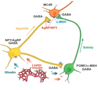
Two neuronal populations have been identified leading to opposing effects on food intake (Cone et al. , 2001 ). The first population expresses pro-opiomelanocortin (POMC) and cocaine- and amphetamine regulated transcript (CART). Activation of POMC neu- rons mediates satiety (anorexigenic). POMC is further cleaved into α- and β-melanocyte stimulating hormone (α- and β-MSH), which upon activity-dependent release stimulate the melanocortin receptor types 3 and 4 (MC3R, MC4R) in target areas and leading to reduced food intake and satiety (Boston et al. , 1997 ; Cone, 2005 ; Ellacott & Cone, 2004 ).

In contrast, neuropeptide Y (NPY) containing neurons in the ARC, expressing the agouti-related peptide (AgRP) mediate orexigenic signals (Aponte et al. , 2011 ). Con- sequently, NPY release increases food intake with a concomitant decrease in energy expenditure (Ollmann et al. , 1997 ; Stanley & Leibowitz, 1984 ). AgRP is a potent inverse agonist on MC3R and MC4R, thus preventing activation by their ligand α-MSH (Smith et al. , 2007 ). Additionally, AgRP expressing neurons co-express the inhibitory transmit- ter γ-aminobutyric acid (GABA) and form unidirectional synapses on POMC neurons, thus simultaneously inhibiting these anorexigenic neurons (Cowley et al. , 2001 ). This interaction may be seen as a important evolutionary blue-print that favors hunger over satiety by tonic inhibition of anorexigenic signals and thus can also lead to overcon- sumption in times of higher food availability (Atasoy et al. , 2012 ; Bates & Myers, 2003 ).

The identification of this network in the ARC led to the question of how signals are

further transferred and integrated to elicit adaptive behavior.

(19)

1 Introduction

Insulin

Figure 1.1: Melanocortin system in the ARC. Two opposing cell types in the ARC sense and process peripheral signals to second order neurons, which express MC4Rs. Insulin and leptin inhibit appetite signaling NPY/AgRP neurons and excite satiety signaling POMC neurons, lead- ing to α-MSH release and the activation of second order neurons via MC4Rs. In contrast, ghrelin excites NPY/AgRP neurons, which form GABAergic synapses on POMC and second order neu- rons, leading to inhibition of satiety signals. NPY inhibits second-order neurons and AgRP potently antagonizes MC4Rs and activation by α-MSH. ARC, arcuate nucleus of the hypotha- lamus; α-MSH, α-melanocyte-stimulating hormone; AgRP, agouti-related peptide; POMC, pro- opiomelanocortin; MC4R, melanocyte receptor type 4; NPY, neuropeptide Y; GABA, γ-amino butoric acid. Modified from (Gao & Horvath, 2007 ).

Downstream targets of POMC and NPY/AgRP neurons

Various nuclei of the CNS exhibit dense innervation by POMC and NPY/AgRP projec- tions, for example the paraventricular nucleus of the hypothalamus (PVH), the VMH, the dorsal medial hypothalamus (DMH) and the LH. MCR and/or NPY-receptor expression could be observed in all of the mentioned hypothalamic nuclei, thus identifying them as strong candidates for melanocortin signaling (Kishi et al. , 2003 ; Mountjoy et al. , 1994 ; Sahm et al. , 1994 ). An elegant study highlighted the PVH neurons as direct downstream targets for ARC POMC neurons and are therefore called second-order neurons in the melanocortin system (Balthasar et al. , 2005 ). The PVH has been studied extensively in the control of food intake and satiety. A study with lesioned PVH in rats, revealed that NPY and POMC signaling is still sufficient to regulate food intake (Dube et al. , 2006 ).

However, Atasoy et al. ( 2012 ) provide data, in which a PVH subpopulation is necessary

in processing the signals that are generated in first place in the ARC to other nuclei in

(20)

the CNS. Furthermore, a large body of literature suggests a role of the PVH in the NA control of energy homeostasis (Leibowitz, 1988 ; Wellman, 2000 , 2005 ). Injections of NA and specific agonists and antagonists of adrenergic receptors (adrenoceptors; AR) into the PVH led to marked effects on feeding behaviour in rats.

In general, signals in relation to eating behaviour from hypothalamic nuclei are pro- cessed to nuclei in the brainstem, the Nucleus Tractus Solitarius (NTS) and the dorsal motor nucleus of the vagus (DMV) and are integrated with mechanosensory signals from the periphery (Suzuki et al. , 2010 ). One important signal is glucose, which mod- ulates electrical activity of POMC and AgRP neurons in the ARC and brainstem nuclei (Diggs-Andrews et al. , 2010 ; Fioramonti et al. , 2007 ; Ibrahim et al. , 2003 ; Mizuno &

Oomura, 1984 ; Parton et al. , 2007 ; Ritter et al. , 2011 ; Thorens, 2011 ; Wang et al. , 2008 ).

A prerequisite to elicit adaptive behaviours in response to changes in glucose concen-

tration is the perception of extracellular glucose levels, a mechanism termed "glucose

sensing" (Levin et al. , 1999 ; Routh, 2002 ).

(21)

1 Introduction

PVH 3V

LH

VMH ARC ME LH DMH

insulin, leptin, ghrelin, FFA, glucose

DMV PVH

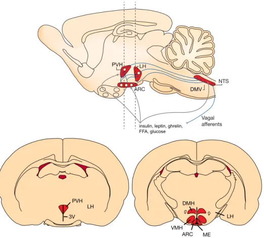
Figure 1.2: The melanocortin system and its downstream targets in the integration of peripheral

signals. Upper scheme The peripheral signals ghrelin, insulin and leptin, FFA and glucose act on

neurons in the brainstem and the hypothalamus. In the brainstem the NTS and DMV receive

sensory information from vagal afferents and process signals to the hypothalamus. Here the

respective information is integrated with the peripheral signals and processed back to the brain-

stem to elicit adequate behavior, i.e. food intake, satiety or energy expenditure. Lower scheme

Coronal sections illustrated by dashed lines above show the positions of the relevant nuclei in

the hypothalamus, which are involved in energy homeostasis. FFA, free fatty acids; NTS, nu-

cleus tractus solitarius; DMV, dorsal motor nucleus of the vagus; PVH, paraventricular nucleus

of the hypothalamus; LH, lateral hypothalamus; ARC, arcuate nucleus of the hypothalamus; 3 V,

3 rd ventricle; ME, median eminence; VMH, ventromedial hypothalamus; DMH, dorsomedial

hypothalamus. Modified from (Morton et al. , 2006 ; Schwartz et al. , 2000 ).

(22)

1.1.2 Glucose sensing

Glucose represents an important nutrient also mediating modulatory effects on neurons within the CNS (Marty et al. , 2007 ). In either high or low concentrations, glucose is able to elicit adaptive behaviours via afferent fibers from the periphery (Berthoud, 2008 ; Yuan & Yang, 2002 ). In the brainstem, this information is processed and integrated with signals from the CNS in terms of energy homeostasis (Watts & Donovan, 2010 ). Well studied examples include the cephalic phase of insulin secretion, initially elicited by activation of taste receptors in the oral cavity (Berthoud & Mogenson, 1977 ; Berthoud &

Powley, 1990 ; Berthoud et al. , 1981 ) to control carbohydrate metabolism.

The CNS constitutes an organ with an specifically high demand in glucose, as it rep- resents the sole energy store of the brain (Levin et al. , 2002 ). Thus, concentrations of CNS glucose must not fall under certain levels ( ∼ 5 mM) and at critical times of lower concentrations, hepatic glucose production and adaptive behaviors, such as food intake or reduced energy expenditure are triggered (Marty et al. , 2007 ). These mechanisms postulate sites in the CNS that control food intake and energy expenditure in response to extracellular glucose concentrations and thus have the ability to sense extracellu- lar glucose (Routh, 2002 ). First evidence that the CNS inherits sites of glucodetection was revealed in the 1950 s (Mayer, 1953 ). Pioneering electrophysiological experiments in the 1960 s suggested the existence of certain neuronal populations, which could change their firing in response to changes in extracellular glucose concentrations (Anand et al. , 1964 ). Further experiments could reveal two distinctly different populations of glucose- sensitive neurons, which either increase or decrease firing frequencies in response to el- evation in extracellular glucose concentrations. Accordingly, they were termed "glucose- excited" (GE) and "glucose-inhibited" (GI) neurons (Belgardt et al. , 2009 ; Burdakov et al.

, 2005 b; Thorens, 2011 ). Until today, this classification has been expanded by the elec- trophysiological identification of neurons, responding to either excessively high or low concentrations of glucose high glucose-excited (HGE) and high glucose-inhibited (HGI;

Fioramonti et al. 2007 ).

In favor of the identified CNS nuclei containing glucose-responsive neurons, great

efforts have been made to unravel the mechanisms that couple extracellular glucose con-

(23)

1 Introduction

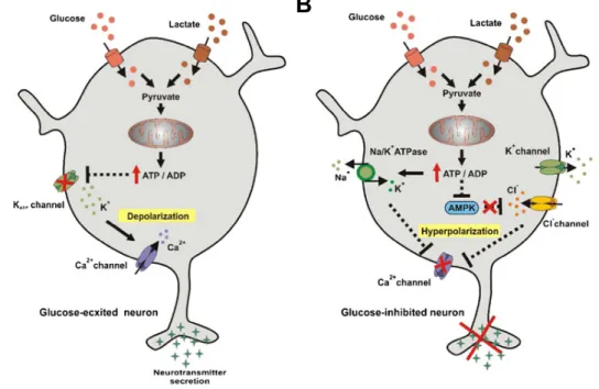
centrations to certain intracellular membrane properties, that increase or decrease action potential (AP) frequencies. Mechanisms involving the adenosine-triphosphate (ATP)- dependent potassium channel (K ATP ) along with the expression of glucose-transporters (GLUTs) and glucokinase (GK) have been first identified in pancreatic β-cells and are also expressed in the CNS (Ashcroft et al. , 1984 ; Levin et al. , 1999 ). Briefly, extracellular glucose enters the cell via low affinity GLUTs. Subsequently, glucose is phosphorylated by GK and enters glucolysis leading to increased ATP concentrations. As a result, ATP closes K ATP channels resulting in membrane potential depolarization and concomitant increase in firing frequencies (Ashford et al. , 1990 ; Dallaporta et al. , 2000 ; Lee et al. , 1999 ; Miki et al. , 2001 ; van den Top et al. , 2007 ). In contrast, glucose-inhibited neu- rons control their firing by activating an ATP driven Na + /K + - exchanger in response to elevated intracellular ATP concentrations. However, a second mechanism suggests the involvement of adenosine monophosphate-activated protein kinase (AMPK) pathways, which results in the opening of Cl - channels and subsequent hyperpolarization of the neuron (Oomura et al. , 1974 ; Silver & Ereci ´nska, 1998 ; Song & Routh, 2005 ; Song et al.

, 2001 ). An overview of the proposed underlying mechansims is given in Figure 2 and reviewed by Jordan et al. ( 2010 ).

Various sites containing glucose-sensing populations have been identified including all key regulatory nuclei of energy homeostasis, such as the ARC, the PVH, VMH and LH (Burdakov et al. , 2005 a). Further glucose sensing sites have been detected in the brainstem including, the NTS, the area postrema (ArP), the DMV and NA neurons in the basolateral medulla (Adachi et al. , 1995 ; Burdakov et al. , 2005 a; Ritter et al. , 2011 ; Routh, 2002 ). A large body of work focused on NA neurons in the brainstem projecting to hypothalamic sites, which are involved in glucoprivic feeding responses (Ritter et al.

, 2011 ). Further studies mark the main NA nucleus in the CNS, the LC, as a potential

source for the NA modulation of energy homeostasis (Wellman, 2000 ). Therefore, NA

and its role in the control of energy homeostasis is described in the following sections.

(24)

A B

Figure 1.3: Neuronal glucose-sensing mechanisms. (A) In GE neurons, extracellular glucose en- ters the cell and is phosphorylated to Pyruvate by the kinase glucokinase (GK). Increasing intra- cellular ATP concentrations lead to the closure of K ATP channels, thus increasing excitability and transmitter release. (A) In the proposed model for GI neurons, ATP activates a Na + /K + -ATPase decreasing membrane potential and transmitter release. Other models implicate AMPK medi- ated opening of Cl channels, other mechanisms involve K ATP channel opening and decrease in Ca 2+ conductance. GE, glucose-excited; GI, glucose-inhibited, K ATP , ATP-dependent potassium channel; AMPK, adenosine monophosphate activated protein kinase; GK, glucokinase. Adopted and modified from Jordan et al. ( 2010 ).

1.2 The catecholamine noradrenaline in the control of energy homeostasis and food intake

Several drugs in the treatment of obesity target catecholaminergic (catecholamine; CA)

neurotransmitter systems, among them the NA system (Hainer et al. , 2006 a; Rosmond,

2004 ). Strong side effects of these drugs are a general problem. Amphetamines, which

also target the NA system have anorexigenic effects but exhibit strong addictive poten-

tials (Di Dalmazi et al. , 2013 ). In order to develop specific drugs with no or minimized

side effects, a detailed understanding of these neurotransmitter systems in the modula-

tion of eating behaviour is necessary. The present study focuses on the NA system in

the control of energy homeostasis.

(25)

1 Introduction

1.2.1 Noradrenaline

NA, adrenaline (adrenergic; A) and dopamine (dopaminergic; DA) belong to the CA class of monoamines with NA and DA representing the two primary CAs in the mam- malian brain (Bloom, 2010 ). Almost five decades ago, these substances were identi- fied by formaldehyde histofluorescence and soon matched to distinct neuronal popula- tions(Carlsson et al. , 1962 ; Dahlström & Fuxe, 1964 ; Vogt, 1954 ).

NA can act either as a circulating hormone or neurotransmitter dependent on the site of biosynthesis and release. In the periphery, the medullae of the adrenal glands release NA and A into the blood, a process which is mainly associated with an adaptive behav- ior to stress, often referred to as "fight or flight" response (Jansen et al. , 1995 ). However, the classical view of NA release in the CNS underlies the postganglionic sympathetic neurons controlling cardiovascular responses to maintain blood pressure and a variety of other responses with the interplay of A (Esler et al. , 1985 ).

It became clear that NA as well as A are also released by various neurons serving as a classical synaptic neurotransmitter in the CNS (Fuxe, 1965 ). Various studies indicate, that projections of NA neurons can also be non-synaptic, thus releasing it nonspecifi- cally within areas of brain-tissues where it rather acts in a hormone-like manner (Smeets

& González, 2000 ). Release of NA in general is known to contribute to a variety of func- tions such as long-term synaptic plasticity, pain modulation, motor control, local blood flow, sleep wake cycles, arousal, task performance optimization and energy homeostasis (Aston-Jones & Cohen, 2005 ; Benarroch, 2009 ; Samuels & Szabadi, 2008 a,b).

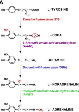
All CAs are synthesized by an specific enzymatic machinery. The neurotransmit- ter NA is synthesized in three steps starting with the amino acid tyrosine, which in a first step is converted to L- 3 , 4 -dihydroxyphenylalanine (L-DOPA) by the enzyme tyrosine-hydroxylase (TH). L-DOPA represents the direct precursor for DA and DA is converted into NA by the enzyme dopamine-β-hydroxylase (DBH). Methylation by phenylethanolamine-N-methyltransferase (PNMT) finally converts NA to A, a process which is predominantly taking place in the medullae of the adrenal glands as A neuron groups are rather small compared to NA neuron groups (see figure 1 . 4 A; Smeets &

González 2000 ).

(26)

A

Figure 1.4: Catecholaminergic biosynthesis. (A)Pathway for catecholamine biosynthesis and its enzymatic steps. The steps of conversion from L-tyrosine to L-noradrenaline are typical for sympathetic and some brain neurons, and the conversion of L-noradrenaline to L-adrenaline is typical for the adrenal medullary cells and some peripheral and central neurons. Modified from Kvetnansky et al. ( 2009 ).

The functional organization of the NA system consists of a peripheral (sympathetic ganglia) and a central part. Both share the common feature that their cell bodies are clustered in a small number of nuclei in the lower brainstem (Moore & Bloom, 1979 ).

First described in rats, seven NA nuclei have been identified in the brainstem and most of them have also been identified in primates and humans. Subdivided into three groups, the caudal (or medullary), central (medullo-pontine) and rostral (pontine) group, NA nuclei have been labeled from A 1 to A 7 . However, 50 % of all noradrenergic neurons are located in the A 6 cell group, the LC.

Projections of the NA system are divided into two major groups: the caudal group

(A 1 ,A 2 ,A 5 ,A 7 ) forms the ventral noradrenergic bundle (VNB) and central group (LC)

gives rise to the dorsal noradrenergic bundle (DNB). Together, both bundles innervate

almost the entire CNS. These wide projection pattern of the NA system reflects the

(27)

1 Introduction

diverse functional role of NA neurons. The anatomy of the NA system is reviewed in Szabadi ( 2013 ).

The Locus Coeruleus - noradrenergic (LC-NA) system, is the main source of NA in the CNS. By innervating most structures, NA release from the LC modulates a large variety of systems (Berridge & Waterhouse, 2003 ). The control of sleep-wake cycles, promoting wakefulness, arousal and modulating task performance, are regarded as the major functions of the LC in the CNS. These contributions have been studied extensively in vitro and in vivo. The activation patterns of neurons in the LC have been matched to different states of wakefulness and arousal. During rapid eye movement (REM) sleep, neurons in the LC remain silent. They become tonically active during times of waking and increase firing with increasing arousal (Berridge et al. , 2012 ). In times of very high arousal (even stress and fear related) demanding optimal task performance, LC neurons are rhythmically active. An elegant study, using optogenetics, revealed the necessity of LC firing in maintaining wakefulness and also shows that LC activity is finely tuned in the control of attentional behavior (Carter et al. , 2010 ). Besides these roles, it is also known, that the LC contributes to autonomic function via the sympathetic nervous system (SNS). By innervating preganglionic sympathetic neurons in the spinal cord, the LC controls blood pressure and sweat glands and may also be involved in mediating iris reflexes in response to light stimuli (Samuels & Szabadi, 2008 b). Additionally, retrograde labeling using pseudorabies viruses injected into brown adipose tissue (BAT) of different species has allowed the identification of the LC, implicated in the regulation of BAT sympathetic nerve activity (SNA) (Bamshad et al. , 1999 ; Cano et al. , 2003 ; Oldfield et al.

, 2002 ). Upon cold exposure it was observed that LC neurons show increased spike activity, concomitant with higher thermogenesis in BAT (Kiyohara et al. , 1995 ; Miyata et al. , 1995 ). This clearly points towards a role of the LC in the control BAT activity.

NA exerts multiple potent effects on target neurons including the modulation of membrane potential, neuronal excitability, intracellular cascades and synaptic plasticity.

In vitro studies indicate that these effects are rather complex and may critically rely

on synaptic concentration as well as on the availability and affinity of certain receptor

subtypes in any specific region (Hein, 2006 ; Philipp & Hein, 2004 ).

(28)

1.2.2 Noradrenergic receptors

The effect of NA and A is mediated via signaling of three major classes of receptors, α 1 -, α 2 - and β-adrenergic receptors (Adrenoceptors; ARs). These receptors are widely dis- tributed in the body and the CNS (Young & Kuhar, 1980 ). Each of the three major types are further divided into three different subtypes (Docherty, 1998 ). Based on pharmaco- logical characteristics, adrenoceptors were originally divided into α- and β-adrenergic re- ceptors (Ruffolo, 1985 ; Ruffolo & Hieble, 1994 ). α-ARs were initially subdivided into α 1 - and α 2 -ARs based on the assumption that the first is expressed postsynaptically whereas the latter is expressed only presynaptically to inhibit transmitter release (Langer, 1974 ).

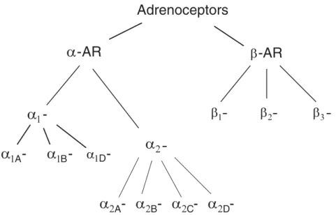
However, this classification soon became obsolete. It was shown that α 2 -ARs can be expressed pre - as well as postsynaptically (Rogawski & Aghajanian, 1982 ; Wellman et al. , 1993 ). Thus, pharmacological characterizations were used and led to the present classification scheme. An overview of the current nomenclature is given in figure 1 . 5 .

Figure 1.5: Current nomenclature of adrenoceptors based on pharamcological properties. ARs are divided in two major classes α-ARs and β-ARs. Modified from Woodcock ( 2007 ).

Several studies have revealed multiple actions of NA on intrinsic ionic currents and cellular properties (Hein, 2006 ). All of the three described major classes of AR belong to the class of G-protein coupled receptors (GPCRs) (Bloom, 1979 ; Insel, 1989 ; Ramos &

Arnsten, 2007 ).

(29)

1 Introduction

α 1 -adrenergic receptors

In general, the class of α 1 -ARs exerts its excitatory effect via coupling to G q proteins, thereby activating phospholipase C (PLC) and phosphotidyl-inositol (PI) intracellular signaling (Macrez-Leprêtre et al. , 1997 ). This leads to activation of protein kinase C (PKC) and the release of intracellular Ca 2 + via inositol-1,4,5-triphosphate (IP 3 ) (Benar- roch, 2009 ; Birnbaum et al. , 2004 ). Further mechanisms have been implicated which also lead to excitatory effects on neurons. An elegant study by Pan et al. ( 1994 ) demonstrated the contribution of decreased K + conductances as well as increased voltage gated Ca 2 + conductances (VGCC) to neuronal excitation. Dodt et al. ( 1991 ) could show that NA in- hibits Ca 2 + -activated K + -currents via activation of α 1 -ARs and thus leading to increased firing in response to excitatory stimuli. Mice carrying deletions for each of the α 1 -AR subtype could reveal different systems they contribute to in the CNS. Various pheno- types were reported as most of the α 1 -ARs are abundantly expressed (Tanoue et al. , 2002 ). The analyzed mice suggest that the subtypes of α 1 -ARs are involved in the con- trol of locomotion, cognition, control of motor activity as well as contribute to memory consolidation and fear-motivated exploratory activity. Consequently, patophysiology in relation to α 1 -ARs exhibit various impairments and neurodegenerative phenotypes (Zuscik et al. , 2000 ). However, the detailed mechanisms by which α 1 -ARs contribute to the different CNS functions remain largely unknown.

α 2 -adrenergic receptors

The α 2 -ARs, which exhibit the highest affinity to NA, are coupled to G i proteins thus

having inhibitory effects on target neurons. Activation of the G i protein reduced the ac-

tivity of the enzyme adenylcyclase (AC), which decreases the intracellular concentration

of the second messenger cyclic adenosine monophosphate (cAMP; Bünemann et al. 2001 ;

Hein 2006 ). Protein kinase A (PKA), a kinase regulating the activity of several cellular

proteins including L-type Ca 2 + channels can be activated by cAMP (Rosenbaum et al. ,

2009 ). Additionally, activation of G protein coupled inwardly rectifying potassium chan-

nels (GIRKs) is a primary response to activation of G i coupled receptors, which leads to

potent inhibition of target neurons (Limbird, 1988 ; Lüscher & Slesinger, 2010 ). α 2 -ARs

(30)

can be expressed either pre - or postsynaptically, where they serve different functions while sharing the same intracellular mechanism. Presynaptically expressed α 2 -ARs are commonly autoreceptors (Callado & Stamford, 2000 ). Here, the synaptic availability of NA is controlled by auto-inhibition via the α 2 -AR itself. In addition to their function as inhibitory autoreceptors, α 2 -ARs can also regulate a number of other neurotransmitters in the central and peripheral nervous system (Richter et al. , 2012 ). In the brain, α 2 A - and α 2 C -ARs inhibit DA release in basal ganglia and serotonin ( 5 -HT) secretion in mouse hippocampus and brain cortex (Bücheler et al. , 2002 ; Scheibner et al. , 2001 ). In the LC, postsynaptically expressed α 2 -ARs have been shown to inhibit baseline activity and increase responsiveness to novel stimuli (Sara, 2009 ).

Further functional contributions of α 2 -ARs are shown for pain perception, procession of sensory information, control of blood pressure and body temperature and also neu- roprotectional effects are described (reviewed in Hein ( 2006 ); Rommelfanger & Wein- shenker ( 2007 ); Weinshenker ( 2008 ). To mention a few, sensorimotor gating deficits, such as schizophrenia, attention deficit disorder and post-traumatic stress disorder are consequences of α 2 -AR impairment (Brede et al. , 2004 ).

β-adrenergic receptors

β-ARs activate G s proteins, which opposingly to α 2 -ARs lead to an increase in cytosolic cAMP concentration, thus leading to excitatory effects (Benovic et al. , 1988 ). β-ARs are mostly known for their role in the regulation of cardiovascular, airway, uterine, and pe- ripheral metabolic functions. Presynaptically expressed β-ARs on some peripheral and central nerve endings have been show to facilitate stimulation evoked neurotransmitter release. However, their major physiological significance is not known. Despite the wide expression of β 1 - and β 2 -ARs within the CNS, specific knockouts did not lead to any significant deficits in CNS function (Nicholas et al. , 1996 ).

The present study aims to gain further knowledge about the NA modulation of en-

ergy homeostasis. Therefore, general effects of NA and underlying receptor subtypes

have been described. The following section provides an overview of NA modulation of

(31)

1 Introduction

energy homeostasis at two sites, the ARC and the LC, both of which are in focus of this thesis.

1.2.3 The hypothalamus in the noradrenergic control of energy homeostasis

In the 1960 s, first evidence appeared that NA directly affects food intake. Exogenous NA and A injected into various sites of the rat’s forebrain was able to elicit feeding and drinking responses dependent on the injection site (Booth, 1967 ; Grossman, 1960 ).

An important mapping study by Leibowitz ( 1978 b) showed that infusions of NA into the medial hypothalamus, specifically into the PVH elicits remarkable bouts of food intake and that these responses were diminished by injecting NA anteriorly, laterally and dorsally to the PVH. Further studies revealed the presence of α 1 - and α 2 -ARs in the hypothalamus (Leibowitz et al. , 1982 ; Young & Kuhar, 1980 ). Pharmacological ex- periments showed that these receptors mediate the effect of exogenous NA. Clonidine, a specific agonist of α 2 -ARs reliably mimicked the effect of NA injection. Consistently, this effect could be abolished by the administration of the specific α 2 -AR antagonists Rauwolscine and Yohimbine (Goldman et al. , 1985 ; Leibowitz, 1988 ). In contrast, a vari- ety of agonists of α 1 -ARs led to reductions in food intake (for review see Wellman et al.

1993 ). The specific α 1 -AR agonist SKF- 89748 was able to reduce food intake in rats dose dependently (Morien et al. , 1993 ). Administration of benoxathian, a specific α 2 -AR antagonist, prevented the reduction of food intake after the systemic administration of the α 2 -AR agonist phenylpropanolamine (Wellman & Davies, 1991 ). In order to confirm these results, electrophysiological studies revealed a subpopulation of neurons in the PVH, which were either inhibited or excited by bath application of NA and/or specific agonists and antagonists (Inenaga et al. , 1986 ; Kow & Pfaff, 1989 ). Importantly, block- ing synaptic transmission could demonstrate that these effects are due to cell intrinsic expression of ARs. Taken together, neurons in the PVH are differentially regulated by α 1 - and α 2 -AR subtypes, with the first leading to the suppression and the latter to the stimulation of food intake.

Anatomical studies aimed to reveal the specific sources in the brainstem which re-

lease NA in terms of energy homeostasis (Ritter et al. , 2000 ; Wellman, 2000 , 2005 ).

(32)

Various NA nuclei show dense innervation of hypothalamic sites (Calaresu & Ciriello, 1980 ; Kataoka et al. , 1975 ; Loughlin et al. , 1986 ). As described earlier, the NA system innervates most parts of the CNS via the DNB, which carries the efferent projections of the LC, and the VNB. The identification of these fiber systems led to further pioneer- ing experiments investigating the specific functions of the NA system with respect to feeding behavior. Almost 40 years ago, Ahlskog & Hoebel ( 1973 ) showed that chem- ical and electrolytic lesions of the VNB results in overeating and obesity. In contrast the interruption of the DNB, originating from the LC resulted in lowered body weight (Hoebel et al. , 1989 ). Genetic knockout (KO) of NA can be accomplished by the deletion of DBH. Consistently with the contribution of NA to feeding behavior, mice exhibited a smaller phenotype concomitant with reduced food intake. However, these mice could still increase feeding in response to overnight fasting (Cannon & Palmiter, 2003 ). Prior to this, a similar outcome has been noted by Rossi et al. ( 1982 ), where inhibition of DBH by the drug FLA- 63 led to reduced eating behavior.

The discovery of the melanocortin system in the hypothalamus also led to experi- ments focusing on the effect of NA in the ARC or the medial hypothalamus in general.

In this context, the ARC is regarded as the first order relay, containing neurons which adapt their activity to peripheral signals such as insulin, leptin, ghrelin, glucose and FFA and innervate downstream targets, especially the PVH, to either suppress or in- duce food intake (Gao & Horvath, 2007 ). A large body of literature suggests a role of NA in modulation of the action of these peripheral signals or vice versa (Brunetti et al.

, 1999 ; Date et al. , 2006 ; Francis et al. , 2004 ; Levin et al. , 1998 ). While leptin inhibits NA release into the hypothalamus, insulin selectively downregulates the expression of α 2 -AR specifically in the ARC (Brunetti et al. , 1999 ; Kawakami et al. , 2008 ; Levin et al.

, 1998 ). Additionally, NA might also affect neurons that are located presynaptically to

the PVH thereby changing excitatory and inhibitory synaptic input on neurons in the

PVH (Han et al. , 2002 ). The ARC is a strong candidate in exerting these effects on PVH

neurons and NA has been shown to activate ARs in the ARC (Kang et al. , 2000 ). How-

ever, these experiments lacked the identification of the respective neurons expressing

the ARs.

(33)

1 Introduction

Taken together, the role of NA signaling in the PVH with respect to food intake has been thoroughly investigated. Various studies also indicate direct or indirect modu- latory effects of NA in the ARC and NA dependent postsynaptic currents have been measured in PVH neurons. POMC and AgRP neurons are located presynaptically to the PVH and are thus potent targets for NA in the regulation of energy homeostasis.

The present study aims to analyze the effect of NA on these neurons. Moreover, this leads to the question which NA nuclei project and release NA into the ARC. The LC contains 50 % of the NA neurons and therefore is regarded as a potential source for NA release into the ARC. Therefore the putative role of the LC in energy metabolism is a further subject of this thesis.

1.2.4 The Locus Coeruleus in the control of energy homeostasis and glucose metabolism

The LC NA system innervates BAT via the SNS and thus may contribute to the con- trol of BAT activity. Besides its prominent role in thermoregulation, recent work has revealed that BAT is also involved in the control of glucose and lipid metabolism in rodents and may thus contribute to energy homeostasis (Bartelt et al. , 2011 ; Nedergaard et al. , 2011 ; Waldén et al. , 2012 ). BAT is a tissue with excessively high glucose uptake, a feature which accidentally led to its identification in humans while screening for tu- mors (Hany et al. , 2002 ). Importantly, NA release into BAT stimulates the expression of GLUT genes resulting in the uptake of glucose from blood vessels. Subsequently, glu- cose is pyruvated and finally oxidized in the mitochondria (Bartelt et al. , 2011 ). Given the sympathetic efferents of the LC into BAT and the effect of NA on glucose uptake, it is important to mention that studies support the hypothesis that the LC may also contribute to glucose metabolism (REF).

In order to maintain glucose homeostasis, the brainstem integrates information of

the hypothalamus and visceral afferents arising in the periphery. A large body of evi-

dence suggests that NA is the neurotransmitter which triggers food intake as response

to glucoprivation (Emanuel & Ritter, 2010 ; Fraley & Ritter, 2003 ; Hudson & Ritter, 2004 ;

Levin et al. , 1999 ; Ritter et al. , 2000 , 2001 , 2006 ). In this context, NA is suggested to

(34)

be mostly released by the A 1 and A 2 NA cell groups (Li et al. , 2009 ; Rinaman, 2011 ).

However, about 50 % of NA somata are located in the LC, which gives rise to the DNB.

As aforementioned, lesioning of DNB fibers leads to phenotypes, which suggest a role in energy homeostasis. Identification of the melanocortin system and glucose-sensing neurons led to studies trying to uncover certain areas, that may sense extracellular glu- cose and may be involved in energy homeostasis. In order to find these areas, in-situ hybridizations for mRNA encoding proteins involved in the glucose-sensing machin- ery have been conducted. The expression of K ATP channels and glucokinase has been anatomically matched with the LC, suggesting the existence of glucose-responsive neu- rons (Dunn-Meynell et al. , 1998 ; Finta et al. , 1993 ; Lynch et al. , 2000 ). Importantly, two studies in the 1990 s by Murai et al. ( 1997 a) and Illes et al. ( 1994 ) provides evidence of glucose-sensing behavior of LC neurons in the rat. In both studies, glucose free medium induced outward currents in a subset of LC neurons, which in the latter study could be blocked by application of the K ATP channel blocker tolbutamide. The same authors could also show that metabolic inhibition due to hypoxia/anoxia induces outward currents, also sensitive to tolbutamide. In this context, hypoxia is thought to exert this effect via depletion of intracellular ATP, thus opening K ATP channels (Grigg & Anderson, 1989 ).

These results indicate the existence of GE neurons in the LC. In contrast, injection of 2-desoxy-glucose (2-DG) , a glucose variant which can’t enter glycolysis mimicking con- ditions of glucoprivation, induced c-fos expression in the LC, suggesting the existence of GI neurons (Ritter et al. , 1998 ). However, this is not contradictory as c-fos expression due to 2-DG fails to label GE neurons, which should decrease electrical activity.

Taken together, a large body of literature provides evidence for a role of the NA system in the control of energy homeostasis. The NA modulation of neurons in the PVH has been well established. In the hypothalamus, the main center in the control of energy homeostasis is the ARC and a series of studies supports evidence for an action of NA in the ARC. However, a detailed analysis of the effect of NA, especially on POMC and NPY/AgRP neurons remains elusive.

The brainstem contains the majority of NA neurons in the CNS. Various nuclei ex-

hibit dense efferent innervation of hypothalamic sites. The LC contains 50 % of NA

(35)

1 Introduction

somata and efferent projections to the ARC have been identified. This points towards a potential source for NA release into the ARC and a contribution to the control of energy homeostasis. A role of the LC in glucose metabolism via autonomic function has been proposed and LC neurons express a number of proteins, which identify these neurons as putative glucose-sensors. However, detailed electrophysiological data has not been described in this context.

1.3 Thesis objectives

The present study aims to expand the knowledge of NA modulation of energy home- ostasis. Because various drugs in the treatment of obesity, at least in part, target the NA system, it is important to understand in detail the targets in the CNS and the mechanisms, by which NA modulates eating behaviour and energy expenditure. The presented experiments help to define the model of NA control of energy homeostasis, which is critical to develop specific drugs in the treatment of obesity with minimized side effects. Two systems are in the focus of this study:

1st: The Arcuate nucleus of the hypothalamus

1 . Basic electrophysiological characteristics of POMC and AgRP neurons in the ARC have been analyzed

2 . The effect of different concentrations of NA on POMC and NPY/AgRP neurons in the ARC has been investigated

3 . The specific underlying receptors, expressed by POMC and NPY/AgRP neurons have been identified by pharmacological tools

4 . Age- and diet-dependent effects on NA signaling in the ARC have been investi- gated for POMC neurons

2nd: The Locus Coeruleus

1 . Basic electrophysiological characterization aimed to create a baseline for future

experiments in our labaratory

(36)

2 . Responses to changes in extracellular glucose have been investigated in LC neu-

rons, as a potential source for NA release into the ARC

(37)

2 Materials and Methods

2.1 Animal care

Care of all animals was within institutional animal care committee guidelines. All ani- mal procedures were approved by local government authorities (Bezirksregierung Köln, Cologne, Germany) and were in accordance with NIH guidelines. Mice were housed in groups of 3 – 5 animals at a temperature of 22 – 24 C with a 12 h light/ 12 h dark cy- cle. After weaning (P 21 ), mice were either fed regular chow food (NCD; Teklad Global Rodent 2918 ; Harlan) containing 53 . 5 % carbohydrates, 18 . 5 % protein, and 5 . 5 % fat ( 12 % of calories from fat) or a high-fat diet (HFD; C 1057 ; Altromin) containing 32 . 7 % carbohydrates, 20 % protein, and 35 . 5 % fat ( 55 . 2 % of calories from fat). All animals had access to water and chow ad libitum. The different mouse strains used for this study were kindly provided by Tim Klöckener, Sulay Tovar and Linda Verhagen of the Brüning group.

2.2 Brain slice preparation

The animals were anesthetized with halothane (B 4388 ; Sigma-Aldrich, Taufkirchen, Ger-

many) and subsequently decapitated. The brain was rapidly removed and a block of

tissue containing the hypothalamus or brainstem was immediately cut out. Coronal

slices ( 250 – 300 µm) were cut with a vibration microtome (HM- 650 V; Thermo Sci-

entific, Walldorf, Germany) under cold ( 4 C), carbogenated ( 95 % O 2 and 5 % CO 2 ),

glycerol-based modified artificial cerebrospinal fluid (GaCSF; Ye et al. 2006 ) to enhance

the viability of neurons. GaCSF contained (in mM): 250 Glycerol, 2 . 5 KCl, 2 MgCl 2 ,

2 CaCl 2 , 1 . 2 NaH 2 PO 4 , 10 HEPES, 21 NaHCO 3 , 5 Glucose and was adjusted to pH 7 . 2

with NaOH resulting in an osmolarity of ∼ 310 mOsm. Brain slices were transferred into

carbogenated artificial cerebrospinal fluid (aCSF). First, they were kept for 20 min. in a

35 C ’recovery bath’ and then stored at room temperature ( 24 C) for at least 30 min

(38)

prior to recording. For the recordings, slices were transferred to a Sylgard-coated (Dow Corning Corp., Midland, MI, USA) recording chamber ( ∼ 3 ml volume) and, if not men- tioned otherwise, continuously perfused with carbogenated aCSF at a flow rate of ∼ 2 ml · min 1 . aCSF contained (in mM): 125 NaCl, 2 . 5 KCl, 2 MgCl 2 , 2 CaCl 2 , 1 . 2 NaH 2 PO 4 , 21 NaHCO 3 , 10 HEPES, and 5 Glucose and was adjusted to pH 7 . 2 with NaOH resulting in an osmolarity of ∼ 310 mOsm.

2.3 Patch-clamp recordings

Current-clamp recordings in neurons of the hypothalamus and the brainstem were per- formed in the perforated patch-clamp configuration. In the hypothalamus, neurons which express POMC or NPY/AgRP were investigated. In the brainstem, NA neurons in the LC were investigated.

Neurons were visualized with a fixed stage upright microscope (BX 51 WI, Olympus, Hamburg, Germany) using 40 × and 60 × water-immersion objectives (LUMplan FL/N 40 × , 0 . 8 numerical aperture, 2 mm working distance; LUMplan FL/N 60 × , 1 . 0 numer- ical aperture, 2 mm working distance, Olympus) with infrared differential interference contrast optics (Dodt & Zieglgänsberger, 1990 ) and fluorescence optics.

POMC and NPY/AgRP neurons were identified by their anatomical location in the ARC and by their GFP fluorescence that was visualized with an X-Cite 120 illumination system (EXFO Photonic Solutions, Ontario, Canada) in combination with a Chroma 41001 filter set (EX: HQ 480 / 40 × , BS: Q 505 LP, EM: HQ 535 / 50 m, Chroma, Rockingham, VT, USA). Putative NA neurons were identified by their location ventrolateral to the 4 th ventricle and/or by their GFP expression. Electrophysiological properties were analyzed to confirm the identity (i.e. slow and regular firing).

Electrodes with tip resistances between 4 and 6 MΩ were fashioned from borosilicate glass ( 0 . 86 mm inner diameter; 1 . 5 mm outer diameter; GB 150 - 8 P; Science Products) with a vertical pipette puller (PP- 830 ; Narishige, London, UK).

Recordings in the ARC were made at room temperature. Recordings of LC NA

neurons were made at ∼ 30 - 32 C using an inline solution heater (SH 27 B; Warner In-

struments, Hamden, CT, USA) operated by a temperature controller (TC- 324 B; Warner

(39)

2 Materials and Methods

Instruments). All recordings were performed with an EPC 10 patch-clamp amplifier (HEKA, Lambrecht, Germany) controlled by the program PatchMaster (version 2 . 32 ; HEKA) running under Windows. Data were sampled at intervals of 100 µs ( 10 kHz) and low-pass filtered at 2 kHz with a four-pole Bessel filter. Cell capacitance was determined by using the capacitance compensation (C-slow) of the EPC 10 . Cell input resistances (R M ) were calculated from voltage responses to hyperpolarizing current pulses. The calculated liquid junction potential of 14 . 6 mV between intracellular and extracellular solution was compensated or subtracted offline (calculated with Patcher’s Power Tools plug-in from http://www.mpibpc.mpg.de/groups/neher/index.php?page=software for IGOR Pro 6 [Wavemetrics, Lake Oswego, OR, USA]).

2.3.1 Perforated-patch clamp recordings

Perforated-patch experiments were conducted using protocols modified from Horn &

Marty ( 1988 ) and Akaike & Harata ( 1994 ). Recordings were performed with ATP and GTP free pipette solution containing (in mM): 128 K-gluconate, 10 KCl, 10 HEPES, 0 . 1 EGTA, 2 MgCl 2 adjusted to pH 7 . 3 with KOH resulting in an osmolarity of ∼ 300 mOsm.

ATP and GTP were omitted from the intracellular solution to prevent uncontrolled per- meabilization of the cell membrane (Lindau & Fernandez, 1986 ). The patch pipette was tip filled with internal solution and back filled with 0 . 02 % tetraethylrhodamine-dextran (D 3308 , Invitrogen, Eugene, OR, USA) added to the internal solution containing the ionophore to achieve perforated patch recordings.

Amphotericin B (A 4888 ; Sigma) and Gramicidin (G 5002 ; Sigma) were dissolved in dimethyl sulfoxide (DMSO; D 8418 , Sigma) following the protocols of Rae et al. ( 1991 )and Kyrozis & Reichling ( 1995 ). The used DMSO concentration ( 0 . 1 – 0 . 3 %) had no obvious effect on the investigated neurons. All ionophores were added to the modified pipette solution shortly before use. The final concentration of nystatin and amphotericin B was

200 µg · ml 1 , the final concentration of gramicidin was ∼ 10 – 75 µg · ml 1 .

Abbildung

Figure 1.1: Melanocortin system in the ARC. Two opposing cell types in the ARC sense and process peripheral signals to second order neurons, which express MC4Rs
Figure 1.2: The melanocortin system and its downstream targets in the integration of peripheral signals
Figure 1.3: Neuronal glucose-sensing mechanisms. (A) In GE neurons, extracellular glucose en- en-ters the cell and is phosphorylated to Pyruvate by the kinase glucokinase (GK)
Figure 1.4: Catecholaminergic biosynthesis. (A)Pathway for catecholamine biosynthesis and its enzymatic steps
+7

Referenzen

ÄHNLICHE DOKUMENTE

We thus aimed to study a potentialfunctional interaction between Rhcg andthe multi-unit protein vacuolar H + -ATPase (H + -ATPase), which constitutes the main H +

In the setting of congestive heart failure, mRNA expression levels of ANP and BNP in cardiomyocytes are markedly increased and plasma ANP concentrations

Consistent with this result, we observed a rise of ATP levels produced by complex V, the final OXPHOS enzyme, in GBE-treated control and APP cells (Fig. Taken together,

Analysis using tetraploid aggregation indicates that a defect in S6K1 -/- placental trophoblast cells leads to growth retardation and developmental delay phenotypes of S6K1 -/-

While behavioral rhythms are regulated by a central circadian pacemaker, accumulating evidence suggests that peripheral clocks strongly contribute to the regulation

Kryazhimskii, A., Watanabe, Ch., and Tou, Y., 2002, The reachability of techno- labor homeostasis via regulation of investments in labor and R&D: a model-based analysis,

To address the mechanism involved in signaling of Gpr111 as well as effects of Gpr111 depletion on in vitro differentiation, brown and white adipocytes were isolated from

Figure 35 Histological analysis of adipose tissue of Ednra AT-KO animals and their corresponding Ednra floxed littermates after sustained cold exposure for one week Panel of