In vivo analysis of the role of FADD in the regulation of intestinal epithelial homeostasis
Inaugural-Dissertation zur
Erlangung des Doktorgrades
der Mathematisch-Naturwissenschaftlichen Fakultät der Universität zu Köln
vorgelegt von Patrick-Simon Welz
aus Lübeck
Köln, 2012
2 Berichterstatter: Prof. Dr. Manolis Pasparakis
Prof. Dr. Jonathan C. Howard
Tag der mündlichen Prüfung: 04.04.2012
3
Diese Arbeit ist meiner Mutter Claudia Barbara Welz gewidmet.
4 Die Einleitung von Zelltod kann ein wichtiger Bestandteil einer Immunreaktion gegen infizierte oder anderweitig beschädigte Zellen sein. FADD ist ein Adapterprotein, welches verschiedene der Immunreaktion zugehörige Signalwege mit der Einleitung von apoptotischem Zelltod verbindet. FADD abhängige Signalwege wurden hauptsächlich in Immunzellen studiert, während die in vivo Rolle von FADD in epithelialem Gewebe, welches sich an der Grenze zu einer großen Anzahl von Stressfaktoren von außerhalb des Organismus befindet, weitestgehend unbekannt ist. Das einzellschichtige intestinale Epithel bildet eine Barriere, die die Bakterien im Darmlumen von den Immunzellen in der Darmschleimhaut des Darmtraktes trennt.
Die Integrität dieser Barriere ist von essentieller Bedeutung für die intestinale Homöostase, was die Regulierung des Zelltods von intestinalen Epithelzellen (IEZ) zu einer wichtigen Angelegenheit macht.
In dieser Doktorarbeit wurde die Rolle von FADD im intestinalen Epithel untersucht.
Wir konnten zeigen, dass FADD essentiell für die intestinale Homöostase ist, indem RIP3 abhängige Nekrose in IEZ verhindert wird. Mäuse, denen FADD speziell in den IEZ fehlt (FADD
IEZ-KOMäuse), entwickelten spontan eine schwere Kolitis sowie eine Dünndarmentzündung. Die Nekrose von IEZ im Dickdarm von FADD
IEZ-KOMäusen wurde unterbunden durch die Deletion von RIP3 oder durch die Expression von mutiertem CYLD, welchem die Deubiquitinierungsaktivität fehlt. Wie im Dickdarm, so war auch die Nekrose von IEZ im Dünndarm abhängig von RIP3. Wie auch immer, die Deubiquitinierungsaktivität von CYLD war nicht notwendig für das Auftreten von Nekrose in den IEZ im Dünndarm, was impliziert, dass verschiedene Signalwege die Nekrose in den IEZ des Dickdarms und des Dünndarms vermitteln. Die Entwicklung der Kolitis war abhängig von der Expression von TNF und MyD88 vermittelten Signalen, die durch die Darmbakterien induziert wurden. Entgegengesetzt dazu war die Entwicklung der Entzündung im Dünndarm der FADD
IEZ-KOMäuse unabhängig von TNF, den Darmbakterien und MyD88 abhängigen Signalen. Folglich sind verschiedene Mechanismen verantwortlich für die Entwicklung der Entzündung des Dickdarms und des Dünndarms von FADD
IEZ-KOMäusen.
Die in dieser Doktorarbeit gezeigten Ergebnisse demonstrieren, dass FADD nicht nur
ein potentieller Mediator von apoptotischem Zelltod im intestinalen Epithel ist,
sondern dass es auch essentiell für das Überleben der IEZ unter homöostatischen
Bedingungen ist, indem RIP3 abhängige Nekrose verhindert wird.
5 The initiation of cell death can be an integral part of the immune response to infected or otherwise damaged cells. FADD is an adaptor protein connecting various immune response related signalling pathways to the induction of apoptotic cell death. FADD dependent signalling has mainly been studied in immune cells, while the in vivo role of FADD in epithelial tissues, which are at the border to various stress factors from outside the organism, is largely unknown. The intestinal epithelium is a single cell layer that forms a barrier, separating the luminal microbiota from the mucosal immune cells in the gastro-intestinal tract. The integrity of this barrier is essential for intestinal homeostasis, making the regulation of intestinal epithelial cell (IEC) death in response to infections or other stress-factors an important issue.
In this thesis, the role of FADD in the intestinal epithelium was studied. We found that FADD is essential for intestinal homeostasis by preventing excessive RIP3 dependent IEC necrosis. Mice lacking FADD specifically in the intestinal epithelium (FADD
IEC-KOmice) developed severe spontaneous colitis and enteritis. IEC necrosis in the colon of FADD
IEC-KOmice was abrogated by deletion of RIP3 or by the expression of a CYLD mutant lacking the deubiquitinase activity. Like in the colon, necrosis of FADD deficient IECs in the small intestinal epithelium was also depending on RIP3. However, the deubiquitinating activity of CYLD was not required for small intestinal IEC necrosis to occur, suggesting different pathways to mediate IEC necrosis in the colon and small intestine of FADD
IEC-KOmice. Development of colitis was partially depending on the expression of TNF and MyD88 mediated signalling induced by the microbiota. On the contrary, enteritis development in FADD
IEC-KOmice was independent of TNF, the commensal bacteria and MyD88 dependent signalling. Thus, different mechanisms are responsible for the development of inflammation in the colon and small intestine of FADD
IEC-KOmice.
The results presented in this thesis demonstrate that FADD is not only a potential
mediator of apoptotic cell death in the intestinal epithelium, but it is also essential for
IEC survival under homeostatic conditions by preventing RIP3 dependent IEC
necrosis.
I Contents
Abbreviations ... 6
1. Introduction ... 9
1.1. Regulated cell death ... 9
1.2. Immunity and cell death ... 9
1.2.1. FADD ...10
1.2.2. Immunity and extrinsically induced apoptosis ...12
1.2.2.1. TNF-induced extrinsic apoptosis ...12
1.2.2.2. FASL-induced apoptosis ...14
1.2.2.3. TRAIL-induced apoptosis ...15
1.2.3. Cell autonomous immunity and apoptosis ...16
1.2.4. Immunity and regulated necrosis ...16
1.3. The gastrointestinal tract ...20
1.3.1. The intestinal epithelium ...21
1.3.2. Immunity, epithelial cell death and inflammation in the intestinal tract ...23
1.4. Cre/LoxP conditional gene targeting ...26
1.5. Project description ...26
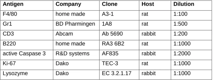
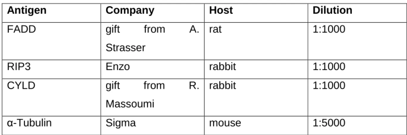
2. Material and Methods ...28
2.1. Material ...28
2.1.1. Chemicals ...28
2.1.2. Material for mouse work ...28
2.1.3. Material for histology ...28
2.1.4. Material for biochemistry ...29
2.1.5. Molecular biology reagents and equipment ...29
2.1.6. Laboratory equipment ...29
2.1.7. Cell culture ...30
2.1.8. Software ...30
2.1.9. Buffers and solutions ...30
2.1.9.1. Washing buffers ...30
2.1.9.2. Buffers and solutions for immunostainings ...31
2.1.9.3. Preparation of protein extracts ...31
2.1.9.4. Buffers and solutions for Western Blot analysis ...31
2.1.9.5. Buffers and solution for DNA extraction, genotyping PCRs and Southern Blot ....33
2.2. Methods ...35
2.2.1. Animal handling and mouse experiments ...35
II
2.2.1.1. Mouse maintenance ...35
2.2.1.2. Generation of conditional and knock-out mice ...35
2.2.1.3. Endoscopy ...35
2.2.1.4. Antibiotic treatment, generation of germ-free mice and conventionalisation ...36
2.2.1.5. Sacrifice of mice ...36
2.2.1.6. Tissue preparation ...37
2.2.2. Histology ...37
2.2.2.1. Preparation of intestinal tissue for histological analysis ...37
2.2.2.2. Haematoxylin and Eosin staining of intestinal tissue sections ...37
2.2.2.3. Immunostainings...38
2.2.2.4. Electron microscopy ...39
2.2.3. Biochemistry ...39
2.2.3.1. IEC isolation from colon and small intestine ...39
2.2.3.2. Preparation of IEC protein extracts ...40
2.2.3.3. Assessment of protein concentration by Bradford assay ...40
2.2.3.4. Western Blot analysis ...40
2.2.4. Molecular Biology ...42
2.2.4.1. Preparation of genomic DNA ...42
2.2.4.2. Southern Blot ...42
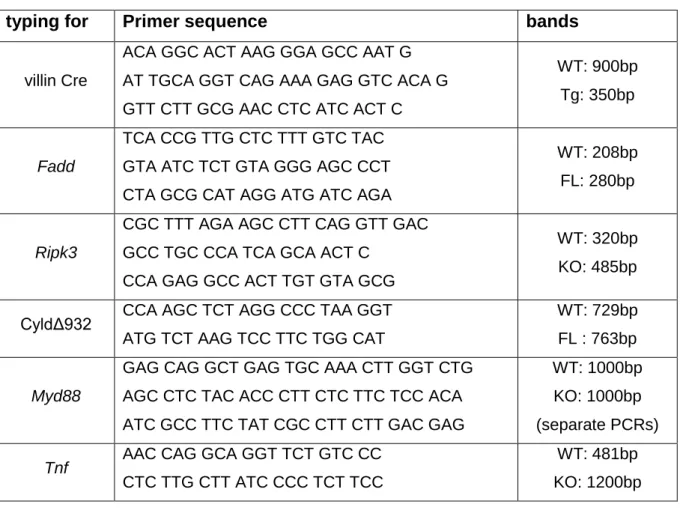
2.2.4.3. Genotyping PCRs ...43
2.2.4.4. Agarose gel electrophoresis ...45
2.2.4.5. Extraction of RNA ...45
2.2.4.6. cDNA synthesis ...45
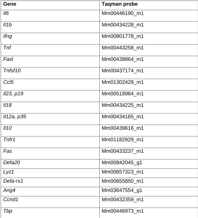
2.2.4.7. Quantitative real time PCR ...46
2.2.5. Cell Biology ...48
2.2.5.1. Culture of mouse embryonic fibroblasts (MEFs)...48
2.2.5.2. Cell death assay with MEFs ...48
2.2.6. Statistics ...48
3. Results ...49
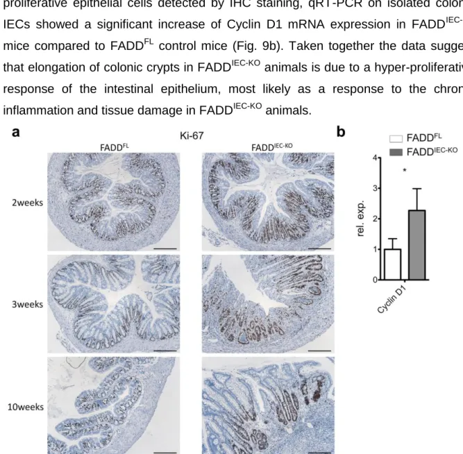
3.1. Deletion of FADD in IECs results in chronic spontaneous colitis and enteritis ...49
3.1.1. Generation of mice deficient for FADD only in the intestinal epithelium ...49
3.1.2. FADD
IEC-KOmice develop spontaneous chronic colitis ...49
3.1.3. FADD
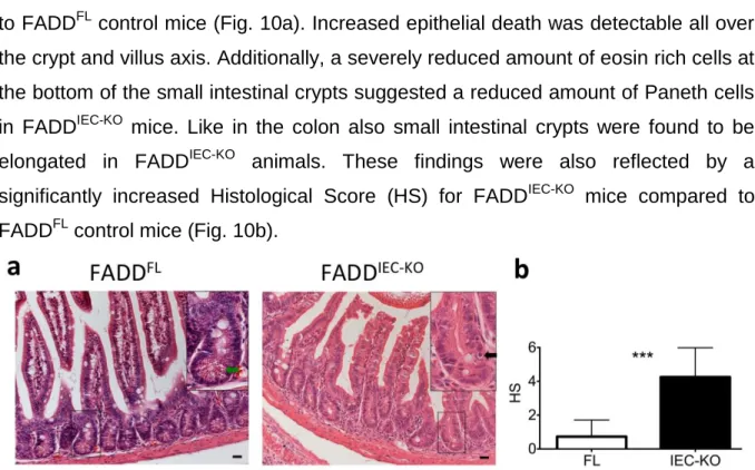
IEC-KOmice develop spontaneous chronic enteritis and have reduced numbers of Paneth cells ...54
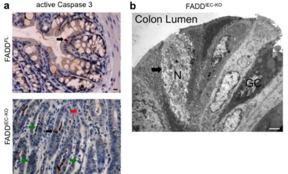
3.2. FADD deficient IECs are sensitised towards cell death with necrotic features ...57
3.2.1. FADD
IEC-KOmice show increased colonic epithelial cell death...57
3.2.2. FADD
IEC-KOmice show increased epithelial cell death in the small intestine ...58
III 3.3. Development of colitis in FADD
IEC-KOmice depends on RIP3-dependent regulated
necrosis ...59
3.3.1. IEC death in the colon and development of colitis in FADD
IEC-KOmice is RIP3 dependent ...59
3.3.2. Cell death and inflammation in the colon of FADD
IEC-KOmice depends on CYLD ....61
3.3.3. Cell death and inflammation in the colon of FADD
IEC-KOmice depends partially on TNF ...63
3.4. Role of the microbiota in the development of colitis in FADD
IEC-KOmice ...64
3.4.1. The development of chronic colitis but not the increased cell death in FADD
IEC-KOmice depends on MyD88 ...64
3.4.2. The development of chronic colitis in FADD
IEC-KOmice depends on microbiota induced signals ...66
3.5. Development of enteritis in FADD
IEC-KOmice depends on RIP3-dependent regulated necrosis ...69
3.5.1. IEC death in the small intestine and development of enteritis in FADD
IEC-KOmice is RIP3 dependent ...69
3.5.2. Cell death and inflammation in the small intestine of FADD
IEC-KOmice is independent of CYLD ...71
3.5.3. Cell death and inflammation in the small intestine of FADD
IEC-KOmice is not induced by TNF ...73
3.6. Role of the microbiota in the development of enteritis in FADD
IEC-KOmice ...75
3.6.1. Enteritis and IEC death in FADD
IEC-KOmice does not depend on MyD88 ...75
3.6.2. Enteritis and IEC death does not depend on the presence of the microbiota ...77
3.6.3. Differential regulation of cell death and inflammation in the colon and small intestine of FADD
IEC-KOmice ...79
4. Discussion ...81
4.1. FADD protects IECs from RIP3 dependent necrosis ...81
4.2. Regulation of RIP3 dependent IEC necrosis by FADD and CYLD ...82
4.3. Possible inducers of RIP3 dependent regulated necrosis in IECs and their physiological impact ...83
4.3.1. Extrinsically induced necroptosis in IECs ...84
4.3.2. Cell autonomous immunity as a possible trigger for RIP3 dependent IEC necrosis 86 4.3.3. Paneth cell death and endoplasmic reticulum (ER) stress ...87
4.3.4. Other possible inducers of RIP3 dependent regulated necrosis ...88
4.4. RIP3 dependent IEC necrosis and inflammation ...89
4.4.1. Development of colitis in FADD
IEC-KOmice ...89
4.4.2. Development of enteritis in FADD
IEC-KOmice ...91
5. Concluding Remarks ...93
Bibliography ...94
Acknowledgements ... 107
IV
Teilpublikation ... 108 Erklärung ... 125 Curriculum Vitae ... 1266
Abbreviations
Ang4 - Angiogenin, ribonuclease A family, member 4 (Protein) Atg5 - autophagy-related 5
Atg12 - autophagy-related 12 BCL-2 - B-cell lymphoma 2
c-FLIP - cellular FLICE-like inhibitory protein CMV - Cytomegalovirus
CYLD - Cylindromatosis (Protein)
DAMP - danger-associated molecular pattern
DD - Death Domain
Defa20 - Defensin, alpha, 20 (Protein)
Defa-rs1 - Defensin, alpha, related sequence 1 (Protein) DISC - Death-Inducing Signalling Complex
DR - Death Receptor
DRL - Death Receptor Ligand DSS - dextran sodium sulphate e.g. - for example
ER - endoplasmic reticulum
Fadd - Fas (TNFRSF6)-associated via death domain (Gene) FADD - Fas (TNFRSF6)-associated via death domain (Protein) Fas - TNF receptor superfamily member 6 (Gene)
FAS - TNF receptor superfamily member 6 (Protein) Fasl - TNF superfamily, member 6 (Gene)
FASL - TNF superfamily, member 6 (Protein) H&E - Haematoxylin and Eosin
HCS - Histological Colitis Score HMGB1 - high-mobility group protein B1
HOIL-1L - heme-oxidized IRP2 ubiquitin ligase 1 homolog HOIP - HOIL-1-interacting protein
HRP - horseradish peroxidase
HS - Histological Small Intestinal Score
IBD - inflammatory bowel disease
IECs - intestinal epithelial cells
7 IHC - immunohistochemical
IKK - Inhibitor of kappaB kinase
IRAK1 - interleukin-1 receptor-associated kinase 1 IRE1 - Inositol-requiring enzyme-1
IRF3 - interferon regulatory factor 3 JNK - c-Jun N-terminal kinase LTα - Lymphotoxin α
LUBAC - linear ubiquitin chain assembly complex Lyz1 - Lysozyme 1 (Protein)
MAPK - mitogen-activated protein kinases MBD4 - methyl-CpG binding domain protein 4 MEFs - mouse embryonic Fibroblasts
MEICS - murine endoscopic index of colitis severity MOMP - mitochondrial outer membrane permeabilization
Myd88 - Myeloid differentiation primary response gene 88 (Gene) MyD88 - Myeloid differentiation primary response gene 88 (Protein) NCCD - Nomenclature Committee on Cell Death
NEMO - NF-kappa-B essential modulator
neo - neomycin
NF-κB - nuclear factor kappa B
PAMP - pathogen-associated molecular pattern PCD - programmed cell death
PRR - pattern recognition receptor qRT-PCR - quantitative real time PCR RCD - regulated cell death
RHIM - Receptor-Interacting Protein Homotypic Interaction Motif
RIP1 - Receptor (TNFRSF)-interacting serine-threonine kinase 1 (Protein) RIP3 - Receptor (TNFRSF)-interacting serine-threonine kinase 3 (Protein) Ripk3 - Receptor (TNFRSF)-interacting serine-threonine kinase 3 (Gene) ROS - reactive oxygen species
SHARPIN - SHANK-associated RH domain interacting protein TAB1 - TAK1-binding protein 1
TAB2 - TAK1-binding protein 2
TAK1 - TGF-beta-activated kinase 1
Tnf - Tumour necrosis factor (Gene)
8 TNF - Tumour necrosis factor (Protein)
Tnfr1 - tumour necrosis factor receptor superfamily, member 1a (Gene) TNFR1 - tumour necrosis factor receptor superfamily, member 1a (Protein) TLR - Toll-like receptor
TLR3 - Toll-like receptor 3 TLR4 - Toll-like receptor 4
TRADD - TNFRSF1A-associated via death domain TRAF2 - TNF receptor-associated factor 2
TRAF5 - TNF receptor-associated factor 5
TRAIL - tumour necrosis factor (ligand) superfamily, member 10 TRAIL1 - tumour necrosis factor receptor superfamily, member 10a TRAIL2 - tumour necrosis factor receptor superfamily, member 10b TRIF - TIR domain-containing adaptor inducing interferon-beta UPR - unfolded protein response
XBP1 - X-box-binding protein 1
XIAP - X-linked inhibitor of apoptosis
9
1. Introduction
1.1. Regulated cell death
Regulated cell death (RCD) in multicellular organisms occurs on the one hand during development and in the maintenance of tissue homeostasis and on the other hand as a mechanism for the removal of stressed, damaged or infected cells as well as in the termination of immune responses by inducing death of activated immune cells. While RCD involved in developmental processes or in the regulation of turnover procedures and tissue homeostasis, also commonly described as programmed cell death (PCD), evolved in multicellular organisms, early stress induced RCD-like mechanisms evolved already in unicellular organisms like bacteria (Ameisen, 2004).
Three main forms of cell death have been described and categorized, mainly according to morphological criterias: cell death type I or apoptotic cell death, cell death type II or autophagic cell death and cell death type III or necrotic cell death, which was for a long time being regarded as a solely non-regulated accidental form of cell death. Recently, the Nomenclature Committee on cell death (NCCD) proposed to use new definitions based on molecular features of the different RCD pathways (Galluzzi et al., 2011), which will be referred to in this work. According to the NCCD, regulated cell death is defined as a process initiated and executed by a “dedicated molecular machinery and can therefore be inhibited by pharmacological and/or genetic interventions”.
1.2. Immunity and cell death
In response to an infection the immune system reacts by activating pathways that
promote cell survival as well as pathways that promote cell death. The primary
response is focussed on the defence against the infection by activation of
proinflammatory pathways, the recruitment of immune cells and production of
antimicrobial factors. If the infection cannot be cleared, cell death can be induced to
remove the infected cells (Lamkanfi and Dixit, 2010). Such cell death can be
triggered extrinsically by certain cytokines, the death receptor ligands (DRLs), or as a
cell autonomous response induced by pattern recognition receptors (PRRs). Fas
(TNFRSF6)-associated via death domain (FADD) is a signal transducer involved in
both, extrinsically induced cell death and PRR triggered cell death. Therefore, FADD
10 is an important mediator of cell death triggered by the immune response (Locksley et al., 2001; Wilson et al., 2009).
1.2.1. FADD
FADD was discovered in 1995 by two groups (Boldin et al., 1995; Chinnaiyan et al., 1995). The FADD protein contains two known domains: a Death Domain (DD), and a Death Effector Domain (DED). The DD of FADD is necessary for binding to other DD containing proteins, like the death receptors, receptor (TNFRSF)-interacting serine- threonine kinase 1 (RIP1) or TNFRSF1A-associated via death domain (TRADD). The DED is also involved in protein-protein interactions, for example in the recruitment of procaspase 8 or in humans additionally procaspase 10 (Boldin et al., 1995;
Chaudhary et al., 1997; Chinnaiyan et al., 1995; Hsu et al., 1996; Muzio et al., 1996;
Schneider et al., 1997). By recruiting procaspase 8, FADD serves as a signalling transducer which can connect various upstream signalling pathways to the induction of cell death. Subsequent cleavage of the catalytically inactive procaspase 8 leads to the active isoform of the cysteine aspartate specific protease Caspase 8, which can induce apoptosis by activating downstream molecules involved in the execution of cell death. Cell death pathways depending on FADD as well as those being regulated by FADD are described in the following chapters.
Beside of its role as a signal transducer towards cell death as part of the immune response, FADD has also been associated with other signalling pathways. FADD deficient T cells die upon mitogenic activation, which led to the speculation that FADD has a role in T cell proliferation and cell cycle progression (Newton et al., 1998; Zhang et al., 2001). In addition, mitogen activated FADD deficient T cells have a hyperautophagic appearance and FADD has been found to interact with an autophagy-related 5 - autophagy-related 12 (Atg5-Atg12) complex (Bell et al., 2008), implicating FADD in the autophagic machinery. FADD has also been suggested to interact with Atg5 in interferon-γ stimulated HeLa cells (Pyo et al., 2005), further promoting a potential role of FADD in autophagy.
An involvement of FADD in IL-1β induced signalling by binding to interleukin-1
receptor-associated kinase 1 (IRAK1) and myeloid differentiation primary response
gene 88 (MyD88) has been reported. This interaction was proposed to lead to the
11 inhibition of further downstream signalling to NF-κB and expression of inflammatory cytokines (Bannerman et al., 2002; Zhande et al., 2007).
FADD is not only located in the cytoplasm, but also in the nucleus (Gomez-Angelats and Cidlowski, 2003). One potential nuclear role of FADD is the interaction with the DNA mismatch repair machinery. FADD has been shown to interact with DNA mismatch repair enzyme methyl-CpG binding domain protein 4 (MBD4) (Screaton et al., 2003). The function of this interaction between FADD and MBD4 has not been resolved yet, but DNA damage induced MBD4 dependent signalling can lead to apoptosis, potentially involving FADD.
Additionally, FADD has been found in microvesicles after adenosine receptor controlled secretion (Tourneur et al., 2008). However, the potential function of this process has not been identified yet.
FADD deficiency in mice leads to embryonic lethality at embryonic day 11.5, suggesting FADD dependent signalling to be important for development (Yeh et al., 1998; Zhang et al., 1998). FADD deficient mice exhibit cardiac failure and abdominal haemorrhage, implicating a role for FADD in those tissues. However, cell type specific roles of FADD have not been studied into great detail yet.
Although loss of FADD expression has been linked to tumourigenesis in certain
cases (Tourneur et al., 2004; Tourneur et al., 2003), surprisingly little is known about
the potential involvement of FADD in human diseases. Mutations resulting in
decreased FADD protein levels were found to be linked to the autoimmune
lymphoproliferative syndrome (ALPS), hepatopathy, encephalopathy, and cardiac
malformations (Bolze et al., 2010). FADD mutations have only rarely been found in
colon or stomach cancers (Soung et al., 2004). Overexpression of FADD has been
reported to be involved in certain cancers (Gibcus et al., 2007), and the
phosphorylation status of FADD has been linked to prostate cancer (Matsumura et
al., 2009; Shimada et al., 2005). However, up to now, despite its important role in
cellular signalling, direct linkages between FADD and human diseases seem to be
rare.
12
1.2.2. Immunity and extrinsically induced apoptosis
Apoptotic cell death can be induced extrinsically during an immune response by the DRLs, which are cytokines of the tumour necrosis factor (TNF) ligand superfamily.
DRLs can be expressed by different types of immune- and non-immune cells and are either secreted or presented at the surface of these cells. The DRLs act through their cognate receptor of the TNF receptor (TNFR) superfamily. These receptors, the death receptors (DRs), all contain a DD in their cytoplasmic part. DRLs binding to the extracellular domain of DRs induce formation of a receptor-bound complex via the intracellular DD, by recruiting certain DD containing signal transducers. Apoptosis triggered by DRLs and their cognate DR is defined as “extrinsic apoptosis” (Galluzzi et al., 2011). Beside of being able to induce death, DRs can trigger as diverse functions as the expression of proinflammatory cytokines or prosurvival factors as well as proliferative signals (Wilson et al., 2009).
DRL/DR signalling modules known to be able to induce extrinsic apoptosis are tumour necrosis factor (TNF) and Lymphotoxin α (LTα) binding to Tnfrsf1a (tumour necrosis factor receptor superfamily, member 1a) (TNFR1), FAS Ligand (TNF superfamily, member 6) (FASL) binding to FAS (TNF receptor superfamily member 6) as well as TNFSF10 (tumour necrosis factor (ligand) superfamily, member 10) (TRAIL) binding to TNFRSF10A (tumour necrosis factor receptor superfamily, member 10a) (TRAILR1) or TNFRSF10B (tumour necrosis factor receptor superfamily, member 10b) (TRAILR2) (Schutze et al., 2008). The DRL/DR signalling pathways are highly conserved between humans and mice, beside of the mouse TRAILR, which is a single ortholog of the human TRAILR1 and TRAILR2.
1.2.2.1. TNF-induced extrinsic apoptosis
Upon activation by TNF or LTα, trimeric TNFR1 recruits the signalling adaptor
TRADD via its DD. TRADD serves as a signalling scaffold for the assembly of the
receptor bound complex I (Ermolaeva et al., 2008), which further contains RIP1, TNF
receptor-associated factor 2 (TRAF2) or TNF receptor-associated factor 5 (TRAF5)
(Tada et al., 2001) and the ubiquitin ligases cIAP1 and cIAP2 (Shu et al., 1996). The
receptor bound complex can activate signalling to nuclear factor kappa B (NF-κB)
through the IκB kinase (IKK) complex, to p38 mitogen-activated protein kinases
(MAPK) and to c-Jun N-terminal kinase (JNK). Different forms of ubiquitination on
several proteins within this receptor-bound complex I are important for the
13 transduction of the signal (Walczak, 2011). Recently it has been shown, that cIAP dependent recruitment of the linear ubiquitin chain assembly complex (LUBAC), consisting of SHANK-associated RH domain interacting protein (SHARPIN) and heme-oxidized IRP2 ubiquitin ligase 1 homolog (HOIL-1) as well as HOIL-1- interacting protein ( HOIP), is essential for TNF mediated activation of NF-κB and JNK.
LUBAC catalyses linear ubiquitin onto RIP1 and NF-kappa-B essential modulator (NEMO), an essential component of the IKK complex, which ultimately mediates NF- κB activation. Linear ubiquitination of these proteins stabilises the receptor bound complex and promotes NF-κB- and JNK activation. (Gerlach et al., 2011; Haas et al., 2009; Ikeda et al., 2011; Tokunaga et al., 2011; Tokunaga et al., 2009).
Activation of TNFR1 signalling can also lead to the formation of two secondary cytoplasmic complexes after TNFR1 internalisation, complex IIa and IIb (Micheau and Tschopp, 2003). Contrary to the outcome of complex I signalling, which is mainly cell survival, the formation of these cytosolic complexes can lead to cell death.
Dissociation of TRAF2 and RIP1 from TRADD allows binding of FADD to TRADD and subsequent recruitment of Caspase 8 through FADD to this cytosolic complex IIa. Alternatively, RIP1 can bind FADD and thereby recruit Caspase 8 to cytosolic complex IIb. Another component of complex II is Receptor (TNFRSF)-interacting serine-threonine kinase 3 (RIP3), although it is not clear whether RIP3 is constantly associated, directly or indirectly, to the FADD containing complex (Cho et al., 2009;
O'Donnell et al., 2011) or whether this association is transient (Oberst et al., 2011).
The apoptotic cell death effector of complex II is Caspase-8. Formation of homotypic procaspase-8 dimers, by recruitment of two single procaspase-8 molecules to FADD, induces the activation of Caspase 8 by reciprocal cleavage of the procaspase 8 isoform. Once Caspase 8 is activated, signalling to apoptosis is mediated through the activation of a caspase cascade (Medema et al., 1997). The caspase cascade is activated through Caspase 8 by cleavage mediated activation of the executioner caspases 3, 6 and 7 either directly (Type I cells) or via a mitochondrial amplification loop (Type II cells) (Barnhart et al., 2003; Wilson et al., 2009). Executioner caspases mediate cell death by cleaving cytoskeletal and nuclear proteins critical for maintenance of cell structure, as well as enzymes involved in metabolism and repair (Alenzi et al., 2010).
TNFR1 dependent signalling to apoptosis can be regulated on different levels. On
one hand, signalling towards apoptosis can be inhibited by different targets of the
14 transcription factor NF-kB. For example, different isoforms of cellular FLICE-like inhibitory protein (c-FLIP), which is a Caspase 8 homologue lacking the caspase activity, compete with procaspase 8 for binding to FADD. Heterodimers of c-FLIP and procaspase 8 cannot induce procaspase 8 cleavage and therefore inhibit complex II induced apoptosis (Irmler et al., 1997). Expression levels of c-FLIP can be increased by survival signals like NF-κB and degradation of c-FLIP can be induced by stress pathways through JNK signalling (Wilson et al., 2009). Other targets of NF-κB are the cIAPs, which negatively regulate complex II formation by ubiquitin mediated stabilisation of complex I (Micheau and Tschopp, 2003; Wang et al., 2008). On the other hand, signalling to cell death can also be promoted, for example by the deubiquitination of RIP1 by Cylindromatosis (CYLD). Deubiquitination of RIP1 is an important event for the formation of the pro-death complex II to occur (Wang et al., 2008; Wertz et al., 2004; Wright et al., 2007). Therefore, the cell fate, survival or death, in response to TNFR1 signalling depends on a very tight regulation of signalling to complex I and complex II.
1.2.2.2. FASL-induced apoptosis
In contrast to TNFRI, binding of FASL to FAS leads to the recruitment of FADD instead of TRADD via the DD to the receptor (Schutze et al., 2008). Thus, FADD is part of the receptor-bound complex, which is essential for FAS-dependent signalling to cell death. In the absence of FADD signalling to cell death, triggered by FASL/FAS, is completely blocked in vitro (Holler et al., 2000; Zhang et al., 1998).
After binding of FASL to FAS, higher order complexes of the receptor are formed (Henkler et al., 2005), which promote the recruitment of FADD, procaspase-8, different c-FLIP isoforms, RIP1 and the cIAPs (Boldin et al., 1996; Budd et al., 2006;
Deveraux et al., 1998; Galluzzi et al., 2011; Holler et al., 2000; Irmler et al., 1997).
Internalisation of the receptor bound complex to the endosomal compartment is
followed by the full activation of the signalling complex, leading to the induction of
apoptosis (Algeciras-Schimnich et al., 2002; Lee et al., 2006). Also a secondary
cytosolic complex II has been reported that might further enhance apoptosis (Lavrik
et al., 2008). FAS dependent signalling has also been implicated in cell proliferation
and tumourigenesis presumably through signalling to JNK and Jun (Chen et al.,
2010; Strasser et al., 2009).
15
1.2.2.3. TRAIL-induced apoptosis
Like FAS mediated signalling, TRAIL induced signalling through TRAILR1 and TRAILR2 in humans, or TRAILR in mice, also leads to the recruitment of FADD to the receptor via the DD. Subsequently, FADD recruits procaspase-8 and c-FLIP to form the receptor bound complex (Kuang et al., 2000; Wiley et al., 1995). Other than in FAS dependent apoptosis, internalisation of the TRAILR complex is not necessary for the induction of apoptosis in type I cells (Guicciardi and Gores, 2009). TRAIL induced signalling can also lead to the activation of NF-kB, p38 MAPK and JNK via a secondary cytosolic signalling complex containing RIP1, TRADD, TRAF2, FADD, Caspase 8 and NEMO (Varfolomeev et al., 2005).
Figure 1: Death receptor induced apoptosis
Upon binding to their cognate receptor the DR-ligands TNF, FASL and TRAIL are able to induce several intracellular signalling pathways. TNF induced signalling leads to the assembly of a receptor- bound complex I, which can either lead to the activation of prosurvival and proinflammatory signalling through NF-κB, JNK and p38 or to pro-death signalling. Cell death signalling is induced by the assembly of an either TRADD or deubiquitinated RIP1 dependent secondary cytoplasmic complex II.
In this complex II FADD serves as a central adaptor-protein, leading to the recruitment of procaspase- 8 homodimers or procaspase-8/c-FLIP heterodimers. In case of FASL and TRAIL induced signalling FADD is directly recruited to the receptor, leading to the assembly of the death inducing complex at the receptor. In procaspase-8 homodimers reciprocal cleavage leads to the formation of the enzymatically active Caspase 8 that is able to induce apoptosis. Ub=ubiquitin
16
1.2.3. Cell autonomous immunity and apoptosis
Extrinsically triggered cell death is not the only way how an immune response can induce death of potentially harmful cells. Also cell autonomous immune mechanisms can lead to apoptosis of the affected cell. Recognition of pathogen-associated molecular pattern (PAMPs) by pattern recognition receptors (PRRs) can induce signalling that can terminate in the death of the cell.
In response to viral infection, cells can induce their own death. The PRR Toll-like receptor 3 (TLR3), located at the endosomal membrane, senses virus derived double-stranded RNA or its synthetic homolog poly(I:C) (Kenny and O'Neill, 2008).
TLR3 signalling can induce the expression of cytokines through the activation of NF- κB, MAPK or interferon regulatory factor 3 (IRF3) signalling (Takeuchi and Akira, 2010). TLR3 can also trigger apoptosis through the adaptor protein TIR domain- containing adaptor inducing interferon-beta (TRIF) which recruits RIP1, Receptor (TNFRSF)-interacting serine-threonine kinase 3 (RIP3), FADD, Caspase 8 and c- FLIP (Kaiser and Offermann, 2005).
In addition to viral infections, infections with bacterial pathogens can also induce PRR dependent cell death. Toll-like receptor 4 (TLR4), a PRR located at the cytoplasmic membrane sensing lipopolysaccharides from the cell walls of gram- negative bacteria as well as some antigens from gram positive bacteria (Park et al., 2004), is also able to induce TRIF dependent apoptosis through the FADD, Caspase 8 signalling axis after internalisation to the endosome (De Trez et al., 2005; Ma et al., 2005; Ruckdeschel et al., 2004). TLR4 signalling through the adaptor protein MyD88 can additionally induce the expression of cytokines by activating NF-κB and MAPK signalling and exhibit cytoprotective effects (Takeuchi and Akira, 2010).
Thus, in TLR3 and TLR4 induced signalling TRIF functions as a signalling platform combining sensing of pathogens to FADD dependent apoptosis.
1.2.4. Immunity and regulated necrosis
As described above, cytokine induced apoptosis can be an important part of the
immune response. Apoptosis leads to plasma membrane blebbing, to cell body
shrinkage (pyknosis), to nuclear condensation and fragmentation (karyorrhexis) as
well as formation of membrane-bound cell fragments (apoptotic bodies). Apoptotic
bodies get rapidly phagocytoced. Since the cytoplasmic content is retained in
17 vesicles, release of intracellular danger-associated molecular pattern (DAMP) is prevented. Thus, cells dying by apoptosis do normally not induce inflammation.
Necrosis, on the other hand, has long been considered an accidental, unregulated form of cell death. Necrotic cell death appears less organized and ordered and differs in many regards from apoptotic cell death. In contrary to apoptotic death, necrosis leads to cell swelling, a rather disorganized hydrolysis of chromatin and rupture of the cell membrane. Due to the loss of membrane integrity during necrosis, intracellular DAMPs like DNA-binding protein high mobility group box 1 (HMGB1) or uric acid are released, leading to proinflammatory responses and subsequently to increased tissue damage (Lamkanfi and Dixit, 2010).
Recent findings challenged the notion that necrosis is a merely accidental and unregulated form of cell death. First reports about TNF being able to induce necrotic cell death date back to the late 1980s and early 1990s (Fady et al., 1995; Laster et al., 1988). Later on, caspase inhibitors like CrmA or z-VAD were shown to promote necrotic cell death in L929 cells (Vercammen et al., 1998), suggesting that necrosis is negatively regulated by caspases. Beside of TNF, also FAS and TRAIL were found to be able to induce regulated necrosis in primary T-cells lacking Caspase 8 or FADD and this form of necrosis was found to depend on RIP1 (Holler et al., 2000). DR signalling induced regulated necrosis was termed necroptosis, after the RIP1 kinase inhibitor Necrostatin-1 was discovered to block this pathway (Degterev et al., 2008;
Degterev et al., 2005). Necroptosis induced by FAS or TRAIL, but not TNF, also
depends on FADD (Holler et al., 2000; Zhang et al., 1998). Therefore, the inability to
activate caspases in FADD deficient cells only sensitized towards TNF mediated
necroptosis, while FAS or TRAIL induced necroptosis is blocked in those cells in
vitro. RIP1 was the only signalling transducer known to participate in the induction of
necroptosis until Cylindromatosis (CYLD) was found to be involved in necroptosis in
vitro (Hitomi et al., 2008). The deubiquitinase CYLD has been proposed to
deubiquitinate RIP1 and thereby to promote formation of the secondary cytosolic
death promoting complex in TNF induced signalling (Wang et al., 2008; Wright et al.,
2007). However, the detailed mechanism how CYLD is regulating necroptosis is not
known. One year after the discovery of CYLD as a regulator of necroptosis, three
groups identified RIP3 as the essential switch for TNF induced necroptosis. RIP3
deficiency completely blocks necroptosis. RIP3 was found to be part of complex II
together with RIP1, FADD, Caspase 8 and c-FLIP (Cho et al., 2009; He et al., 2009;
18 Zhang et al., 2009). Under normal conditions, RIP3 dependent necrosis seems to be inhibited by a mechanism including c-FLIP
Land Caspase 8 (Oberst et al., 2011). Not much is known about the signalling events downstream of the RIP1, RIP3, FADD, Caspase 8, c-FLIP complex towards necroptosis, but it has been suggested that RIP3 activation regulates key metabolic enzymes leading to increased production of reactive oxygen species (ROS) (Zhang et al., 2009). To summarise, DRLs can induce necroptosis either through complex II in TNFR1 signalling, or by the receptor- bound death inducing complexes in FAS- and TRAIL signalling, when the activation of Caspase 8 is blocked. Blockade of Caspase 8 releases the inhibition of RIP3 dependent signals, which lead to necrotic cell death, most likely by metabolic changes that favour the production of ROS.
In addition to DRL induced necroptosis, also signalling induced by stimulation of TLR3 and TLR4 has been shown to be able to induce regulated necrosis (Feoktistova et al., 2011; He et al., 2011; Zhang et al., 2009). Induction of regulated necrosis by TLR3 or TLR4 depends on TRIF, which can interact with RIP1 and RIP3 via the Receptor-Interacting Protein Homotypic Interaction Motif (RHIM) domain. Like in DRL induced necroptosis, Caspase 8 activity inhibits TLR3 and TLR4 induced regulated necrosis (Ma et al., 2005). Thus, Caspase 8 activation has to be blocked for TLR induced regulated necrosis to occur. Both, DRL induced necroptosis as well as TLR induced regulated necrosis depend on RIP3 as the central switch towards the induction of necrotic cell death.
No direct marker for the detection of RIP3 dependent regulated necrosis exists up to now, making it difficult, if not impossible, to unambiguously detect regulated necrosis in vivo other than by genetic manipulation. Thus, not much is known about the physiological roles of necroptosis in humans. However, several different human and mouse cell types and cell lines, like epithelial colon cancer cells, Fibrosarcoma cells, T lymphocytes, macrophages, or mouse embryonic fibroblasts (MEFs) have been described to be able to undergo necroptosis or TLR induced regulated necrosis once caspase activation is blocked (Degterev et al., 2005; He et al., 2011; He et al., 2009;
Holler et al., 2000; Zhang et al., 2009), indicating that RIP3 dependent regulated
necrosis can occur in a variety of different cell types in mice as well as in humans. In
mice, a physiological role for regulated necrosis has been reported in photoreceptor
detachment (Trichonas et al., 2010) and several ischemic models, like ischemic brain
injury (Degterev et al., 2005). Additionally, infection with Vaccinia Virus induces TNF
19 mediated necroptosis, confirming a role for necroptotic cell death in the clearance of virally infected cells (Cho et al., 2009). However, RIP3 deficient mice do not show any developmental phenotype (Newton et al., 2004), therefore RIP3 dependent necrotic death does not seem to play a role during mouse development.
Both, extrinsically induced necroptosis through DRLs as well as cell autonomously induced regulated necrosis through TLR3 and TLR4 have an important function in the removal of infected cells. Nowadays it becomes clear, that even more pathways involved in the immune response to viral infections are able to induce RIP3 dependent necrosis. Cells infected with Cytomegalovirus (CMV) have been shown to die by RIP3 dependent necrosis (Upton et al., 2008, 2010). CMV possesses the inhibitor M45/vIRA, which blocks RHIM domain dependent signalling. Proteins like RIP1, TRIF and RIP3 contain RHIM domains and interaction between RHIM domains is essential for the induction of regulated necrosis by CMV (Upton et al., 2010).
Therefore, CMV can block RIP3 dependent regulated necrosis by the M45 inhibitor, while infection with CMV containing a mutated RHIM inhibitor M45 leads to RIP3 dependent regulated necrosis of the infected cell. This RIP3 dependent form of necrosis proceeds independent of RIP1, TRIF and TNF and is therefore different from RIP1 dependent DR induced necroptosis and TRIF dependent TLR3 and TLR4 induced regulated necrosis (Upton et al., 2010). However, up to now it is neither known how this RIP3 dependent necrosis is induced nor how it is molecularly regulated.
Up to now, RIP3 dependent necrosis seems to be an alternative pathway to overcome inhibition of Caspase 8 dependent cell death in various conditions.
Whether RIP1 or RIP3 dependent necrosis also occurs independent of Caspase 8 inhibition remains to be shown.
20
Figure 2: Signalling pathways leading to RIP3 dependent regulated necrosisTNFR, FAS and TRAILR1/2 as well as TLR3- and most likely also TLR4 induced signalling can lead to RIP1 and RIP3 dependent regulated necrosis. Normally regulated necrosis is blocked by a not fully understood mechanism involving procaspase-8 and c-FLIP. Thus, regulated necrosis takes place, when activation of Caspase 8 is blocked, for example by the pancaspase inhibitor z-VADfmk.
1.3. The gastrointestinal tract
The gastrointestinal tract consists of the small intestine and the large intestine or colon. The small intestine can further be subdivided in the duodenum, the jejunum, and the ileum, and is followed by the caecum. The colon is subdivided into the ascending colon, the descending colon and the rectum. The main function of the gastrointestinal tract is the digestion of food and absorption of nutrients and water, the latter mainly taking place in the colon.
The outer part of the intestine contains two muscle layers, an outer longitudinal and an inner circular muscle, the combined action of both being responsible for the intestinal peristalsis leading to the directional transport of the luminal contents. Inside of the two muscle layers lies the submucosa, which contains blood and lymphatic vessels as well as nerves, which are embedded in an irregular connective tissue. The submucosa is followed by the mucosa. The mucosa consists of three different parts:
the muscularis mucosa is a thin smooth muscle layer separating the mucosa from the
21 submucosa, the lamina propria which contains the immune cells of the gastrointestinal tract in a loose connective tissue, and the intestinal epithelium.
1.3.1. The intestinal epithelium
The intestinal epithelium is a single cell layer lining the intestinal lumen, thereby forming a barrier separating luminal contents of the intestine, which contain the commensal bacteria, from the lamina propia, which is home to the mucosal immune cells. Impairments in this barrier may result in an inappropriate contact of bacterial antigens and mucosal immune cells, which could lead to the activation of immune cells and a subsequent inflammatory response in mice (Catalioto et al., 2011). Also patients suffering from inflammatory bowel disease (IBD), like Ulcerative Colitis (UC) or Crohn’s Disease (CD) exhibit impairments in the intestinal epithelial barrier function (Roda et al., 2010). But the intestinal epithelium is not only a passive physical barrier, it also has important active roles in the maintenance of intestinal homeostasis. The intestinal epithelium is thought to actively establish an immunosuppressive environment in the mucosa to prevent the intestinal immune cells from overreacting to commensal bacterial antigens. Additionally, the intestinal epithelium actively shapes the environment at the epithelial luminal boarder by establishing layers of mucus and by the secretion of antimicrobial peptides (Wells et al., 2011).
The architecture of the epithelium and the presence of specialised intestinal epithelial
cells (IECs) differ between the colon and the small intestine. While the mucosa of the
small intestine protrudes into the lumen with the villi and invaginates into crypts, the
colon only contains invaginating crypts but lacks protrusions (see Fig. 4). The crypts
and villi enlarge the surface area where the uptake of nutrients takes place. The
crypts of both, the colon and the small intestine, harbour the slowly proliferating stem
cells at the bottom followed by the more rapidly dividing transit amplifying cells further
up in the crypt, which together maintain the cells of the intestinal epithelium. The
intestinal epithelium is a highly proliferative tissue being completely renewed every 3
to 5 days (van der Flier and Clevers, 2009). New IECs derive from the stem cells,
which divide about once per day giving rise to the transit amplifying cells, which
divide a further 4 to 5 times before they lose their ability to divide by undergoing cell
cycle arrest. The IECs further differentiate while moving up the crypt until they get
shed off at the tip of the crypt or villus. Four different specialised intestinal epithelial
22 cell types can be found: the absorptive enterocytes, the secretory goblet cells, the enteroendocrine cells and the small intestinal specific Paneth cells (Radtke and Clevers, 2005). Enterocytes are the most abundant cells of the intestinal epithelium.
They possess a brush border at the apical side and secrete hydrolases and absorb nutrients. Goblet cells are the most common cells of the secretory cell types. They secrete mucins, which constitute the protective mucus layer that prevents direct contact of commensal bacteria with the epithelium (McGuckin et al., 2009). The rather rare enteroendocrine cells secrete certain hormones, which regulate the secretion of digestive fluids and contraction of the intestinal muscles. Paneth cells are another type of highly secretory cells in the intestinal epithelium. They can be found at the bottom of small intestinal crypts in between the stem cells. They secrete antimicrobial peptides and proteins, like lysozyme or defensins, which contribute to host defense against bacterial pathogens and also seem to have a role in shaping the composition of the intestinal flora. Paneth cells also provide important factors for the stem cell niche of the small intestine (Bevins and Salzman, 2011).
At the end of the life cycle of an IEC at the tip of the small intestinal villus or colonic crypt, the cell gets shed off and expelled into the intestinal lumen. This homeostatic cell death is thought to be induced by the loss of anchorage and is named anoikis.
Morphologically, cell detachment coincides with phenotypic alterations resembling apoptotic cell death (Vereecke et al., 2011). To prevent barrier leakage while cells get shed off, neighbouring cells have been reported to dynamically rearrange cytoskeletal elements leading to the formation of a ring like structure of actin and myosin around those cells that are about to be expelled. Contraction of these ring like structures build by the neighbouring cells leads to extrusion of the cell in their middle, while at the same time preventing the formation of holes in the epithelial barrier, at the places where cells get shed off (Madara, 1990; Rosenblatt et al., 2001).
Additionally, an impermeable substance was found to plug epithelial discontinuities at
the villus tip as a response to cell shedding, further preventing the formation of
harmful gaps within the intestinal epithelium due to homeostatic cell shedding
(Watson et al., 2005).
23
Figure 4: The intestinal epithelium at the mucosal luminal border of the gastrointestinal tract (modified from(Johansson and Hansson, 2011)).The intestinal epithelium forms a barrier separating the luminal microbiota from the mucosal immune cells. IECs also establish the protective mucus layer to prevent a too close contact with the microbes.
On the other hand IECs also regulate mucosal immune cell behaviour by creating an immunosuppressive environment. The crypts in the colon (right panel) are invaginations increasing the epithelial surface. A thick mucus layer prevents direct contact between the epithelium and the microbiota. The small intestine (left panel) consists of the crypts and the protruding villi. Paneth cells at the bottom of the crypts express important antimicrobial peptides and are important for the establishment of the stem cell niche.
1.3.2. Immunity, epithelial cell death and inflammation in the intestinal tract
Homeostatic cell death in the intestinal epithelium is a tightly regulated process, preventing the epithelial barrier to become leaky. Excessive IEC death could be exceptionally harmful for the organism. Breakdown of the intestinal epithelial barrier would enable microbes to invade the mucosa, where antigen sensing by the mucosal immune cells could cause a severe inflammatory condition. On the other hand, infected or damaged IECs could become harmful, when the infection cannot be brought under control and thus spreads to other cells or otherwise harms the organism. Thus, immune responses regulating survival or death of infected or damaged IECs are a delicate process.
As described above, cytokines like the DRLs can regulate cell survival and cell death as part of the immune response. Not much is known about FASL and TRAIL induced signalling in the intestine, but TNF induced signalling has been shown to play an important role in the regulation intestinal inflammation in IBD patients. TNF levels
Mucosa