D OUBLE F ERTILIZATION IN A RABIDOPSIS THALIANA
Dissertation zur Erlangung des
Doktorgrades der Naturwissenschaften (Dr. rer. nat.) der Fakultät für Biologie und Vorklinische Medizin
der Universität Regensburg
vorgelegt von F
RANKV
OGLERaus
A
LTDORF BEIN
ÜRNBERGim Jahr
2015
Das Promotionsgesuch wurde eingereicht am:
27.04.2015
Die Arbeit wurde angeleitet von:
DR.STEFANIE SPRUNCK
Unterschrift:
Für Dani und Feli
i |
Table of Contents
CHAPTER 1 General Introduction ... 1
1.1 The Phenomenon of Cell Polarity ... 1
1.2 Fundamental Principles of Cell Polarity ... 3
1.2.1 Cell polarity requires the spatiotemporal regulation of small GTPases ... 3
1.2.2 The dynamic reorganization of the cytoskeleton is important for cell polarity... 4
1.2.3 Polar secretion and rapid turnover by exo- and endocytosis determines cell polarity .. 5
1.2.4 Posttranslational protein modification by phosphorylation regulates cell polarity ... 6
1.2.5 Cell polarity underlies the principles of positive feedback loops and mutual antagonisms ... 7
1.2.6 Cell polarization is triggered by external cues or induced spontaneously ... 9
1.3 Cell Polarity in Flowering Plants ... 10
1.4 Cell Polarization and Polar Tip Growth of Pollen Tubes and Root Hairs ... 12
1.4.1 Pollen activation ... 14
1.4.2 Stages of root hair and pollen tube growth ... 15
1.4.3 Molecular mechanisms of tip growth ... 16
1.4.4 External factors regulating pollen tube growth ... 17
1.5 Double Fertilization ... 18
1.5.1 Pollen-stigma interactions ... 18
1.5.2 Pollen tube growth along the transmitting tract ... 19
1.5.3 Ovular pollen tube guidance ... 22
1.5.4 Micropylar pollen tube guidance ... 23
1.5.5 Pollen tube growth termination and gamete interaction ... 23
1.6 Aims of the Work... 27
CHAPTER 2
K
nockin’ on Pollen’s Door: Live Cell Imaging of Early Polarization Events in Germinating Arabidopsis Pollen ... 292.1 Introduction ... 29
2.2 Material and Methods ... 31
Table of Figures ………...vii
Abbreviations …….………...ix
|ii
2.2.1 Plant material ... 31
2.2.2 Molecular cloning and generation of transgenic lines ... 31
2.2.3 Pollen mounting and live cell imaging ... 32
2.2.4 Morphological modeling of pollen germination ... 33
2.2.5 Pollen staining and microscopy ... 33
2.2.6 TIRF microscopy ... 34
2.2.7 Image processing and quantitative analysis ... 34
2.3 Results ... 35
2.3.1 Live cell imaging of pollen germination and PT growth ... 35
2.3.2 Pollen tube growth kinetics ... 35
2.3.3 Changes in PT shape after pollen germination ... 38
2.3.4 ARO1-GFP is associated to vesicles ... 39
2.3.5 ARO1-GFP decorated vesicles peak at the future germination site during pollen activation ... 40
2.3.6 Patterns of abnormal PT growth correlate with deviating ARO1-GFP signals ... 42
2.3.7 The actin cytoskeleton polarizes prior to germination and undergoes characteristic changes during PT growth ... 45
2.3.8 Sperm cell transport starts when the switch to rapid tip growth has taken place ... 47
2.4 Discussion ... 48
2.4.1 Germinating Arabidopsis pollen reveal characteristic tube morphologies and growth kinetics, accompanied with F-actin and vesicle polarization ... 48
2.4.2 New insights into Arabidopsis pollen activation, provided by live imaging of vesicle dynamics and F-actin ... 51
2.5 Conclusions ... 52
2.6 Summary ... 53
CHAPTER 3 Functional Conserved Armadillo Repeat Only Proteins Act in the Network of Polarity Signaling and Polarized Secretion of Arabidopsis Root Hairs ... 55
3.1 Introduction ... 55
3.2 Material and Methods ... 58
3.2.1 Plant material and plant growth ... 58
3.2.2 Bioinformatic analyzes ... 58
3.2.3 Analysis of T-DNA insertion lines and reverse transcriptase PCR ... 58
3.2.4 Generation of constructs ... 59
3.2.5 Root hair mutant characterization ... 60
3.2.6 Drug treatments ... 60
3.2.7 Complementation assays ... 61
iii |
3.2.8 Microscopy ... 61
3.2.9 In vivo pull-down and Western Blotting ... 61
3.2.10 Yeast two-hybrid screening ... 62
3.2.11 Transient gene expression in tobacco leaves ... 62
3.2.12 Accession numbers ... 63
3.3 Results ... 63
3.3.1 The promoters of the closely related ARO2 and ARO3 genes are active in root hair cells ... 63
3.3.2 Double knock-out of ARO2 and ARO3 causes short and swollen root hairs ... 65
3.3.3 The ARO2 and ARO3 proteins act during polar root hair elongation ... 68
3.3.4 Trichoblast enriched ARO3-GFP localizes to the tip of growing root hairs ... 68
3.3.5 Tip-localization of ARO3-GFP is BFA-sensitive ... 70
3.3.6 The ARO protein family is functionally conserved ... 73
3.3.7 The subcellular localization of polarity markers is altered in the aro2 aro3-1 mutant 77 3.3.8 A RhoGAP and Pleckstrin homology-domain containing protein, Exo70E2 and RHD3 are ARO3 interactors ... 78
3.3.9 ARO3 colocalizes with its interactors in different subcellular locations ... 80
3.4 Discussion ... 83
3.4.1 Differential expression but functional conservation among ARO proteins in polar tip growth... 83
3.4.2 ARO protein function recapitulates phylogeny ... 84
3.4.3 ARO3 functions in establishing polar tip-growth of root hair cells ... 84
3.4.4 ARO3 interactors suggest a mechanistic role in the negative regulation of ROP and in targeted vesicle secretion ... 85
3.5 Summary ... 87
CHAPTER 4 Brassinosteroids Promote Arabidopsis Pollen Germination and Growth ... 89
4.1 Introduction ... 89
4.2 Material and Methods ... 91
4.2.1 Plant material and growth conditions ... 91
4.2.2 In vitro pollen tube growth experiments ... 92
4.2.3 Live cell imaging of pollen tube growth ... 93
4.2.4 Gene expression profiles according to the GENEVESTIGATOR microarray database ... 93
4.2.5 Molecular cloning and microscopy of reporter lines ... 93
4.2.6 Pollination experiments and in vivo pollen tube growth ... 94
4.3 Results ... 94
|iv
4.3.1 Epibrassinolide promotes in vitro germination of Arabidopsis pollen ... 94
4.3.2 Pollen tube growth rates are stimulated by epiBL... 95
4.3.3 The promoter of CYP90A1/CPD is highly active in the transmitting tissue of the Arabidopsis pistil but not in pollen ... 98
4.3.4 Pollen tube growth is retarded in BR-deficient pistils ... 101
4.4 Discussion ... 103
4.4.1 Arabidopsis pollen reacts on epiBL in a dose-dependent manner ... 103
4.4.2 BRs are beneficial but not essential for pollen tube germination and growth ... 105
4.4.3 BRs: provided by the transmitting tract of Arabidopsis to stimulate pollen tube growth? ... 106
4.5 Summary ... 107
CHAPTER 5 Male–female Communication Triggers Calcium Signatures During Fertilization in Arabidopsis ... 109
5.1 Introduction ... 109
5.2 Material and Methods ... 111
5.2.1 Generation of CerTN-L15 expressing plants ... 111
5.2.2 Generation of the double marker line LHR ... 112
5.2.3 Growth conditions of Arabidopsis thaliana ... 112
5.2.4 Root imaging ... 113
5.2.5 Semi-in vivo pollen tube growth assay ... 113
5.2.6 Modified semi-in vivo assay ... 113
5.2.7 Microscopy ... 114
5.2.8 Image processing and data analysis ... 115
5.3 Results ... 116
5.3.1 Sensor based recording of cell-specific calcium changes ... 116
5.3.2 Pollen tube apex triggers calcium signatures in synergids ... 116
5.3.3 Sperm cell delivery causes calcium signals in female gametes ... 120
5.3.4 Egg cell-specific calcium signature during plasmogamy ... 120
5.4 Discussion ... 124
5.5 Summary ... 128
CHAPTER 6 Egg Cell–Secreted EC1 Triggers Sperm Cell Activation During Double Fertilization ... 129
6.1 Introduction ... 129
6.2 Results and Discussion ... 129
6.3 Material and Methods ... 136
v |
6.3.1 Plant materials and growth conditions ... 136
6.3.2 Expression studies by RT-PCR ... 137
6.3.3 In situ hybridization ... 138
6.3.4 Preparing ovules and siliques for microscopy ... 138
6.3.5 Pollen tube reception and double fertilization ... 139
6.3.6 Confocal Laser Scanning Microscopy (CLSM) ... 139
6.3.7 Generation of constructs and plant transformation ... 139
6.3.8 Western Blot analysis of EC1-GFP ... 141
6.3.9 Sperm cell activation assay ... 141
6.3.10 Spinning Disc Microscopy (SDM) and data evaluation ... 142
6.3.11 Bioinformatics and sequence analysis ... 143
6.4 Summary ... 144
CHAPTER 7 Comprehensive Summary, Discussion and Outlook ... 145
Supplemental Data ……….149
Publications ………...151
References ………...153
Acknowledgements ………..179
Author Contributions Statement ………181
|vi
vii |
Table of Figures
Figure 1.1 | Cell polarity – whence and whither? ... 2
Figure 1.2 | The functional cycle of G proteins. ... 4
Figure 1.3 | Cell polarity regulation by phosphorylation. ... 7
Figure 1.4 | Polarity control in leaf epidermal cells. ... 8
Figure 1.5 | Different growth forms of the human pathogen Candida albicans. ... 9
Figure 1.6 | Zygote and early embryo development in Arabidopsis thaliana. ... 10
Figure 1.7 | The pollen tube and the male germ unit of Arabidopsis thaliana. ... 13
Figure 1.8 | Wild-type root hair development. ... 15
Figure 1.9 | Organization of the Arabidopsis pistil and ovule. ... 20
Figure 1.10 | Schematic representation of the double fertilization process in Arabidopsis thaliana. ... 24
Figure 2.1 | Arabidopsis pollen imaging in the micro-germination setup. ... 36
Figure 2.2 | Pollen tube growth kinetics and morphology changes. ... 37
Figure 2.3 | ARO1-GFP localizes on vesicles at the pollen tube tip, accumulating in the inverted cone-shaped region. ... 40
Figure 2.4 | Polarization of vesicle trafficking in activated pollen predetermines the site of pollen tube emergence. ... 41
Figure 2.5 | Exceptional pollen germination events confirm the correlation between local vesicle accumulation and pollen tube emergence. ... 43
Figure 2.6 | The actin cytoskeleton undergoes characteristic changes during pollen germination. ... 46
Figure 2.7 | The male germ unit is transported into the PT after the transition to rapid tip growth. ... 47
Figure 2.8 | Scheme summarizing subcellular changes observed during different phases of pollen germination and tube growth. ... 49
Figure 3.1 | Expression of ARO genes in Arabidopsis roots. ... 64
Figure 3.2 | Phenotyping of aro2 aro3 double knock-out mutants. ... 66
Figure 3.3 | Characterization of the aro2 aro3-1/2 double mutant phenotypes. ... 67
Figure 3.4 | Localization of the ARO2-GFP and ARO3-GFP fusion proteins in the root. ... 69
Figure 3.5 | Similar to ARO1-GFP in pollen tubes, ARO3-GFP is tip-localized in root hairs during polar tip growth. ... 71
Figure 3.6 | The polar localization of ARO3-GFP in root hairs is BFA dependent. ... 72
Figure 3.7 | Root hair and pollen tube mutant complementation by ectopic ARO expression. .. 74
Figure 3.8 | Summary of ARO functional redundancy. ... 76
|viii
Figure 3.9 | Subcellular localization of polarity markers in the aro2 aro3-1 mutant or in wild- type root hairs. ... 78 Figure 3.10 | ARO3 interaction partners in Arabidopsis. ... 80 Figure 3.11 | Transient colocalization of protein interaction pairs in tobacco leaves. ... 82 Figure 4.1 | Epibrassinolide promotes in vitro pollen germination and tube growth in a dose-
dependent manner. ... 96 Figure 4.2 | Pollen tube growth rates are significantly stimulated by epibrassinolide. ... 98 Figure 4.3 | CYP90A1/CPD and BRI1 are highly expressed in female reproductive tissues but
show very low expression in pollen. ... 99 Figure 4.4 | The promoter of CYP90A1/CPD is highly active in the reproductive tract of
Arabidopsis. ... 100 Figure 4.5 | Pollen tubes grow shorter in BR-deficient pistils of cyp90a1-1. ... 102 Figure 5.1 | [Ca2+]cyto signatures in synergid cells during pollen tube arrival and discharge. ... 117 Figure 5.2 | [Ca2+]cyto signature in the synergid cell depends on physical interaction with the
pollen tube apex. ... 119 Figure 5.3 | [Ca2+]cyto signatures in female gametes during double fertilization., ... 121 Figure 5.4 | A second [Ca2+]cyto transient in egg cells is associated with successful fertilization. 123 Figure 6.1 | The EC1 gene family is specifically expressed in the egg cell. ... 130 Figure 6.2 | Triggered secretion of small cysteine-rich EC1 proteins. ... 131 Figure 6.3 | The EC1 gene family is essential for gamete fusion and for blocking supernumerary
sperm cell delivery. ... 133 Figure 6.4 | EC1 peptides activate the sperm endomembrane system. ... 135
ix |
Abbreviations
Abbreviations indicating gene and protein names are explained in the text at the first time of ap- pearance and abbreviatinos used in Figures are explained in the respective captions.
3AT 1,2,4-Triazole EST Expressed Sequence Tag
aa Amino acid(s) F-actin Filamentous actin
AD Activation Domain FL Fluorescence
ANOVA Analysis Of Variance FRET Förster Resonance Energy Transfer
ATP Adonsine Triphosphate g Gramm(s)
ATPase Adonsine Triphosphatase GA3 Gibberellic Acid
BF Brightfield GABA Gamma-Aminobutyric Acid
BFA Brefeldin A gDNA Genomic DNA
bHLH Basic Helix-Loop-Helix GDP Guanosine Diphosphate
bp Base pair(s) GPI Glycosylphosphatidylinositol
BP Band Pass GTP Guanosine Triphosphate
BR Brassinosteroid GTPase Guanosine Triphosphatase
CCD Charge Coupled Device GUS β-Glucuronidase
cDNA Complementary DNA h Hour(s)
CHX Cycloheximide hap Hours after pollination
CLSM Confocal Laser Scanning Microscopy HRP Horseradish Peroxidase
cm Centimeter(s) HyD Hybrid Detector
Col-0 Columbia-0 IAA Indoleacetic Acid
CRIB Cdc42/Rac Interactive Binding IQR Interquartile Range
Ct Cycle threshold kDa Kilo Dalton
cyto Cytosolic kg Kilogramm(s)
DAPI 4',6-Diamidino-2-Phenylindole L Liter(s)
DB DNA-Binding Domain LatB Latrunculin B
DEPC Diethylpyrocarbonate LC Liquid Chromatography
DIC Differential Interference Contrast LED Light-Emitting Diode
DMSO Dimethyl Sulfoxide LUT Look Up Table
DNA Desoxyribonucleic Acid m Meter(s)
DOC Sodium Deoxycholate M Molar
DTT Dithiothreitol MAP Mitogen Activated Protein
ECM Extracellular Matrix MAPK MAP Kinase
EDTA Ethylenediaminetetraacetic Acid MAPKK MAP Kinase Kinase
(e)GFP (Enhanced) Green Fluorescent Protein MES 2-(N-Morpholino)ethanesulfonic Acid
EMCCD Electron-Multiplying CCD mg Milligramm(s)
EMT Epithelial-Mesenchymal Transition MGU Male Germ Unit
epiBL Epibrassinolide µg Microgramm(s)
ER Endoplasmatic Reticulum µL Microliter(s)
| x
µm Micrometer(s) TGN Trans-Golgi Network
µM Micromolar TIRF Total Internal Reflection
min Minute(s) Fluorescence
miRNA MicroRNA TOF Time Of Flight
mL Milliliter(s) Tris Tris(hydroxymethyl)-
mM Millimolar aminomethane
mm Millimeter TRITC Tetramethylrhodamine
(m)RFP (Monomeric) Red Fluorescent Protein Isothiocyanate
mRNA Messenger Ribonucleic Acid UHR-Q-TOF Ultra-High Resolution
MS Mass Spectrometry Quadrupole-Time Of Flight
MS medium Murashige and Skoog medium UTR Untranslated Region
MW Molecular Weight UV Ultra Violett
MYB Myeloblastosis v/v Volume/volume
NA Numerical Aperture WS4 Wassilewskija-4
NAA 1-Naphthaleneacetic Acid WT Wild-Type
NLS Nuclear Localization Signal w/v Weight/volume
nm Nanometer(s) X-Gluc 5-Bromo-4-chloro-3-indolyl-
nM Nanomolar beta-D-glucuronic Acid
nt Nucleotide(s) YFP Yellow Fluorescent Protein
PAGE Polyacrylamide Gel Electrophoresis PAT Phosphinotricin-Acetyltransferase PBS Phosphate Buffered Saline PCR Polymerase Chain Reaction PGM Pollen Germination Medium PMSF Phenylmethanesulfonyl Fluoride PMT Photomultiplier Tube
PT Pollen Tube
PVDF Polyvinylidene Fluoride Rf Retardation factor RNA Ribonucleic Acid RNAi RNA interference ROI Region Of Interest ROS Reactive Oxygen Species RT-PCR Reverse Transcriptase PCR
S Svedberg
s Second(s)
SDS Sodium Dodecylsulfate
SE Standard Error
SI Self Incompatibility SV Secretory Vesicles TCA Trichloracetic Acid T-DNA Transfer DNA
1 |
CHAPTER 1 General Introduction
1.1
The Phenomenon of Cell PolarityCell polarity is generally defined as the asymmetric distribution of organelles, molecules such as proteins, lipids or nucleic acids, or ions within a cell (Cove, 2000; Ebnet, 2015). Cell po- larity is a fundamental feature of almost all cells (Thompson, 2013), no matter if they are prokar- yotic or eukaryotic and whether they belong to unicellular or multicellular organisms. Cell polari- ty is the intrinsic property to embrace different shapes (Csikász-Nagy et al., 2013) and has already been described by Thomas H. Morgan and Jaques Loeb as a fundamental and universal biological phenomenon (Morgan, 1905; Loeb, 1906). A prerequisite for cell polarity is the presence of poles that are distinct and opposite, e.g. an anterior and posterior pole, with an asymmetrical and or- dered distribution of structures along a center axis (Wolpert, 2013). This feature allows at any point to draw an arrow pointing to one of these poles.
In some cell types like animal neurons with axons and dendrites, cell polarity is morphologically very obvious, whereas in others such as plant root tip cells, polarity is conferred by the asymmet- ric distribution of efflux carriers. Cell polarity can also appear only temporarily in some points of an organisms’ life cycle, as it is for example during budding in baker’s yeast.
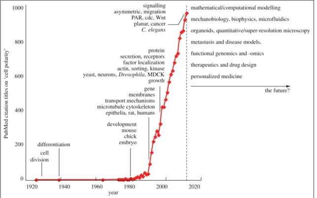
The first entry of ‘Cell Polarity’ in the title of a publication in the PubMed database ranges back to 1926 (Csikász-Nagy et al., 2013). This first publication dealt about fibroblast polarity in rab- bits (Bunting and Eads, 1926). Strikingly, the next entry dates back to 1939 and in this publica- tion cell polarity in grass root hairs was described (Sinnott and Bloch, 1939). As Figure 1.1 shows, by the 1980’s research on cell polarity drastically increased. This is likely related to pio- neering work in developmental, cell and molecular biology, as for example carried out by the No- ble Prize laureates Edward B. Lewis, Christiane Nüsslein-Volhard and Eric F. Wieschaus, who discovered cell polarity genes like gooseberry, hedgehog, patch and armadillo in Drosophila (Nüsslein-Volhard and Wieschaus, 1980; Riggleman et al., 1989; Pfeifer and Wieschaus, 1990).
A very recent hypothesis even put the phenomenon of cell polarization in context with the evolu- tion of the eukaryotic cell by endosymbiosis and stresses a rethinking of its origin by mere en- gulfment of eubacteria by archeae (Axelrod and Bergmann, 2014). According to their hypothesis, Buzz and David Baum suggested that archeae could have formed multiple cytoplasmic protru- sions that surrounded symbiotic eubacteria. Thereby this primary extracellular compartment would have increased and became the cytoplasm, and the archeal cell body would have been re- duced to the nucleus of the eukaryotic cell. Cytoplasmic membrane protrusions could have fused to bring the eubacteria into the cell. Also the endoplasmic reticulum (ER) could have originated from fused membrane protrusions. Polar distribution of mRNAs into individual compartments of this highly compartmentalized cell would have been a natural consequence and could have formed the basis for cell polarity (Axelrod and Bergmann, 2014).
|2
Despite being a general phenomenon of almost all cells, cell polarity or its defects play a crucial role in human diseases. For example the loss of apical-basal cell polarity in epithelial cells, also known as epithelial-mesenchymal transition (EMT), is related to metastatic tumor growth and tumor recurrence, which are among the most observed causes of death in breast cancer (Alvarez et al., 2013; Macara and McCaffrey, 2013). Also polar hyphal growth of the human pathogenic fungus Candida albicans initiated upon the contact with serum, is directly linked to human dis- eases like candidiasis or candidaemia (Sudbery, 2011; Arkowitz and Bassilana, 2014).
Human nutrition is directly or indirectly related to the yield of crop plants such as maize, wheat, or rice. However, many processes during plant fertilization and seed formation rely on cell polari- ty. These processes are described below in detail. Thus, the understanding and control of cell po- larity represents not only an interesting question from a research perspective, but also is tightly linked to human welfare.
Figure 1.1 | Cell polarity – whence and whither?
Yearly number of publications in the PubMed database with titles harboring the search term ‘Cell Polarity’
from 1926 to 2013. Prominent key words for each decade are indicated, as well as possible future trends.
Figure adopted from Csikász-Nagy et al. (2013).
3 |
1.2
Fundamental Principles of Cell PolarityAlthough cell polarity can be found among all cells ranging from bacteria to higher eukar- yotes and controls diverse biological processes, the underlying mechanisms are highly similar which allows to abstract fundamental principles.
1.2.1 Cell polarity requires the spatiotemporal regulation of small GTPases
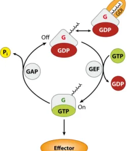
Most often, these small GTPases are members of the Ras-GTPase superfamily and only contain one G domain. The G domain is subdivided into four to five conserved fingerprint mo- tifs, G1 to G5 (Vetter and Wittinghofer, 2001; Wittinghofer and Vetter, 2011). The G1 motif (also called P-loop) binds the α- and β-phosphates of GTP and GDP, and the G2 and G3 motif (also called switch regions I and II) bind the γ-phosphate of GTP. The G4 and, if present, the G5 motifs are responsible for binding the guanine ring. Thereby the GTPase can cycle between an active, GTP-bound form and an inactive, GDP-bound form. As these GTPase have high nucleo- tide binding affinities but only low intrinsic hydrolysis activities, the interaction with regulatory proteins is crucial for GTPase function with downstream effectors. GUANINE NUCLEOTIDE EX- CHANGE FACTORS (GEFs), GTPASE ACTIVATING PROTEINS (GAPs) and GUANINE NUCLEOTIDE
DISSOCIATION INHIBITORS (GDIs) are effective GTPase regulators (Figure 1.2). GEFs facilitate the exchange of GDP by GTP and thus function as activators, while GAPs facilitate the GTP hy- drolysis to GDP and thereby function as inactivators (Vetter and Wittinghofer, 2001; Witting- hofer and Vetter, 2011). GDIs impede the release of GDP from the GTPases, inhibit GTPase activity and extract the GTPases from plasma membranes into the cytosol.
In bacteria, only recently it has been shown that the two small GTPases SofG and MglA play an important role in cell polarity and motility in Myxococcus xanthus (Leonardy et al., 2010; Patryn et al., 2010; Zhang et al., 2010; Bulyha et al., 2013). The response regulator RomR, which is regulated by the Frz chemosensory system, interacts with MglA-GTP and recruits it to both poles of the rod-shaped bacterium (Keilberg and Søgaard-Andersen, 2014). Its cognate GAP MglaB selectively inactivates MglA, so that the future leading pole is characterized by MglA-GTP and a higher local concentration of RomR. The lagging pole in contrast is marked by a high MglB con- centration and a low concentration of RomR. Asymmetrically localized MglA and its interplay with the GTPase SofG serve in the correct polar localization of the ATPases PilT and PilB to the leading and to the lagging pole respectively. PilB and PilT function in type-IV-pilus (T4P) de- pendent motility by providing energy for T4P retraction and extension.
In eukaryotes, Rho (for RAS HOMOLOGY) GTPases of the Ras superfamily play a crucial role in cell polarity. Whereas fungi and animals have developed diverse Rho GTPase families, such as Rho, Rac or Cdc42, in plants only one, called ROP (RHO OF PLANTS), with a high similarity to human Rac GTPases can be found (Craddock et al., 2012; Hall, 2012). The probably best under- stood eukaryotic small GTPase in terms of polarity establishment and signaling is the CELL DIVI-
|4
SION CYCLE protein Cdc42 from the budding yeast, Saccharomyces cerevisiae. Its function is high- ly conserved, and mammalian Cdc42 genes are at least partially able to rescue yeast cdc42 dele- tions (Shinjo et al., 1990). Loss-of-function mutants for Cdc42 in yeast are unable to establish cell polarity and stay symmetrical (Thompson, 2013). Its C-terminal CAAX motif can bind gera- nyl-geranyl molecules, which partially anchors Cdc42 to the plasma membrane. Upon interaction with its GEF Cdc24 or its GAPs Bem2, Bem3, Rga1 or Rga2, Cdc42 is activated or inactivated respectively (Howell and Lew, 2012; Bi and Park, 2012). The polar localization of its GEF Cdc24 plays an essential role in cell polarity establishment and maintenance. Also the GAPs are localized polar and their inhibition by phosphorylation furthermore contributes to the temporal Cdc42 activation. Another polarity regulator is the RHO GDI Rdi1 that effectively extracts Cdc42 from membranes and thus facilitates its rapid cycling between the plasma membrane and the cytoplasm. The spatiotemporal regulation of Cdc42 is an important trigger for downstream cell polarization. Upstream, Cdc42 is controlled by the action of the GTPase Bud1/Rsr1, its GAP Bud2 and its GEF Bud5. Transmembrane glycoproteins such as Bud8, Bud9 and Bud10/Axl2 serve as polar localized landmarks in axial or bipolar budding and finally determine the site of Cdc42-mediated cell polarity (Howell and Lew, 2012; Bi and Park, 2012).
1.2.2 The dynamic reorganization of the cytoskeleton is important for cell polarity Both, in eukaryotes and in prokaryotes, the small GTPase-regulated organization of the cytoskeleton formed by actin or actin-like proteins is necessary for cell polarity. In the bacterium Caulobacter crescentus, the BACTOFILIN cytoskeleton proteins BacA and BacB specifically localize to the site of the cell where a thin protrusion, the stalk, is localized. The polar localization of BacA and BacB serves as a landmark for the recruitment of the stalk-building cell wall biosynthe- sis enzyme PbpC (Kühn et al., 2010; Hughes et al., 2013). In M. xanthus it has been shown that by disturbing the assembly of the actin-like cytoskeleton protein MreB, the polar localization of
Figure 1.2 | The functional cycle of G proteins.
Small GTPases from the Ras superfamily bind lipids at their C-terminus and show partial membrane associa- tion. Regulation occurs by interaction with guanine nucleotide exchange factors (GEFs), GTPase activating proteins (GAPs), and guanine nucleotide dissociation inhibitors (GDIs), which activate (GEF) or inactivate (GAPs, GDIs) GTPases by facilitating the exchange of guanine nucleotides, the hydrolase activity or the re- lease from membranes. Figure adopted from Witting- hofer and Vetter (2011).
5 |
the small GTPase MglA is perturbed and motility is inhibited (Mauriello et al., 2010). The small GTPase SofG has been shown to interact with the cytoskeleton protein BACTOFILIN BacP that forms two polar patches at both sites of the rod (Bulyha et al., 2013). However, SofG is specifi- cally recruited to the BacP patch at the leading pole of the cell and together with MglA promotes T4P-dependent motility.
The yeast Cdc42 GTPase interacts with two formins, Bni1 and Bnr1 that mediate actin nuclea- tion and thus initiate the filament formation (Bi and Park, 2012). Cells in which these formins are deleted are synthetically lethal and show strong polarity defects. If the actin filament is dis- rupted by treatment with the drug Latrunculin A, also the polarized localization of the GTPase Bud1/Rsr1 is abolished (Kozubowski et al., 2008), highlighting the important role of actin organ- ization in cell polarity establishment.
1.2.3 Polar secretion and rapid turnover by exo- and endocytosis determines cell po- larity
The polar distribution of new cell wall and plasma membrane material in cells that elongate at one site such as budding yeast, animal neurons or plant pollen tubes and root hairs is essential for polar growth that is initiated by small GTPase-mediated actin transport. In yeast, actin cables polarized by the Cdc42-activated formin Bni1, guide vesicles via a myosin-V-dependent transport to the site of bud formation (Bi and Park, 2012). Bound to the vesicle membrane, additional Cdc42 is transported to the bud site, thereby supporting the positive feedback loop of cell polari- zation. Once vesicles reach at their target membrane, protein complexes such as the hetero- octameric exocyst complex act in membrane tethering. Finally membrane fusion occurs via a SNARE (SOLUBLE N-ETHYLMALEIMIDE-SENSITIVE FACTOR ATTACHMENT PROTEIN RECEPTOR) mediated process. The evolutionary conserved exocyst complex composes of the Sec proteins 3, 5, 6, 8, 10 and 15, identified as their mutants show defects in secretion, and the Exo70 and Exo84 proteins (Heider and Munson, 2012). Whereas in yeast and animals all of these proteins are en- coded by single-copy genes (Heider and Munson, 2012), in plants often more copies are found (Žárský et al., 2009; Cvrčková et al., 2012). Especially the Exo70 family has a high number of members indicating functional diversification among these proteins in plants. The exocyst com- plex is supposed to form dynamically in each round of exocytosis (Bi and Park, 2012). In Dro- sophila epithelia, the transmembrane protein CRUMBS (Crb) is secreted polar to the apical mem- brane via the exocyst machinery in a Rab GTPase dependent manner (Thompson, 2013). Simul- taneously, endocytosis of Crb via the AP2/Clathrin machinery at basolateral membranes is re- quired for cell polarity establishment and maintenance. Dynamic rapid exo- and endocytosis and cycling of components between the plasma membrane and the cytoplasm are further important determinants of cell polarity in eukaryotes. FRAP experiments have shown that Cdc42 undergoes fast cycling between both localizations, the plasma membrane and the cytoplasm (Wedlich- Soldner et al., 2004). This cycling is thought to be important for restricting the local Cdc42 acti-
|6
vation and inactivation (Bi and Park, 2012). In plants, rapid actin-dependent endocytic cycling has been demonstrated for the polar localized auxin plant growth hormone efflux carrier PIN
FOMED 1 (PIN1) (Kleine-Vehn and Friml, 2008).
In the M. xanthus bacterium, polar slime secretion is a prerequisite for adventurous (A) motility in gliding. If the slime is not secreted asymmetrically, A motility is abolished (Yu and Kaiser, 2007). Deletion of the polar localized small GTPase MglA or its GEF MglB in M. xanthus (Keil- berg and Søgaard-Andersen, 2014), results in a loss of polar slime secretion and consequently in the loss of A motility (Yu and Kaiser, 2007).
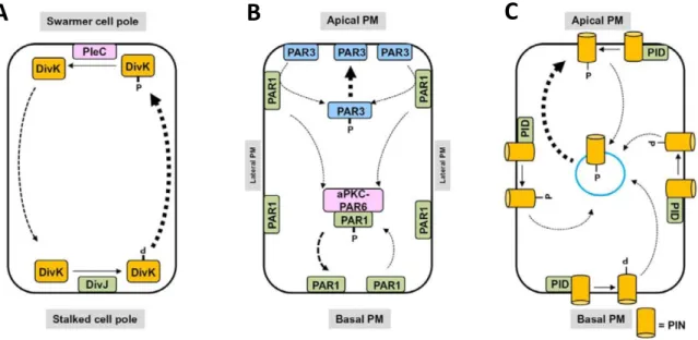
1.2.4 Posttranslational protein modification by phosphorylation regulates cell polarity The aquatic bacterium Caulobacter crescentus is polar organized. After division it produces a motile swarmer cell and a sessile stalked cell (Sommer and Newton, 1991; Ganguly et al., 2012).
Prior to division, the cell division gene and response regulator DivK rapidly shuttles between both poles in a phosphorylation dependent manner (Figure 1.3 A). At the stalked pole, DivK is phosphorylated by the DivJ kinase and moves to the swarmer pole. There, the PleC phosphatase dephosphorylates DivK which then shuttles back to the stalked pole of the cell. Furthermore there is a gradient of the phosphorylated response regulator CtrA towards the swarmer pole. This gradient is generated by CckA phosphatases located at the stalked, and CckA kinases located at the swarmer pole (Tsokos and Laub, 2012). Consequently, after division the swarmer cell has a high fraction of dephosphorylated DivK and phosphorylated CtrA, and the stalked cell a high fraction of phosphorylated DivK and dephosphorylated CtrA.
The Par proteins play an essential role in cell polarity establishment in animals (Thompson, 2013). At the apical pole of D. melanogaster follicle cells, Par3 is localized in a complex with Cdc42, Par6 and the ATYPICAL PROTEIN KINASE C aPKC, whereas at the basolateral domains the kinase Par1 is located (Figure 1.3 B). Par1 phosphorylates Par3 and thereby restricts its localiza- tion to the apical plasma membrane. Conversely, Par1 is restricted to the basolateral plasma membrane by aPKC-Par6 complex mediated phosphorylation (Ganguly et al., 2012). Polarity in some Arabidopsis thaliana plant cells is achieved by the polar localization of PINs (Figure 1.3 C), the auxin plant hormone efflux carriers (Dettmer and Friml, 2011). The main driving force for polarity maintenance is PIN phosphorylation and dephosphorylation and endocytic recycling (Geldner, 2008). The serine-threonine AGC kinase PINOID (PID) is responsible for PIN phos- phorylation and the PROTEIN PHOSPHATASE 2A (PP2A) for dephosphorylation. The endosomal PIN recycling process is controlled by ADENOSYL RIBOSYLATION FACTOR GUANOSINE EX- CHANGE FACTORS (ARF-GEFs) and the Rab5-GTPase ARA7 (Dhonukshe et al., 2008). First, PIN proteins are secreted to the membranes in a non-polar manner and phosphorylation and re- cycling regulates subsequent PIN polarization. Predominantly, phosphorylated PINs show a pref- erence for apical and dephosphorylated PINs for basal membranes (Ganguly et al., 2012). PIN
7 |
polarization is thus dependent on the level of activity of the antagonistic processes of phosphory- lation and dephosphorylation (Kleine-Vehn and Friml, 2008).
1.2.5 Cell polarity underlies the principles of positive feedback loops and mutual antagonisms
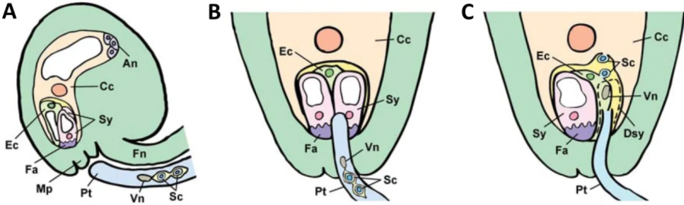
Especially recent mathematical modeling approaches have revealed the importance of feedback loops that action in a cooperative, non-linear fashion to establish a single local spot for cell polarization (Thompson, 2013). Two partially redundant and interlinked feedback loops are thought to function in cell polarization in yeast. In a fast process, an autocatalytic cluster of Cdc42 in complex with its GEF Cdc24, the scaffold protein Bem1 and the kinase Cla4 is able to generate a positive feedback loop that leads to Cdc42 polarization by self-recruiting (Altschuler et al., 2008; Johnson et al., 2012; Bi and Park, 2012). Another feedback loop is generated after cy- toskeleton polarity establishment by the actin-dependent Cdc42 polarization. This second slower feedback loop stabilizes the polarization process initiated by the first autocatalytic clustering (Slaughter et al., 2009). Besides the requirement of positive feedback loops, the importance of mutual antagonism between polarity components has been demonstrated by mathematic model- ing of Caenorhabditis elegans zygotes (Goehring et al., 2011) and Drosophila melanogaster follicle cells (Fletcher et al., 2012). At the anterior pole of the C. elegans zygote, Par3 forms a complex
Figure 1.3 | Cell polarity regulation by phosphorylation.
Cell polarity regulation by phosphorylation-mediated polar protein trafficking in (A) bacteria, (B) mammals and (C) plants. (A) in the bacterium Caulobcater crescentus, DivK is phosphorylate by DivJ at the stalked cell pole and localizes to the swarmer cell pole. There, DivK is dephosphorylated by PleC and thus redirected to the stalked cell pole. (B) in mouse epithelial cells, phosphorylation regulates polar localization of the Par pro- teins. Par1 is localized basolateral and Par3 apical. Par3 is phosphorylated by Par1 and its localization is phos- phorylation-dependent. Also the basolateral localization of Par1 is phosphorylation dependent and regulated by the aPKC-Par6 complex. (C) in Arabidopsis thaliana, PID-mediated phosphorylation of PIN specifically tar- gets it to the apical membrane. Figure modified from Ganguly et al. (2012).
A B C
|8
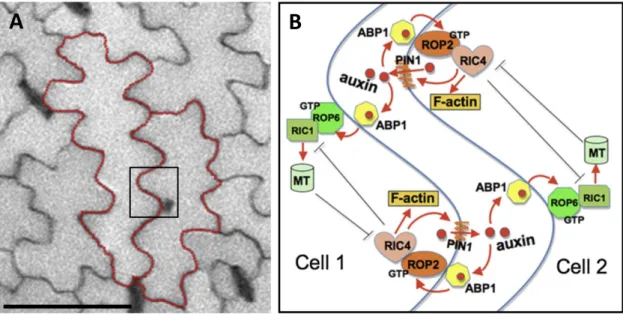
with the PDZ-domain protein Par6, the serine/threonine kinaseaPKC and Cdc42. The kinase Par1, the RING domain protein Par2 and the LETHAL GIANT LARVAE (LGL) protein form a complex at the posterior pole (Thompson, 2013). By mutual antagonistic phosphorylation of Par3 by Par1 and of Par1 by aPKC, the respective polarity components are removed from the plasma membrane, thereby keeping the anterior-posterior restricted distribution in equilibrium (Goehring et al., 2011; Thompson et al., 2013). The same polarity components are required to generate an apical-basolateral cell polarity in D. melanogaster epithelia cells. Here it was also shown that mutual antagonistic regulation by phosphorylation of apical-localized BAZOOKA (the homologue to Par3 from C. elegans) by Par1 and basolateral-localized Lgl by aPKC is important to generate and sustain cell polarity (Fletcher et al., 2012; Thompson et al., 2013). Plant leaf epi- dermal pavement cells exhibit a jigsaw piece-like morphology with spots of local outgrowth, the lobes and spots with suppressed outgrowth, the indentations (Figure 1.4). The regulation of po- larized local outgrowth and local outgrowth suppression is achieved by the mutual antagonisms between the small GTPases ROP2 and ROP6 in an auxin and PIN1 mediated positive feedback loop in Arabidopsis (Xu et al., 2010).
A B
Figure 1.4 | Polarity control in leaf epidermal cells.
(A) Overview image of the Arabidopsis thaliana leaf epidermis showing single jig-saw piece-like pavement cells highlighted by red outlines. (B) Interdigitated growth of leaf epidermis pavement cells in Arabidopsis thaliana is regulated by feedback loops and auxin signaling and is inter- and intracellular coordinated. Central roles play the small GTPases ROP2 and ROP6 which control lobe and indent formation respectively in complemen- tary pathways. ROP2 acts in a self-activating local feedback loop together with PIN1 and auxin and regulates actin cytoskeleton organization via RIC4. Localized extracellular auxin regulates the antagonizing ROP6 path- way and microtubule formation via RIC1 in adjacent cells. ROP2 functions in a positive feedback loop and both, the ROP2 and ROP6 pathways act mutually antagonizing. Scale bar = 25 µm in (A). Figure modified from Xu et al. (2010).
9 |
1.2.6 Cell polarization is triggered by external cues or induced spontaneously
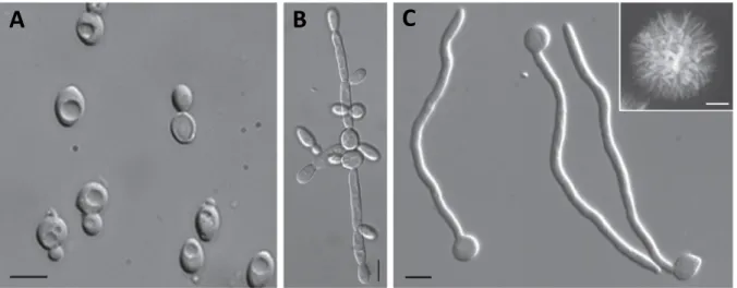
Both, spontaneous or external-triggered polarization events are fundamental properties of polarized cells (McCaffrey and Macara, 2012). For example, the opportunistic human pathogenic fungus Candida albicans can grow in its predominantly unpolar yeast form that only during bud- ding shows spontaneous cell polarization (Figure 1.5). Stimuli that represent stresses such as nu- trient starvation, temperature variation or hypoxia, or the sensing of serum, trigger the switch to its highly polarized and oriented hyphal growth form (Arkowitz and Bassilana, 2014). Some cells are mainly polarized only upon external cues. The eggs of the worm C. elegans initially show a high degree of symmetry. The entry of the sperm during fertilization leads to symmetry breaking and triggers the polarization of the zygote (Thompson, 2013). Thereby the position of the sperm centrosome marks the new posterior pole. Also the zygote of the brown alga Fucus distichus is mainly polarized by external cues after fertilization (Cove, 2000). There, the first cell division leads to the formation of a rhizoid cell and a cell that differentiates to the later thallus. If the zy- gote is exposed to a light gradient, the rhizoid cell will always emerge from the shaded side. On a subcellular level, this position is marked by the occurrence of an F-actin assembly at the site of rhizoid outgrowth (Cove, 2000).
Figure 1.5 | Different growth forms of the human pathogen Candida albicans.
Morphology of the fungal pathogen Candida albicans in its yeast (A), pseudohyphal (B) and hyphal (C) forms.
The inset in (C) shows a hyphal colony grown for 5 days on Spider medium. Bars = 5 µm in (A) to (C) and 1 mm in inset of (C). Note the high morphological similarity of growing hyphae (C) and germinating pollen tubes (Fig- ure 2.1). Figure adopted from Sudbery (2011).
A B C
|10
1.3
Cell Polarity in Flowering PlantsCell polarity is a fundamental necessity for the organization of the entire plant body. At every point in the life cycle of a flowering plant, cell polarity is required to control development, growth and morphogenesis (Yang, 2008). Comparable to other organisms, plants exhibit a broad range of polar organized cell types. On one side of the spectrum are cells such as root tip cells which show only a low level of morphological polarity. But as mentioned above, they exhibit a high level of molecular polar organization marked by the distribution of PIN auxin efflux carriers.
On the other side, morphologically highly polarized cell types can be found, such as, for example trichomes. They are formed by the leaf epidermis and exhibit a complex 3-dimensional architec- ture. With respect to the rigid cell wall that surrounds the mature plant cell and limits its exten- sion capability, polarized cell division plays an important role in plant body construction and de- velopment. Also different to animal systems, tightly cell wall-encapsulated plant cells are not able to migrate within a tissue (Dettmer and Friml, 2011). Thereby cell polarization by means of asymmetric cell division becomes an important developmental phenomenon.
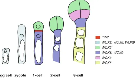
Notably, already the first cell division in a flowering plants’ life cycle is highly asymmetric and results in the formation of two cells that undergo completely different cell fates (Figure 1.6). In Arabidopsis, after gamete fusion in the process of double fertilization (see section 1.5), the zygote undergoes polarized elongation (Lau et al., 2012). A large vacuole is located at the micropylar pole, whereas the nucleus is positioned at the chalazal pole (Zhang and Laux, 2011). The first cell division occurs transversely to the future embryo axis and result in a small apical cell and a large basal cell. All cells derived from the apical cell differentiate into cells of the embryo. The large ba-
Figure 1.6 | Zygote and early embryo development in Arabidopsis thaliana.
Schematic drawing showing the polarized egg cell and zygote as well as apical-basal axis formation during early embryo development. In the egg cell and elongated zygote, large vacuoles reside at the basal (micropylar), and nuclei at the apical (chalazal) poles of the cell. The zygote undergoes an asymmetric cell division and forms a small apical cell (ac) and a large basal cell (bc). The cells derived from divisions of the apical cell together with the uppermost cell of the dividing basal cell (hypophysis, hy) form become embryonic cells, whereas the re- maining daughter cells of the basal cell form the suspensor. Polar localization of PIN7 in the basal cell and cell- type specific expression of WOX genes is indicated by the color code. Figure modified from Zhang and Laux, (2011).
11 |
sal cell will give rise to a cell file of which the uppermost, the hypophysis, contributes to the root meristem of the embryo. The remaining cells differentiate into the embryo-supplying suspensor.
On the molecular level, asymmetric cell division of the zygote is dependent on polarized auxin transport. As judged from the DR5rev:GFP reporter activity, auxin accumulates in the small api- cal cell (Friml et al., 2003). This accumulation is regulated by the PINFROMED 7 (PIN7) auxin efflux carriers that are localized polar in the apical membrane of the basal cell and pump auxin into the small apical cell and thus regulate its development. PIN protein expression and auxin homeostasis is furthermore regulated by the WUSCHEL HOMEOBOX (WOX) proteins. In the egg cell, WOX2 and WOX8 are both expressed, whereas after asymmetric cell division, WOX2 is pre- sent only in the apical, and WOX8 only in the basal cell (Haecker et al., 2004). Zygote elongation and asymmetric cell division is regulated by the MAPKKkinase YODA (YDA). Notably, in the yda-1 mutant, the zygote divides almost symmetrically (Lukowitz et al., 2004). Upstream of YDA acts the SHORT SUSPENSOR (SSP) interleukin-1 receptor-associated kinase / Pelle-like kinase, and its loss phenocopies the yda mutant (Bayer et al., 2009). Interestingly, only the SSP allele that is derived from the paternal sperm cell line functions in embryo patterning as shown by crossing experiments. In a model of SSP function it is hypothesized that the polar delivery of the SSP mRNA by the sperm cell fusion with the egg cell triggers zygote elongation and asymmetric cell division via the YDA/MAPK pathway (Bayer et al., 2009). This situation of zygote polarity estab- lishment controlled by sperm cell entry is reminiscent of the polarization that occurs in the C.
elegans egg described above. Although far away from being entirely understood, polarity estab- lishment in the Arabidopsis zygote represents an outstanding example of the close relation be- tween polarity, cell fate determination and developmental regulation.
But already before double fertilization and zygote formation, cell polarity plays an important role in cell fate determination in the female gametophyte. Already the synergid, egg and central cells display a polar organization (Sprunck and Groß-Hardt, 2011). In the synergid and the central cell the nucleus is oriented towards the micropylar pole, whereas a large vacuole is located at the chalazal pole. In contrast, the egg cell is orientated the other way round: a large vacuole is located at the micropylar and the nucleus at the chalazal pole. Mutant analyzes suggest that polar nucleus localization is tightly linked to cell fate of the female gametophytic cells. In female gametophyte mutants such as ATROPOS, CLOTHO, EOSTRE or LACHESIS, nuclei of synergid cells are often mislo- cated at the chalazal pole, which ultimately results in the expression of egg cell genes (Groß-Hardt et al., 2007; Pagnussat et al., 2007; Moll et al., 2008). These examples demonstrate the tight in- terconnectivity between cell polarity and cell fate determination.
On a macroscopic scale, cell polarity is more obvious for leaf epidermis pavement cells or for tri- chomes, compared to the cells of the female gametophyte. As shown in Figure 1.4, pavement cells form many highly polarized structures of lobes and indentations that join together with the indentations and lobes of the neighboring cells, like the pieces of a jigsaw puzzle. As described above, in pavement cells the spatially controlled auxin-mediated interplay between local feedback-
|12
regulated ROP signaling and cytoskeleton organization controls lobe and indentation formation, and thus cell polarity (Xu et al., 2010).
Trichomes are leaf hairs that are formed by single epidermis cells in duty to protect the plant against herbivore attacks, water loss by transpiration, or UV irradiation. Therefore, trichomes are present on most aerial parts of the Arabidopsis plant (Marks, 1997; Pattanaik et al., 2014). On a genetic level, trichome development is regulated by a network including the TRANSPARENT TES- TA GLABRA 1 (TTG1) WD40 repeat protein, the functionally redundant GLABRA 3(GL3) and ENHANCER OF GLABRA 3(EGL3) bHLH transcriptions factors, and the MYB transcription fac- tors GLABRA 1 (GL1) and MYB23 (Oppenheimer et al., 1991; Walker et al., 1999; Payne et al., 2000; Zhang et al., 2003; Kirik et al., 2005). Trichome outgrowth and proper trichome morpho- genesis establishment is regulated by the GLABRA 2 (GL2) homeodomain, and the TRANSPARENT
TESTA GLABRA 2 (TTG2) WRKY, (Rerie et al., 1994; Johnson et al., 2002) as well as by the MYB82 transcription factors (Liang et al., 2014). Normally, trichomes in Arabidopsis develop three to four branches (Marks, 1997). However, the knock-out of the RPN1A subunit of the 26S proteasome results in a higher number of trichome branches, likely by interfering with gibberellic acid and cytokinin signaling pathways (Yu et al., 2015). Also, small endogenous non-coding mi- croRNAs (miRNAs) play a role in trichome development regulation by modulating the expres- sion of SQUAMOSA PROMOTER BINDING PROTEIN LIKE (SPL) genes (Yu et al., 2010). The ex- pression of a mimicry target of miR156 results in a reduction of trichome density, whereas the constitutive expression of miR156 leads to ectopic trichome development. Thus, a complex net- work of transcriptional regulation, protein level regulation and hormone signaling is required to control cell polarity in trichome development. Although we have a quite elaborate picture of ge- netic root hair developmental control, the underlying cell biological events are only poorly char- acterized (Kulich et al., 2015). Only very recently it was shown that the exocyst vesicle tethering complex subunit Exo70H4 plays an important role in UV irradiation-induced secondary cell wall formation in trichomes (Kulich et al., 2015).
However, detailed cell biological and molecular studies on cell polarity and polar cell growth in flowering plants have mainly been conducted on pollen tubes and root hairs where we have a quite elaborated, but still incomplete picture of the underlying mechanisms (for review see: Kost, 2008; Yang, 2008; Chebli et al., 2013; Guan et al., 2013; Rounds and Benzanilla, 2013; Ketelaar, 2013; Cai et al., 2015). The molecular basis of cell polarity establishment and polar tip growth of pollen tubes and root hairs in Arabidopsis represent the focus of the subsequent section.
1.4
Cell Polarization and Polar Tip Growth of Pollen Tubes and Root HairsIn plant polar growth research, flowering plant pollen tubes and root hairs have developed to the cell types of choice as they represent protrusions of single cells that grow rapidly and highly polarized (Yang, 2008). The outgrowth of pollen tubes and root hairs occurs from only one single
13 |
point of the cell and the expansion of the protuberance takes place only at an annular region shortly below the very apex of the tip-growing tube. Thus, the growth of pollen tubes and root hairs can be described as heterotropic monotropic annular tip growth (Geitmann, 2009, 2010).
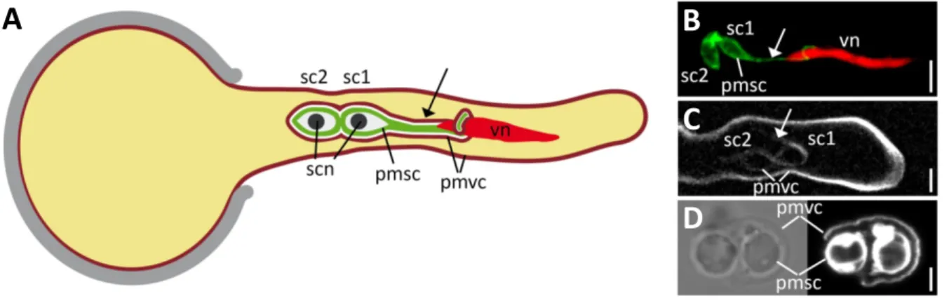
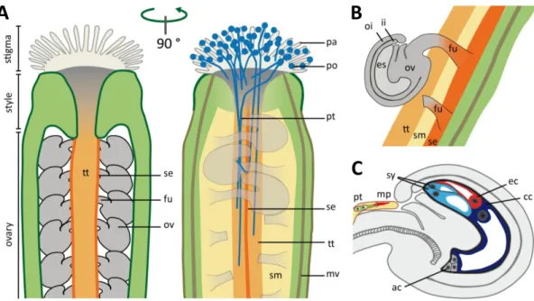
In both cell types the highly polarized growth is related to a specialization in their biological func- tion. Root hairs are formed in the root differentiation zone and mainly serve in anchorage and nutrient uptake as well as in plant-microbe interactions by enormously increasing the root surface and the root diameter (Grierson et al., 2014). On the other hand, the pollen tube of flowering plants represents the mature three-cellular male gametophyte (McCormick, 2004). During mi- crosporogenesis, diploid pollen mother cells undergo meiosis which results in the generation of haploid microspores. The microspore undergoes a first mitosis to form the bicellular pollen grain, consisting of a large vegetative and a small generative cell. In a second mitosis, the generative cell divides into the two sperm cells. The two sperm cells are enclosed by the cytoplasm and sur- rounded by the plasma membrane of the vegetative cell. One of the sperm cells forms a plasma membrane projection that is physically connected to the nucleus of the vegetative cell. Together the two sperm cells and the vegetative nucleus form the so called male germ unit (MGU) (McCue et al., 2011). After landing on the stigma, the large vegetative cell forms the tip-growing pollen tube that has the function to transport the two sperm cells to the female gametophyte to confer double fertilization, the fusion of one sperm cell with the haploid egg cell, and the fusion of one sperm cell with the diploid central cell nucleus (Figure 1.7).
Figure 1.7 | The pollen tube and the male germ unit of Arabidopsis thaliana.
A) Scheme of a growing Arabidopsis pollen tube and the male germ unit (MGU) which comprises two physically connected sperm cells (sc1 and sc2). The plasma membrane of the distal sperm cell (pmsc) forms a projection (arrow) to the vegetative nucleus of the tube cell (vn). Both sperm cells are enclosed by the membrane of the vegetative cell (pmvc, dark red). Sperm cell nuclei (scn) are in black. (B) Confocal laser-scanning micrograph of a growing pollen tube with GFP-labelled sperm cell membranes (pmsc) and a red fluorescent vegetative nucleus (vn). (C) Spinning-disc confocal micrograph of a pollen tube expressing a fluorescent membrane protein that la- bels the membrane of the vegetative cell (pmvc) which also encloses the two sperm cells. (D) Sperm cells re- leased from a pollen tube in vitro. The membrane-selective styryl dye FM4-64 labels the sperm cell membranes (pmsc) and the membrane of the vegetative cell (pmvc), which ruptured during pollen tube burst. Note that ob- jects shown in (A) are not to scale. Scale bars = 5 µm in (B) and (C) and 2 µm in (D). Figure modified from Sprunck et al. (2014).
B
C
D
A
|14
After microsporogenesis, mature tricellular pollen grains are partially dehydrated and metabolical- ly inactivated until they get in contact with the receptive stigma of a flower after pollination (Ed- lund et al., 2004). During this contact phase, a water ‘bridge’ between the pollen exine and the cell wall of the papilla cell is established, the so called ‘foot’ (Wolters-Arts et al., 1998).
1.4.1 Pollen activation
After rehydration in a process called pollen activation, the pollen grain undergoes sym- metry breaking and polarization, which finally leads to the formation of the pollen tube (Raghavan, 2003). So far we have only little knowledge about the cell biological and molecular basis of symmetry breaking and growth-site selection in pollen grains. In a recent study it was demonstrated by using cell wall component specific staining techniques, electron microscopy and mutant analysis, that in Arabidopsis pollen prior to germination, a structure termed germination plaque consisting of cellulose, callose and partly de-esterfied pectin is formed at the site of pollen tube emergence (Hoedemaekers et al., 2014). The BURSTING POLLEN (BUP) gene encodes a Gol- gi-located glycosyltransferase responsible for the proper organization of the germination plaque and the tip of the growing pollen tubes. Furthermore, after pollen hydration and approximately 6 minutes before germination, a cytosolic Ca2+ gradient is established towards the future germina- tion site (Iwano et al., 2004). Fluorescence microscopy revealed that in Pyrus communis and Nar- cissus pseudonarcissus pollen the actin cytoskeleton is polarized towards the germination site (Ti- wari and Polito, 1988; Heslop-Harrison and Heslop-Harrison, 1992). It seems very likely that, comparable to the role of the small GTPase Cdc42 during symmetry breaking in yeast, in the pollen grain the small GTPase ROP1 plays a central role in polarization (Yang and Lavagi, 2012).
The ROPINTERACTIVE PARTNER 1 (RIP1/ICR1) was identified as molecular interactor of ROP1 by yeast two-hybrid screening. Its location switches from the nucleus to the future germination site before pollen tube emergence (Li et al., 2008). By recruiting ROP1 to the plasma membrane, it is thought that RIP1/ICR1 is involved in the positive feedback regulation of ROP1 localization to the site of pollen polarization. Very recently it was shown that the cytoplasmic ROP-interactive CRIB motif-containing protein RIC1 is localized to the plasma membrane at the pollen tube emergence site during germination and at the tip of growing pollen tubes (Zhou et al., 2015).
RIC1 regulates F-actin dynamics and thus pollen tube growth by directly binding to and severing of F-actin in a Ca2+-dependent manner. ROP1 was already shown to interact with two other RICs, namely RIC3 and RIC4 and thereby to activate two counteracting downstream pathways (Gu et al., 2005). RIC4 promotes the assembly of F-actin, whereas RIC3 promotes the Ca2+- mediated disassembly of F-actin at the pollen tube tip (Gu et al., 2005; Lee et al., 2008).
We have shown that prior to pollen germination, vesicles marked with the ARMADILLO REPEAT
ONLY 1 (ARO1) protein, an important player in polar elongation of growing pollen tubes (Gebert et al., 2008), accumulate at the pollen tube germination site shortly before outgrowth (Vogler et al., 2015, Chapter 2). However, this accumulation does not represent a constant in-
15 |
crease, but rather some short and peaked maxima 12 to 3 minutes before pollen tube formation.
We could also show that about 15 minutes before pollen tube formation, F-actin reorganization events take place. Especially at the flanks and the opposite pole of the germination site, massive cortical actin arrays appear, possibly forming a mechanical counter bearing for the future protru- sion formation and F-actin polarization.
1.4.2 Stages of root hair and pollen tube growth
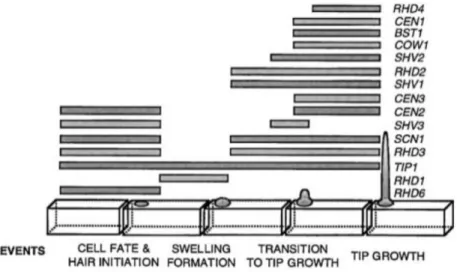
Due to large genetic screening approaches (e.g. Schiefelbein and Sommerville, 1990; Par- ker et al., 2000) we currently have a much better understanding of cell polarization events that take place shortly after protrusion formation in root hairs than in pollen tubes (Figure 1.8). This imbalance of knowledge is mainly caused by two facts. First, as they grow slower (Ketelaar et al., 2008), root hairs are much easier to investigate on a mechanistic level than pollen tubes. Second, pollen tubes are haploid and involved in sexual reproduction. Therefore already weak recessive mutations can lead to male sterility and thus hamper the screening for genetic interactions of higher order mutants. Root hair development underlies several genetically distinct steps (Schiefel- bein and Sommerville, 1990; Parker et al., 2000). First, a local swelling, the so called bulge is formed by the trichoblast (i.e. the root hair forming cell) protoplast. This roundish swelling then enlarges and at a certain point transitions into an elongating structure. In the last step, the root hair finally grows by polar tip growth. For each of these steps, characteristic mutants could have been identified (summarized in Bibikova and Gilroy, 2003; Grierson et al., 2014).
Using morphological modeling and quantitative live-cell imaging, we could show that also pollen tube growth can be subdivided into comparable distinct steps, thereby providing a methodologi- cal and knowledge framework for large-scale genetic screening approaches (Vogler et al., 2015, Chapter 2).
Figure 1.8 | Wild-type root hair development.
Characteristic events during the development of root hairs in Arabidopsis. Genes important for certain steps of root hair development are indicated on the right. Figure modified from Parker et al. (2000).
|16
1.4.3 Molecular mechanisms of tip growth
Besides similarities in mechanisms of protrusion formation, a large body of publications has revealed significant similarities between cell biological and molecular mechanisms underlying polar tip growth of flowering plant pollen tubes and root hairs (e.g. Šamaj et al., 2006; Campa- noni and Blatt, 2007; Kost, 2008). Both exhibit an inverted cone shaped vesicle-rich zone at the very apex that, due to the lack of large light-scattering organelles, is named the ‘clear zone’ (Hep- ler and Winship, 2015). In both cell types vesicles move at the flanks towards the apex and return in the center back to the shank in a so called ‘reverse fountain’ streaming pattern (Cheung and Wu, 2008; Chebli et al., 2013). The actin cytoskeleton is highly polarized in longitudinal bundles in both cell types (Hepler et al., 2001) and both show a steep tip-focused Ca2+ gradient that tight- ly interplays with reactive oxygen species (ROS) and ROP/RIC signaling, local pH changes, actin dynamics, and phospholipid signaling (Cárdenas et al., 2008; Cárdenas, 2009). In both cell types the polar localization and tip-restricted action of a small ROP GTPase is regulating the tubular shape and the elongation process. In pollen tubes, ROP1 is the central molecular polarity switch (Li et al., 1999), whereas in root hairs ROP2 fulfills this function (Jones et al., 2002). A recent comparative microarray transcriptomics approach has revealed that root hairs and pollen tubes cluster closely together in regard of their expression profiles (Becker et al., 2014). Given the sig- nificant mechanistic and transcription profile similarities, it is not surprising that gene knock-outs can cause polarity defects in both cell types. Thus, the loss of the ankyrin and DHHC-CRD do- main containing TIP GROWTH DEFECT 1 (TIP1) protein, the exocyst subunit Exo70A1, the ARF GAP RPA or the PHOSPHATIDYLINOSTITOL MONOPHOSPHATE-5-KINASE 4 (PIP5K4) results in both, aberrant pollen tubes and root hairs (Schiefelbein et al., 1993; Song et al., 2006; Synek et al., 2006; Ischebeck et al., 2008; Sousa et al., 2008; Wada et al., 2015). In other cases genes im- portant for polar tip growth in one cell type can have close homologs that serve an identical func- tion in the other cell type. For example the Rab GTPase RabA4b is localized polar in root hairs and RabA4d in pollen tubes (Preuss et al., 2004; Szumlanski and Nielsen, 2009) and the AGC kinases AGC1.5 and AGC1.7 are important for directional growth of pollen tubes, whereas AGC1.6/RSH3 (for ROOT HAIR SPECIFIC 3) is acting specifically in root hairs (Won et al., 2009;
Zhang and McCormick, 2009).
We have shown that in Arabidopsis two close homologs of ARMADILLO REPEAT ONLY 1 (ARO1) are involved in the polar elongation of root hairs (Vogler et al., submitted, Chapter 3). ARO pro- teins are conserved plant specific Armadillo Repeat Motif (ARM) proteins (Gebert et al., 2008).
The Arabidopsis ARO protein family comprises four members among which ARO1, ARO2 and ARO3 exhibit relatively high similarities, whereas ARO4 is the most distant related family mem- ber (Gebert et al., 2008; Vogler et al., submitted). In contrast to ARO1 that is expressed exclu- sively in gametophytes, ARO2-ARO4 are expressed in almost all sporophytic parts of the plant but are excluded from pollen tubes (Gebert et al., 2008). The double homozygous knock-out of ARO2 and ARO3 leads to mutant root hairs with a phenotype comparable to that of ARO1-
17 |
deficient pollen tubes. The ectopic expression of ARO2 or ARO3 in aro1-3/+ mutant pollen, as well as vice versa the ectopic expression of ARO1 in aro2 aro3 double mutant root hairs is able to restore wild-type morphologies, thereby proofing functional conservation and redundancy among ARO proteins. To our knowledge this represents the first report of functional complementation of pollen tube and root hair growth by both, ectopic expression of a gametophyte specific gene in mutant root hairs and ectopic expression of sporophyte specific genes in mutant pollen tubes.
ARO functional redundancy even more emphasizes the close similarity between pollen tubes and root hairs.
1.4.4 External factors regulating pollen tube growth
Polar pollen tube growth depends on the concerted interplay between signaling compo- nents, cytoskeleton organization and polarized membrane trafficking, to which the ARO proteins functionally contribute. However, external components and cues are necessary to induce pollen germination and to support polar growth. In vitro germination of Arabidopsis pollen has been shown to be dependent on the presence and concentration of sucrose and Ca2+, K+ and borate ions in the germination medium, as well as on the pH, temperature and the water availability (Brewbaker and Kwach, 1963; Li et al., 1999; Boavida and McCormick, 2007; Bou Daher et al., 2009; Yetisen et al., 2011). Organic molecules such as the polyamine spermidine, and flavonols like kaempferol also promote in vitro pollen germination in Arabidopsis, Petunia and Nicotiana tabacum (Mo et al., 1992; Ylstra et al., 1992; Taylor and Grotewold, 2005; Rodriguez-Enriquez et al., 2013). In pollen of Torenia fournieri and N. tabacum, germination was supported by the addition of plant hormones like the auxin analog indoleacetic acid (IAA) or gibberellic acid (GA3) (Chen and Zhao, 2008; Wu et al., 2008). However, adverse, no, or only weak promoting effects on in vitro pollen germination were shown for the plant hormones abscissic acid, ethylen and zeatin (Sfakiotakis et al., 1972; Wu et al., 2008).
For in vitro pollen germination, experimentally the most demanding is Arabidopsis pollen, due to high variances as a result of pollen quality (Boavida and McCormick, 2007; Rodriguez-Enriquez, 2013). By adding the brassinosteroid phytohormone epibrassinolide (epiBL) to germination me- dia we were able to significantly enhance and stabilize Arabidopsis in vitro pollen germination in a dose-dependent manner (Vogler et al., 2014, Chapter 4). Besides promoting pollen germination by nine fold, epiBL also significantly enhances in vitro Arabidopsis pollen tube elongation rates by almost five fold.
In vitro pollen tube growth rates are not only elevated by the plant hormones IAA, GA3 and ep- iBL (Wu et al., 2008) but furthermore by organic molecules such as gamma-aminobutyric acid (GABA) (Palanivelu et al., 2003), myo-inositol (Schneider et al., 2006), N-methanesulfinyl- azadecalins (Qin et al., 2011), by peptides such as the STYLE INTERACTOR FOR LEPRKS (STIL) (Wengier et al., 2010) or by proteins such as the stigma specific small cysteine-rich STIG1 pro- teins from Nicotiana tabacum or Lycopersicon esculentum (Goldmann et al., 1994; Tang et al.,