I NVESTIGATIONS O F M ITOCHONDRIA F UNCTION IN A
H UMAN C ELLULAR M ODEL
DISSERTATION
ZUR ERLANGUNG DES DOKTORGRADES DER NATURWISSENSCHAFTEN (DR. RER. NAT.)
DER FAKULTÄT FÜR BIOLOGIE UND VORKLINISCHE MEDIZIN DER UNIVERSITÄT REGENSBURG
vorgelegt von Kerstin Kuffner
aus Viechtach
im Jahr
2020
Das Promotionsgesuch wurde eingereicht am: 20.02.2020
Die Arbeit wurde angeleitet von: Prof. Dr. Christian Wetzel
Unterschrift: ______________________________
Table of contents
Acknowledgements ... 9
Abbreviations ... 10
Abstract ... 12
Zusammenfassung ... 14
1 Introduction ... 16
1.1 Mitochondria – the vital force in our cells ... 16
1.1.1 Mitochondria structure and the site of action ... 16
1.1.2 Mitochondria in the cellular metabolism... 19
1.1.3 Fission and fusion: a dynamic network ... 21
1.2 Major Depressive Disorder – a multifactorial disease ... 24
1.2.1 Classification and diagnostics ... 24
1.2.2 Early and new theories for manifestation ... 26
1.3 The risk factor stress - impact on mitochondria and mood ... 28
1.3.1 The stress response in the human body ... 28
1.3.2 Benefits of acute stress ... 29
1.3.3 Sustained stress and its deleterious consequences ... 30
1.4 Mitochondria and disease – what we know so far ... 33
1.4.1 Mitochondriopathies ... 33
1.4.2 Mitochondrial disruption in neurodegenerative diseases ... 34
1.4.3 Dysfunction of mitochondria and psychiatric disorders ... 35
1.5 From fibroblasts via induced pluripotent stem cells through to induced neurons - a cellular model for MDD ... 36
1.6 Bioenergetic imbalance in MDD – hypothesis and aim of the thesis ... 38
2 Materials ... 41
2.1 Lab supplies ... 41
2.2 Reagencies, chemicals and kits ... 41
2.4 Antibodies ... 43
2.5 Culture media and buffer compositions ... 43
3 Methods ... 45
3.1 Study participants ... 45
3.2 Adult human dermal fibroblasts ... 46
3.2.1 Culturing conditions ... 46
3.2.2 Passaging, freezing and thawing procedure ... 46
3.2.3 Automated cell counting with CASY Cell Counter ... 47
3.3 Reprogramming of adult human dermal fibroblasts into induced pluripotent stem cells using Epi5 plasmids ... 47
3.1 Coating with Vitronectin ... 47
3.2 Preparation of Epi5 plasmids ... 47
3.3 Preparation and electroporation of human fibroblasts ... 48
3.4 Coating for iPSCs and NPCs ... 48
3.5 Induced pluripotent stem cells ... 49
3.5.1 Culturing conditions ... 49
3.5.2 Passaging, freezing and thawing procedure... 49
3.6 Neural induction and expansion of neural progenitor cells ... 50
3.7 Immunocytochemical staining ... 51
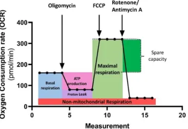
3.8 Assessment of respiratory properties: Seahorse XFp Flux Analyzer ... 51
3.10 Normalization of respiratory experiments ... 53
3.10.1 Normalization by Hoechst staining ... 53
3.10.2 Normalization by Bichinoic Acid Assay ... 53
3.11 Luminescent assay for ATP content ... 54
3.12 Live cell imaging ... 55
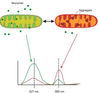
3.12.1 Mitochondrial membrane potential measurements with JC-1 ... 55
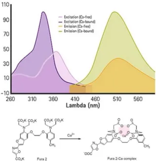
3.12.2 Analysis of intracellular Ca
2+with Fura-2 dye ... 56
3.13 Determination of the mitochondrial content ... 57
3.13.1 gDNA extraction ... 57
3.13.2 Quantitative Real-Time Poly Chain Reaction ... 58
3.14 Data analysis and statistics ... 60
4 Results ... 61
4.1 Patient and control cohort ... 61
4.2 Differences in mitochondrial metabolism in peripheral cells: Fibroblasts show alterations in mitochondria-related functions ... 62
4.2.1 Mito Stress Test: ETC function and glycolysis ... 63
4.2.2 ATP content ... 69
4.2.3 Bioenergetic properties and mitochondria-related homeostasis in fibroblasts ... 70
4. 3 Mitochondrial content: mtDNA copy number in fibroblasts ... 74
4.4 Successful reprogramming of fibroblasts into iPSCs and induction of NPCs ... 75
4.4.1 Cultivation and reprogramming of human dermal fibroblasts ... 75
4.4.2 Quality control for pluripotency ... 76
4.4.3 Differentiation of neural progenitor cells ... 77
4.5 Alterations of the mitochondrial metabolism in cells of the CNS: Neural progenitor cells depict changes in energy supply ... 78
4.5.1 Mito Stress Test: ETC and glycolysis ... 79
4.5.2 ATP content ... 84
4.5.3 Bioenergetics and mitochondria-related homeostasis in NPCs ... 85
5 Discussion ... 88
5.1 Fibroblasts from MDD patients show lower respiratory rates and altered bioenergetic functions ... 88
5.2 Enhanced metabolism in fibroblasts due to hormonal and metabolic stressors .... 90
5.3 Similar mitochondrial densities in MDD and control fibroblasts ... 93
5.4 Decreased mitochondria-related metabolism in MDD NPCs ... 94
5.5 Effects of the synthetic hormonal stressor dexamethasone in NPCs ... 96
6 Conclusion and future perspectives ... 98
List of references ...101
List of figures ...114
List of tables ...116
Appendix ...118
Acknowledgements
Danksagung
Allen voran möchte ich mich bei meinem Doktorvater Prof. Dr. Christian Wetzel bedanken. Vielen Dank, Christian, für deine Unterstützung, die Ratschläge und Ermutigungen. Danke aber auch für das entgegengebrachte Vertrauen und die Freiheiten, die du mir geschenkt hast. Ohne dich wäre diese Arbeit nicht die zu der sie geworden ist.
Ein weiterer Dank geht an meine Mentoren und Prüfer Prof. Dr. Stephan Schneuwly, Prof. Dr. Iris- Tatjana Kolassa und Prof. Dr. Oliver Bosch. Zudem möchte ich dem ganzen GRK2174 unter der Leitung von Prof. Dr. Inga Neumann und meiner 2. PI innerhalb des GKRs PD Dr. Caroline Nothdurfter, sowie Prof. Dr. Rainer Rupprecht als Leiter des Lehrstuhls für Psychiatrie und Psychotherapie danken.
Des Weiteren möchte ich mich bei Dr. Leopold Größer, Dr. Konstantin Drexler und Prof. Dr. Mark Berneburg der Dermatologie des UKR für die Durchführung der Hautstanzen bedanken. Ein weiterer Dank für die Kollaborationen geht auch an Prof. Dr. Markus Riemenschneider der Neuropathologie des UKR und Prof. Dr. Richard Warth für die Leihgabe des Seahorse XFp Flux Analyzers.
Ein herzliches Dankeschön geht auch an Dr. Vladimir Milenkovic. Vielen Dank, Vladi, für die Weitergabe deiner technischen und wissenschaftlichen Expertise, aber auch die Aufmunterungen durch deine Witze und deinen Charme. Du trägst maßgeblich zu der guten Stimmung im Labor bei!!
Bei den technischen Assistentinnen – allen voran Tati – möchte ich mich bedanken. Heike, Doris und Anett – auch eure Hilfe schätze ich sehr.
Zudem gilt mein Dank Christoph und Julian, die mich während meines ersten Jahres unterstützt – und bestens unterhalten – haben. Danke auch an Kati für die Zusammenarbeit im Labor und die immer nette Gesellschaft im Büro während meines zweiten Jahres. Treuste und definitiv unterhaltsamste (lauteste?) Begleiterin war Steffi, die keine Gelegenheit ausgelassen hat mit mir Späße zu machen und herzlich zu lachen. Du ,,Gaudibletzal‘‘! Das werde ich vermissen!
DANKE euch allen Laborkollegen - ihr habt mir diese Zeit zu einer wunderschönen gemacht!
Bei meinen Kollegen des GRK 2174 möchte ich mich auch bedanken, insbesondere aber bei Lisa und Felix, welche nicht nur Kollegen waren, sondern zu echten Freunden geworden sind.
Zu guter Letzt geht natürlich auch ein riesen Dankeschön an meine Eltern und Thassilo, die mich vor allem im letzten Jahr immer wieder aufgebaut und ermutigt haben, nicht aufzugeben. Danke für euer offenes Ohr, euren Zuspruch und eure Geduld. Danke, dass ihr immer für mich da wart und an mich geglaubt habt.
Abbreviations
ACL ATP-citrat-lyase
ACTH adrenocorticotropic hormone
AD Alzheimer’s Disease
ADP adenosine diphosphate
ADs antidepressants
ALS Amyotrophic Lateral Sclerosis
APP amyloid precursor protein
ATP adenosine triphosphate
Aβ amyloid-β-peptide
B2M beta-2-microglobulin
BCA bichinoic acid
BDNF brain-derived neurotrophic factor
BMI body-mass-index
BPD Bipolar Disorder
BSA bovine serum albumin
CON control
CPEO Chronic Progressive External Opthalmoplegia
CRP C-reactive protein
Cyt c Cytochrome c
DEX dexamethasone
DNA desoxiribonucleic acid
drp1 dynamin-like protein 1
ECAR extracellular consumption rate
EHS Engelbreth-Holm-Swarm
ER endoplasmic reticulum
ETC electron transport chain
FCCP carbonyl cyanide-4-(trifluoromethoxy)phenylhydrazone
FCS fetal calf serum
GABA gamma-aminobutric-acid
GAL galactose
GC(R) gluccocortocoide (recptor)
GLS glutaminase
GSH glutathione
GTP guanosintripohosphate
HAM-D Hamilton Depression Scale
HD Huntington’s Disease
HPA axis Hypothalamus-Pituitary-Adrenal axis HTRA2 high-temperature requirement protein A2
IL interleukin
IMM inner mitochondrial membrane
iN induced neuron
iPSC induced pluripotent stem cell
IMS Intermembrane space
KSS Kearns Sayre Syndrome
LRRK2 leucine-rich-repeat kinase 2
MAM mitochondria-associated membrane
MAOIs monoamine oxidase inhibitors
MAPK mitogen-activated protein kinase
MDD Major Depressive Disorder
MELAS Mitochondrial Encephalomyopathy-Lactic Acidosis and Stroke- like Episodes
MERF myoclonic epilepsy and ragged red fibres
Mfn 1/2 Mitofusin 1/2
MILS maternally inherited Leigh Syndrome MMP mitochondrial membrane potential MnSOD mitochondrial superoxide dismutase MSRA methionine sulfoxide reductase A
mt mitochondrial
Mt-TL1 mitochondrially encoded tRNA leucine 1
N non-treated
NMDA N-Methyl-D-aspartic acid
NPC neural progenitor cell
OCR oxygen consumption rate
OMM outer mitochondrial membrane
OPA1 optic atrophy 1
OXPHOS oxidative phosphorylation
P phosphate
PBMC peripheral blood mononuclear cell
PBS phosphate buffered saline
PD Parkinson’s Disease
PFA paraformaldehyde
PGC-1α proliferator-activated receptor-coactivator 1-α
PINK PTEN-induced kinase 1
PrimFibM Primary Fibroblast Medium
PTEN phosphatase and tensin homologue
RNA ribonucleic acid
ROS reactive oxygen species
RT-PCR real-time poly chain reaction
SEM standard error of the mean
SIRT sirtuins
SMAC second mitochondrial activator of caspases
SN substantia nigra
SOD 1/2 Superoxide dismutase 1/2
SSRIs selective serotonin reuptake inhibitors
SZ Schizophrenia
TCA cycle tricarboxylic acid cycle TCAs tricyclic antidepressants
TIM translocase of the inner membrane
TNF tumour necrosis factor
TNFR1 tumour necrosis factor receptor 1 TOM translocase of the outer membrane
TRX-2 thioredoxin-2
TSPO Translocator Protein 18 kDa
UCP uncoupling protein
VDAC voltage-gated anion channel
Abstract
Major Depressive Disorder (MDD) is a severe disease with almost 300 million people suffering worldwide (WHO 2019). By 2030, MDD is suggested to be identified as the global leading cause of disease burden. Only about 60% of patients react to a classical antidepressant (ADs) treatment and even if the subject is successfully treated and remission is achieved, the depressive disorder still imposes a considerable burden.
Rarely, all of the symptoms disappear during remission and still depict an impairment to the patients’ daily life (Lépine and Briley 2011). The treatment of symptoms cannot be the sole ‘cure’ for MDD, rather a treatment of the cause is needed. Yet, the cause of MDD cannot be broken down to one single mechanism or factor, it is rather a variety of malfunctions leading to the manifestation of MDD.
This study hypothesized the involvement of mitochondria and a bioenergetic imbalance for MDD patients. The mitochondrial respiration, the adenosine triphosphate (ATP) content, as well as the mitochondrial membrane potential (MMP) and the Ca
2+homeostasis were investigated in human cellular model. The mitochondria-related functions were assessed in somatic cells (human dermal fibroblasts) and neural progenitor cells (NPCs) derived from iPSCs of 16 MDD patients and 16 gender- and age- matched healthy control subjects.
Individuals with MDD showed significantly impaired mitochondrial functioning in fibroblasts under standard culturing conditions: basal and maximal respiration, spare respiratory capacity, non-mitochondrial respiration and ATP-related turnover is lower in Seahorse XFp Flux Analyzer respiratory measurements. Moreover, MDD fibroblasts harbor lower ATP levels determined by a bioluminescence assay. Measurements with the cationic dye JC-1 reveal a significantly more negative MMP in MDD fibroblasts, whereas the assessment of cytosolic Ca
2+with the ratiometric dye Fura-2 does not result in any significant differences.
Additionally, fibroblasts were exposed to metabolic stress (galactose, 7 days) and hormonal stress (dexamethasone, 7 days). After the exposure to stress, the differences in bioenergetics in MDD fibroblasts were widely abolished and fibroblasts of both groups showed an overall significantly increased metabolism.
In order to investigate the bioenergetics on a neuronal cellular level, the fibroblasts were
episomally reprogrammed into iPSCs and differentiated into NPCs. Despite this process,
the mitochondria-related alterations discovered in MDD fibroblasts were also detectable in NPCs: A significantly lower basal and non-mitochondrial respiration and proton leak- related oxygen consumption were discovered in respiratory measurements. Whereas ATP levels did not exhibit any significant differences, MDD NPCs showed a significantly more positive MMP and a trend towards altered Ca
2+homeostasis could be shown. Similar to the observations in fibroblasts, the differences between MDD patients and controls are not present after the exposure to hormonal stress (dexamethasone, 7 days).
The results of the present study underpin the theory of a bioenergetic imbalance in MDD.
It is likely that a mitochondrial dysfunction contributes to the pathophysiology of MDD and suggests that these alterations contribute to the biomolecular manifestation of depressive symptoms.
Those differences in electron transport chain (ETC) function and the altered
mitochondria-related properties depict a basis for further investigations of disease-
causing mechanisms. Furthermore, it might open new ways to gain insight into ADs-acting
pathways, which could demonstrate a promising tool for new therapeutical approaches.
Zusammenfassung
Die Unipolare Depression, mit weltweit mehr als 300 Millionen Betroffenen, ist eine schwerwiegende Krankheit (WHO 2019). Es wird prognostiziert, dass die Unipolare Depression bis zum Jahr 2030 die weltweit führende Krankheitsbelastung darstellt. Auf eine Behandlung mit klassischen Antidepressiva sprechen nur in etwa 60% der Patienten an und selbst wenn eine Remission erreicht wird, erlegt die Depression den Betroffen noch immer eine Last auf. Selten sind die Patienten während der Remission symptomfrei und leiden unter den damit einhergehenden täglichen Einschränkungen (Lépine und Briley 2001). Die alleinige Behandlung der Symptome kann nicht die ,,Heilung‘‘ der Depression sein - vielmehr wird eine Behandlung der Ursache benötigt. Der Grund für die Entstehung einer Depression kann jedoch nicht an einem einzigen Mechanismus oder Faktor festgemacht werden. Es ist eher eine Vielzahl an Fehlfunktionen, welche schlussendlich zur Manifestation einer Depression führen.
Im Zuge dieser Studie wird eine Beteiligung der Mitochondrien als einer dieser Faktoren vermutet und es wird angenommen, dass ein bioenergetisches Ungleichgewicht in depressiven Patienten vorliegt. In einem humanen Zellmodell werden die mitochondriale Respiration, der Gehalt an Adenosin-Triphosphat (ATP), wie auch das mitochondriale Membranpotential (MMP) und der Ca
2+-Haushalt untersucht. Diese mit Mitochondrien assoziierten Funktionen werden in somatischen Zellen (humanen dermalen Fibroblasten) und neuralen Vorläuferzellen von 16 depressiven Patienten und 16 alters- und geschlechtsangepassten gesunden Kontrollen betrachtet.
Die Fibroblasten von Personen mit einer Unipolaren Depression zeigen signifikant eingeschränkte mitochondriale Funktionen unter Standard-Kulturbedingungen: Basale und maximale Respiration, respiratorische Reservekapazität, nicht-mitochondriale Respiration und die ATP-produktionsabhängige Atmung sind verringert in Seahorse XFp Flux Analyzer Messungen. Außerdem besitzen Fibroblasten von Patienten einen niedrigeren ATP Gehalt, welcher mit Hilfe eines Biolumineszenz Assays bestimmt wurde.
Messungen mit dem kationischen Farbstoff JC-1 resultieren in einem signifikant
negativeren MMP in Patientenfibroblasten, wohingegen keine signifikanten Unterschiede
in den Messungen des zytosolischen Ca
2+mittels des ratiometrischen Farbstoffes Fura-2
festgestellt werden können.
Zudem wurden die Fibroblasten eine Woche lang metabolischem Stress in Form von Galaktose und hormonellem Stress durch die Zugabe des synthetischen Glukokortikoids Dexamethason ausgesetzt. Durch die Anwendung dieser Stressprotokolle wurden die bioenergetischen Unterschiede in den Patientenfibroblasten weitestgehend aufgehoben und Patienten- als auch Kontrollfibroblasten zeigen generell eine gesteigerte metabolische Aktivität.
Um die Bioenergetik auf einem neuralen Zelllevel zu untersuchen, wurden die Fibroblasten zu induzierten pluripotenten Stammzellen reprogrammiert und zu neuralen Vorläuferzellen differenziert. Trotz dieses Prozesses können die Veränderungen in der Mitochondrienfunktion, welche vor der Reprogrammierung in den Patientenfibroblasten gemessen wurden, auch in den neuralen Vorläuferzellen nachgewiesen werden: Eine signifikant verringerte basale und nicht-mitochondriale Atmung wurden in Respirationsexperimenten ermittelt. Obgleich der ATP-Gehalt der neuralen Vorläuferzellen sich zwischen Patienten und Kontrollen nicht unterscheidet, konnte ein signifikant verringertes MMP und ein Trend zu einer veränderten Ca
2+-Homöostase gezeigt werden. Ähnlich den Beobachtungen in Fibroblasten, sind die Unterschiede in Patienten und Kontrollen auch bei neuralen Vorläuferzellen nach hormonellem Stress (Dexamethason, 7 Tage) nicht mehr vorhanden.
Die Ergebnisse dieser Studie untermauern die Theorie eines bioenergetischen Ungleichgewichts bei einer Unipolaren Depression. Es ist sehr wahrscheinlich, dass eine mitochondriale Dysfunktion zur Pathophysiologie der Unipolaren Depression beiträgt und es gibt Hinweise, dass diese Veränderungen einen Einfluss auf die biomolekulare Manifestation der depressiven Symptome haben.
Die veränderte Funktion der Elektronentransportkette und die abweichenden
Eigenschaften bezüglich der Mitochondrien legen einen Grundstein für weitere
Untersuchungen von Mechanismen, welche Depressionen verursachen. Des Weiteren
könnten diese Entdeckungen neue Wege und Möglichkeiten bieten, einen Einblick in
Wirkmechanismen von Antidepressiva zu gewinnen, welche wiederum eine
vielversprechende neue Methode darstellen neue therapeutische Ansätze zu eröffnen.
1 Introduction
1.1 Mitochondria – the vital force in our cells 1.1.1 Mitochondria structure and the site of action
The human body harbors trillions of mitochondria. For a eukaryotic cell’s function mitochondria are vital. Hundreds to thousands of small bacteria-sized (0.5-1 µM) mitochondria ensure the cell’s energy supply. The number of mitochondria varies among cell type, tissue and organ, depending on the energetic needs. Muscle and cardiac cells, neurons and liver cells, for instance, contain up to 2,000 mitochondria, whereas red blood cells do not have them at all. However, mitochondria do not only vary in number, but also in size and shape. Mitochondria have – among many others – one key function: the provision of the cell with energy in form of ATP, which is pivotal for the maintenance of the cell’s proper function and survival. To fulfill this crucial task, mitochondria have a special structure: They are double-membraned, with an outer (OMM) and an inner membrane (IMM), an intermembrane space (IMS), cristae and the matrix (Alberts et al.
2002).
Figure 1 The organization of the mitochondrion. The outer membrane encloses the inner membrane possessing cristae, where the ETC machinery is located. The matrix of the mitochondrion contains granules, ribosomes and the mtDNA.
The mitochondrion is enclosed by OMM that is equipped with porins for the exchange of metabolites. Between the OMM and the IMM is the IMS. The smooth OMM, consisting of a lipid-double layer, has numerous embedded proteins. Large integral proteins called porins form the connection between the cell’s cytosol and the IMS. Those porins are the major junction to the cells‘ surrounding and they are responsible for the transport of nucleotides, ions and metabolites (Alberts et al. 2002). One of these important gateways from outside to the IMS of mitochondria is the voltage-gated anion channel (VDAC) that extends over the OMM. VDAC is described as a large aqueous pathway from a single 30 kDa protein; a channel, that does not only have the purpose of the exchange of small hydrophilic molecules across the OMM. VDAC displays multi-faceted features and is able to form complexes with proteins and enzymes and responds to the protein concentration of the cytoplasm (Colombini 2004). Hence, VDAC, in association with the Translocator Protein 18 kDa (TSPO), for example, depicts a noteworthy equipment of the OMM setup and therefore the entire mitochondrial structure (Shoshan-Barmatz, Pittala, and Mizrachi 2019).
Enzymes and proteins also run through the IMM. The TOM complex in the OMM and the TIM complex situated in the IMM are multimeric protein assemblies which enable protein transport across the double membrane. Most importantly, the proteins of the ETC are embedded into the IMM. To extend the membrane and enlarge its surface, the IMM has cristae. The cristae are foldings of the IMM and just like the number, size and shape of the mitochondria itself, they differ widely along cell types. Muscle cells and neurons, for instance – cells types hat require a high energetic demand - show large cristae in order to gain some extra space for the machinery of oxidative phosphorylation (OXPHOS): the process which creates chemical energy (Alberts et al. 2002).
The matrix contains the mitochondrial DNA (mtDNA). The mtDNA is a small, circular and
multi-copy genome. It includes 37 mitochondrial genes whereof 13 mt genes are coding
for essential components of the mitochondrial ETC and the ATP synthase. Each
mitochondrion can contain 2–10 copies of mtDNA (Robin and Wong 1988).
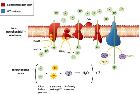
Figure 2 The ETC and the mechanism of OXPHOS in mitochondria. Through the transfer of electrons at the Complexes I-IV of the respiratory chain the reduction equivalents and the pumping of protons from the mitochondrial matrix the mitochondrial membrane potential is built up. This electrochemical gradient drives the ATP synthase that converts ADP and inorganic phosphate the cell’s most valuable energy carrier ATP.
The respiratory chain is the ‘’site of action’’ and through OXPHOS the final products of the tricarboxylic acid (TCA) cycle and glycolysis are metabolized. Via OXPHOS basically the metabolism of sugars is completed and the mitochondrion fulfills its major function in the cell: the energy supply in form of ATP. In the respiratory chain, the reduction equivalents NADH and FADH
2derived from the TCA cycle are oxidized through serial redox reactions.
Besides H
2O and CO
2,36 molecules of ATP are synthesized. The respiratory chain is built
up of four protein complexes and the ATP synthase located at the IMM. At Complex I
(NADH Ubiquinone-Oxidoreductase) two electrons are transferred from NADH/H
+to
ubiquinone. Two further electrons are transferred from FADH
2to ubiquinone at Complex
II, called the Succinate-Ubiquinone-Oxidoreductase. At the third Complex (Ubiquinone-
Cytochrome-c-Oxidoreductase) two molecules of Cytochrome c (Cyt c) are reduced by the
transfer of two electrons from ubiquinol. Finally, at Complex IV (Cyt c-Oxidase), Cyt c is
reoxidized under the reduction of O
2to H
2O. Besides the transport of electrons along the
reduction equivalents, protons are pumped from the matrix of the mitochondrion into the
IMS. Therefore, an electrochemical gradient is built up over the IMM. This proton motive
force drives the ATP synthase and enables the generation of ATP from ADP+P
i(Alberts et
al. 2002).
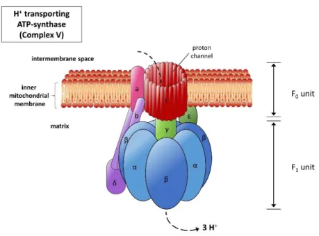
Figure 3 The ATP synthase. The ATP synthase is located at the IMM and consists of two subunits, which are in turn built up of several subunits. The hydrophobic F0 (consisting of a, b, c) is situated in the membrane and forms a proton channel: it converts the flux of the protons through the membrane into a rotation, with whose help the ATP synthase is driven. The hydrophilic F1-subunit (put together by the alpha, beta, gamma, delta and epsilon unit), sticks out of the IMM into the matrix, where from ADP und Pi ATP are synthesized. The OSCP-subunit also belongs to the F1-complex and plays an important role when it comes to stabilization of the F1-subunit against the rotation of the F0-subunit.
1.1.2 Mitochondria in the cellular metabolism
The mitochondrion’s key function, the OXPHOS, cannot be seen as a separate mechanism, independently passing off in the mitochondrion. Mitochondria are involved in a network of metabolic pathways and the products of these important metabolic pathways are transformed into energy and eventually, mitochondria are the organelles keeping the cell alive. Metabolites from lipid degradation, glycolysis and the TCA cycle convert in the mitochondrion.
Oxidative metabolism of both glucose and free fatty acids result in the generation of the
key metabolite Acetyl-CoA. During the oxidation of one molecule glucose into two
molecules pyruvate, net 2 ATP and 2 NADH are generated. Pyruvate is converted by
enzyme complex pyruvate dehydrogenase into two molecules of Acetyl-CoA, which is
trafficked from the cytosol into the mitochondrion. During β-oxidation fatty acids are
broken down in a four-step reaction: The β-carbon is oxidized to ketone followed by the
cleavage of the α- and the β-carbon which yields after all one molecule Acetyl-CoA and the
reduction equivalents NADH and FADH
2. In the TCA cycle, Acetyl-CoA is oxidized in an
eight steps process in the mitochondrial matrix outside the cristae. Each molecule of
Acetyl-CoA yields 3 molecules of NADH and one molecule of FADH
2. These reduced
nucleotide coenzymes subsequently transfer electrons to molecular oxygen via the ETC and ATP is generated from ADP and inorganic phosphate (Alberts et al. 2002).
Mitochondria are the main source of endogenously produced reactive oxygen species (ROS). ROS are a byproduct of OXPHOS which is produced by the ETC through the reduction of O
2to superoxide. In order to prevent oxidative stress by overwhelming amounts of ROS, cells harbor an antioxidative defense (Roma et al. 2017). The mitochondrial enzymes superoxide dismutase (SOD) and thioredoxin-2 (TRX2) scavenge free radicals and strive for an equilibrium in the cell. Another endogenous antioxidant system with mitochondrial involvement is the glutathione (GSH) peroxidase system (Tanaka et al. 2002).
Figure 4 Mitochondria in the cellular metabolism. Mitochondria are involved in crucial cellular functions including biosynthesis as well as degradation of metabolites, bioenergetics and energy supply and balance and signaling of ROS. The key processes – glycolysis, β-oxidation of fatty acids, the TCA cycle and one-carbon metabolism – in the cell and in the mitochondrion and their interplay are depicted.
However, ROS is necessary for the cell and its production is imperative for redox homeostasis. Balanced ROS levels are vital for the cell‘s homeostasis. ROS signalling is not only part of the immune system, but it also mediates cell proliferation and apoptotic pathways to ensure proper regulation of the cell cycle and programmed cell death (Roma et al. 2017). Ca
2+homeostasis is the second key player in cell survival and apoptosis. Ca
2+as second messenger molecule mediates intracellular signalling cascades as well as cell-
cell-signalling (Clapham 2007). Mitochondria regulate cytosolic Ca
2+levels and consequently Ca
2+-mediated signalling. Mitochondrial Ca
2+levels are crucial for the rate of OXPHOS. Uncoupling proteins 1, 2 and 3 (UCP1, UCP2, UCP3) are embedded in the IMM and belong to the superfamily of mitochondrial ion transporters (Ricquier and Bouillaud 2000) . UCP1 accounts for heat production by inducing a H
+leak that uncouples OXPHOS, whereas UCP2 and UCP3 contribute to many cellular processes including mitochondrial free-radical production, apoptosis, hormone secretion and they are implicated in glucose and fatty acid metabolism (Dejean et al. 2004; Harper et al. 2002; Krauss et al. 2003;
Mookerjee et al. 2010). Most importantly, UCP2 and UCP3 were shown to be fundamental for mitochondrial Ca
2+transport (Graier, Trenker, and Malli 2014).
Besides those transport systems, Ca
2+signalling and buffering is accomplished by interfaces of the mitochondria and the endoplasmic reticulum (ER) and are entitled mitochondria-associated ER membranes (MAMs). Proteins in MAM either are involved in physical connection between ER and mitochondria or modify the tethering complexes in MAMs. Mitofusin 1/2 (Mfn1/2), a mitochondrial fusion GTPase, which situated to the OMM, is part of the MAM complex. MFN1/2 plays a role in mitochondrial fusion and together with OPA1, another mitochondrial fusion GTPase, which located at the IMM, regulates the merging of mitochondria (De Brito and Scorrano 2008; Cipolat et al. 2004;
Detmer and Chan 2007).
1.1.3 Fission and fusion: a dynamic network
Mitochondria are highly dynamic organelles and change their shapes and distribution constantly. Mitochondria’s shapes range from small vehicles, short rods and reticular networks spanning the entire cell. Their rapid changes in shape and location, allow mitochondria a fast adaption to energetic needs and they are essential to mitochondrial health, wherefore damaged organelles or precipitates are restored and removed.
Mitochondrial fission and fusion events are balanced and have been identified as a critical process in mitochondrial morphology and function. As mentioned beforehand, and most importantly, fission and fusion control the shape length and number of mitochondria.
Additionally, the shape of mitochondria affects the ability of cells to distribute their
mitochondria to specific subcellular locations. By continuous merger and division, the
exchange of lipid membranes and intra-mitochondrial content is ensured (Charmandari,
Tsigos, and Chrousos 2005; Gold et al. 2002; Kyrou, Chrousos, and Tsigos 2006; Detmer
and Chan 2007). Defects in fission and fusion come with severe diseases. Dysregulation of those two processes are associated with an energetic defect, giving the hint that mitochondrial structure and function are tightly related (Bertholet et al. 2016; Lee and Yoon 2016). Mfn1 and Mfn2 are responsible for OMM fusion, whereas OPA1 regulates the fusion of the IMM. Fission events in mammals are mediated by the dynamin-like protein 1 (drp1), which is predominantly a cytosolic protein (Meeusen et al. 2006; Yonashiro et al. 2009). Fibroblasts that lack both, Mfn1 and Mfn2, show reduced respiratory capacity and individual mitochondria show great heterogeneity in shape and membrane potential.
Cells that lack OPA1 show similar defects, with an even greater reduction in respiratory capacity (Chen, Chomyn, and Chan 2005).
Figure 5 Fission and Fusion events. (a) Wild-type cells with intact mitochondrial dynamics. Functioning mitochondria are shown in green, whereas mitochondria with defects and lack of mtDNA are shown in orange. By fusion events the deficiencies in non-functional mitochondria can regain its function and mtDNA by fusing with a neighbouring mitochondrion. The fused mitochondrion then undergoes fission, with both daughter mitochondria receiving mtDNA nucleoids. (b) Cells with fusion-deficient properties. The mitochondria are fragmented since fission events are still happening. Cells lack mtDNA nucleoids accumulate because there is no pathway for defective mitochondria to regain mtDNA. Fusion-deficient cells can maintain mtDNA nucleoids, but such nucleoids serve a much smaller mitochondrial mass.
Hence, mitochondrial dynamics are extremely important for mitochondrial function.
Mitochondria should not be seen as separate organelles but as a population of organelles
with heterogeneous functions. Information and contents are being exchanged and
deficient mitochondria can be rescued by others. A few mitochondria might be non-
functional owing to the loss of essential components. However, this dysfunction is
transient, since mitochondrial fusion provides a pathway for these defective
mitochondria to regain essential components (figure 5). An essential prerequisite for proper mitochondrial function is mitochondrial DNA (mtDNA) which is organized in nucleoids. The mtDNA genome encodes important subunits of the respiratory Complexes I, III and IV, and is therefore indispensable for OXPHOS. When mitochondrial fusion is abolished, a large fraction of the mitochondrial population loses mtDNA nucleoids (Chen, McCaffery, and Chan 2007). When mitochondria divide, most daughter cells inherit at least one mtDNA nucleoid. If this inheritance fails, fusion events compensate for that and the mtDNA is restored in the daughter cell. Since in cells with disturbed mitochondrial dynamics, the exchange of contents does not take place, the restoration of mtDNA nucleoids probably accounts for the heterogeneity in mitochondrial MMP and the reduced respiratory capacity. Cells with a lack of fusion capability, however, still harbour a significant number of mtDNA nucleoids. Due to fission events, the functional mitochondrial mass gets reduced. Furthermore, not only the mtDNA gets restored by mitochondrial fusion, but also other components like substrates, metabolites and specific lipids can be recovered in disturbed mitochondria (Detmer and Chan 2007). Concluding, the mitochondrial mass is strongly influenced by mitochondrial dynamics and might reflect the cell’s health or disease state. The cellular mtDNA content is regulated by the nuclear DNA that encodes for in mtDNA replication, transcription, translation and repair.
Molecular defects in the genes responsible for mtDNA biogenesis and therefore a reduction of mtDNA content. It is known that mtDNA depletion and a lack of the maintenance of mtDNA integrity can lead to a series of disorders. Those diseases include, for instance, Leigh-like encephalopathy with dystonia, deafness and lactic acidosis combined with encephalomyopathy, but also mitochondrial hepatoencephalopathy with hepatic and neurologic symptoms during infancy (Dimmock et al. 2008; Ostergaard et al.
2007; Sarzi et al. 2007; Wong et al. 2008).
Studies also reported alterations of the mitochondrial mass in mood disorders. MtDNA
copy number is reduced in leucocytes of Schizophrenia (SZ) and Bipolar Disorder (BPD)
patients and could be correlated to the severity of disease and the anti-psychotic
treatment. They suggested a link between mitochondrial dysfunction and psychosis-like
symptoms (Kumar et al. 2018). Decreased number of mtDNA in leucocytes was revealed
for MDD (Kim et al. 2013). Decreased mtDNA copy numbers were also discovered with
increasing duration of the Posttraumatic Stress Disorder (PTSD) symptoms, whereas the
copy number is highest in the initial phase of PTSD (Bersani et al. 2016). Moreover, for
patients with MDD, anxiety disorder or adjustment-disorder an increased mtDNA copy
numbers in peripheral blood is reported and it is positively correlated to the severity of disease (Karabatsiakis et al. 2014). It might be assumed that higher mtDNA copy numbers compensate for dysfunctional mitochondrial metabolism and the resulting energetic deficiencies (Wang et al. 2017).
1.2 Major Depressive Disorder – a multifactorial disease 1.2.1 Classification and diagnostics
Sadness, emptiness, hopelessness, a loss of interest and pleasure – MDD has many faces.
The severe disease is of high heterogeneity and multifaceted in the clinical picture.
Symptoms include changes in mood, cognition, interest, but also troubles in decision- making, anxiety, anhedonia, problems in volition and motivation and changes with respect to psycho-motility and energy. Furthermore, appetite and therefore weight and the circadian rhythm can be affected. According to Diagnostic and Statistical Manual of Mental Disorders V (DSM V) classification two out of three main symptoms - a depressed mood, anhedonia and the lack of drive and some of the side symptoms, respectively somatic symptoms include troubles in cognition, reduced self-esteem, feelings of guilt and/or hopelessness, changes in appetite, sleep disturbances and the expression of meaninglessness of life – so suicidal ideation – must be present for at least 2 weeks nearly every day. MDD is diagnosed based on physician-administered or patient self- administered interview and is still subjective since it depends on the individual clinical judgment (Bilello 2016; Young et al. 2016). MDD state is evaluated through a questionnaire that determines the severity of depression. The Hamilton Depression Scale (HAM-D) ranges from 0 (no depression) to 10-20 scores (mild depression) to 21-30 scores (medium severe depression). More than 30 points are considered as a severe MDD.
Classification according to ICD-10 Classification of Mental and Behavioral Disorders.
Clinical Descriptions and Diagnostic guidelines (ICD-10) provides the following categories:
- F32.0: Mild depressive episode: Two or three of the symptoms are present. The subject is generally impaired and is seeking for help, however he/she is still able to fulfill occupational and private routine.
- F32.1: Medium severe depressed episode: Usually four or more of the symptoms
are present. The subject has severe troubles continuing daily routine.
- F32.2: Severe depressed episode without psychotic symptoms: Several of the symptoms are present to a torturous extent. Subjects typically exhibit a loss of self- esteem and worthlessness and affected persons utter suicidal thoughts and actions. Persons additionally suffer from somatic symptoms, e.g. agitation, sleep disturbances or loss of appetite or libido.
- F32.3: Severe depressed episode with psychotic symptoms: State of disease is described as F32.2, but additionally subjects suffer from hallucinations and delusional ideas, psychomotor inhibitions or stupor, so that daily social activities are impossible and persons are in danger of life due to suicide and a lack of imbibition and ingestion.
- 32.8: Other depressive episode.
- 32.9: Depressed episode, not specified.
Figure 6 Symptoms of MDD and its increasing impact on daily performance. A combination of symptoms concerning emotional status, vegetative and cognitive function must be present over a period of 2 weeks. Increasing number of a combination of manifestations causes cumulative functional impairment. Main symptoms for depression are marked with a red dot. The color green indicates emotional symptoms, blue color stands for neurovegetative symptoms and orange depicts neurocognitive symptoms.
MDD has a severe impact on the affected persons. It is a disease with tremendous societal,
economic and emotional burden and results in an impairment in social or occupational
functioning and therefore impairs daily living (Kessler and Bromet 2013). MDD is
considered as a global burden with 1 to 17% of affected people in adults, 3 to 6% in adolescents, and 12 to 38% in elderly (Gururajan et al. 2016; Kessler and Bromet 2013;
Rantamaki and Yalcin 2016). MDD is one of the leading causes of mental disability worldwide, with an estimated lifetime prevalence of up to 20–30% (Weissman et al.
1996). Moreover, women are affected more frequently than men (Kessler and Bromet 2013). MDD is highly comorbid with somatic diseases like diabetes, asthma and arthritis, but also with neurodegenerative disorders like Alzheimer’s disease (AD), Parkinson’s diseases (PD) and Huntington’s disease (HD) (Kupfer, Frank, and Phillips 2012; Réus et al. 2016).
MDD is a multifactorial disease including, genetic and epigenetic factors, the exposure to stress or traumatic events and the interaction with other psychological or environmental factors and even personality itself plays a role in the manifestation of MDD (Rantamaki and Yalcin 2016; Schneider and Prvulovic 2013). It is assumed, that for MDD patients neuronal circuits are affected and neuronal connectivity is altered, leading to functional neuroanatomical modifications (Rantamaki and Yalcin 2016). From a molecular point of view, neurobiological, but also neuroendocrine, neurotrophins as well as oxidative stress and inflammatory processes contribute to the manifestation of MDD and several theories for the development of the disease merged the last decades.
1.2.2 Early and new theories for manifestation
The oldest hypothesis that is postulated with regard to the manifestation of MDD is the
monoaminergic theory of depression. It claims that the underlying pathophysiologic basis
for the manifestation of MDD is a lack of monoamines like serotonin, norepinephrine
and/or dopamine in the synaptic cleft. Tricyclic antidepressants (TCAs) and selective
serotonin reuptake inhibitors (SSRIs) are the most common treatments for the MDD
(Bartl et al. 2014; Eisenhofer, Kopin, and Goldstein 2004). These medications elevate the
level of neurotransmitters in the brain by either prolonging their presence in the synaptic
cleft or blocking their presynaptic auto-receptors. Depressive symptoms are effectively
reversed by monoamine oxidase inhibitors (MAOIs) and TCAs, however with a delayed
onset. Drugs acting on the monoaminergic system usually need at least 2 to 8 weeks
before a therapeutic effect can be observed and each drug is only efficient in around two
third of MDD patients (Sonnenberg et al. 2008). Besides that, the observed discrepancy
between pharmacological and biochemical function of ADs and their clinical mood altering responses remains unclear (Leonard 2007).
Therefore, additional neurotransmitter systems came into play and newer theories postulate that monoamine deficiency cannot be the sole underlying reason for the development of depression. The neuroplasticity theory of depression includes other transmitters like Glutamate and γ–butyric acid (GABA), but also the Brain-Derived- Neurotrophic factor (BDNF). The glutamatergic system may be also implicated in MDD (Hashimoto 2009). Fast-acting drugs like Ketamine, which is an antagonist of the NMDA- receptor, revealed antidepressant effects (Dutta, McKie, and Deakin 2015). Moreover, it is likely that the glutamatergic system is involved in the manifestation of MDD, since increased glucocorticoid levels, which are released under conditions of chronic stress that associated with depression, lead to enhanced glutamatergic transmission, elevated NMDA receptor expression and increased extracellular glutamate levels (Lu et al. 2003).
Glutamate and GABA are both involved in synaptic plasticity and various studies implicated GABA in the pathophysiology of depression (Krystal, Sanacora, and Duman 2013; Luscher, Shen, and Sahir 2011).
Synaptic plasticity is an important process in the brain including synaptogenesis, alterations in dendritic function, neurite extension, synaptic remodelling and modification of synaptic neurotransmissions. ADs treatment can – at least partly - reverse this disturbed plasticity which contributes to a pathological state (Pittenger and Duman 2008; Wood et al. 2004). The modulation of receptors for serotonin and noradrenaline through ADs treatment, different downstream pathways get activated that have a common function which is the regulation of gene expression, e.g. BDNF, which is crucial for the formation of neuronal networks (Yu and Chen 2011).
Numerous studies also suggest that inflammation may contribute to symptoms relevant
to a number of psychiatric disorders and particularly depression. Inflammation and
depression are linked and there is strong evidence that one goads the other, in the sense
that inflammatory responses can lead to depression and depression can lead to
inflammation. It is known that patients with inflammatory diseases are more likely to
show greater rates of MDD. Moreover, patients with high inflammation have been shown
to react poorly to conventional ADs therapies (Felger 2018). A large number of people
with MDD show elevated peripheral inflammatory biomarkers, even in the absence of a
medical illness. Several studies have reported increased circulating inflammatory
cytokines, such as interleukin (IL)-1, IL-6, and tumor necrosis factor (TNF), their soluble
receptors, and acute phase reactants, like C-reactive protein (CRP), in patients with MDD (Maes 1999; Maes et al. 1992; Sluzewska 1999). And coming back to neurogenesis and alterations in brain signaling pattern, inflammatory mediators have been found to alter glutamate and monoamine neurotransmission as well as glucocorticoid receptor (GR) resistance (Amodeo, Allegra Trusso, and Fagiolini 2018).
A dysfunction of GRs, elevated levels of glucocorticoid hormones and the accompanied disturbed feedback mechanism of the hypothalamus-pituitary-adrenal (HPA) axis is strongly associated with MDD (Vreeburg et al. 2009). ADs act on the upregulation of the HPA axis and moderate the hyperactivity by ameliorating many of the neurobiological disturbances in depression and relief depressive symptoms (Anacker et al. 2001).
1.3 The risk factor stress - impact on mitochondria and mood 1.3.1 The stress response in the human body
The human stress response is a homeostatic mechanism that provides a better chance of survival when the body is under threat. It mobilizes neural and hormonal networks to optimize cognitive, cardiovascular, immunological and metabolic function.
The HPA axis is the key pathway in the human stress response and reacts immediately to a real or perceived stressor. It is the central mediator of the ‘’fight or flight’’ reaction.
Figure 7 The Hypothalamic-Pituitary-Adrenal Axis. An internal or external stressor induced the releases of CRH from the hypothalamus which causes a release of ACTH from the pituitary gland. ACTH acts on the adrenal glands and elevated levels of Cortisol follows. Cortisol action is regulated by a negative feedback mechanism on the hypothalamus. Source: https://images.agoramedia.com/everydayhealth/gcms/What-Is-Cortisol-722x406.jpg
The HPA axis is a complex combination of organs and transmitters with a feedback mechanism. Internal, e.g. the circadian rhythm, and external signals, like stress, trigger the hypothalamic release of corticotropin releasing hormone (CRH), which acts on the anterior pituitary, which in turn stimulates the synthesis and secretion of adrenocorticotropic hormone (ACTH) in the pituitary gland. ACTH then acts on the adrenal cortex to stimulate the production and secretion of Cortisol, a glucocorticoid (GC).
The GCs that are released, mediate through GRs which are expressed throughout the entire body and trigger negative feedback mechanism by also targeting the hypothalamus and anterior pituitary. The production and release of CRH and ACTH are inhibited and thereby limit both, the intensity and duration, of the GC increase (Oakley and Cidlowski 2013). Hence, the stress response is crucial for our homeostasis and survival and comes along with beneficial adaptations on a cellular and molecular level.
1.3.2 Benefits of acute stress
Not just the release of CRH, ACTH and GCs are typical for the stress response, but also the release of catecholamines, adrenaline and noradrenaline, as well as pro-inflammatory factors TNF-α, IL-1 and IL-6. Altogether, the secreted substances ensure the fast response that includes effective blood supply in the brain, the cardiac muscle and the skeletal muscle. Furthermore, the energy production is enhanced by recruiting substrates like glucose, fatty acids and amino acids from storages. Besides that, the optimal ATP availability to vital tissues is orchestrated (Charmandari, Tsigos, and Chrousos 2005).
Mitochondria are the pivotal organelle when it comes to biosynthetic activities and the
supply of energy. Together with their involvement in Ca
2+metabolism, signaling, the
regulation of thermogenesis and the generation of ROS, as well as the decision makers
over cell survival or apoptosis, they are the first organelles to react to stressors
(Klinedinst and Regenold 2014). Oxygen consumption and total energy expenditure are
increased during the initial phase of the acute stress. Several signalling pathways are
activated to meet energy demands during stress situations. Firstly, a higher number of
mitochondria are recruited and they increase their volume. The expression and the
activity of OXPHOS units are enhanced and the uncoupling of the respiratory chain and
consequently energy in the form of heat is released. Besides that, the ROS level is
regulated which is in turn important for signalling and defence and finally, an apoptotic
cascade is triggered, depending on the nature of the stressor (Goldenthal and Marin- Garcia 2004).
Mitochondrial function is affected and enhanced through GR action. GRs are expressed in mitochondria in several cell types (Demonacos et al. 1995). There is evidence that short- term exposure to stress and the concentration of GCs is associated with induction of mitochondrial biogenesis and enzymatic activity of the ETC units. For instance, by acute exposure of skeletal muscles to the synthetic GC dexamethasone, the transcription primarily of nuclear genes, but also mtDNA-encoded genes, affecting mitochondrial function and biogenesis, are initiated (Mikes et al. 2002). Mitochondrial biogenesis and OXPHOS and consequently adaptive thermogenesis in adipose tissue as well as in the skeletal muscle are stimulated by the catecholamines that are released. In brown adipose tissue energy saved from OXPHOS of substrates is dissipated as heat instead of the storage in form of ATP: this process is denominated as ‘uncoupling’. The process of uncoupling is beneficial for the cell, as it attenuates mitochondrial ROS production and protects against cellular damage (Brand and Esteves 2005). Another signalling pathway that is activated during acute stress is the peroxisome proliferator-activated receptor-coactivator 1-α (PGC-1α) signalling pathway. Cytokines, such as TNF-a, IL-1, which are immediately released, trigger the transcriptional activity of PGC 1-α via direct phosphorylation and activation the mitogen-activated protein kinase (MAPK) pathway. This results in stabilization and activation of PGC-1α protein, which is the master regulator of mitochondrial biogenesis. PGC 1-α switches on gene expression that drives mitochondrial biogenesis and OXPHOS, again directing the metabolism towards mitochondrial uncoupling and energy expenditure (Michael et al. 2001; Wu et al. 2006).
Taken together, the acute effects of stress on the human body and the downstream effects are beneficial and necessary for the reaction to a stressor in general. But what happens when the perceived or actual stressor is persistent and the human body is steadily exposed to stress?
1.3.3 Sustained stress and its deleterious consequences
Chronic exposure to stress results in reversal of the beneficial effects. The long-term
cortisol exposure becomes maladaptive, which can lead to a broad range of problems
including the metabolic syndrome, obesity, cancer, mental health disorders,
cardiovascular disease and increased susceptibility to infections (Björntorp and Rosmond
2000; Pufall 2015a; Steckler, Holsboer, and Reul 1999; Strüber, Strüber, and Roth 2014a).
However, starting with the initial process leading to these serious diseases: the activation of the HPA axis and the release of GCs, adrenaline and cytokines. Prolonged exposure to GCs causes respiratory chain dysfunction, increased ROS generation, mitochondrial structural abnormalities, apoptosis and cell death. Even if the release of GC is crucial and necessary in a stressed state, an overload of cortisol – regardless if naturally secreted or synthetic GCs administered through pharmacological treatments – is associated with various serious diseases (Charmandari, Tsigos, and Chrousos 2005; Chrousos 2000; Gold et al. 2002; Kyrou, Chrousos, and Tsigos 2006). The beneficial PCG1-α signalling, which is necessary for mitochondrial biogenesis, can become maladaptive and is detrimental to the cell in response to persistent stressors. PGC-1α overexpression for instance, caused hepatic insulin resistance, manifested by higher glucose production and diminished insulin suppression of gluconeogenesis (Liang et al. 2009). Another study was able to show, also in a murine model, that the overexpression of PGC1-α leads to cardiomyopathy (Russell et al. 2004). Abnormally increased mitochondrial biogenesis leads to an elevated level of ROS. Increased oxidative damage in form of elevated levels of stress markers and genetic modification, namely telomere shortening, was revealed in children of mother with chronical psychological stress (Epel et al. 2004). In this context, especially the NF-κβ pathway is highly sensitive to changes in the intracellular redox environment and thus can be activated by oxidative stress. Mitochondrial gene expression can be negatively regulated in response to cellular TNF-α stimulation (Cogswell et al. 2003). Moreover, exceeded TNF-α release induces apoptosis in several cell types. TNF-α mediated apoptosis requires binding of the cytokine to its receptor TNFR1. This triggers a downstream activation of caspase 8 increasing Cyt c release which is followed by a loss of the MMP and the induction of apoptosis (Micheau and Tschopp 2003). Moreover, mtDNA is sensitive to oxidative damage. Mutations in the mtDNA can lead to altered transcription and expression of the ETC enzymes, which causes pathologic changes of mitochondrial function (Shokolenko et al. 2009).
It is known that chronic stress alters ingestion behavior. Nutrient overload and a
carbohydrate- and lipid-rich diet leads to an excess of GC and is associated with abdominal
obesity. It is very likely that overweight is accompanied by the development of diseases
affecting metabolism, e.g. metabolic syndrome, type-2 diabetes and cardiovascular
disease (Krempler et al. 2002; Patti et al. 2003). Disruptions of the OXPHOS and mtDNA
abnormalities cause changes in PGC 1-α expression and UCP2. PGC 1- α and UCP2 play a
crucial role in proton leak and thermogenesis and they are linked diabetes and the metabolic syndrome (Brand and Esteves 2005).
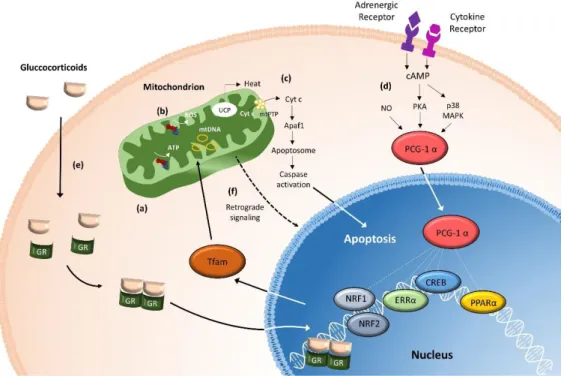
Figure 8 Mitochondrial functions in the stress response. This figure summarizes important mitochondrial functions including (a) the energy production through OXPHOS, (b) the generation of ROS and (c) the induction of apoptosis by opening of the mitochondrial permeability transition pore (mtPTP). By the release of Cyt c and the activation of Apaf1, the Apoptosome is triggered downstream and caspase activation follows. Alongside, the mitochondrion’s most important signaling pathways are shown. (d) By adrenergic receptor activation, cAMP and PKA are activated.
By cytokine receptor activation, the p38 MAPK pathway is triggered, which in results an activation of the major upstream regulator of mitochondrial function-related gene expression, the PGC-1α. Exposure to stressors, such as cold temperature, fasting, exercise, cachexia or chronic infection activates PGC-1α. (e) The release of GC during the stress response nuclear and mitochondrial genes regulating mitochondrial biogenesis and function are activated and the GRs can enter the mitochondrion and regulate mtDNA transcription. (f) A ‘mitochondria-specific stress response’ takes place by a retrograde signalling from the mitochondrion to the nucleus.