Regulation of gene silencing:
From microRNA biogenesis to post-translational modifications of TNRC6 complexes
DISSERTATION
zur Erlangung des
DOKTORGRADES DER NATURWISSENSCHAFTEN (Dr. rer. nat.) der Fakultät Biologie und Vorklinische Medizin
der Universität Regensburg
vorgelegt von Johannes Danner
aus Eggenfelden
im Jahr 2017
Das Promotionsgesuch wurde eingereicht am: 12.09.2017 Die Arbeit wurde angeleitet von:
Prof. Dr. Gunter Meister
Johannes Danner
Summary
‘From microRNA biogenesis to post-translational modifications of TNRC6 complexes’ summarizes the two main projects, beginning with the influence of specific RNA binding proteins on miRNA biogenesis processes. The fate of the mature miRNA is determined by the incorporation into Argonaute proteins followed by a complex formation with TNRC6 proteins as core molecules of gene silencing complexes.
miRNAs are transcribed as stem-loop structured primary transcripts (pri-miRNA) by Pol II. The further nuclear processing is carried out by the microprocessor complex containing the RNase III enzyme Drosha, which cleaves the pri-miRNA to precursor-miRNA (pre-miRNA). After Exportin-5 mediated transport of the pre-miRNA to the cytoplasm, the RNase III enzyme Dicer cleaves off the terminal loop resulting in a 21-24 nt long double-stranded RNA. One of the strands is incorporated in the RNA-induced silencing complex (RISC), where it directly interacts with a member of the Argonaute protein family. The miRNA guides the mature RISC complex to partially complementary target sites on mRNAs leading to gene silencing. During this process TNRC6 proteins interact with Argonaute and recruit additional factors to mediate translational repression and target mRNA destabilization through deadenylation and decapping leading to mRNA decay.
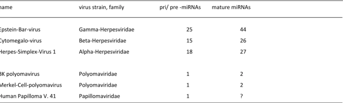
Viral miRNA Biogenesis. Surprisingly, miRNAs were identified in human herpes, papilloma and polyoma viruses. These miRNAs regulate viral and host gene expression and influence infection efficiency. The miRNA biogenesis is strictly regulated and by northern blotting different expression profiles of infected cell lines were detected.
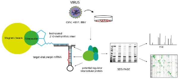
To identify RNA-binding-proteins involved in post-transcriptional regulation of miRNA biogenesis, a mass spectrometric pull down assay with in-vitro transcribed pre-miRNA was established. The obtained data generated together with bioinformatical analyses a valuable set of potential regulatory candidates. The interaction of a subset of potential regulators was verified by repeating the pull-down with overexpressed Flag-/Ha-tagged proteins. For further functional characterization, the influence of RBPs on pre-miRNA processing was analyzed in knockout cell lines in which candidate RBPs have been depleted. Overexpression of the potential candidates further confirms a strong impact on the miRNA biogenesis.
Taken together, mass spectrometric approaches identified RNA-binding-Proteins involved in viral miRNA biogenesis.
Post-translational modifications of TNRC6 proteins. TNRC6 and Ago proteins play a central role
in the gene silencing mechanism. The Interaction of both proteins is based on two Tryptophan’s
components of the gene silencing machinery.
To assess whether gene silencing is regulated by post-translational modifications, TNRC6 proteins were analyzed by mass spectrometry. To analyze endogenous proteins, we established monoclonal antibodies against TNRC6A-C for immunopurificaiton of TNRC6 proteins from cell lysates. The validity and specificity of the antibodies was further verified by mass spectrometric selected reaction monitoring analyses. Followed by a detailed mass spectrometric analysis, multiple endogenous phosphorylation sites on TNRC6 proteins were detected. The obtained data identified conserved phosphorylation sites both among the TNRC6 paralogs and within different species. Functional analyses of phospho-mimicking and non-phospho mutants showed low effects on the downstream gene silencing processes. Localization studies and Ago-binding assays also indicate no effects of the phospho-sites on TNRC6 function.
Taken together, post-translational modifications on TNRC6 proteins with potential, but so far
unknown function in gene silencing were identified.
Zusammmenfassung
"From microRNA biogenesis to post-translational modifications of TNRC6 complexes" fasst die beiden Hauptprojekte dieser Doktorarbeit zusammen.
miRNAs werden als primäre transkripte (pri-miRNA) von der RNA Polymerase II transkribiert. Die weitere Verarbeitung erfolgt durch den Mikroprozessor-Komplex, der das katalytisch aktive Enzym Drosha enthält, welches die pri-miRNA zu Vorläufer-miRNAs (pre-miRNA) spaltet. Nach dem Exportin-5-vermittelten Transport der pre-miRNA in das Zytoplasma, spaltet das RNase III- Enzym Dicer die terminale Schleife der pre-miRNA ab, was zu einer 21-24 nt langen doppelsträngigen RNA führt. Einer der beiden Stränge wird in den RNA-induzierten Silencing- Komplex (RISC) eingebaut, wo er direkt mit einem Mitglied der Argonaute-Proteinfamilie wechselwirkt. Der reife RISC-Komplex bildet durch komplementäre Basenpaarung der miRNA zur mRNA den Gene-silencing Komplexe. Während dieses Prozesses interagiert ein TNRC6-Protein mit Argonaut und durch Rekrutierung von zusätzlichen Faktoren wird die Translation reprimiert und die Ziel-mRNA destabilisiert und abgebaut.
Virale miRNA Biogenese. Überraschenderweise wurden miRNAs bei humanen Herpes-, Papillom- und Polyomaviren identifiziert. Diese miRNAs regulieren die Virus- und Wirtsgenexpression und beeinflussen den viralen Lebenszyklus.
Die miRNA-Biogenese ist streng reguliert und durch Nothern Blotting wurden verschiedene miRNA Expressionsprofile von infizierten Zelllinien nachgewiesen. Zur Identifizierung von RNA-bindenden Proteinen, die an der post-transkriptionelen Regulation der miRNA-Biogenese beteiligt sind, wurde eine massen-spektrometrische Pull-Down-Anwendung mit in vitro transkribierter Pre- miRNA etabliert. Die gewonnenen Daten, die zusammen mit bioinformatischen Analysen erzeugt wurden, sind ein wertvoller Datensatz von potenziellen regulatorischen Proteinen. Die Wechselwirkung einer Teilmenge von potentiellen Regulatoren wurde durch Wiederholen des Pull-downs mit überexprimierten Flag-/ Ha-markierten Proteinen verifiziert. Für eine weitere funktionelle Charakterisierung wurde der Einfluss von RNA-bindenden Proteinen (RBP) auf die pre-miRNA-Verarbeitung in Knockout-Zelllinien analysiert.
Zusammenfasst, wurden in massenspektrometrischen Analysen RNA-bindende Proteine identifiziert, die an der viralen miRNA-Biogenese beteiligt waren.
Posttranslationale Modifikationen von TNRC6-Proteinen. TNRC6- und Ago-Proteine spielen eine
zentrale Rolle im Gen-Silencing-Mechanismus. Die Interaktion beider Proteine basiert auf zwei
Aminosäureabschnitte und dienen als Bindeplattform für viele Komponenten der Gen-Silencing- Maschinerie.
Um zu beurteilen, ob Gen-Silencing durch posttranslationale Modifikationen reguliert wird, wurden TNRC6-Proteine durch Massenspektrometrie analysiert. Um endogene Proteine zu analysieren, wurden monoklonale Antikörper gegen TNRC6A-C für Immuno-Aufreinigungen von TNRC6-Proteinen aus Zelllysaten etabliert. Die Gültigkeit und Spezifität der Antikörper wurde durch massenspektrometrische ausgewählte Analysen weiter verifiziert. Nach einer detaillierten Analyse wurden mehrere endogene Phosphorylierungsstellen in TNRC6-Proteinen nachgewiesen.
Die erhaltenen Daten identifizierten konservierte Phosphorylierungsstellen sowohl unter den humanen TNRC6-Paralogen als auch innerhalb verschiedener TNRC6 proteine anderer Tiere.
Funktionsanalysen von Phospho-Mimik- und Nicht-Phosphorylierbaren-Mutanten zeigten geringe Auswirkungen auf die nachgeschalteten Gen-Silencing-Prozesse. Lokalisierungsstudien und Ago- Bindungsversuche zeigen auch keine Wirkungen der phosphorylierten Aminosäuren auf die TNRC6-Funktion an.
Zusammengefasst wurden posttranslationale Modifikationen an TNRC6-Proteinen identifiziert
und charakterisiert.
Publications
Schraivogel D., Schindler S.G., Danner J., Kremmer E., Pfaff J., Hannus S., Depping R. & Meister G.
Importin-β facilitates nuclear import of human GW proteins and balances cytoplasmic gene silencing protein levels. Nucleic Acids Res. 2015
Johannes Danner, Balagopal Pai, Ludwig Wankerl and Gunter Meister. Peptide-Based Inhibition of miRNA-Guided Gene Silencing. Methods Mol. Biol. 2017
Miguel Quévillon Huberdeau, Daniela M. Zeitler, Judith Hauptmann, Astrid Bruckmann, Lucile Fressigné, Johannes Danner, Sandra Piquet, Nicholas Strieder, Julia C. Engelmann, Guillaume Jannot, Rainer Deutzmann, Martin J. Simard and Gunter Meister. Phosphorylation of Argonaute proteins affects mRNA binding and is essential for microRNA-guided gene silencing in vivo.
EMBO Journal 2017
Johannes Danner, Thomas Treiber, Nora Treiber, Emma Kraus, Eduard Hochmuth, Astrid Bruckmann, Christina Paulus, Michael Nevels, Hans-Helmut Niller, Adam Grundhoff and Gunter Meister.
Seduction of viral miRNA biogenesis during viral life cycle Manuscript in preparation
Mariangela Morlando, Sama Shamloo, Johannes Danner, Astrid Bruckmann, Gunter Meister and Irene Bozzoni.
The Interplay between lnc31, pre-lnc31 and YBox1 in differentiating muscle cells (working title)
Manuscript in preparation
Presentations and Posters
Parts of this thesis were presented at the following meetings/ conferences
Symposium - From functional Genomics to Systems Biology 2014 in Munich with Poster presentation
Exploiting peptide and antibody purification strategies to analyze post-translational modification of endogenous Argonaute and GW182
Biosysnet Group member meeting 2014 in Munich with Talk
Regulation of microRNA biogenesis on human latent EBV and lytic CMV Biosysnet Retreat 2015 in Wildbad Kreuth with Poster presentation
Dissection of herpesviral microRNA biogenesis Microymposium 2015 in Vienna with Poster presentation
Exploiting peptide and antibody purification strategies to analyze post-translational modification of endogenous Argonaute and TNRC6 proteins
Microymposium 2016 in Vienna with Poster presentation
Post-translational modifications of endogenous Argonaute and TNRC6 proteins RNA Society meeting 2017 in Prague with Poster presentation
Hyper-phosphorylation of an unstructured loop of Argonaute proteins triggers
dissociation from mRNAs
Summary
Zusammmenfassung Publications
Presentations Contents
1 INTRODUCTION ... 1
1.1 MAMMALIAN MICRORNA BIOGENESIS ... 2
1.1.1 Processing of primary miRNAs by the microprocessor complex ... 2
1.1.2 Dicer cleavage of pre-miRNAs and RISC loading ... 6
1.2 GENE SILENCING AND TRANSLATIONAL REPRESSION ... 10
1.2.1 Interplay of TNRC6 and Ago... 10
1.2.2 Canonical post-transcriptional gene silencing ... 17
1.2.3 Translational repression and other ways of mRNA decay ... 19
1.2.4 Regulation of miRNA mediated gene silencing by post-translational modifications and interacting modifying enzymes ... 20
1.3 RNA BINDING PROTEINS ... 24
1.4 MIRNA CONTAINING VIRUSES ... 26
1.4.1 Viral life cycle ... 26
1.4.2 Function of miRNAs in human herpes viruses ... 27
1.4.3 Polyoma and Papilloma viruses ... 29
2 RESULTS ... 31
2.1 PART I:IDENTIFICATION OF RBPS THAT REGULATE THE VIRAL MIRNA BIOGENESIS... 32
2.1.1 Aims of part I ... 32
2.1.2 Viral miRNA expression profile of EBV, CMV and HSV1 ... 32
2.1.3 Identification of pre-miRNA binding proteins by mass spectrometric approaches ... 35
2.1.4 pri-miRNAs sequence alignments with RBP consensus motifs ... 41
2.1.5 Validation of specific pre-miRNA-RBP interactions ... 43
2.1.6 Influence of RBP candidates on viral miRNA processing ... 44
2.2 PART II:POST-TRANSLATIONAL MODIFICATIONS OF TNRC6 PROTEINS ... 48
2.2.1 Aims of part II ... 48
2.2.2 Purification and characterization of TNRC6 containing complexes ... 48
2.2.3 Phosphorylation of mammalian TNRC6 proteins ... 58
2.2.4 Characterization of TNRC6 phospho-mutants ... 65
3 DISCUSSION ... 69
3.1 PART I:DISSECTION OF VIRAL MIRNA BIOGENESIS ... 70
3.1.3 Validation and influence of specific pre-miRNA protein interactions ... 73
3.1.4 Future perspectives and a model for the viral miRNA biogenesis ... 74
3.2 PART II:POST-TRANSLATIONAL MODIFICATIONS OF TNRC6 PROTEINS ... 76
3.2.1 Immunopurification and enrichment of TNRC6-Ago-complexes from different species with monoclonal TNRC6 antibodies ... 76
3.2.2 Quantification of TNRC6 levels by SRM ... 77
3.2.3 Detection of endogenous phosphorylation sites of mammalian TNRC6 proteins ... 78
3.2.4 Characterization of selected TNRC6 phospho-mutants ... 80
3.2.5 Model and Outlook for the PTM project ... 81
4 MATERIALS AND METHODS ... 83
4.1 MATERIALS ... 84
4.1.1 Consumables and chemicals ... 84
4.1.2 Instruments and technical equipment ... 84
4.1.3 Bacterial strains, cell lines and viruses ... 85
4.1.4 DNA oligonucleotides ... 86
4.1.5 Plasmids ... 87
4.1.6 Antibodies ... 88
4.1.7 Heavy peptides for SRM measurements ... 89
4.2 METHODS ... 90
4.2.1 Molecular biological methods ... 90
4.2.2 Cell biological methods ... 94
4.2.3 RNA based methods ... 96
4.2.4 Proteinbiochemical methods ... 99
4.2.5 Mass spectrometry ... 103
4.2.6 Computational methods and statistical analyses ... 105
5 APPENDIX ... 106
5.1 SUPPLEMENTARY INFORMATION ... 107
5.1.1 Herpesviral miRNAs and their function ... 107
5.1.2 Virus hairpin pull-down - data sets and analysis ... 109
5.1.3 MS results ... 119
5.1.4 MSA, in silico and MS phospho-analysis of TNRC6 ... 122
5.1.5 DNA oligonucleotides for northern blot ... 129
5.1.6 DNA Oligonucleotides ... 131
5.2 LIST OF FIGURES ... 137
5.3 LIST OF TABLES ... 139
5.4 LIST OF ABBREVIATIONS ... 140 6 REFERENCES ... 142 6.1 EIDESSTATTLICHE ERKLÄRUNG... 162
Introduction 1
1 I NTRODUCTION
1.1 Mammalian microRNA biogenesis
miRNAs are the core molecule for selective regulation of gene expression by initiating translational repression and mRNA decay.
miRNAs can be multiply located within the genome and they are organized as individual single unit or as cluster (V. N. Kim, Han, and Siomi 2009; Chaulk et al. 2011; Libri et al. 2013; Y.-K. Kim, Kim, and Kim 2016). Most of the miRNA genes are organized within different genomic organization patterns, mainly in intronic regions of mRNAs (Monteys et al. 2010), non-coding RNAs (nc-RNAs) (Libri et al. 2013) or independent intergenic transcription units (P. Ramalingam et al. 2014).
miRNAs can be derived from other non-coding RNAs like snoRNAs, lncRNAs (Röther and Meister 2011) or tRNAs (Hasler et al. 2016), from splicing (mirtron pathway) or out of short hairpins (Y.-K.
Kim, Kim, and Kim 2016).
Transcriptional regulation of pri-miRNA. The initial biogenesis and simultaneously a highly regulated step is the transcription of primary miRNA (pri-miRNA) transcripts. pri-miRNA transcripts are mainly RNA Polymerase II generated and hence contain 5' caps with 7-methyl guanosine and a 3' poly (A) tail (Cai, Hagedorn, and Cullen 2004; Y Lee et al. 2004; He et al. 2007;
Raver-Shapira et al. 2007; Tarasov et al. 2007). At a co-transcriptional level, transcription factors like p53, MYC, ZEB1/2 or MYOD are known to promote or block transcription by Pol II (Pol III). For instance, p53, MYC and MYOD1 promote transcription of the miR-234-cluster, miR-17-cluster and miR-1cluster. In contrary MYC and ZEB1/ 2 inhibit transcription of mir-15a-cluster and mir-200- cluster (Rnas et al. 2008; V. N. Kim, Han, and Siomi 2009; Krol, Loedige, and Filipowicz 2010; Ha and Kim 2014; Louloupi et al. 2017).
1.1.1 Processing of primary miRNAs by the microprocessor complex
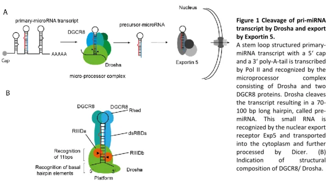
Primary miRNA processing is the first catalytic cleavage step of the canonical biogenesis pathway.
The pri-miRNA transcript is incorporated into the microprocessor complex. This complex consists
of the minimal components RNAse III enzyme Drosha and a dimer of DGCR8 (Figure 1) (Gregory
et al. 2004; Denli et al. 2004; Han et al. 2004; Landthaler, Abdullah Yalcin and Tuschl 2004; Kwon
et al. 2016). The pri-miRNA transcript forms a double-stranded RNA (dsRNA) stem-loop-structured
hairpin with a stem length of usually 35 base pairs (bp) and ssRNA bulges. A single-stranded (ss)
loop raises as apical structure and an adjacent ssRNA basal junction marks the end of the stem
(Han et al. 2006; Nguyen et al. 2015). DGCR8 contains two double-stranded RNA-binding domains
(dsRBDs) (Roth, Ishimaru, and Hennig 2013; Quick-cleveland et al. 2015; Nguyen et al. 2015; Kwon
Introduction 3
et al. 2016). Drosha is structurally very similar to Dicer (low sequence conservation), but exhibits unique compartments like a zinc-finger motif (Figure 1) (Nguyen et al. 2015).
Drosha and DGCR8 function together as distance measuring system for specific hairpin structured RNAs (Kwon et al. 2016). Therefore, the DGCR8 dimer positions at the upper part of the stem and Drosha at the lower part of the stem. Catalytic Drosha cleavage occurs after positioning of the microprocessor at the height of 11 bp of the stem (Han et al. 2006; Nguyen et al. 2015). This aims in a typical stem-loop-structured 60-70 bp long hairpin known as precursor miRNA (pre-miRNA) with a 2 nucleotide 3' overhang (Morlando et al. 2008). Drosha deletions result in the loss of canonical processed miRNAs (Y.-K. Kim, Kim, and Kim 2016). Because of the main intronic origin of the pri-miRNA transcripts, splicing and microprocessor cleavage are interconnected and influence each other's efficiency (Y.-K. Kim and Kim 2007; Kataoka, Fujita, and Ohno 2009).
Figure 1 Cleavage of pri-miRNA transcript by Drosha and export by Exportin 5.
A stem loop structured primary- miRNA transcript with a 5‘ cap and a 3‘ poly-A-tail is transcribed by Pol II and recognized by the microprocessor complex consisting of Drosha and two DGCR8 proteins. Drosha cleaves the transcript resulting in a 70- 100 bp long hairpin, called pre- miRNA. This small RNA is recognized by the nuclear export receptor Exp5 and transported into the cytoplasm and further processed by Dicer. (B) Indication of structural composition of DGCR8/ Drosha.
The pre-miRNA hairpin-structure is then exported to the cytoplasm with a canonical RNA export
mechanism with Exportin-5 (Exp5) in a Ran-GTP depended manner (Yi et al. 2003; Bohnsack,
Czaplinski, and Gorlich 2004; Y.-K. Kim, Kim, and Kim 2016). The loading of the pre-miRNA into
Exp5-RanGTP remains unclear, but additional factors of the microprocessor complex like ILF-3
could have a major role (Libri et al. 2013).The structural mannerism of the pre-miRNA results in a
specific recognition of Exp5-RanGTP (Okada et al. 2009). After nuclear exporting, the complex
decomposes and the released pre-miRNA is bound by a multi-protein complex containing Dicer
(K. Miyoshi et al. 2009). Interestingly, after knockout of Exp5 cytoplasmic transport still occurs
suggesting redundant or alternative mechanisms (Y.-K. Kim, Kim, and Kim 2016).
Figure 2 Examples of regulatory RBPs/RNAs.
(A) Schematic overview of a representative pri-miRNA structure containing a mature miRNA indicated in red. Interaction sites for potential regulators as well as conserved sequence motifs are highlighted. Regulatory RBPs/ miRNAs influencing the miRNA biogenesis by direct binding to the hairpin structured pri-miRNA are indicated in red by inhibiting or in green by promoting the process. (B) Regulatory RBPs/ miRNAs influencing the miRNA biogenesis are indicated in red by inhibiting or in green by promoting the process.
Regulatory mechanisms. As already suggested, the miRNA biogenesis is not only tightly regulated at a transcriptional level, but even more at the following biogenesis steps (Libri et al. 2013; Ha and Kim 2014; S. Li, Wang, Fu, and Dorf 2014). The efficiency of pri-miRNA and pre-miRNA processing underlies the sequence and/ or resulting structural characteristics of the hairpin (Auyeung et al.
2013). These specific RNA compositions are recognized by hairpin-interacting RNA-binding proteins (RBPs) which promote or block the processing steps (Han Wu et al. 2010; Trabucchi et al.
2009; Gu et al. 2011; X. Zhang et al. 2011; Connerty, Ahadi, and Hutvagner 2015; Du et al. 2015).
Recently a large proteomics-based hairpin-pull-down screen identified several hundred potential interactors which regulate Drosha processing (Treiber et al. 2017). In the following part few regulatory RBPs which influence processing are briefly described.
Regulation of processing by proteins that interact with the pri-miRNA. The pri-miRNA transcript contains several conserved sequence elements that are important for processing. The loop contains a UGUG motif and at the basal flanking sites an UG and a CNNC motif (Ha and Kim 2014;
Roden et al. 2017). The splicing factor Srp20 (Ajiro et al. 2015) and the DEAD-box RNA helicase p72 (DDX17) interact with the CNNC motif and promote Drosha processing (Figure 2 A) (Sabin et al. 2009; Guil and Cáceres 2007).
The RBP TAR DNA-binding protein 43 (TDP43) positively interferes with Drosha processing by
binding to the terminal loop sequence of pre-miR-143 and pre-miR-547 (Kawahara and Mieda-
Sato 2012; Ha and Kim 2014). Interestingly the serine/ arginine-rich SR protein (SF2/ASF) promotes
processing by altering the structure of pri-miR-7. The mature miR-7 regulates the mRNA transcript
Introduction 5
of SF2 down by gene silencing, suggesting an negative feedback loop for steady-state production of miR-7 (Han Wu et al. 2010). The RBP Rbfox3 regulates both inhibition and improvement of processing depending on the pri-miRNAs. It was suggested that many pri-miRNAs are regulated and particularly shown that interaction of Rbfox3 with the loop region of pri-miR-15a resulted in processing. In contrary binding to the stem of pri-miR-485 resulted in an inhibition of the microprocessor recruitment (K. K. Kim et al. 2014). The RBPs hnRNPA1 and KSRP (KH-type splicing regulatory protein) interact with the terminal loop of several pri-miRNAs e.g. pri-miR18a, pri-miR- 16, pri-miR-21 and promote their processing (Figure 2 A) (Michlewski et al. 2008; Michlewski and Cáceres 2010; Guil and Cáceres 2007; X. Zhang et al. 2011; Briata et al. 2012) In contrary hnRNPA1 negatively regulates pri-let-7a processing while competing with KSRP for the stem binding site.
The nuclear factors 45 and 90 inhibit processing by interaction to pri-let-7a or pri-miR-21 (Sakamoto et al. 2009). The RNA editing enzymes ADAR1 and ADAR2 are known to transform an adenosine to an inosine within specific pri-miRNAs, which inhibits Microporocessor hairpin interaction (Figure 2 A)(Cho, Myung, and Chang 2017).
Regulation of pri-miRNA processing by proteins that interact with the microprocessor complex.
The RBP p68 (DDX5) together with p72 (DDX17) are recruited to Drosha by interaction to the hairpin and promote pri-miRNA processing (Figure 2 B). It is known that several additional factors interact with p68/p72 and further block or promote the processing. For Instance, promoting interactors are BRCA1 (breast cancer susceptibility gene 1), SNIP1 (SMAD nuclear interacting protein), ARS2 (arsenite resistance protein 2) or the TGF-β induced transcription factors SMAD1- 3 and 5 (Figure 2 B). In contrary, the estrogen receptor alpha inhibits processing by an interfering interaction to p68/p72 (Davis et al. 2008; Sabin et al. 2009; Kawai and Amano 2012; Vos et al.
2015; Thillainadesan et al. 2012; Suzuki et al. 2009; Fukuda et al. 2007). Interestingly, the helicases p68/p72 are involved in the processing of nearly one-third of the known pri-miRNAs, according to studies within knock-out mice (Fukuda et al. 2007)
Regulation of pri-miRNA processing by miRNAs. Several examples where miRNAs are transported back to the nucleus for the regulation of pri-miRNA processing are known. The interaction to the pri-miRNA is formed by complementary base pairing within the flanking regions of the primary transcript. For example, in C. elegans the processing of pri-let-7 is promoted by an auto-regulatory mechanism of let-7 which interacts with the pri-let-7 3’ flanking region. The mechanism of how the miRNA-Alg-1 complex promotes processing is not fully understood (Figure 2 A) (R. Tang et al.
2012; Zisoulis et al. 2012).
Drosha/ DGCR8 regulatory modifications. The functionality of the microprocessor complex is
additional regulated by PTMs. Drosha localization in the nucleus is regulated by phosphorylation
of S300 and S302 by GSK3. DGCR8 exhibits higher stability when phosphorylated by ERK.
Sumoylation at K707 by SUMO1 stabilizes DGCR8 by inhibiting ubiquitination (C. Zhu et al. 2015;
Fletcher et al. 2017). Further Drosha is stabilized by acetylation which inhibits ubiquitination. In contrary the affinity to pri-miRNAs and hence processing is increased by deacetylation of DGCR8 by HDAC1 (histone deacetylated by histone deacetylase1) (X. Tang et al. 2010; X. Tang et al. 2011;
X. Tang et al. 2013; Casseb et al. 2016).
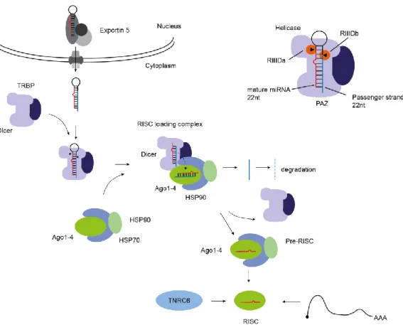
1.1.2 Dicer cleavage of pre-miRNAs and RISC loading
In the cytoplasm the pre-miRNA is released from the Exp5-RanGTP complex and immediately incorporated into Dicer. Dicer is structurally similar to Drosha a RNase III enzyme. It contains also two RNase III catalytic cleavage sites. For proper positioning and function, additional co-factors are needed. The cofactor TAR RNA binding protein 2 (TRBP) and the protein activator of PKR (PACT) contain both double-stranded-RBDs and additionally promote the substrate interaction (Gregory et al. 2005; Chendrimada et al. 2010; Yoontae Lee et al. 2006; H. Y. Lee et al. 2013). TRBP functions as a pre-miRNA length determining compartment through a defined positioning of the hairpin by interaction with the helicase domain of Dicer (Fukunaga 2005). The function of PACT remains elusive and is still unclear (Figure 3) (H. Y. Lee et al. 2013; Y. Kim et al. 2014; Ha and Kim 2014). TRBP mainly interacts with the apical loop and the upper part of the stem. After the pre- miRNA is positioned, Dicer interacts with the precursor and cleaves off the terminal loop. This occurs within the catalytically active centre of the RIIId domains. The cleavage product is a 21-24 nt long double-stranded RNA with 2 nucleotides 3’-overhangs, a 5’-phosphate and a 3’-hydroxyl group (H. Zhang et al. 2004; MacRae et al. 2006; Taylor et al. 2013; Wilson et al. 2015; Fareh et a l. 2016; Song and Rossi 2017). The ssRNA terminal loop as additional cleavage product is degraded.
After cleavage occurred, the RISC loading complex is assembled. Therefore, Dicer interacts with Ago via the Piwi and the RNase III domain. Additionally Ago recruits co-chaparones with the components heat shock protein 90/70 (Hsp90) and FK506-binding immunophilins Fkbp4/5. The HSP70/HSP90 complex loads the RNA duplex into Ago in an ATP dependent manner (Iwasaki et al.
2010). During the loading process one strand of the miRNA heteroduplex is selected and imparted
to Ago while the chaperones stabilize the opening and incorporation process into Ago (Figure 3).
Introduction 7
Figure 3 Dicer cleavage of the pre-miRNA and RISC complex loading.
This small RNA is recognized by the nuclear export receptor Exp5 and transported to the cytoplasm and further processed within the catalytic domains of the Dicer/TRBP complex. Dicer/TRBP form a multi-subunit complex where TRBP is responsible for positioning of the pre-miRNA. This results in a double-stranded RNA with 22 nt length. The mature strand is incorporated into Ago during RISC loading and leads to the mature RISC complex assembly.
An unwinding of the dsRNA strand is mediated by the N domain of Ago and the selection of one strand which is maybe supported by TRBP has to be performed (Kwak and Tomari 2012). However, statistical and thermodynamically rules suggest that the strand with a stable 5' end is preferentially loaded (Dueck and Meister 2014; T. Miyoshi et al. 2010; Natalia J Martinez et al.
2013; Iwasaki et al. 2010; Nakanishi et al. 2016). After loading Dicer and the co-chaperones dissociate which leads to the mature RISC complex (Kawamata and Tomari 2010; Dueck and Meister 2014; K. Miyoshi et al. 2009).
Regulation of processing by proteins that interact with the pre-miRNA. Rbfox2 another RBP is suggested to be important for cancer and neurodegeneration induced by the mis-regulation of miR-107 and miR-20b. This miRNAs are suppressed by the interaction with Rbfox2 under certain conditions. This leads to a inhibition of Dicer cleavage and hence processing (Yu Chen et al. 2016).
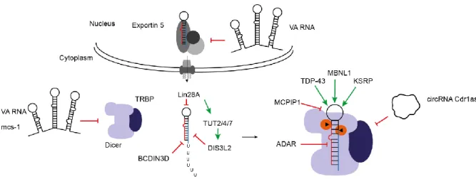
At the Dicer cleavage stage, several RBPs seem to compete for the binding to the terminal loop of
the pre-miRNAs. For instance MBNL1 competes with Lin28 and hence U tailing and degradation is
blocked (Androsavich and Chau 2014; Rau et al. 2011).
Surprisingly also base modifications of pre-miRNAs regulate Dicer interaction. For Instance Dicer recognition of the 5' monophosphate is blocked by methylation of the 5’ end of the pre-miR-145 by the human RNA-methyltransferase BCDIN3D (Xhemalce, Robson, and Kouzarides 2012; Park et al. 2011).
Regulatory mechanisms during Dicer cleavage. As already suggested the miRNA biogenesis is not only tightly regulated at transcriptional level, but even more at the pre-miRNA biogenesis step by many RBPs which interact with the pre-miRNA and the Dicer/TRBP complex (Figure 4) (Libri et al.
2013; Ha and Kim 2014; S. Li, Wang, Fu, and Dorf 2014). Here, few examples which influence Dicer cleavage are presented. The most prominent example of negative influence on pre-miRNA processing is the stem cell factor lin28 that interacts with the members of the let-7 family (also observed for miR-107, miR143, etc.). The RBP lin28 consisting of a Cold shock and a CCHC-type Zincfinger domain interacts with the GGAG motif of the terminal loop of the pre-let-7 members except let-7a-3/c-2. Through the interaction the enzymes terminal uridyltransferases TUT4 (ZCCHC11) or TUT7 which uridylates the pre-miRNAs are recruited (Figure 4) (L. Wang et al. 2017;
Triboulet, Pirouz, and Gregory 2015). This short poly (U) tail at the 3’ end of the pre-miRNAs interferes negatively with Dicer cleavage and leads to 3’ to 5’ degradation of the pre-miRNA by DIS3L2. The inhibition of pre-let-7 biogenesis blocks important developmental processes, causing the cells to stay at a stem cell level (Newman, Thomson, and Hammond 2008; Rybak et al. 2008;
Heo et al. 2008; Heo et al. 2009; Viswanathan, Daley, and Gregory 2008; Thornton et al. 2014;
Shyh-Chang and Daley 2013; Triboulet, Pirouz, and Gregory 2015; Hao-ming Chang et al. 2013).
Interestingly, lin28 is a phospho-protein which is modified by ERK/MAPK at several residues and hence stabilized in pluripotent stem cells (Tsanov et al. 2017; Xiangyuan Liu et al. 2017). In contrary of inhibition, TUT4, TUT2 and TUT 7 are reported to monouridylate a specific set of pre- miRNAs including pre-let-7 at the 3’. This additional uridylation promotes Dicer cleavage in non- stem cells which lack Lin28 (Heo et al. 2012).
Regulation of pre-miRNA processing by other RNAs. Other RNAs can block dicer pre-miRNA interaction and recognition. For instance the adenoviral RNA VA1 competes with the pre-miRNAs for Dicer interaction and hence inhibits the processing (Libri et al. 2013).
A recent study reports a dysregulation of miR-7 and miR-671 induced by a downregulation of
circRNA Cdr1as that interacts with the named miRNAs. Interestingly, the data illustrates the
downregulation of the miR-7 by the loss of the circRNA, hence an influence on pre-miRNA
processing is suggested (Figure 4) (Piwecka et al. 2017).
Introduction 9
Figure 4 Regulation of Dicer cleavage.
This small RNA is recognized by the nuclear export receptor Exp5 and transported to the cytoplasm and further processed by Dicer. This results in a double-stranded RNA with 22 nt length. The mature strand is incorporated into Ago during RISC loading and leads to the mature RISC complex. During this stepwise process many RBPs positively (green arrows) or negatively (red arrows) influence this process. Regulatory RNAs influencing the miRNA biogenesis are indicated in red by inhibiting the process.
Regulation of pre-miRNA export. The export mechanism is known to be blocked by few RNAs that bind to Exportin-5. For instance the adenoviral non-coding RNA VA1 inhibits miRNA export to the cytoplasm by competing with the endogenous pre-miRNAs for binding to Exportin- 5. Hence less pre-miRNAs are transported to the cytoplasm (Figure 4) (Y.-K. Kim, Kim, and Kim 2016; Libri et al.
2013; Lu and Cullen 2004; Grimm et al. 2006).
Dicer/ TRBP modifications and further functions. The Dicer/ TRBP complex is stabilized by TRBP phosphorylation by the MKK1/Erk pathway which causes selectively enhanced miRNA processing for growth-promoting miRNAs (Paroo et al. 2009). An additional phosphorylation occurs at S283/286 by S6 kinase which leads also to enhanced miRNA processing and links the miRNA biogenesis machinery to the mTOR pathway (C. Xu et al. 2016). A recent study reports the phosphorylation of TRBP by MAPK, which stabilizes the complex to Lin28a. This interaction leads to reduced let-7 levels and hence to an induced neuronal dendritic spine growth (Amen et al.
2017).
Further sumoylation of TRBP at K52 inhibits ubiquitination at K48, stabilizes the complex and
promotes RISC loading (C. Chen et al. 2015). In C. elegans oocytes it was observed that Dicer is
phosphorylated by ERK which causes inhibition of Dicer activity. This inhibition is reactivated
before fertilization starts in the oocytes (Drake et al. 2014). Of note, Dicer seems to have a certain
nuclear role in double-stranded DNA repair when phosphorylation is induced by DNA damage at
S1016, S1728 and S1852 (Burger et al. 2017).
1.2 Gene silencing and translational repression
The mature RISC complex contains a miRNA incorporated in Ago. It finds its mRNA target during a less understood scanning mechanism through complementary base pairing of the seed sequence with the 3’ untranslated region (3’UTR) of the targets. The state of Ago during scanning in terms of protein interactors and regulatory modifications remains speculative. The minimal RISC is supposed to interact with mRNA and/or the TNRC6 proteins. However, it is still unclear whether interactions are at the same time, sequential or simultaneous. Further, it is assumed that the target scanning process and also translational repression and /or storage takes place or is next to structured protein networks of various size called p-bodies (Patel, Barbee, and Blankenship 2016;
Zipprich et al. 2009; J. Liu et al. 2005; Wilczynska and Bushell 2015; S. Lee and Vasudevan 2013;
Kamenska et al. 2016). As a consequence of Ago-miRNA-mRNA-TNRC6 complex assembly, translational repression and mRNA decay are initiated. These processes are mainly induced by proteins and enzymes that are recruited sequentially or in parallel by TNRC6. Ago functions conclusively as initial target finding enzyme and mediates through binding to TNRC6 gene silencing (Jonas and Izaurralde 2015; Dueck and Meister 2014).
1.2.1 Interplay of TNRC6 and Ago
1.2.1.1 TNRC6 functions as core binding platform of the gene silencing process
TNRC6 proteins belong to the family of GW proteins. The best known member is GW182 found in D. melanogaster (human homolog TNRC6A). Mammals express two additional paralogs TNRC6B, C and many uncharacterized isoforms. In general, TNRC6 proteins are structurally and functionally conserved from an evolutionary point of view. They may have evolved by the development of multicellularity and whole genome duplication to three paralogs within the vertebrates (Zielezinski and Karlowski 2015; Mauri et al. 2017). TNRC6 proteins consists of two main regions, the N-terminal Ago binding domain (ABD) and the C-terminal silencing domain (SD). Glycine- Tryptophan (GW, W, GWG, WG) repeats are randomly distributed over the whole proteins, especially in the ABD and the SD. Ago proteins interact through specific binding of two binding pockets located in the PIWI domain with two Ws located in the TNRC6 ABD (see Figure 6 and Table 1) (Braun et al. 2011; Jonas and Izaurralde 2015; Huntzinger and Izaurralde 2011; Pfaff et al. 2013).
This specific interaction is conserved within the mammalian Ago 1-4 proteins. Interestingly, there
are very limited regions within this various GW repeats in the ABD where Ago proteins interact.
Introduction 11
However, the specificity or selectivity of this process is not understood. It is suggested that a specific distance of 10 amino acids combined with a specific amino acid pattern determines the binding process (Pfaff et al. 2013; Hauptmann et al. 2015). Altogether, three binding hot spots on TNRC6A are known. These hotspots may interact also with Agos at the same time (Elkayam et al.
2017). It is further known that the TNRC6 paralogs may contain a different number of Ago interaction sites. They are partly conserved, e.g. TNRC6B possesses two and TNRC6A has three interaction sites (Takimoto, Wakiyama, and Yokoyama 2009; Nishi et al. 2013; Pfaff et al. 2013;
Hauptmann et al. 2015a; Baillat and Shiekhattar 2009). TNRC6 proteins are suggested to function redundantly and to interact with all Ago1-4 proteins without preferential combinations.
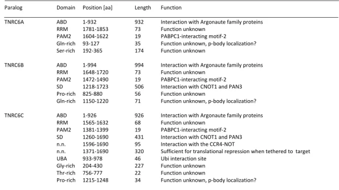
Table 1 Domain organization of mammalian TNRC6 paralogs (adapted from Uniprot database).
Paralog Domain Position [aa] Length Function
TNRC6A ABD 1-932 932 Interaction with Argonaute family proteins
RRM 1781-1853 73 Function unknown
PAM2 1604-1622 19 PABPC1-interacting motif-2
Gln-rich 93-127 35 Function unknown, p-body localization?
Ser-rich 192-365 174 Function unknown
TNRC6B ABD 1-994 994 Interaction with Argonaute family proteins
RRM 1648-1720 73 Function unknown
PAM2 1472-1490 19 PABPC1-interacting motif-2 SD 1218-1723 506 Interaction with CNOT1 and PAN3 Pro-rich 825-880 56 Function unknown
Gln-rich 1150-1220 71 Function unknown, p-body localization?
TNRC6C ABD 1-926 926 Interaction with Argonaute family proteins
RRM 1565-1632 68 Function unknown
PAM2 1381-1399 19 PABPC1-interacting motif-2 SD 1260-1690 431 Interaction with CNOT1 and PAN3 n.n. 1596-1690 95 Interaction with the CCR4-NOT
n.n. 1371-1690 320 Sufficient for translational repression when tethered to target UBA 933-978 46 Ubi interaction site
Gly-rich 204-430 227 Function unknown Thr-rich 756-777 22 Function unknown
Pro-rich 1215-1248 34 Function unknown, p-body localization?
The central part of TNRC6 contains a number of gene silencing independent domains. A typical ubiquitin-associated (UBA)-like domain which is involved in the proteasomal degradation (Figure 6). The UBA-like domain folds into a trimeric helix bundle with hydrophobic regions for ubiquitination probably by the E3 ubiquitin ligase TRIM65 (S. Li, Wang, Fu, Berman, et al. 2014;
Buchberger 2002; V. S. and A. F. Lau 2009).
Next to the UBA a nuclear localization signal (NLS) and a nuclear export signal (NES) are located
within the middle region in TNRC6A (Figure 6). Recently the structural composition of the TNRC6A
NLS interacting with importin α was solved (Chaston et al. 2017). TNRC6B and C contain just a NES
at a similar position; the location of the NLS is unknown. The NLS and NES recruit additional
proteins like importin β which leads to nuclear shuttling (Nishi et al. 2013; Schraivogel et al. 2015).
All TNRC6 proteins contain several domains where specific residues are enriched e.g. a glutamine- and proline-rich region in TNRC6B. The location of these domains is partly conserved. However, their function is unknown, but mechanisms in p-body assembly/location are suggested for the Q- rich region within TNRC6B (Lazzaretti, Tournier, and Izaurralde 2009).
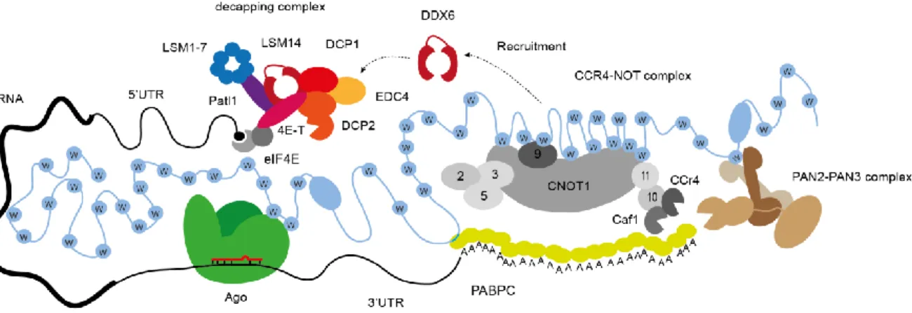
The C-terminal SD contains a PAM2 motif, a RRM and many Ws important for the interaction with the CCR4-Caf1-NOT and the trimeric Pan2-Pan3 complex (Figure 6. A interaction of TNRC6 with the decapping complex is not known. Interestingly, the SD mediates translational repression and mRNA decay when proximal mRNA is present (Zipprich et al. 2009; Lazzaretti, Tournier and Izaurralde 2009; Eulalio, Tritschler, et al. 2009). While Ago has the target recognition and gene silencing initiation function, TNRC6 serves as a binding platform and as mediator for all downstream processes (see Figure 6 and Table 1).
Figure 5 Schematic model based on functional and structural aspects of the miRNA-mediated gene silencing process.
A functional miRNA-mediated gene silencing complex requires at least one Argonaute protein, one TNRC6 protein, several Poly-A-binding proteins (PABPC1), and the PAN2–PAN3 and CCR4–caf1-NOT deadenylase complexes. The decapping complex is recruited by DDX6 and consists of the core subunits EDC4, DCP1-2 and others. Translational repression and destabilization of the target mRNA leads to 5’-to-3’ decay through exonucleases like XRN1 which is in direct neighbourhood within the p-bodies.
The RRM of D. melanogaster GW182 (human homolog TNRC6A) lacks the ability to bind RNA.
Nevertheless, it is required for the full function of gene silencing and is therefore suggested to
may bind additional unknown protein interactors. The atypical RRM and UBA are the only defined
structured parts of TNRC6 proteins.
Introduction 13
Figure 6 Schematic model of TNRC6 domain organization based on functional and structural aspects of the miRNA- mediated gene silencing process.
(A), (B), (C), (D) domain organization of TNRC6A-C. Vertical black bars represent the relative positions of tryptophan’s.
Different domains and regions are marked in different colour, abbreviations can be found within the text. (D) Ago proteins bind to two Ws in the ABD. Importin beta may bind to the NLS/NES region to transport TNRC6 into the nucleus.
CRM1 (not shown) interacts in the same region as imp beta.Pan2-Pan3 trimer interacts through Pan3 dimer with a W from TNRC6. CCR4-Caf1-NOT complex interacts with TNRC6 with several Ws interactions of cNOT1 and cNOT9.
Decapping complex is not interacting with TNRC6.
The PAM2 motif interacts with the first PABPC1 linked to the poly (A) tail and thus leads to an indirect mRNA positioning (Jonas and Izaurralde 2015) (Figure 6). The interaction with the CCR4- Caf1-NOT deadenylation complexes is thought to be similar to the Ago-TNRC6 interaction, meaning that the interaction relies on tryptophan insertion into binding pockets to the respective protein partner. After recruiting the complex by Ago-TNRC6, deadenylation will be completed and the mRNA will be finally degraded. In case of miRNA independency, translational control through deadenylation also occurs (Collart, Panasenko, and Nikolaev 2013; Gupta et al. 2016).
Binding occurs with the main subunit cNOT1 that possesses a similar function as TNRC6. cNOT1 is also considered as a scaffold binding platform for the catalytic deadenylases Caf1 and CCR4a and other subunits(Figure 6).
The interaction of cNOT1 with TNRC6 relies on several Ws together with the regions CCR4- interacting-motif1/ 2 (CIM-1/ CIM-2). Furthermore, NOT9 contacts two Ws of TNRC6 with unknown position and interacts with cNOT1. Both interactions are limited to the silencing domain which causes the downstream silencing effects (see Figure 6 and Table 1) (Braun et al. 2011;
Chekulaeva, Filipowicz and Parker 2009; Chekulaeva et al. 2011a; Fabian et al. 2013a; Ying Chen et al. 2014).
The trimeric Pan2-Pan3 deadenylation complex consists of the catalytic deadenylase Pan2 and two Pan3 proteins. This complex mediates mRNA association both through PABPC1 interaction and by direct binding to the poly(A) tail through a zinc finger domain (see Figure 6 and Figure 6) (Wolf et al. 2014; Jonas et al. 2014). It is thought that the Pan3 dimer assembly leads to a formation of a W binding pocket which further stabilizes and strengthens the interaction to TNRC6 (see Figure 6)(Braun et al. 2011; Christie et al. 2013; Jonas et al. 2014; Wolf et al. 2014).
All three TNRC6 paralogs function redundantly and promote post-translational gene silencing (PTGS). Individual K.O.s indicated no reduction in tethering assays. Inhibition of all three paralogs leads to a strong de-repression comparable to de-repression assays conducted with the T6B peptide (Hauptmann et al. 2015a; Danner et al. 2017). As binding platforms, TNRC6 proteins are required to be unstructured to allow a flexible and dynamic change of interaction partners as well as to assemble within bigger structured gene silencing compartments (see Figure 6) (Jonas and Izaurralde 2015).
There are many reports in literature that report on additional binding partners of TNRC6 proteins.
These are not yet fully related to a functional subunit or understood. For instance, recent reports
suggest additional interactions with LIM1, which binds to the ABD, and is suggested to have a
regulatory role within the gene silencing mechanism (S. Li, Wang, Fu, Berman, et al. 2014; E. Wu
et al. 2016; Bridge et al. 2017).
Introduction 15
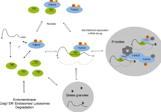
1.2.1.2 Subcellular localization of TNRC6-Ago complexes
The mammalian miRNA mediated gene silencing is suggested to occur in the cytoplasm. Therefore, the associated functional proteins are also mainly located in the cytoplasm. The function determines localization, hence depending on main or auxiliary function, the subcellular position of the proteins to other cellular compartments can switch. For instance it is reported that gene silencing occurs at terminal axons or that TNRC6 proteins have putative functions in the nucleus (Figure 7) (Kalantari, Chiang, and Corey 2016; N. R. Sharma et al. 2016; Schratt et al. 2006). Hence many different cell lines and tissues exhibit a particular localization and expression pattern of TNRC6 and Ago Proteins (Schraivogel et al., n.d.; Rüdel et al. 2008; Keith T. Gagnon, Liande Li, Bethany A. Janowski 2012; Hauptmann et al. 2015b).
Figure 7 Subcellular localization of the gene silencing process.
A dynamic fast switching system triggered by regulatory stimuli determines the intracellular localization of Ago-TNRC6 complexes from single molecules to highly structured networks.
TNRC6 and Ago proteins seem to shuttle into the nucleus as single molecules or as complex. Ago
is imported through canonical redundant mechanisms and has potential chromatin associated
functions (Ameyar-Zazoua et al. 2012). TNRC6 is mainly imported into the nucleus by importin
alpha/beta and exported by CRM1 dependent mechanisms (Schraivogel et al.,2015). It is
suggested that TNRC6 may act as binding platform for different nuclear processes such as splicing and transcription. However, this assumption is speculative and relies on proteomic screens which were performed lacking conclusive functional assays and necessary controls (Kalantari et al. 2016).
According to IF stainings, TNRC6 proteins localize within the cytoplasm in small complexes and large structured processing-bodies (p-bodies) when bound to Ago. The functional subunit, called p-bodies was, extensively studied in the last decades and many mRNA related functions were found such as mRNA decay (deadenylation, decapping complexes, exonucleases), translational repression (TNRC6-Ago), PTGS (TNRC6-Ago, deadenylase and decapping complexes) and nonsense mediated decay (UPF1/2/3 etc.) (Kulkarni, Ozgur, and Stoecklin 2010; S. Lee and Vasudevan 2013).
The architecture of p-bodies exhibits a structural binding network and fast, dynamic and flexible changing mRNA-complexes (Figure 7). Purification of p-bodies is difficult, therefore, most studies use IFs to detect p-bodies in overexpressed conditions (Rüdel et al. 2008). Optical detection of small endogenous p-bodies yield unreliable data and estimation of size is difficult. It is suggested that the real processing bodies are smaller, dynamic and fast changing/adapting network systems which can also store mRNA-RBP complexes. However, this remains speculative (Meister et al.
2005; Pillai et al. 2005; Leung, Calabrese, and Sharp 2006; Eulalio et al. 2007; Eulalio, Behm- Ansmant, and Izaurralde 2007; Rajgor et al. 2014; S. Lee and Vasudevan 2013; Pitchiaya et al.
2017).
Overexpressed TNRC6 itself co-localizes mainly with p-body markers like Lsm4. Interestingly Ago proteins show weaker co-localization with p-bodies (Schraivogel et al. 2015; Nishi et al. 2013).
Above all in an endogenous manner, it was even shown that p-body location is not required for TNRC6-Ago interaction (Lazzaretti, Tournier, and Izaurralde 2009). This findings suggests, that Ago proteins and PTGS are located around the p-bodies (N. R. Sharma et al. 2016; Pitchiaya et al. 2017).
Ago can also be found in other compartments, e.g. in extracellular signalling vesicles (exosomes) (McKenzie et al. 2016) and under certain stress conditions in stress granules (Figure 7) (Anderson and Kedersha 2008; Detzer et al. 2011; Rieckher and Tavernarakis 2017; Buchan and Parker 2009).
In general, gene-silencing complexes such as the RISC loading complex are associated with the
endomembrane system consisting of the ER, Golgi complexes, endosomes and lysosomes (Figure
7) (Y. J. Kim, Maizel, and Chen 2014; D Gibbings et al. 2012; Derrick Gibbings et al. 2013; N J
Martinez and Gregory 2013; Barman and Bhattacharyya 2015).
Introduction 17
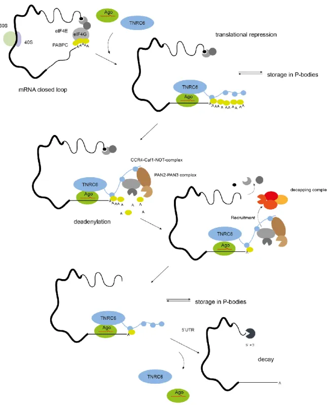
1.2.2 Canonical post-transcriptional gene silencing
The initiation of translational repression and mRNA degradation is induced by binding of the miRNA seed sequence to the target. Binding occurs through complementarity of the bases 2 - 8 of the miRNA with the 3’ UTR of the mRNA or in rare cases in other regions like the 5’ UTR (Hafner et al. 2010; Hausser et al. 2013; G. Li et al. 2016; Ørom, Nielsen, and Lund 2008). Thereby, repression is triggered while the translational initiation closed loop structure is assembled (Figure 8). This structure is formed by the poly (A) tail bound to the cytoplasmic poly (A)-binding Protein (PABPC1) which interacts with eIF4G within the 5’cap structure (Jackson, Hellen, and Pestova 2010).
The target finding process is suggested as highly regulated through internal or external signals (Giraldez et al. 2005; van Rooij et al. 2007; Avraham and Yarden 2012). Furthermore, the question of target capability of many miRNAs remains unsolved, because a single miRNA can bind many mRNAs and the other way round, a mRNA can interact with many miRNAs. The occurrence of proper target finding is not well understood, as it also strongly depends on the mRNA/transcript expression profile (and turnover) of specific cell and tissue types (Rüegger and Großhans 2012;
Dueck et al. 2012; Jacobsen et al. 2013; S. Wu et al. 2010).
To induce mRNA degradation, GW182 (or human paralogs TNRC6A-C) interacts through the minimal miRISC with the mRNA. Additionally, PABPC1 binds the PAM2 motif within TNRC6 to form a stable structured complex (Figure 8) (in D. melanogaster with additional W interactions) (Chekulaeva 2011). The detailed mechanism of the interaction of PABPC1 to TNRC6 is controversial discussed. It is assumed that translational repression is independent of this interaction because deadenylation through CCR4-Caf1-NOT and Pan2-Pan3 still occurs which leads to mRNA decay (Fabian et al. 2009; Braun et al. 2011; Jinek, Coyle, and Doudna 2011; Zekri, Kuzuoğlu-Öztürk, and Izaurralde 2013; Fabian et al. 2013b). Nevertheless, the interaction of TNRC6 with PABPC1 may decompose the closed loop structure through eIF4G dissociation (Figure 7) (Zekri et al. 2009; Fabian et al. 2009).
Nevertheless, within the canonical pathway the translational repression complex is formed, the closed loop structure is opened and subsequently the deadenylase complexes consisting of the CCR4-Caf1-NOT and Pan2-Pan3 are recruited. Both units interact with several Ws of the C-terminal silencing domain of TNRC6 (Makino et al. 2015; Zipprich et al. 2009; Chekulaeva et al. 2011b;
Lazzaretti, Tournier, and Izaurralde 2009; Eulalio, Huntzinger, et al. 2009; Eulalio, Tritschler, and
Izaurralde 2009).
Figure 8 Small RNA mediated gene silencing by the mature RISC complex.
Different steps of the miRNA mediated gene silencing process are combined in a schematic chronological multistep illustration based on structural and functional information. Briefly, the minimal miRISC mediates gene silencing through complementary mRNA target binding together with TNRC6. Binding of RISC to a partially complementary mRNA at the seed sequence region results in repression of translation. This subsequently leads to deadenylation through the deadenylase complexes CCR4-Caf1-Not and Pan2 –Pan3 that are recruited by their mediator TNRC6. The mRNA is further destabilized through 5’decapping initiated by a DDX6 mediated recruitment of the decapping complex consisting of EDC1/2. After completing the mRNA destabilization, translational repression complex detaches which leads to 5’ to 3’ decay through p-body located exonucleases like XRN1.
Introduction 19
Deadenylation and parallel dissociation of PABPC1 is then thought to be initiated by Pan2-Pan3 as first step. Followed by further diminishing of the poly (A) tail by the CCR4-Caf1-NOT complex until completing of deadenylation leads to a parallel DDX6 mediated recruitment of the decapping complex (Fabian and Sonenberg 2012; Subtelny et al. 2014; Wahle and Winkler 2013; Yamashita et al. 2005). Interestingly, only the CCR4-Caf1-NOT complex leads to complete deadenylation and subsequent decay (Huntzinger et al. 2013; Piao et al. 2010; Behm-Ansmant et al. 2006; Eulalio, Tritschler, and Izaurralde 2009; Yamashita et al. 2005). The Dead-box helicase DDX6 interacts with the MIF4G domain of cNOT1 (Ying Chen et al. 2014). Upon interaction it is activated and is suggested to recruit the decapping complex through interaction with EDC3 and DCP1-DCP2 after its dissociation (Figure 8) (Makino et al. 2015; Tritschler et al. 2009; Mathys et al. 2014; Jonas and Izaurralde 2015). Additionally DDX6 mediates, together with the eIF4E transporter protein 4E-T, pat1 and the associating Lsm1-7 proteins, the mRNA decay machinery to the 5‘ cap via direct interaction to eIF4E (Nishimura et al. 2015; Sharif and Conti 2013; Ozgur et al. 2015; Peter et al.
2015).
The decapping complex which is located within the p-bodies consists of DCP1, EDC4, DDX6 and its catalytical core subunit DCP2 (Figure 7) (Huntzinger and Izaurralde 2011; Jonas and Izaurralde 2015). After decapping and deadenylation the stability of the mRNA falls below a critical limit. This induces a rapid degradation of the mRNAs by decapping complex associated 5’ to 3’ exonulceases such as XRN1. Interestingly, XRN1 depletion results in the accumulation of deadenylated mRNAs bound by the RISC and decapping machinery (Nishimura et al. 2015; Sun et al. 2013; Behm- Ansmant et al. 2006; Nishihara et al. 2013).
The other components dissociate after decapping and deadenylation and the (minimal) RISC may be recycled, degraded or re-initiated in a new gene-silencing round (Quevillon Huberdeau et al.
2017; Golden et al. 2017).
1.2.3 Translational repression and other ways of mRNA decay
Next to direct mRNA decay, translational mRNA repression complexes are stored within p-bodies,
stress granules or other compartments. The regulation, the possible re-activation, and the reason
of storage is not well understood (Kulkarni, Ozgur, and Stoecklin 2010; Ayache et al. 2015; Dudek
et al. 2010; E. Wu et al. 2016). It is suggested that translational repression occurs before mRNA
destabilization. Hence, a block of decapping and deadenylation is necessary to stabilize
translational repression complexes.
The length of the poly (A) tail may play a role in terms of a sequential degradation process, where decapping is initiated after a certain length of the tail is reached (Djuranovic, Nahvi, and Green 2012; Béthune, Artus-Revel, and Filipowicz 2012; Subtelny et al. 2014). This idea is supported by in vitro methods such as constitution assays and Tail-seq. There it was shown that the deadenylation rate is influenced by additional RBPs, the sequence/structure of the mRNA itself, and the length of the poly (A) tail (Stowell et al. 2016; Hyeshik Chang et al. 2014).
It is suggested, that the 3’ UTR may modulate the fate of its mRNA, through length, additional secondary structures and RNA modifications that allow binding of regulatory RBPs which further modify, inhibit or promote translational repression (Mishima and Tomari 2016).
1.2.4 Regulation of miRNA mediated gene silencing by post- translational modifications and interacting modifying enzymes
The regulation of miRNA expression and activity can occur at every level at the biogenesis and the gene silencing pathway, including transcription, miRNA processing, target site binding and the formation of the gene silencing complex (Dueck and Meister 2014; Huntzinger and Izaurralde 2011; Jonas and Izaurralde 2015). In the following part the regulatory post-translational modifications of different steps of the gene silencing pathway, especially TNRC6 and Ago will be introduced.
TNRC6 proteins were identified as autoimmune phospho-proteins. Unfortunately their role as phospho-protein is still unclear (Eystathioy T, Chan EK, Tenenbaum SA, Keene JD, Griffith K 2002).
As large protein(s), they contain numerous serines, threonines and tyrosines, interestingly often directly next to the GW repeats. According to the database phosphosite.org, many residues seem to be phosphorylated (30-50). Few of them are reported with high numbers of records. Many of these records are whole phospho-proteome studies, but they point out few sites to appear more often.
It was further shown that multi dephosphorylated phospho-sites surrounding the PAM2 motif which interacts with the MLLE domain of the PABPC1 strengthens the interaction. Thus, it is suggested that the multi-phosphorylation of this sites regulate/inhibits the interaction to PABPC1.
This would be then important in the first steps of Ago-TNRC6-miRNA-mRNA complex assembly
and maybe for the release of the mRNA (Figure 9) (Huang et al. 2013). PABPC1 exhibits several
Introduction 21
residues which may also be modified and thus be of importance for typical interaction to PAM2 motifs (Brook et al. 2012; Brook and Gray 2012).
The function of all this phospho-sites remains unknown maybe they have importance for nuclear shuttling, recycling, complex stabilization and assembly, degradation or even p-body formation.
However, it was shown that highly phosphorylated Ago proteins interact with TNRC6 proteins in the “normal” way (Golden et al. 2017; Quevillon Huberdeau et al. 2017), leaving the question if also TNRC6 is highly phosphorylated or in addition to PABPC1 interaction non-phosphorylated.
The degradation of TNRC6 proteins by ubiquitin dependent pathways and instant influence on miRNA mediated PTGS seems to be regulated by tripartite motif 65 (TRIM65) which is a E3 ubiquitin ligase (S. Li, Wang, Fu, Berman, et al. 2014; S. Li, Wang, Fu, and Dorf 2014).
TNRC6 expression is regulated at a transcriptional stage through PI3-Akt-mTOR and JAK-stat-Pim that act at a cap-dependent way on the transcripts levels and influence the ribosomal output.
Consequently, the miRNA pathway is influenced. This regulation of the transcript levels occurs during the transition from a stimulated/active to an non-stimulated/quiescent state. This leads Ago into an inactive state and TNRC6 seems to be degraded over time (Olejniczak et al. 2013;
Olejniczak et al. 2016; La Rocca et al. 2015).
Argonaute proteins are shown to be differentially modified with modifications such as phosphorylation, sumoylation, ADP-ribosylation and hydroxylation. In Table 2 the best known modifications from the last years of intensive research are listed. Many of the modifications are exclusively detected in particular conditions like hypoxia or increased stress while others like the pS387 may be stable in different cellular states and standard growth conditions.
The conserved S824-834 phosphorylation cluster is up to five times phosphorylated. It seems to have reduced binding ability to target mRNAs through this heavy negatively charged flexible loop.
An interference with the negatively charged mRNA through proximity to the binding cleft is suggested. This modification may be regulated through the kinases CK1/GSK3 and the phosphatase 6 complex (Golden et al. 2017; Quevillon Huberdeau et al. 2017).
Another report suggests LIMD1 and WTIP as modification dependent RISC complex interactors.
Thereby the AB motif of LIMD1 directly binds to the linker 2 between PAZ and MID domain of AGO2 when the Akt3 dependent S387 is phosphorylated (Bridge et al. 2017; James et al. 2010).
Other groups suggest a role of KRAS to be important for the localization of Ago2 in multi-vesicular
endosomes which is prevented by the stable pS387 (McKenzie et al., 2016).
Table 2 Overview of reported Ago2 modifications (adapted from Wilczynska and Bushell 2015).
Modification [aa] Function pred. Mod. Enzyme Reference
Phosphorylation
S[387] p-body localization of Ago2; MAPKAPK2 (Zeng et al. 2008)
enhancement of miRNA -mediated repression kinase Akt3 (Horman et al. 2013) (Rüdel et al. 2011)
Y[393] Induced in hypoxia; decreased interaction to Dicer EGFR (Shen et al. 2013) inhibition of miRNA maturation
Y[529] Reduced p-body localization; impaired miRNA binding ? (Rüdel et al. 2011) transient loss of miRNA binding by Ago2;
reduced interaction to TNRC6 proteins
S [824-834] Cluster-phos.; reduced target binding GSK3?, CK1? Huberdeau et al.
2017
Phosphatase 6 (Golden et al. 2017)
Sumoylation Increase in protein stability Sumo1/2/3 Sahin et al. 2014;
K[402] Josa-Prado, Henley,
and Wilkinson 2015 Sahin et al.2014 Hydroxylation Stabilization of Ago2 and enhancement [C-P4H(I)] (H. H. Qi et al. 2008) P[700] of miRISC function in hypoxia;
Increase in miRNA abundance
ADP-ribosylation Enhancement of stress granule formation; poly(ADP-ribose) (Leung et al. 2011) relief of miRNA repression
Ubiquitination Specific degradation of Ago2 , Trim71 (Rybak et al. 2009) degradation during T-cell activation (J. Chen, Lai, and
Niswander 2012;
Loedige et al.2013
Recycling mechanisms. The sequential pathway of gene silencing is upon regulation quiet well understood. However, it is completely unknown if there are recycling mechanisms after mRNA destabilization which return Ago-miRNA and TNRC6 back to certain pools for a new cycle after complex decomposition (Figure 9). It is unclear if Ago-miRNA is degraded, stored or unloaded/re- loaded with a new miRNA. The same is true for TNRC6, which interacts with Ago, the deadenylation, decapping complexes and other proteins. For TNRC6 as binding platform there would be even the possibility that a core complex of TNRC6 and deadenylation complex stays for direct and fast action when an Ago protein together with an mRNA target arrives. Further different complex states depending on the fate of TNRC6 including all steps of gene silencing are thinkable.
Certain hypothesis is addressed for Ago proteins and suggests an recycling of the minimal miRISC
depending on the state of phosphorylation (Figure 9) (Quevillon Huberdeau et al. 2017; Golden et
al. 2017; La Rocca et al. 2015). Another report suggest that phospho-regulation of Ago2 through
Introduction 23
Akt3 seems to be important for interaction to GW182 and p-body localization (Horman et al.
2013).
Figure 9 Recycling mechanism of gene silencing and translational repression.
After Ago-miRNA-TNRC6-Ago complex is formed, mRNA is destabilized and released from a hyper-phosphorylated Ago for decay. The phosphorylation pattern of TNRC6 proteins may change during gene silencing according to the sequential function.