AND A SSOCIATED B ACTERIA IN U NTAPPED B IOTOPES
DISSERTATION
zur Erlangung des Doktorgrades der Naturwissenschaften (Dr. rer. nat.) der Fakultät für Biologie und Vorklinische Medizin der
Univeristät Regensburg vorgelegt von
Alexander Josef Probst aus Rotthalmünster
2014
Das Promotionsgesuch wurde eingereicht am: 31.03.2014 Diese Arbeit wurde angeleitet von: Prof. Dr. Reinhard Wirth Prüfungsausschuss: Vorsitzender: Prof. Dr. Jürgen Heinze 1. Gutachter: Prof. Dr. Reinhard Wirth 2. Gutachter: Prof. Dr. Reinhard Sterner 3. Prüfer: Prof. Dr. Robert Huber
Unterschrift: Alexander J. Probst
Image on title page: From microbial diversity to coding potential of a genome Circular, phylogenetic tree based on 16S rRNA gene sequences of the microbial diversity present in the Mühlbacher Schwefelquelle (Burgweinting, Germany). Each leaf represents one prokaryotic family of which signatures were either detected in SM1 Euryarchaeal biofilms (red circle) or spring water (blue circle) which are colored by higher taxonomic levels. Outer eight rings represent the genome structure of the SM1 Euryarchaeon metagenome recovered from the same spring.
AND A SSOCIATED B ACTERIA IN U NTAPPED B IOTOPES
DISSERTATION
for achieving the doctoral degree of natural sciences (Dr. rer. nat.)
at the faculty of Biology and Preclinical Medicine at the University of Regensburg
by
Alexander Josef Probst
from Rotthalmuenster 2014
Running title:
Uncultivated Archaea in untapped biotopes
Key words:
microbial ecology, archaeome, subsurface microbial life, cleanroom microbiome, human skin microbiome, SM1 Euryarchaeon, sulfate-reducing bacteria, biofilm, string-of-pearls community,
astrobiology, planetary protection,16S rRNA, microarray, PhyloChip, SR-FTIR, metagenomics
This dissertation was performed under the auspices of
Prof. Dr. Reinhard Wirth (PhD supervisor)
Prof. Dr. Robert Huber (1st mentor)
Dr. Tamas Torok (2nd mentor)
Dr. Christine Moissl-Eichinger (project supervisor)
at the Naturwissenschaftlichen Fakultaet III, Biology and Preclinical Medicine under fulfillment of all
requirements of the Regensburger Graduate School of Life Science (RiGeL).
‘If you’re going through hell, keep going.’
— Winston Chruchill
‘Uuuuuuuuur Ahhhhrrrrrr Uhrrrr Ahhhhrrrrrr Aaaarhg...’
— Chewbacca
Acknowledgements
First and foremost, special thanks go to Dr. Christine Moissl-Eichinger for supervising my projects and giving me freedom to unfold my mind. I am very grateful to Prof. Dr. Reinhard Wirth for being my doctoral father, for the endorsement of every idea I had, and all the support in my career. I thank Prof. Dr. Robert Huber for mentoring my PhD thesis and the many advices in these three years. I am indebted to Dr. Tamas Torok for his great generosity to mentor my PhD project from abroad, to spending the many hours listening to my research, and to giving me valuable advices. I am also grateful to Prof. Dr. Reinhard Sterner as my second assessor.
A great “thank you” go to the group of Prof. Dr. Thomas Rattei in Vienna. Thomas, I am very happy for all your support and for all the things you thought me. Thomas Weinmaier, thank you for sharing your bioinformatics expertise with me and for having become an awesome friend.
Many thanks go to Todd Z DeSantis for teaching me microbiome research, bioinformatics and biostatistics.
I thankfully acknowledge Prof. Dr. Gary Andersen for introducing me to the wide field of environmental microbiology. I also thank Gary’s group, particularly Dr. Yvette Piceno and Lauren Tom for introducing my to microarray data acquisition.
A fantastic and very interdisciplinary cooperation with Dr. Hoi-Ying Holman’s BSISB group is much appreciated. I thank Hoi- Ying for introducing me to infrared spectromicrosopy and Dr. Giovanni Birarda for amazing analysis times and a great friendship. Dr. Hans Bechtel is acknowledged for continuous support of the project via beamline maintenance.
I am grateful to Dr. Simonetta Gribaldo for phylogenomic analysis and hosting me at the Pasteur Institute. Kasie Raymann, I thank you for great times in Paris, great science and a great friendship.
Great thanks go to Prof. Dr. Jillian Banfield for a joint collaboration in metagenomics, and to Dr. Ivan Berg for the continuous discussions on metabolism.
I deeply acknowledge people from the Biotechnology and Planetary Protection group for continuous support and many joint projects. Dr. Dr. Kasthuri Venkateswaran for the many articles we published together and the introduction to science in general and Dr. Parag Vaishampayan for great collaborations and our weekly meetings. Last but not least, I thank Myron LaDuc for scientific discussions, proofreading, and a great friendship.
I thank the entire Department for Microbiology and Archaea Center for continuous support and encouragement during the last three years. Prof. Dr. Michael Thomm, thank you for hosting me at your department and the great parties, Dr. Annett Bellack for proofreading and friendship, Anna Auerbach for all the amazing lab work and the good times and the many other people, with whom I worked in the lab: Sandra Meck, Alexandra Perras, Alexander Mahnert, Maximilian Mora, and Charlotte Völkel.
I am grateful to Stephanie Weber and Christiane Absbacher for proofreading my thesis.
Research funds and travel support from diverse institutions is acknowledged: Studienstiftung des deutschen Volkes (German National Academic Foundation), DFG, ESA, NASA, DOE, UR, BaCaTec, EANA, ASM, ISME, and Graduate Research Academy RNA Biology.
In general, I would like to thank all people who contributed to the publications that made this cumulative dissertation possible.
I would like to thank my family and particularly my parents for unreserved support throughout the entire thesis and my whole life.
Acknowledgements
List of Publications
Publications listed here have appeared or will appear in peer-reviewed journals or are publications by the National Aeronautics and Space Administration (NASA). The 22 publications have been cited >550 times, 11 are first author publications by Alexander J. Probst and all accepted publications have a cumulative impact factor of 90 (March 2014).
Publications 01-05 appeared prior to entering the Ph.D program. Copies of all publications are available on the supporting DVD (./List_of_publications).
Publications
01. Kasthuri Venkateswaran, Alexander Probst, Parag A Vaishampayan, and Sudeshan Ghosh (2010): NASA New Technology Report:
Isolation of Resistance-bearing Microorganisms. NASA Tech Briefs, March 2010
02. Parag Vaishampayan, Alexander Probst, Srinivasan Krishnamurthi, Sudeshna Ghosh, Shariff Osman, Alasdair McDowall, Arunachalam Ruckmani, Shanmugam Mayilraj, and Kasthuri Venkateswaran (2010): Bacillus horneckiae sp. nov., isolated from a spacecraft assembly clean room. Int J Syst Evol Microbiol, pp. 1031-1037, Vol. 60
03. Alexander Probst, Parag Vaishampayan, Shariff Osman, Christine Moissl-Eichinger, Gary Andersen, and Kasthuri Venkteswaran (2010):
Diversity of Anaerobic Microbes in Spacecraft Assembly Clean Rooms. Appl Environ Microbiol, pp. 2837-2845, Vol. 76, No. 9
04. Alexander Probst, Rainer Facius, Reinhard Wirth, and Christine Moissl-Eichinger (2010): Validation of the Nylon-Flocked Swab for Efficient Recovery of Bacterial Spores from Smooth and Rough Surfaces. Appl Environ Microbiol, pp. 5148-5158, Vol. 76, No. 15 05. Terry C. Hazen, Eric A. Dubinsky, Todd Z. DeSantis, Gary L. Andersen, Yvette M. Piceno, Navjeet Singh, Janet R. Jansson, Alexander Probst, Sharon E. Borglin, Julian L. Fortney, William T. Stringfellow, Markus Bill, Mark S. Conrad, Lauren M. Tom, Krystle L. Chavarria, Thana R. Alusi, Regina Lamendella, Dominique C. Joyner, Chelsea Spier, Jacob Baelum, Manfred Auer, Marcin L. Zemla, Romy Chakraborty, Eric L. Sonnenthal, Patrik D'haeseleer, Hoi-Ying N. Holman, Shariff Osman, Zhenmei Lu, Joy D. Van Nostrand, Ye Deng, Jizhong Zhou, and Olivia U. Mason (2010): Deep-Sea Oil Plume Enriches Indigenous Oil-Degrading Bacteria. Science, pp. 204-208, Vol. 330, No. 6001 06. Reinhard Wirth, Annett Bellack, Markus Bertl, Yvonne Bilek, Thomas Heimerl, Bastian Herzog, Madeleine Leisner, Alexander Probst, Reinhard Rachel, Christina Sarbu, Simone Schopf, and Gerhard Wanner (2011): The Mode of Cell Wall Growth in Selected Archaea Follows the General Mode of Cell Wall Growth in Bacteria — An Analysis using Fluorescent Dyes. Appl Environ Microbiol, pp. 1556-1562, Vol. 77, No. 5
07. Alexander Probst, Rainer Facius, Reinhard Wirth, Marco Wolf, and Christine Moissl-Eichinger (2011): Recovery of Bacillus spores from rough surfaces: A challenge to cleanliness control of space missions. Appl Environ Microbiol, pp. 1628-37, Vol. 77, No. 5
08. Moogega Cooper, Myron T. La Duc, Alexander Probst, Parag Vaishampayan, Christina Stam, James N. Benardini, Yvette M. Piceno, Gary L. Andersen, and Kasthuri Venkateswaran (2011). Comparison of Innovative Molecular Approaches and Standard Spore Assays for Assessment of Surface Cleanliness. Appl Environ Microbiol, pp. 5438-5444, Vol. 77, No. 15
09. Daniel McDonald, Morgan N. Price, Julia Goodrich, Eric P. Nawrocki, Todd Z. DeSantis, Alexander Probst, Gary L. Andersen, Sean R.
Eddy, Adam Arkin, Rob Knight, and Philip Hugenholtz (2011). An Improved Greengenes Taxonomy for Bacteria and Archaea with explicit ranks. ISME J, pp. 610- 618, Vol., No. 3
10. Agnes Weiner, Simone Schopf, Gerhard Wanner, Alexander Probst, and Reinhard Wirth (2012). Positive, neutral and negative interactions in cocultures between Pyrococcus furiosus and different methanogenic Archaea. Microbiol Insights, pp. 1-10, Vol. 4
11. Alexander Probst, Alexander Mahnert, Christina Weber, Klaus Haberer, and Christine Moissl-Eichinger (2012). Detecting inactivated endospores in fluorescence microscopy using propidium monoazide. Int J Astrobiol, p. 117-123, Vol. 11, No. 2
12. Parag Vaishampayan*, Alexander J. Probst*, Myron T La Duc*, Emilee Bargoma, James N. Bernadini, Gary L. Andersen, Kasthuri Venkateswaran (2012). New perspectives on viable microbial communities in low-biomass cleanroom environments. ISME J, pp. 312-324, Vol. 7 - *authors contributed equally
13. Kasthuri Venkateswaran*, Myron T. La Duc*, Parag Vaishampayan*, Shariff Osman, Kelly Kwan, Emilee Bargoma, Alexander Probst, Moogega Cooper, James N. Benardini, and James A. Spry (2012): Genetic Inventory Task: Final Report, Volume 2, JPL Publication 12-12, Jet Propulsion Laboratory, California Institute of Technology, Pasadena, CA - *main contributors
14. Alexander J. Probst, Hoi-Ying N. Holman, Todd Z. DeSantis, Gary L. Andersen, Hans A. Bechtel, Maria Sonnleitner, Kasthuri Venkateswaran, and Christine Moissl-Eichinger (2012). Tackling the minority: Sulfate-reducing bacteria in an archaea-dominated subsurface biofilm. ISME J, pp. 635-651, Vol. 7
15. Alexander J. Probst, Anna K. Auerbach, and Christine Moissl-Eichinger (2013). Archaea on human skin. PLoS One, e65388, Vol. 8 16. Christine Moissl-Eichinger, Rüdiger Pukall, Alexander J. Probst, Michaela Stieglmeier, Petra Schwendner, Maximilian Mora, Simon Barczyk, Maria Bohmeier, and Petra Rettberg (2013). Lessons Learned from the Microbial Analysis of the Herschel Spacecraft during Assembly, Integration, and Test Operations. Astrobiology, pp. 1125-1139, Vol. 13, No. 12.
17. Jordan E. Krebs*, Parag Vaishampayan*, Alexander J. Probst*, Lauren M. Tom, Viggó Thór Marteinsson, Gary L. Andersen, and Kasthuri Venkateswaran (2014). Microbial community structures of novel icelandic hot spring systems revealed by PhyloChip G3 analysis.
Astrobiology, epub ahead of print, Vol. 14, No. 3 - *authors contributed equally
18. Alexander J. Probst, Pek Yee Lum, Bettina John, Eric A Dubinsky, Yvette M Piceno, Lauren M Tom, Gary L Andersen, and Todd Z DeSantis (2014). Microarray of 16S rRNA gene probes for quantifying population differences across microbiome samples. In: Microarrays:
Current Technology, Innovations and Applications. Editor: Zhili He, Horizon Scientific Press and Caister Academic Press, accepted 15. Anja Bauermeister, Alexander Mahnert, Anna Auerbach, Alexander Böker, Niwin Flier, Christina Weber, Alexander J. Probst, Christine Moissl-Eichinger, Klaus Haberer (2014). Quantification of encapsulated bioburden in spacecraft polymer materials by cultivation-dependent and molecular methods. PLoS One, accepted
20. Matthew Miezeiewski, Todd Schnaufer, Michele Muravsky, Su Wang, Ivette Caro-Aguilar, Susan Secore, David S. Thiriot, Charlie Hsu, Irene Rogers, Todd Z. DeSantis, Justin Kuczynski, Alexander J. Probst, Christel Chehoud, Rachel Steger, Janet Warrington, Jean-Luc Bodmer, Jon H. Hinrichs (2014). An in virto culture model to study the dynamics of colonic microbiota in syrian golden hamster and their susceptibility to infection with Clostridium difficile. ISME J, submitted
21. Alexander J. Probst, Giovanni Birarda, Hoi-Ying N. Holman, Todd Z. DeSantis, Gerhard Wanner, Gary L. Andersen, Alexandra K.
Perras, Sandra Meck, Jörg Völkel, Hans A. Bechtel, Reinhard Wirth, and Christine Moissl-Eichinger (2014). Coupling genetic and chemical microbiome profiling reveals Heterogeneity of Archaeome and Bacteriome in Subsurface Biofilms that are Dominated by the Same Archaeal Species. PLoS One, submitted
22. Alexander J. Probst, Thomas Weinmaier, Kasie Raymann, Alexandra Perras, Joanne B. Emerson, Thomas Rattei, Gerhard Wanner, Andreas Klingl, Ivan Berg, Bernhard Viehweger, Marcos Yoshinaga, Kai-Uwe Hinrichs, Simonetta Gribaldo, Brian C. Thomas, Sandra Meck, Anna Auerbach, Matthias Heise, Jillian F. Banfield, and Christine Moissl-Eichinger (2014). Grappling with dark matter: Biology of an uncultivated subsurface archaeon. Nat Comm, in preparation.
Patent
Giovanni Birarda, Alexander J Probst, Hoi-Ying N Holman (2014). “Rapid and label-free infrared imaging for microbial community screening and profiling”. U.S. patent application serial number 61/908,014 filed on 22 November 2013, submitted
Conference Proceedings
Contributions at international conferences included >30 posters and talks (date: March 2014) held at e.g. International Society for Microbial Ecology (ISME), American Society for Microbiology (ASM). A full list of all contributions can be requested from the author (alexander.j.probst@gmail.com).
List of Publications
Table of Contents
I. Abstract 12
II. General Introduction 13
1. Exploring the uncultivated biodiversity: breaking the three-domain mold? 13
1.1. Carl R. Woese and George E. Fox: proposal of the three domains of life 13
1.2. 16S rRNA gene based phylogenetics 14
1.3. Phylogenomics and the two-domain hypothesis 16
1.4. Refining the tree of life: Metagenomics and single cell sequencing 17
2. Astrobiology and the search for (extra)terrestrial life 19
2.1. Panspermia as the origin of life? 20
2.2. Planetary protection a.k.a. applied astrobiology 20
2.3. Deciphering microbiomes: The transfer of technologies from microbial ecology to planetary protection21
2.4. Obstacles in cleanroom microbiome research 23
3. The SM1 Euryarchaeon — facts, heuristics, hypotheses 24
3.1. The discovery of the SM1 Euryarchaeon and its life styles 24
3.2. The extraordinary traits of the SM1 Euryarchaeon 27
3.3. Knowledge gaps in SM1 Euryarchaeon research 28
4. Scope of this thesis and publication guide 30
4.1. Expanding the current knowledge base on cleanroom microbiomes 30 4.2. Investigating the SM1 Euryarchaeon’s biofilm and its potential bacterial partners 30
III. Publications 32
Overview 32
1. New perspectives on viable microbial communities in low-biomass cleanroom environments 33
Abstract 33
Introduction 34
Material and Methods 34
Results 38
Discussion 42
2. Archaea on human skin 45
Abstract 45
Introduction 46
Materials and Methods 46
Results and Discussion 49
3. Tackling the minority: sulfate-reducing bacteria in an archaea-dominated subsurface biofilm 55
Abstract 55
Introduction 56
Material and Methods 57
Results 63
Discussion 70
4. Coupling genetic and chemical microbiome profiling reveals heterogeneity of archaeome and bacte- riome in subsurface biofilms that are dominated by the same archaeal species 72
Abstract 72
Introduction 73
Materials and Methods 74
Results 77
Discussion 82
5. Grappling with dark matter: Biology of an uncultivated subsurface archaeon 85
Abstract 85
Introduction 86
Results and Discussion 86
Conclusion 97
Material and Methods 98
IV. General discussion 104
1. Planetary protection research shaping the human skin archaeome 104 Table of Contents
1.1. The cleanroom microbiome research: How to respect the living? 104
1.2. Discovery and definition of the human skin archaeome 106
2. Microbiome profiling beyond nucleic acids? 108
2.1. 16S rRNA gene analysis: The tip of the iceberg or ‘a proxy of the proxy’ 108 2.2. A new technique for microbiome profiling using Synchrotron radiation-based Fourier transform infrared
spectromicroscopy 109
3. “Candidatus Altiarchaeum hamiconnexum” a.k.a. SM1 Euryarchaeon 111
3.1. The time after the SM1 Euryarchaeon ‘pangenome’ 111
3.2. From metabolic predictions to cultivation of the SM1 Euryarchaeon 113 3.3. The interspecies relationship of the string-of-pearls community revisited 117 3.4. Dominance in the dark: conclusions from SM1 Euryarchaeon research 120
V. Zusammenfassung 123
VI. Bibliography 124
VII. Content of supporting DVD 145
VIII. Eidesstattliche Erklärung 146
I. Abstract
Despite the almost ubiquitous nature of Archaea about the planet, and an ever-increasing appreciation for the contribution they make to Earth’s energy cycles, the distribution and metabolic potential of these microorganisms remain poorly understood due to the lack of cultivated representatives. The overarching objective of this study was to significantly bolster the current understanding of the occurrence and function of archaea, and co-existing bacteria, in two so-far poorly explored biotopes. To date, archaea have yet to be isolated and cultivated under laboratory conditions from either of these environments, which necessitated the use of metagenomic and other emerging molecular approaches available in the field of microbial ecology.
The first biotope investigated, spacecraft assembly cleanrooms, are strongly influenced by rigorous cleaning regimens, controlled humidity, and scarce nutrients. While the occurrence of Thaumarchaeota had already been reported in these environments, their origin and mode of ingress remained unknown and unexplored. Over the course of this investigation, Thaumarchaeota were identified to persist on human skin, and human activity was shown to be a source of the observed archaeal presence in spacecraft assembly facilities. In a parallel vein, a viability-dependent assay coupled to molecular microbiome profiling enabled the differentiation of living fraction of bacteria from the entire community profile. The results of these analyses represent a milestone in cleanroom monitoring, as they suggest that the viable fraction of the spacecraft assembly cleanroom microbiome accounts for a mere tenth of its total abundance and diversity.
The second biotope investigated in this study were subsurface environments that are accessible through anaerobic, sulfidic springs. It had previously been demonstrated that, with regard to biodiversity and metabolic potential, the subsurface remains poorly characterized and thus represents a great reservoir for mining novel microbial lineages. More specifically, the upwelling of sulfidic springs near Regensburg has been shown to source extensive quantities of nearly mono-species biofilms of the recently discovered SM1 Euryarchaeon (95% purity). The findings of this investigation, for the first time ever, demonstrated that bacterial contingent of this primarily archaeal biome was actively taking part in sulfate-reduction.
Comparative microbiome analysis of biofilms from two geographically distinct spring locations enabled the observation of extensive variation between communities without substantial change in their function. To resolve the metabolic capability and phylogenomic position of the SM1 Euryarchaeon, metagenomic approaches were utilized in concert with other state-of-the- art techniques. The results of these efforts evidenced a distinct pyhlogenetic position of the SM1 Euryarchaea, for which the name “Candidatus Altiarchaeum hamiconnexum” (candidatus family “Altiarchaeaceae”, candidatus order “Altiarchaeales”) is proposed. The genome of this novel taxon was shown to encode the enzymes responsible for a novel, archaeal acetyl-CoA pathway free of Factor420. Such a pathway affords these double-membraned archaea to grow anaerobically, most likely via acetate oxidation. Genes for the genesis of the biofilms mediated by nano-grappling hooks and diverse extracellular polysaccharides were identified in the genome. It is hypothesized that the biofilm provides a competitive advantage with respect to fitness, as they help to outcompete other subsurface microorganisms by filtering and capturing nutrients that seep into the groundwater.
The findings of the many investigations that make up this thesis significantly contributed to today’s much enhanced understanding of archaea and the roles they play alongside the co-occurring bacteriome in two distinct biotopes. At the same time, the results represent a remarkable example of how well the ecophysiology of novel archaeal lineages can be examined with current state-of-the-art technologies.
I. Abstract
II. General Introduction
1. Exploring the uncultivated biodiversity: breaking the three-domain mold?
1.1. Carl R. Woese and George E. Fox: proposal of the three domains of life
In 1977, Carl Woese
1and George Fox posited that each and every form of life on Earth could be binned into one of the three primary domains, the Eukaryota, the Bacteria, and the Archaea (Woese and Fox, 1977; Woese et al., 1990). The latter two domains were termed the ‘Prokaryotes’.
This descriptor is not to be confused with the broader term ‘microorganisms’, which also encompasses Eukaryotic clades like fungi, protists, and mites. Prior to Woese’s and Fox’s three- domain hypotheses, all Prokaryotes were termed Bacteria reflected by the misleading nomenclature of some archaea like Methanobacterium (Kluyver and Van Niel, 1936) and Halobacterium (Harrison and Kennedy, 1922; Ventosa and Oren, 1996). Renaming these archaea subsequently, i.e.
Halobacterium to Haloarchaeum (DasSarma and DasSarma, 2008), was not possible since it would stand in violation of the ‘General Considerations, Principles and Rules of the International Code of Nomenclature of Bacteria’ (Oren, 2008).
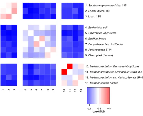
Woese and Fox concluded on their three-domain hypothesis based primarily on the sequence analysis of 18S ribosomal RNA (rRNA; eukaryotic) and 16S rRNA (prokaryotic) isolated from 13 different organisms. The rRNA gene is universally present in all forms of life on this planet. Since Sanger sequencing technology (Sanger et al., 1977) had not yet been available, Woese and Fox refined molecular methods of their time to find differences in sequential nucleotide compositions of rRNA gene transcripts. They digested 18S/16S rRNA with T1 RNase individually for each organism, the end product of which was used to generate oligonucleotide fingerprints via 2D-electrophoresis followed by sequencing the short oligonucleotides. The association coefficient S
ABwas calculated in pairwise manner for each of the sequences generated, corresponding to a similarity value between transcripts (Woese and Fox, 1977). Figure II.1-1 depicts a heatmap generated from these S
ABcoefficients. A clear trend towards separation of three domains of life is resolved due to the low S
ABcoefficients between them. Intra-domain similarity, however, also varied greatly.
1 In December 2012, Carl Woese passed away. For his lifework the reader is referred to the many reviews covering this topic, e.g. Fox, G.E. (2013). Carl R.
Woese, 1928-2012. Astrobiology 13, 1201-1202.
Figure II.1-1 Heatmap based on SAB coefficients provided in Woese and Fox, 1977. This particular image was generated using R (R-Development-Core-Team, 2005). Please note that Methanobacterium thermoautotrophicum is now referred to as
Methanothermobacter thermoautotrophicus. Deep red is indicative of a high SAB value (similarity score), whereas dark blue reflects a low similarity value.
1.2. 16S rRNA gene based phylogenetics
Unlike Woese’s and Fox’s, by way of Sanger sequencing modern day scientists have easy access to the entire ca. 1500 base pair 16S rRNA gene of Prokaryotes (Sanger et al., 1977).
Accurately assessing the entire rRNA gene sequence similarity shared between disparate organisms not only facilitates the generation of much more meaningful dissimilarity values, it also allows to infer phylogenetic relationships based on a single gene (Fox et al., 1977). Particularly the comparison of color intensities of the heatmap presented in Figure II.1-1 and the heatmap presented in Figure II.1-2A verifies the superiority of information that can be gathered from entire 16S/18S rRNA gene compared to the S
ABvalues used by Woese and Fox, 1977.
Today, the widespread application of 16S rRNA gene sequencing and analysis has enabled many researchers to identify and infer the phylogenetic position of microbial isolates as depicted in Figure II.1-2B (Fox et al., 1977). However, Stackebrandt and Goebel have cautioned that while 16S rRNA gene phylogeny can be used to infer relationships for Bacteria and Archaea from kingdom down to species level, it is limiting with respect to strain specificity (Stackebrandt and Goebel, 1994). This circumstance was supported by for instance Ash et al., who reported the inability in resolving the Bacillus cereus group to the species level via 16S rRNA gene sequence analysis (Ash et al., 1991).
Nevertheless, 16S rRNA gene sequence analysis has revolutionized environmental microbiology.
As it is currently estimated that fewer than 1 in every 100 prokaryotic lineages on Earth can be cultivated under laboratory conditions (Amann et al., 1995; Colwell, 1997), the value of methods to collect, process, purify, and analyze 16S rRNA from the environment, and thereby circumventing the
1 2 3 4 5 6 7 8 9 10 11 12 13
13. Methanosarcina barkeri
12. Methanobacterium sp., Cariaco isolate JR−1 11. Methanobrevibacter ruminantium strain M-1 10. Methanobacterium thermoautotrophicum 9. Chloroplast (Lemna)
8. Aphanocapsa 6714 7. Corynebacterium diphtheriae 6. Bacillus firmus
5. Chlorobium vibrioforme 4. Escherichia coli 3. L cell, 18S 2. Lemna minor, 18S
1. Saccharomyces cerevisiae, 18S
0.1 0.3 0.5
SAB-value II. General introduction
need to culture cells, is tremendous. Such techniques have, in many instances, provided the only source of understanding the unseen diversity of bacteria and archaea contributing to Earth’s elemental cycles (Pace et al., 1986; Ward et al., 1990).
Figure II.1-2 A) Heatmap based on rRNA gene similarity. Gene sequences were downloaded from the SILVA database and clustered using the average neighbor algorithm in mothur (Pruesse et al., 2007; Schloss et al., 2009). Dark red is indicative of
high similarity, whereas dark blue reflects low similarity. B) Phylogenetic tree consisting of the same rRNA genes used for A.
While the intra-domain phylogeny is lacking information due to under-sampling of the phylogenetic clades available, the three-domain hypothesis easily resolved. The phylogenetic tree was generated using FastTree V2.0 (maximum likelihood
approximation) and visualized in iTOL (Letunic and Bork, 2007; Price et al., 2010).
As technologies continue to evolve, 16S rRNA gene sequence analysis is nowadays barely used to infer the phylogenetic position of individual microorganisms unless scientist describe novel species.
Instead, this technique has proven important for deciphering the entire microbial communities housed within a given environmental sample. To accomplish this, researchers extract total environmental
‘metagenomic’ DNA and amplify the 16S rRNA genes via polymerase chain reaction (PCR) with archaea- or bacteria-directed primers [a detailed evaluation and review of frequently used 16S rRNA gene primers was recently published by Klindworth and co-workers (Klindworth et al., 2013)]. The resulting 16S rRNA gene amplicons are then either cloned into competent E. coli cells and afterwards sequenced via Sanger methods (Sanger et al., 1977) or directly subjected to next generation sequencing [NGS, (Sogin et al., 2006); for details on NGS please see II.2.3 and a review by Hutchison (Hutchison, 2007)]. The acquisition and analysis of 16S rRNA gene NGS data has been thoroughly standardized through existing software pipelines (Caporaso et al., 2010; Edgar, 2013; Schloss et al., 2009). Consequently, the NGS approach is no longer used to describe the alpha-diversity of samples, but rather community relationships between samples as well as changes in microbial abundance of
1 2 3 4 5 6 7 8 9 10 11 12 13
13. Methanosarcina barkeri, AJ012094 12. Methanobacterium formicicum, AF169245 11. Methanobrevibacter ruminantium, AY196666 10. Methanothermobacter thermautotrophicus, AY196600 9. Chloroplast (Arabidopsis thaliana), FB152799 8. Aphanocapsa muscicola VP3−03, FR798916 7. Corynebacterium diphtheriae, X84248 6. Bacillus firmus, D16268
5. Prosthecochloris vibrioformis, M62791 4. Escherichia coli, X80725 3. Homo sapiens, 18S, AADB02002333 2. Arabidopsis thaliana, 18S, AC006837 1. Saccharomyces cerevisiae, 18S, AY251631
0.2 0.6 16S rRNA gene similarity
0.01
Escherichia coli
Prosthecochloris vibrioformis Bacillus firmus
Corynebacterium diphtheriae Aphanocapsa muscicola VP3−03
Chloroplast (Arabidopsis thaliana)
Methanosarcina barkeri Methanobrevibacter ruminantium
Methanothermobacter thermautotrophicus Methanobacterium formicicum
Homo sapiens (human) Saccharomyces cerevisiae
Arabidopsis thaliana (thale cress)
A
B
single taxa as a function of time and/or geographic location [for a summary and comparison of the various community relationship calculations that exist in the literature, the reader is referred to the study by Kuczynski and coworkers (Kuczynski et al., 2010)]. NGS-generated beta-diversity and gamma-diversity analyses contributed significantly to the Human Microbiome Project and the Earth Microbiome Project, whose objectives were to elucidate the entire spectrum of microorganisms living on the human body and on our home planet, respectively (Gilbert et al., 2010a; Gilbert et al., 2010b;
Wortman et al., 2010). Ergo, it has become commonplace in the literature to refer to the total genetic information pertaining to all of microorganisms (bacteria, archaea, fungi, and some lower Eukaryota) present in a given environmental sample as its ‘microbiome’. When such information is restricted to the domain Bacteria, the term ‘bacteriome’ found great acceptance in the literature. Along these lines of reasoning, the term ‘archaeome‘ — referring to the total genetic information pertaining to all of the archaea detected in an environmental sample — is used in this study, although it has not appeared in the literature so far. For any given sample, its bacteriome and archaeome can determined to the exclusion of the other via 16S rRNA gene PCR/amplicon analysis with domain-directed primers, as mentioned above.
1.3. Phylogenomics and the two-domain hypothesis
Analysis and interpretation of 16S rRNA gene phylogenetics have their limitations, not only in deciphering differences between strains but also at higher taxonomic levels. Ancient evolutionary relationships can barely be recovered by molecular phylogenies in general (Gribaldo and Philippe, 2002), since successive substitutions can disguise early speciation events, particularly when computing phylogenetic relationships from single marker genes like 16S rRNA gene (Gribaldo et al., 2010). As a result, the inferred evolutionary relationship between Archaea, Bacteria, and Eukaryota can consequently lead to different results depending on the gene analyzed (Harris et al., 2003).
Consequently, the ‘conditioned reconstruction’ of the entire genomic information (Lake and Rivera, 2004) and the concatenation of orthologous genes to large alignments (Delsuc et al., 2005; Snel et al., 2005) have each been applied for to better resolve dissimilarities between taxonomic lineages. The latter methodology, in particular, has gained considerable popularity. By calculating dissimilarities between concatenated ribosomal protein sequences, this approach has become the standard for inferring phylogenetic positions of newly sequenced bacterial and archaeal genomes (Castelle et al., 2013; Gribaldo et al., 2010; Kozubal et al., 2013; Rinke et al., 2013; Spang et al., 2010; Williams et al., 2013; Wrighton et al., 2012), although sequences of proteins involved in replication also seemed to appropriately reflect phylogeny (Raymann et al., 2014).
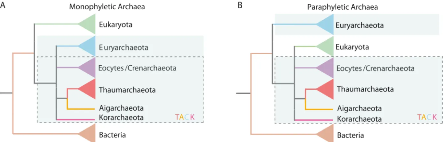
The associated methodologies, pitfalls, artifacts, and major encompasses of evolutionary reconstruction of the tree of life have recently been reviewed (Gribaldo et al., 2010; Williams et al., 2013), and the authors agree upon one major conclusion. Currently, there are two phylogenetic scenarios plausible for the relationship between Archaea, Bacteria, and Eukaryota: A) the ‘three primary domains’ (3D) and B) the ‘two primary domains’ (2D) scenario (Figure II.1-3). While the former is similar to what had been proposed by Woese et al. 1977 and 1990 (Woese and Fox, 1977; Woese et al., 1990), the latter suggests a sister relationship between Eukaryota and the Crenarchaeota (also
II. General introduction
known as ‘eocytes’), where Archaea are the direct ancestor of Eukaryota (Guy and Ettema, 2011;
Lake et al., 1984).
Euryarchaeota
Eocytes/Crenarchaeota Eukaryota
Bacteria
Eocytes /Crenarchaeota Paraphyletic Archaea
Thaumarchaeota Aigarchaeota Korarchaeota Euryarchaeota
Eocytes/Crenarchaeota Eukaryota
Bacteria Euryarchaeota Eocytes /Crenarchaeota Monophyletic Archaea
Thaumarchaeota Aigarchaeota
Korarchaeota TACK TACK
B A
Figure II.1-3 Phylogenetic trees taken from Williams et al., which reflect the three-domain scenario. A) and the two domain scenario. B) (Williams et al., 2013). In the 2D scenario, the Archaea form a paraphyletic group. [TACK = Thaumarchaeota-
Aigarchaeota-Crenarchaeota-Korarchaeota superphylum, (Guy and Ettema, 2011)]
Although Gribaldo et al. argued that the final scenario cannot yet be determined (with adequate confidence), Williams et al. maintain that the 2D scenario presents the most plausible solution as it is well resistant to eukaryotic-archaeal horizontal gene transfer [HGT; (Gribaldo et al., 2010; Williams et al., 2013)]. Nevertheless, many questions remain unanswered — perhaps none greater than the question for the different lipid architecture of Archaea and Eukaryota. This is, however, possibly also explainable by HGT as bacteria may have transferred their lipid biosynthesis machinery to Eukaryota (Gribaldo et al., 2010; Pereto et al., 2004). In any scenario, whether the Archaea are a sister domain of the Eukaryota or their direct ancestors, it is necessary to A) study the biology of Archaea in depth and B) taxonomically sample Earth’s life sufficiently in order to be able to reconstruct proper phylogeny and understand the processes behind Eukaryogenesis (Gribaldo et al., 2010).
1.4. Refining the tree of life: Metagenomics and single cell sequencing
Neither the 2D nor the 3D scenario is supported by adequate empirical evidence, however, as
the 3D paradigm has been widely accepted in the literature of the past 20 years (Woese et al., 1990),
the current thesis refers to Archaea as the third domain of life. Historically, this domain comprised two
major phyla, the Euryarchaeota and the Crenarchaeota (Woese et al., 1990). Nevertheless, in recent
years, many more phyla were proposed, whereas the Korarchaeota (Barns et al., 1996), the
Aigarchaeota (Nunoura et al., 2011), and the Thaumarchaeota (Brochier-Armanet et al., 2008; Pester
et al., 2011; Spang et al., 2010) have found great acceptance in the literature so far. The phylum
Nanoarchaeota is currently represented by a few genomes and only one cultured organism,
Nanoarchaeum equitans (Huber et al., 2002; Podar et al., 2013; Waters et al., 2003). The
Nanoarchaeota have been discussed to be a ‘fast-evolving euryarchaeal lineage’, and their status as
a separate phylum is currently uncertain (Brochier et al., 2005). Additional phyla, like the
Geoarchaeota, have been proposed (Kozubal et al., 2013), but recent re-analysis of the phylogenomic
positioning of the Geoarchaeota revealed this archaeal lineage as Thermoproteales-related (Guy et al.,
2014).
The aforementioned Thaumarchaeota are a remarkable case where single gene phylogenetics (16S rRNA gene) fail to reconstruct evolutionary relationships. Previously, members of this phylum were described as widespread crenarchaeota contributing substantially to nitrogen cycling via ammonia-oxidation in the ocean and soil biotopes (DeLong, 1992; Fuhrman et al., 1992; Karner et al., 2001; Konneke et al., 2005; Leininger et al., 2006). Using phylogenomics, these crenarchaea were separated from others and reclassified as Thaumarchaeota, whose distribution on Earth still necessitates exploration (Brochier-Armanet et al., 2008; Pester et al., 2011; Spang et al., 2010).
At least two of the aforementioned phyla of Archaea (Korarchaeota and Aigarchaeota) have been designated based on genomic data not generated from pure cultures (Elkins et al., 2008;
Nunoura et al., 2011). Such a reconstruction of genomes from enrichment cultures or directly from environmental samples is termed ‘metagenomics’ and was launched by two independent groups in 2004 (Tyson et al., 2004; Venter et al., 2004). Venter’s group generated a huge set of environmental sequences never seen before, which continues to reveal interesting hypotheses as scientists further explore and reanalyze these data. For instance, a recent publication based solely on these sequencing data claimed indications for a ‘fourth domain of life’ (Wu et al., 2011). Nevertheless, these sequences may also belong to unknown viruses (Wu et al., 2011), like giant viruses that even contain ribosomes (Philippe et al., 2013), and their positioning as the fourth domain of life continues in the literature (Colson et al., 2012; Colson et al., 2011; Legendre et al., 2012; Williams et al., 2011). In contrast to generating huge amounts of shotgun sequencing data, Tyson et al. focused on the reconstruction of entire genomes or draft genomes. Since then, multiple genomes of different bacteria and archaea have been reconstructed from the environment, which has lead to the proposal of many Candidate phyla (Castelle et al., 2013; Di Rienzi et al., 2013; Nunoura et al., 2011; Sharon and Banfield, 2013; Wrighton et al., 2012).
Another approach to get an insight into the genomes of the uncultivated majority of microorganisms in the environment is the amplification of DNA from a single-cell (Zhang et al., 1992) followed by sequencing, also termed single-cell genomics (Hutchison and Venter, 2006). This method has recently been performed on large scale (Rinke et al., 2013). Rinke et al. reconstructed 201 different microbial genomes, which had, however, only a completeness of 40% on average (ranging from 10% to 90%). The lack of completeness is one downside of single-cell genomics compared to metagenomics, where multiple genomes of closely related or identical organisms are assembled (Sharon and Banfield, 2013). Rinke et al. ended up proposing a superphylum called DPANN (representative for the lineages Diapherotrites, Parvarchaeota, Aenigmarchaeota, Nanoarchaeota, and Nanohaloarchaeota) at the base of the Archaea, which unified many environmental genomes gathered from nano-sized archaea, including the Nanoarchaeota (Rinke et al., 2013). However, this incorrect placement of the Nanoarchaeota in the phylogenetic tree of Archaea has been noted earlier (Brochier et al., 2005), and Castelle et al. recently reported that not all nano-sized archaea cluster together when using appropriate phylogenomics coupled with increased taxon sampling (Castelle et al., 2013).
These results were confirmed by a recent study that used DNA replication protein sequences instead of ribosomal protein sequences for phylogenetic inference (Raymann et al., 2014). Thus, the enigmatic
II. General introduction
clustering of nano-sized archaea can be attributed to an insufficient taxon sampling of these lineages resulting in a long branch attraction artifact (Felsenstein, 1978; Lartillot et al., 2007).
Many metagenomic studies have not only amplified our knowledge on the phylogeny of Archaea and Bacteria, they have also shed light on the potential metabolic capabilities of the uncultivated majority in our ecosystems (Albertsen et al., 2013; Baker et al., 2010; Baker et al., 2006; Castelle et al., 2013; Erkel et al., 2006; Kantor et al., 2013; Nunoura et al., 2011; Tyson et al., 2004; Wrighton et al., 2012). Particularly, novel archaeal lineages were shown to carry a great proportion of dark matter concerning their genomic coding potential (Baker et al., 2010; Baker et al., 2006; Nunoura et al., 2011). These unexplored genes may encode for novel enzymatic pathways and metabolic capabilities that go way beyond the knowledge gathered from cultivated microorganisms. At the same time, new archaeal lineages also harbor great novelty concerning their cell ultrastructure (Comolli et al., 2009;
Moissl et al., 2005b). For instance, more than 37% of the genes in ARMAN (Archaeal Richmond Mine Acidophilic Nanoorganisms) showed no matches to clusters of orthologous groups in Archaea, and these novel lineages were also described to be one of the first archaea to possess a double- membrane system (Baker et al., 2010; Comolli et al., 2009). These findings highlight the importance of exploring novel, untapped biotopes to understand diversity and function of uncultivated archaea.
Such novel biotopes are mainly found in the subsurface, as it accommodates a great proportion of microbial life that has been barely explored due to the lack of accessibility (Whitman et al., 1998).
Indeed, Castelle et al. and Nunoura et al. showed that subsurface samples can reveal many unexplored archaeal phyla and represent a great reservoir for mining novel archaeal taxa (Castelle et al., 2013; Nunoura et al., 2011). Such a novel, uncultivated archaeal taxon called SM1 Euryarchaeon is found near Regensburg, Germany, in a cold sulfidic spring, where it is constantly washed up from the subsurface (Henneberger et al., 2006). Although this archaeon appeared to be a novel lineage within the Euryarchaeota based on 16S rRNA gene sequence analysis (Rudolph et al., 2001), it remains completely unexplored with regard to its genomic information, metabolic capabilities or phylogenomic placement. Related 16S rRNA gene sequences of this novel archaeal taxon were described to be wide-spread (Rudolph et al., 2004) and also found to be dominant in hydrogen- enriched, anoxic subsurface hydrothermal waters (Chapelle et al., 2002). The latter biotope was intensely discussed to be an analogous biotope of foreign celestial bodies in our solar system like Mars and Europa [moon of Jupiter; (Chapelle et al., 2002)]. Consequently, life thriving in these subsurface environments could potentially resemble extraterrestrial life that mankind still desires to discover — if it exists.
2. Astrobiology and the search for (extra)terrestrial life
Astrobiology is an interdisciplinary field unifying numerous science disciplines and space
technologies to answer mankind’s fundamental questions, such as ‘How did life originate?’, ‘Are there
other habitable planets?’, and ‘Does life exist anywhere else in the universe?’. Many subdisciplines
have evolved in recent decades, all of them shaping the picture of the relatively new field of
Astrobiology
(Morrison, 2001).
2.1. Panspermia as the origin of life?
Approximately 100 years ago, Arrhenius formed the hypothesis that life may not have originated on Earth but on another celestial body in the galaxy and may have been transported to Earth as microbial life (Arrhenius, 1908). This theory was named ‘Panspermia’, and numerous incarnations have been formed, like Lithopanspermia (transmission of microorganisms via meteorites (Tobias and Todd, 1974)) or Radiopanspermia [the distribution of life via starlight (Secker et al., 1994)]. It was concluded that life may be ubiquitous in the galaxy, if a theory like Radiopanspermia was real (Parsons, 1996). A more exotic theory is the Directed Panspermia, which hypothesizes that extraterrestrial intelligent life forms have sent microbial life to Earth (Crick and Orgel, 1973). However, it has been calculated that even within our solar system, terrestrial life could have been spread to other celestial bodies via Lithopanspermia. These celestial bodies should not be considered as isolated, and planets like Mars could therefore harbor microbial life that originated on Earth and vice versa (Melosh, 1988). Nevertheless, the search for extraterrestrial life is a great desire of mankind and continues with many ongoing and future life detection space missions.
2.2. Planetary protection a.k.a. applied astrobiology
As mentioned above, astrobiology holds many subdisciplines, one of them being the field of
‘planetary protection’. In this applied research area, scientists work to prevent the inadvertent contamination of otherwise pristine celestial bodies with terrestrial life forms while conducting various space exploration initiatives (Rummel, 1989). For instance, exceedingly large numbers of terrestrial life forms must not be transported to an extraterrestrial setting (e.g. Mars) via spacecraft, as their presence could confound present and future life detection missions. In addition, they could even colonize the environment and interfere with the natural progression and evolution of indigenous life forms (termed ‘forward contamination’). Conversely, ‘reverse contamination’ refers to a scenario in which extraterrestrial life forms are inadvertently returned to Earth. Any such instance could prove harmful to the planet’s biosphere. Missions to different celestial bodies are categorized based on many attributes pertaining to the scientific interest in the selected destination, and different types of missions have different requirements with regard to cleanliness of spacecraft hardware [for details please see (COSPAR, 2002)].
As extraterrestrial life has yet to be discovered, there are those that have criticized planetary protection as it may be unnecessary but expensive (Fairén and Schulze-Makuch, 2013); others, however, contend that it represents a very important scope of application in astrobiology (Conley and Rummel, 2013). Particularly, safeguarding life detection missions to avoid false positives is of high interest — as recently demonstrated by Mars Science Laboratory’s drill bit debacle
1.
As was the case for the Mars Science Laboratory (also known as ‘Curiosity’ rover), all spacecraft hardware outbound Earth needs to be assembled in a controlled facility, i.e. a cleanroom, in order to minimize contamination risk. This helps to ensure the integrity (with respect to cleanliness) of the hardware surfaces and minimizes the likelihood of stowaway contaminant microbes inadvertently being transferred to extraterrestrial settings of scientific interest. Such cleanroom facilities
II. General introduction
1 Los Angeles Times/McClatchy / September 10, 2012;
typically maintain a constant humidity, are equipped with redundant air-filtering systems, and are subjected to rigorous cleaning regimens. Consequently, these cleanrooms harbor low nutrients for microorganisms. This is ideal, since microorganisms that enter cleanrooms on equipment and humans can be very problematic and even threaten forward contamination (Venkateswaran et al., 2001). The molecular signatures of microorganisms that have been detected in cleanroom facilities belonged to all three domains of life, Bacteria, Archaea, and Eukaryota (La Duc et al., 2012; Moissl et al., 2008; Venkateswaran et al., 2001).
Since the Viking era, scientists have labored fervently to catalog the entire microbial population associated with spacecraft hardware and the cleanroom facilities in which it is assembled (Puleo et al., 1977). As a result, the techniques by which to assess the cleanroom microbiome have evolved significantly, coinciding with a great many other methodological advancements in the field of microbial ecology.
2.3. Deciphering microbiomes: The transfer of technologies from microbial ecology to planetary protection
The field of microbial ecology has experienced two major milestones in microbial community analysis. First and foremost, the ability to directly analyze 16S rRNA (genes) retrieved from an environmental sample has tremendously improved the current understanding of the extent of — and role of — the uncultivated portion of a given environmental microbial community (Pace et al., 1986;
Ward et al., 1990). Historically, the 16S rRNA gene has been proven a suitable marker gene due to its length that can be covered by two (to several) Sanger sequencing reads and due to its 9 (hyper-)variable regions flanked by conserved regions allowing the inference of phylogenetic relationships between microorganisms (Fox et al., 1980; Lane et al., 1985; Sanger et al., 1977; Ward et al., 1990; Woese and Fox, 1977). The second milestone was the advent of so-called ‘metaomics’
and their application to microbial community samples. This discipline covers the analysis of an environmental sample to reconstruct microbial genomes [metagenomics, (Tyson et al., 2004)], microbial transcriptomes [metatranscriptomics; (Poretsky et al., 2005)] and microbial proteomes [metaproteomics; (Wilmes and Bond, 2004)] of an entire community. Other ‘omics’ approaches like metabolomics and metalipidomics have been reviewed by Zhang and coworkers (Zhang et al., 2010).
16S rRNA gene analysis, metagenomics, and metatranscriptomics have all benefited immensely from NGS technologies, such as illumina/solexa (Bennett, 2004; Bennett et al., 2005), 454 pyrosequencing (Nyrén et al., 1993; Ronaghi et al., 1996), and ion torrent (Rothberg et al., 2011).
NGS facilitates a greater sampling depth of molecules that can be assayed in comparison to the
outdated Sanger sequencing technology (Sanger et al., 1977). However, with respect to intrinsic
fidelity, these technologies suffer from short read length and/or homopolymer errors (Huse et al.,
2007; Margulies et al., 2005), which need to be quelled via appropriate bioinformatic tools (Gilles et
al., 2011; Hamady et al., 2008; Kircher et al., 2009; Preheim et al., 2013; Quince et al., 2009; Schiex
et al., 2003). Another technology that facilitated the high throughput of 16S rRNA gene amplicon
analysis is a hybridization based technology called PhyloChip
TMG3 (DeSantis et al., 2007; Hazen et
al., 2010). Here, 500 ng of DNA (PCR amplicons), corresponding to approximately 10
1116S rRNA
gene molecules, are fragmented, labeled, and hybridized onto an array, which carries ca. 1.1 million
probes (25-mers) designed from the entire Greengenes 16S rRNA gene database (DeSantis et al., 2006; Hazen et al., 2010; Probst et al., 2014). Although 10 to 100 fold more molecules of a single sample compared to an entire illumina HiSeq 2000 lane can be analyzed [www.illumina.com; (Probst et al., 2014)], the disadvantage of the PhyloChip technology was believed to be the limitation to already a priori 16S rRNA genes not allowing the discovery of novel phylogenetic taxa (Brodie et al., 2007; La Duc et al., 2009). In a recent publication we introduced a novel, empirical, and unsupervised method for taxon discovery from PhyloChip data (Probst et al., 2014). The new software termed
‘sinfonietta’ correlates probes across different arrays and across taxonomic affiliation to reconstruct 16S rRNA genes from the existing probe collection. Our data previously published in Science Magazine (Hazen et al., 2010) was re-analyzed, and an outlier sample was discovered not seen by the reference-based microarray analysis applied those days (Probst et al., 2014). Ergo, this technology appears to be a very attractive alternative to NGS-based 16S rRNA gene analysis enabling a deeper assay of microbial communities.
Each and every molecular analysis technique has its advantages and pitfalls, and as such, no single technique can be used to explain composition, dynamics, and function(s) of microbial communities. For instance, 16S rRNA gene analysis can be very useful to discover microbiome changes between different samples, but has great limitations in establishing evidence for microbiome function (Langille et al., 2013). Meanwhile, metagenomics enable the reconstruction of genomes and metabolic pathways from the environment but give no information on gene expression, enzyme activity or even the viability of a detected organism. Consequently, these and other high throughput technologies need to be combined with other (molecular) tools to unveil the countless facets at play in a typical environmental microbial community.
With a delay of approximately 10 years, planetary protection practitioners have at least partially applied the aforementioned novel methodologies of microbial community analysis to generate a comprehensive microbial inventory associated with spacecraft hardware and its assembly facilities.
This delay in application is largely a consequence of the extremely low-biomass nature of such environments – many emerging technologies simply require more biomaterial than these ultra-clean surfaces typically harbor (Kwan et al., 2011; Probst et al., 2010a; Probst et al., 2011). Standard cultivation-dependent assays for heat-shock resistant microbes performed by both NASA and ESA are used regularly to estimate the total spore burden of spacecraft. This approach is well established and enables comparisons between recently gathered and previously acquired datasets [e.g., Viking era spacecraft (Puleo et al., 1977)]. In contrast, molecular assays that provide insight beyond the cultivable microbiome are rather fast developing and can become outdated quickly but provide unique insights into the microbiome beyond cultivability (Ward et al., 1990). In 2001, Venkateswaran and co- workers were the first to apply bacteria-directed 16S rRNA gene cloning on spacecraft assembly facility samples, which gave raise to a more enhanced understanding of microbial cleanroom diversity (Venkateswaran et al., 2001). The utilization of PhyloChip technology by LaDuc et al. and Cooper et al. facilitated the discovery of an even greater microbial diversity, discovering parts of the ‘rare biosphere’ present in cleanrooms (Cooper et al., 2011; La Duc et al., 2009). Results of studies focusing on the bacteriome in spacecraft assembly cleanrooms have led to the conclusion that A)
II. General introduction
humans are the predominant source of microbial contaminants, and B) the cleanroom bacteriome very closely resembles the human bacteriome (Moissl et al., 2007).
Using 454 pyrotagsequencing, LaDuc et al. ascertained not only the bacterial diversity but also the archaeal and fungal diversity in samples taken from a rover of a recent Mars mission (La Duc et al., 2012). This study represented the first ever assessment of microbial diversity to encompass all three domains of life from one sample set. Archaea had, however, previously been detected in spacecraft assembly facilities in two independent reports (Moissl et al., 2008; Moissl-Eichinger, 2011).
Current investigations at NASA's Jet Propulsion Laboratory comprise also a metagenomic survey of a cleanroom microbiome, which may provide great information on microbiome function (pers. com.
Parag, Vaishampayan, Jet Propulsion Laboratory, CA, US, funded by NASA-ROSES 2011).
2.4. Obstacles in cleanroom microbiome research
Hitherto, all of the molecular-based studies of the cleanroom environment published have focused on alpha diversity and microbial community structure and development (Cooper et al., 2011;
La Duc et al., 2003; La Duc et al., 2009; La Duc et al., 2012; Moissl et al., 2008; Moissl et al., 2007;
Moissl-Eichinger, 2011; Probst et al., 2010b; Vaishampayan et al., 2010; Venkateswaran et al., 2001).
While these reports were appreciatively descriptive, they failed to overcome two significant obstacles, namely fractional viability of the population and the contribution of its archaeal members. In these studies, it is unclear whether the entire set of molecular data gathered originated solely from viable microbial cells or a mixture of living cells and the remnants of dead microorganisms. Many cultivation- based studies have purified bacterial isolates from cleanroom samples, and the proportion of cultivable species in such samples has been reported to approach 35% (Moissl-Eichinger et al., 2013). This finding should not be taken lightly, as this index is 35 times greater than the 1% estimated for the entirety of Earth’s cultivable microbial species (Amann et al., 1995; Colwell, 1997). Ultimately, however, the living proportion and diversity of bacteria in cleanrooms has yet to be thoroughly assessed.
The second obstacle that remains unresolved is the presence and function of archaea in
cleanroom facilities. Moissl et al. and Moissl-Eichinger have described the occurrence of archaea in all
spacecraft assembly cleanrooms that they studied and described their diversity to be dominated by
Thaumarchaeota (Moissl et al., 2008; Moissl-Eichinger, 2011), a phylum previously classified
crenarchaeotic and comprised of putative ammnonia-oxidizers (Brochier-Armanet et al., 2008; Pester
et al., 2011; Spang et al., 2010). With respect to the bacterial diversity, as mentioned above,
biodiversity resembles the human skin microbiome since humans are thought to be the major source
of microbial contaminants in spacecraft assembly cleanrooms (Moissl et al., 2007; Moissl-Eichinger et
al., 2013). However, the human skin has been considered almost Archaea-free (Grice and Segre,
2011; Hulcr et al., 2012), and Thaumarchaeota have only been reported on two human palms as
transient contaminants (Caporaso et al., 2011). Thus, the current knowledge of the human
microbiome and its impact on cleanroom diversity do not explain the occurrence of archaea in these
facilities.
3. The SM1 Euryarchaeon — facts, heuristics, hypotheses
3.1. The discovery of the SM1 Euryarchaeon and its life styles
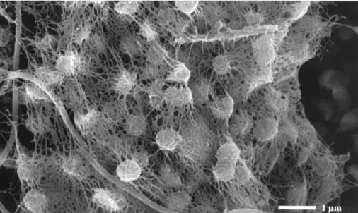
In 2001, Rudolph and coworkers discovered streamers with string-of-peals like morphology floating in the streamlet of a marsh environment of the Sippenauer Moor, Germany. The water was reportedly strong with sulfidic odor and was thus identified to be sulfide-rich and oxygen-limited (Rudolph et al., 2001). Detailed diverse microscopic analyses of these string-of-pearls structures revealed that the interior of each pearl consisted primarily of archaeal cocci, while the outside was formed by filamentous bacteria. These bacteria, later characterized as Thiothrix sp. (Moissl et al., 2002), also formed the string spanning the various pearls. This spatially coordinated association of novel archaea and (potentially) sulfide-oxidizing bacteria was appropriately termed ‘string-of-pearls community’ (Rudolph et al., 2001). This coordination was illustrated in many electron microscopy (EM) and fluorescence in situ hybridization (FISH) micrographs (Moissl et al., 2002; Moissl et al., 2003;
Rudolph et al., 2004; Rudolph et al., 2001); an example is given in Figure II.3-1. The cells of the archaeon were ca. 0.5 µm in diameter and, while embedded in this matrix, displayed hundreds of surface-bound appendages called hami [plural; singular: hamus; (Moissl et al., 2005b), please see also chapter II.3.2.].
Figure II.3-1 Scanning electron micrograph of the inner of a pearl. Small cocci (archaea) are embedded in a dense network of cell surface appendages and surrounded by filamentous bacteria (Rudolph et al., 2001).
Using 16S rRNA gene amplicon cloning, Rudolph et al. also characterized the novel archaeon phylogenetically and demonstrated that it branched deeply within the Euryarchaeota but without any cultivated representative in the entire clade of environmental sequences. Due to its novelty, there was no prior knowledge on the environmental function of these archaea nor on their potential metabolism.
The archaeon was refractory to cultivation under laboratory conditions, and was thus termed ‘SM1 Euryarchaeon’, whereas SM stands for Sippenauer Moor, the first biotope where the archaeon was discovered (Rudolph et al., 2001).
The string-of-pearls community was grown in situ on polyethylene nets in the streamlets of the Sippenauer Moor. This method facilitated the collection of appreciable amounts of SM1 Euryarchaeon
II. General introduction
and associated community biomass with the help of state-of-the art purification techniques (Moissl et al., 2003). Although this community was referred to as the net population (Moissl et al., 2003), it was very similar to the original string-of-pearls community and is henceforth not differentiated herein.
Growing the string-of-pearls community in its actual biotope and harvesting high amounts of biomass sparked many subsequent studies that investigated the archaea and the community at molecular level [(Moissl et al., 2005b; Moissl et al., 2003), please see also chapter III.3. and III.4.].
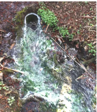
In another study of the group around Robert Huber the distribution of the SM1 Euryarchaeon was analyzed, and many other environmental 16S rRNA genes of the so-called SM1 group were retrieved (Rudolph et al., 2004). The study focused on sulfidic springs in Bavaria but also included samples from Turkey. All springs had representative sequences of the SM1 clade indicative of a certain environmental success of these microorganisms. Importantly, identical 16S rRNA gene sequences of the SM1 Euryarchaeon were retrieved from the Mühlbacher Schwefelquelle, Germany (Figure II.3-2). This sulfur spring is an artificial bore-hole and emits thousands of liters of water per hour; the chemical composition and temperature of its water was very similar to that found at Sippenauer Moor (Rudolph et al., 2004). In the streamlet a similar string-of-pearls community was found, also being comprised of the SM1 Euryarchaeon in the inner of the pearl but surrounded by the IMB1 Epslionproteobacterium; the environmental clade of these microoganisms was later classified Sulfuricurvum (genus) due to the cultivation of a representative (Kodama and Watanabe, 2004).
Sulfuricurva are filamentous bacteria capable of sulfide-oxidation and may therefore fulfill a similar symbiotic role for SM1 Euryarchaea at the Mühlbacher Schwefelquelle as Thiothrix sp. at the Sippenauer Moor (Rudolph et al., 2004).
Figure II.3-2 Mühlbacher Schwefelquelle (Regensburg/Burgweinting, Germany). The bore-hole has an approximate diameter of 25 cm and is man-made (Probst et al., 2013b).