Effects of oxytocin receptor variants on activated signaling cascades and cellular processes
Dissertation
Zur Erlangung des Doktorgrades der Naturwissenschaften (Dr. rer. nat.) der
Fakultät für Biologie und Vorklinische Medizin der Universität Regensburg
Vorgelegt von Magdalena Meyer
aus Roth
Juli 2020
- II -
- III - Die Arbeit wurde angeleitet von Dr. Benjamin Jurek.
Unterschrift:
- IV -
- V -
Oxytocin ist ein Neurohormon, welches eine wichtige Rolle bei sozialen Verhaltensweisen spielt.
Eine Fehlregulation des Oxytocin Systems kann daher zu tiefgreifenden Konsequenzen für die mentale Gesundheit führen. Solche Fehlregulationen können den Liganden selbst betreffen, wobei eine generell erniedrigte Konzentration bei einer Patientengruppe, die an einer Form von Autismus leidet, festzustellen ist. Des Weiteren konnte eine genetische Komponente, sogenannte Einzelnukleotidpolymorphismen (engl. single nucleotide polymorphisms, kurz SNPs), im Oxytocin Rezeptor Gen mit Autismus assoziiert werden. Aufgrund dieser Assoziationen und seiner vielversprechenden prosozialen und angstlösenden Wirkung, wird bereits seit längerem intensiv am Einsatz von Oxytocin als Therapeutikum bei psychosozialen Störungen geforscht. In den bestehenden Studien zeichnet sich jedoch ein eher kontroverses Bild ab. Während die Verabreichung einer einzelnen Dosis Oxytocin, das über ein Nasenspray appliziert wird, meist zu positiven Effekten in den Tests führte, kam es bei der wiederholten Gabe über einen längeren Zeitraum oft zu keiner Verbesserung der Symptome. Um eine langfristig sichere Therapie mit Oxytocin gewährleisten zu können, oder mögliche Alternativen zu identifizieren, müssen die molekularen Wirkmechanismen von Oxytocin und des Oxytocin Rezeptors genauer untersucht werden.
In meiner Doktorarbeit habe ich verschiedene Zelllinien verwendet, um die daran beteiligten Signalwege und zellulären Prozesse aufzuklären. So konnte ich zeigen, dass Oxytocin die Morphologie von Neuronen beeinflussen kann, indem es zu einer Änderung der Neuritenlänge führt. Ob sich dies in einer Verkürzung oder Verlängerung der neuronalen Fortsätze äußert, ist von der Expression und Aktivierung des Transkriptionsfaktors Myocyte Enhancer Factor (MEF) 2A abhängig. In diesem Zusammenhang konnte ich zwei Faktoren identifizieren, welche die Aktivität von MEF2A regulieren: den MAP Kinase Signalweg und die Phosphatase Calcineurin.
Zudem konnte ich durch einen Knock-out von MEF2A dessen zentrale Rolle bei der Funktion von Mitochondrien mit direkter Konsequenz für die Energiebereitstellung in Neuronen belegen.
Des Weiteren habe ich die Auswirkungen einer genetischen Variante des Oxytocin Rezeptors, des
SNPs rs4686302, der in verschiedenen Studien mit Autismus, Empathie und sozialen
Kognitionsstörungen assoziiert wurde, auf molekularer Ebene untersucht. Durch den
- VI -
unmittelbaren Vergleich zweier humaner Zelllinien, wobei eine die SNP- und die andere eine Wildtyp-Variante des Rezeptors exprimierte, konnte ich Abweichungen gezielt analysieren.
Durch eine Sequenzierung des Genoms fiel dabei eine Duplikation des Oxytocin Rezeptor Gens in der SNP-Zelllinie als weitere Variation auf. Auf Basis der beiden genetischen Varianten, konnte ich Unterschiede hinsichtlich der Rezeptorstabilität, des Calcium- und MAP Kinase Signalweges sowie eine umfassend differentielle Genregulation aufzeigen.
Zusammenfassend konnte ich durch meine Arbeit das Wissen über die zellulären Effekte von
Oxytocin und die Auswirkungen von genetischen Variationen, die das Oxytocin Rezeptor Gen
betreffen, erweitern und somit zu einem besseren Verständnis des Oxytocin Systems beitragen.
- VII -
This cumulative dissertation is composed of the following publications or manuscripts in preparation, in which I am first author:
A. Magdalena Meyer, Ilona Berger, Julia Winter, Benjamin Jurek (2018). Oxytocin alters the morphology of hypothalamic neurons via the transcription factor myocyte enhancer factor 2A (MEF-2A). Molecular and Cellular Endocrinology 10.1016/j.mce.2018.06.013 B. Magdalena Meyer, Kerstin Kuffner, Julia Winter, Inga D. Neumann, Christian H. Wetzel
and Benjamin Jurek (2020). Myocyte Enhancer Factor 2A (MEF2A) Defines Oxytocin- Induced Morphological Effects and Regulates Mitochondrial Function in Neurons. Int. J.
Mol. Sci. 2020, 21, 2200; 10.3390/ijms21062200
C. Magdalena Meyer, Vladimir Milenkovic, Christian H. Wetzel, Inga D. Neumann and Benjamin Jurek (in preparation). Functional Characterization of the Oxytocin Receptor Variant A218T.
In the course of this work, I contributed to further publications, which are not part of the dissertation:
D. Benjamin Jurek and Magdalena Meyer (2020). Anxiolytic and Anxiogenic? How the Transcription Factor MEF2 Might Explain the Manifold Behavioral Effects of Oxytocin.
Front. Endocrinol. 11:186; 10.3389/fendo.2020.00186
E. Julia Winter, Magdalena Meyer, Ilona Berger, Sebastian Peters, Melanie Royer, Dominik Langgartner, Stefan O. Reber, Kerstin Kuffner, Anna K. Schmidtner, Katharina Hübner, Finn Hartmann, Anna Bludau, Marta Bianchi, Simone Stang, Oliver J. Bosch, David A.
Slattery, Erwin van den Burg, Inga D. Neumann, and Benjamin Jurek (in preparation).
Chronic oxytocin-driven alternative splicing of CRFR2α induces anxiety.
F. Julia Winter, Magdalena Meyer, Simone Stang, Carl-Philip Meinung, Eva-Maria Rom-
Jurek, Christoph Irlbeck, Katharina Limm, Peter Oefner, Gero Brockhoff, Inga D. Neumann,
Benjamin Jurek (in preparation). A CRISPR-Cas-generated oxytocin receptor knock out
causes morphological alterations in neuronal cells.
- VIII -
- IX - Publication A
The research was designed by Benjamin Jurek and myself. Ilona Berger, Julia Winter, Benjamin Jurek and myself performed and analyzed morphological experiments. Immunocytochemical stainings were performed by Ilona Berger. Cell stimulations and Western Blots were performed by myself. The work was supervised by Benjamin Jurek. The publication was written by Benjamin Jurek and myself.
Publication B
The research was designed by Christian H. Wetzel, Benjamin Jurek and myself. The CRISPR-Cas9 mediated knockout of MEF2A was done together with Julia Winter. Immunofluorescence stainings were performed by Benjamin Jurek. Mitochondrial Respiration Analysis was conducted by Kerstin Kuffner and me. The morphology experiments were analyzed by myself, Julia Winter and Benjamin Jurek. Transfections, Western Blots and cellular assays were performed by myself.
The work was supervised by Benjamin Jurek and Inga D. Neumann. The publication was written by Benjamin Jurek and myself.
Publication C
The research was designed by Christian H. Wetzel, Inga D. Neumann, Benjamin Jurek and myself.
Transduction and establishment of cell lines was performed by myself. Calcium Imaging was performed by Vladimir Milenkovic and myself. Assays and Western Blots were performed by myself. The work was supervised by Inga D. Neumann and Benjamin Jurek. The publication was written by Benjamin Jurek and myself.
Publication D
The publication, a mini-review, was written by Benjamin Jurek and myself.
- X - Publication E
The research was designed by Inga D. Neumann, Benjamin Jurek and Julia Winter. The experimental work was performed by all authors including myself. In detail, I helped with animal experiments, membrane fractioning and data analysis. The work was supervised by Inga D.
Neumann and Benjamin Jurek. The publication was written by Julia Winter, Erwin van den Burg, Inga D. Neumann and Benjamin Jurek.
Publication F
The research was designed by Julia Winter and Benjamin Jurek, and supervised by Benjamin
Jurek. Julia Winter, Simone Stang, Carl-Philip Meinung, Eva Rom-Jurek, Christoph Irlbeck,
Katharina Limm, Benjamin Jurek and myself performed and analyzed experiments. The
publication was written by Julia Winter, Eva Rom-Jurek, Katharina Limm, and Benjamin Jurek.
- XI -
KURZFASSUNG DER ARBEIT ... V LIST OF PUBLICATIONS ... VII PERSONAL CONTRIBUTIONS ... IX TABLE OF CONTENTS ... XI
1 GENERAL INTRODUCTION ... 1
1.1 Oxytocin ... 1
1.2 Oxytocin receptor ... 2
1.3 Oxytocin receptor-coupled signaling ... 4
1.4 Single nucleotide polymorphisms in the oxytocin receptor gene ... 7
1.5 Aim of the thesis ... 9
2 OXYTOCIN ALTERS THE MORPHOLOGY OF HYPOTHALAMIC NEURONS VIA THE TRANSCRIPTION FACTOR MYOCYTE ENHANCER FACTOR 2A (MEF2A) ... 11
2.1 Abstract ... 11
2.2 Introduction ... 11
2.3 Materials and methods ... 13
2.3.1 Cell culture ... 13
2.3.2 Cell viability assay ... 14
2.3.3 Protein isolation ... 14
2.3.4 Western blot ... 14
2.3.5 Cell stimulations ... 15
2.3.6 Morphological assessments ... 16
2.3.7 Statistical analysis ... 17
2.4 Results ... 17
2.4.1 OTR activation mediates OT-induced changes in neurite length and nucleus size ... 17
2.4.2 OT leads to a MAPK-dependent neurite retraction as well as increased cell viability, whereas vasopressin affects neurite length MAPK-independently ... 20
2.4.3 OT increases transcriptional activity of MEF2A ... 21
2.4.4 OT-induced neurite retraction is reversed by MEF2A knock down ... 22
- XII -
2.5 Discussion ... 24
2.6 Conclusion ... 27
3 MYOCYTE ENHANCER FACTOR 2A (MEF2A) DEFINES OXYTOCIN-INDUCED MORPHOLOGICAL EFFECTS AND REGULATES MITOCHONDRIAL FUNCTION IN NEURONS ... 29
3.1 Abstract ... 29
3.2 Introduction ... 29
3.3 Material and methods ... 32
3.3.1 Cell culture ... 32
3.3.2 CRISPR-Cas9 mediated knockout of MEF2A ... 32
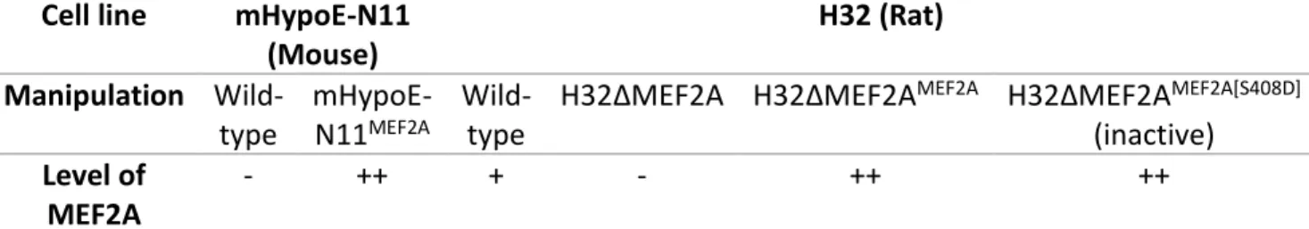
3.3.3 Transfection of H32 and mHypoE-N11 Cells with MEF2A overexpression plasmids ... 33
3.3.4 Cell stimulations ... 33
3.3.5 Protein isolation ... 33
3.3.6 Western blotting ... 34
3.3.7 Immunofluorescence ... 34
3.3.8 Morphological characterization ... 35
3.3.9 Cell viability assay ... 35
3.3.10 Mitochondrial respiration analysis ... 36
3.3.11 CellTiter-Glo 2.0 assay ... 36
3.3.12 Statistical analysis ... 36
3.4 Results ... 37
3.5 Discussion ... 45
3.6 Appendix ... 49
4 FUNCTIONAL CHARACTERIZATION OF THE OXYTOCIN RECEPTOR VARIANT A218T ... 51
4.1 Abstract ... 51
4.2 Introduction ... 51
4.3 Material and methods ... 53
4.3.1 Cell culture ... 53
4.3.2 Transduction of HEK293 cells with OTR gene variants ... 53
4.3.3 Establishment of monoclonal cell lines expressing the OTR ... 54
4.3.4 Whole-genome sequencing ... 54
4.3.5 Cytosolic calcium imaging with Fura-2/AM ... 54
- XIII -
4.3.8 Cycloheximide protein degradation assay ... 56
4.3.9 Statistical analysis ... 57
4.4 Results ... 57
4.5 Discussion ... 70
5 GENERAL DISCUSSION ... 73
5.1 MEF2A as major regulator of OT-induced effects ... 73
5.2 The role of genetic variations affecting the OTR gene ... 76
5.3 Conclusion ... 82
6 ABBREVIATIONS ... 83
7 REFERENCES ... 87
8 ACKNOWLEDGEMENTS ... 101
- XIV -
- 1 -
1 General introduction
This section is intended to convey and expand biological principles, basics and functions relevant to my thesis. It forms the basis for the introductions in 2.2, 3.2 and 4.2.
1.1 Oxytocin
The neuropeptide oxytocin (OT) enjoys a good reputation, reflected by its common description as the love or cuddle hormone. Released during physiological processes that are usually positively associated like uterus contraction during birth, milk ejection during breast-feeding, and orgasms, it promotes feelings of love, trust, bonding and well-being (Jurek and Neumann, 2018). Hence, besides its important role in reproduction, it is involved in many social actions and interactions like mother-child bonding, caregiving and trusting behavior, empathy, pair bonding, sexual behavior, as well as anxiety and aggression.
In accordance with this overall beneficial picture, there seems to be a positive correlation between our states of mental health and endogenous OT levels. People within the first stages of a romantic relationship have higher levels of OT (Schneiderman et al., 2012; Acevedo et al., 2012).
In contrast, low levels of OT have been found in patients suffering from autism spectrum disorder (ASD) or depression (Parker et al., 2017; Yuen et al., 2014).
Given these correlations, and because of its prosocial and anxiolytic effects, it has been suggested as a promising therapeutic agent for many psychosocial disorders, such as anxiety disorder or ASD. Delivered through a nasal spray, OT raised hopes as a non-invasive treatment option to help people who avoid social interaction and persistently experience fear and mistrust.
However, there is a general inconsistency occurring in studies testing its potential as treatment,
especially when tested in long-term administration regimens. Comparing its effects on anxiety in
rodents upon acute single-dose versus chronic long-term administration, the thin line between
advantages and risks becomes quite apparent. An acute infusion of synthetic OT directly into the
paraventricular nucleus (PVN) of male and female rats increased the time they spent in the open
arms or lit compartment in the behavioral tests of the elevated plus maze or light dark box,
reflecting a decreased level of anxiety (Blume et al., 2008; Jurek et al., 2012). A chronic treatment
with 10 ng/h intracerebroventricular OT via osmotic minipumps however, induces anxiogenesis
- 2 -
in male mice (Peters et al., 2014) and, in combination with a mild stressor, in male and female rats (Winter et al., 2020).
A similarly controversial picture of OT emerges from studies examining its efficacy and safety as treatment for ASD patients. ASD is a neurodevelopmental disorder that affects communication and social behavior. People who suffer from ASD have difficulties interacting with other people and show impaired communication skills. For example, they do not to look at or listen to people or show facial expressions and movements not matching with what is being said. They often show restricted interests and repetitive behaviors, and slight changes in their daily routine can make them feel upset and extremely irritated (Freitag and Konrad, 2014). Those psychological traits hamper their abilities to function properly in school, work, and private life. Upon administration of one single dose of OT, several studies confirmed improvements in a wide range of social behaviors, as well as a reduction of repetitive behaviors (Watanabe et al., 2014; Aoki et al., 2014).
However, as an acceptable treatment for ASD, OT must have prolonged efficacy. While some studies claim considerable improvements in key symptoms of ASD by long-term administration of OT (Parker et al., 2017; Tachibana et al., 2013), other studies found no improvements (Guastella et al., 2015; Dadds et al., 2014).
In order to understand the opposing effects provoked by the same compound, we have to develop a deeper understanding of the molecular underpinnings and functions of the OT system.
OT consists of the nine amino acids: Cys-Tyr-Ile-Gln-Asn-Cys-Pro-Leu-Gly-NH
2, and is produced in the supraoptic nucleus (SON), accessory nuclei, and PVN of the hypothalamus (Jurek and Neumann, 2018). It can be released from magnocellular neurons by axonal and somatodendritic release, and into the periphery via axonal projections. Due to its similarity to the closely related nonapeptide vasopressin, OT can bind to the three vasopressin receptor subforms V1a, V1b and V2, although with reduced affinity compared to the main target of the natural ligand – the oxytocin receptor (Chini and Manning, 2007).
1.2 Oxytocin receptor
The oxytocin receptor (OTR) belongs to the family of G protein coupled receptors (GPCRs),
consisting of seven transmembrane helices, three extracellular as well as three intracellular loops
(Gimpl and Fahrenholz, 2001). In humans, the OTR is encoded by the OTR gene located on
- 3 -
chromosome 3 (3p25–3p26x·2) as an inverted 17-kb single-copy gene and consists of four exons and three introns. The coding sequence encompasses only the major part of Exon 3 and the first part of Exon 4 (Peter et al., 1995). The OTR forms homodimers and heterodimers with other GPCRs, which consequently results in an enhanced activation of receptor-coupled signaling cascades (Busnelli et al., 2016).
In the periphery, the OTR is expressed by the myoepithelial cells of the mammary gland, in both, the myometrium and endometrium of the uterus, in cardiomyocytes of the heart, dermal fibroblasts as well as osteoclasts and osteoblasts. In the mammalian brain, the OTR is expressed throughout many brain regions including the hypothalamus, prefrontal cortex, hippocampus, and amygdala (Jurek and Neumann, 2018). However, the OTR is generally expressed at low levels, rendering its detection in some regions quite challenging (Freund-Mercier et al., 1994).
In addition, the OTR membrane expression depends on ligand availability, since an increased ligand availability over prolonged periods can result in receptor desensitization and internalization. The desensitization of the OTR is initiated by the G protein-coupled receptor kinase 2, which phosphorylates and primes the OTR for subsequent ß-arrestin binding. Arrestin binding to the OTR induces uncoupling from the G proteins, blocks further signaling cascades and targets the OTR for internalization. The OTR is internalized in a clathrin pit-dependent mechanism and intracellularly stored in “Ras-related in brain 4/5”-vesicles that get recycled after four hours as shown in HEK293 cells (Conti et al., 2009). Interestingly, selective OTR ligands like the agonist carbetocin have been shown to induce β‐arrestin‐independent internalization without recycling back to the plasma membrane (Passoni et al., 2016).
These processes, resulting in a reduced membrane expression, bear consequences for a therapy that would encompass repeated daily intranasal applications of OT over a longer period.
Besides the natural ligands OT and vasopressin, there are several synthetic OTR agonists and antagonists that can exclusively activate or block the OTR based on their selectivity and affinity.
The OTR itself can occur in a high- or low-affinity state, whereby the conversion between the two
affinity states is reversible (Gimpl and Fahrenholz, 2001; Wiegand and Gimpl, 2012). Two
components, cholesterol and magnesium, have been found to act as strong allosteric modulators,
affecting the stability and binding of ligands to the OTR.
- 4 -
Very recently, the crystal structure of the human OTR has been solved and published in a pre- print (Waltenspühl Y. et al., 2020). In this study, the authors also localized the binding sites for cholesterol and magnesium, providing a structural understanding of the stabilization-effects for the OTR, thereby constituting a giant leap for a better understanding of the OT system and for the future development of therapeutic compounds targeting the receptor.
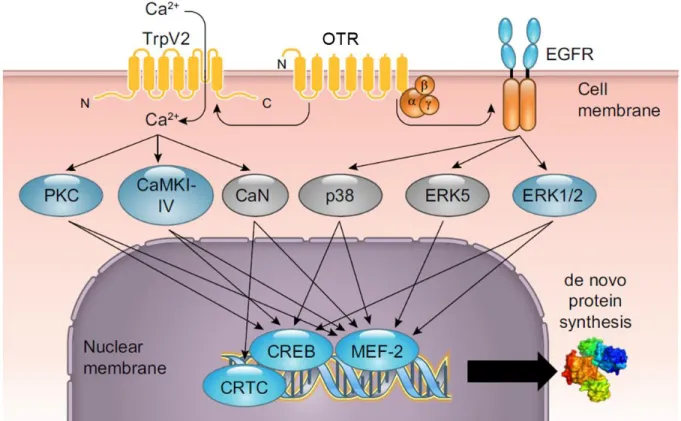
As a GPCR, the OTR is coupled to a trimeric complex of G proteins comprising a Gα and a Gβ/γ unit, which are separated from each other upon ligand binding. Depending on the activated Gα subtype, which can differ between tissues and cell types, the outcome can be either inhibitory or excitatory and different downstream signaling cascades get activated (Figure 1).
1.3 Oxytocin receptor-coupled signaling
Upon OTR activation there is an increase in intracellular Calcium (Ca
2+) caused by a Ca
2+influx from the extracellular space via several Ca
2+channels like transient receptor potential (TRP) cation channels including the canonical forms TRPC1 and TrpC3-TRPC6 as well as the vanilloid forms TRPV2 and TRPV4, but also voltage-gated Ca
2+channels (van den Burg et al., 2015;
Murtazina et al., 2011; Shlykov et al., 2003; Ulloa et al., 2009; Ying et al., 2015). In addition, OTR activation provokes a release of Ca
2+from intracellular stores (Sanborn et al., 1998; Tobin et al., 2011). Upon G protein activation, phospholipase C (PLC) gets activated cleaving phospholipid phosphatidylinositol 4,5-bisphosphate (PIP2) into diacyl glycerol (DAG) and inositol 1,4,5- trisphosphate (IP
3). Those two products are important second messenger molecules that control several cellular processes and serve as substrates for other signaling molecules. IP
3is a soluble signaling molecule that diffuses through the cytoplasm to the endoplasmic reticulum, where it binds to its receptor, a Ca
2+channel, inducing the release of Ca
2+from the endoplasmic reticulum lumen into the cytosol. DAG, the second product of PLC activation, remains in the plasma membrane and mediates the activation of protein kinase C (PKC). The resulting elevation in cytoplasmatic Ca
2+levels from intracellular and extracellular sources is essential for several OTR- coupled signaling pathways.
The basal intracellular Ca
2+concentration and evoked Ca
2+signals have to be tightly regulated
since important aspects of cell function are dependent on this homeostasis, including gene
expression, and energy metabolism (Clapham, 2007). These cell functions depend on Ca
2+signal
- 5 -
propagation to the specific organelles like the mitochondria for synchronization of the ATP generation with cell function, or the nucleus for gene regulatory events (Zhang et al., 2009;
Griffiths and Rutter, 2009). Dysregulations in Ca
2+signaling can have severe consequences including cell necrosis or apoptosis.
The set of downstream signaling cascades comprise the already mentioned PKC, several CaMK subforms, the phosphatase Calcineurin (CaN), as well as the mitogen-activated protein kinase (MAPK) pathway. However, not only intracellular Ca
2+leads to the activation of this pathway. At the same time, a cross-reaction of receptor tyrosine kinases like the epidermal growth factor (EGFR) has been observed eventuating in the recruitment of the membrane-associated proto- oncoprotein GTPase Rat sarcoma (Ras) (Blume et al., 2008). Ras in turn functions as an initiator of the MAPK pathway with its kinases MAP kinase kinase 1 and 2 (MEK1/2) and extracellular signal regulated kinase 1/2 (ERK1/2). Besides that, the OTR has also been linked to an activation of the MAPKs p38 and ERK5 in myometrial cells, but their coupling and downstream targets in neuronal cells remain to be elucidated (Devost et al., 2008; Brighton et al., 2011).
All those signaling pathways converge on transcription factors regulating the transcription of
their target genes. The OT-induced targets we have mostly focused on are the transcription factor
CREB, which is involved in spatial memory formation and CRF gene transcription (Tomizawa et
al., 2003; Jurek et al., 2015) and the myocyte enhancer factor (MEF) 2 (Meyer et al., 2018; Meyer
et al., 2020). The MEF2 family of transcription factors consists of four isoforms, namely MEF2A, -
2B, -2C and -2D. All subforms share a common genetic structure consisting of an N-terminal DNA
binding domain, the MEF2 domain, and a COOH-terminal transcriptional regulatory domain,
whose activity is orchestrated by multiple posttranslational modifications. The phosphorylation
status of the MEF2 proteins regulates the gene transcription and thereby determines the function
of the respective biological process. In case of MEF2A, phosphorylation of the protein at the sites
serine (S) 387, threonine (Thr) 312, and Thr319 stimulates gene transcription, whereas
phosphorylation at S408 has an inhibitory outcome (Potthoff and Olson, 2007).
- 6 -
Figure 1. Overview of oxytocin receptor (OTR)-coupled signaling pathways in neurons. Binding of oxytocin to the OTR activates transient receptor potential vanilloid type 2 (TRPV2) channels and subsequent Calcium (Ca2+)- dependent cascades including protein kinase C (PKC), Ca2+/calmodulin-dependent kinase (CaMK) I, II, IV, and calcinueurin (CaN). OTR activation also induces transactivation of the epidermal growth factor receptor (EGFR) and subsequent MAPK activation involving the extracellular signal regulated kinase (ERK) 1/2, ERK5, and p38. The signaling pathways converge on the transcription factor cyclic AMP responsive element binding protein (CREB) and cyclic-AMP-regulated transcriptional coactivators (CRTC) as well as myocyte enhancer factor 2 (MEF2), leading to the transcription of target genes. Adapted from (Jurek and Neumann, 2018).
The MEF2 proteins show distinct but overlapping sets of functions (Pon and Marra, 2016). They play an important role in muscle and cardiac cell development and affect cell differentiation, proliferation, migration, apoptosis, and metabolism (Potthoff and Olson, 2007). Furthermore, they are implicated in fundamental cellular processes in the central nervous system including neuronal morphology, survival, connectivity, plasticity, and metaplasticity (Akhtar et al., 2012;
Chen et al., 2012; Meyer et al., 2018; Meyer et al., 2020). These processes are strongly
intertwined since neuronal connectivity and plasticity are regulated by adaptations of the cellular
morphology including neurite outgrowth, synapse formation, and cell adhesion, depending on
the temporary requirements. Changes in the cellular morphology, like branching dendrites and
axons or the generation of dendritic spines mediated via the cytoskeleton, serve to make wiring
- 7 -
among networks more efficient (Chklovskii, 2004). Therefore, the balance of these processes is essential for proper cognitive function and has to be maintained.
Dysfunctions concerning the MEF2 family have been associated with ASD, mental retardation, amyotrophic lateral sclerosis, Alzheimer’s, and Parkinson’s disease (Morrow et al., 2008; Lipton et al., 2009; Tu et al., 2017).
Thus, a balanced activity of the OT system is crucial for essential behavioral responses. Genetic or epigenetic factors may influence this by contributing to individual differences in behavioral responses. Among the genetic factors that might lead to a predisposition for the development of certain psychosocial disorders like ASD are single nucleotide polymorphisms in the OTR gene (Bakermans-Kranenburg and van Ijzendoorn, 2014).
1.4 Single nucleotide polymorphisms in the oxytocin receptor gene
A single nucleotide polymorphism (SNP) is a variation in a single nucleotide occurring at a specific position in the genome, which is carried by more than 1 % of individuals in a population. Of the 4-5 million SNPs mapped so far in the human genome, just a few percent are within protein- coding genes, while the majority lies within regulatory or inactive regions (Freedman et al., 2011;
Edwards et al., 2013). Depending on their location in coding or non-coding sequences within a given gene, they can affect the protein in various ways. For instance, SNPs outside the coding sequence like introns, promoters, untranslated regions, and exonal non-coding regions can regulate the gene splicing or transcription factor binding. When they are located within the coding region of an exon, they can be either synonymous or non-synonymous. Synonymous SNPs do not affect the protein sequence whereas non-synonymous SNPs result in an amino acid change in the protein. A change in the amino acid sequence affects the protein structure and its function. Thus, non-synonymous SNPs are believed to have the highest impact on the phenotype.
How non-synonymous SNPs could affect the phenotype is not fully understood, however, mutations present in genes can affect transcriptional and translational output (Robert and Pelletier, 2018).
Genome-wide association studies (GWAS) that identify genetic variants linked with a specific
phenotype, e.g. a disease, are a quite recent achievement, further facilitated by the declining
costs and proceeding methods of high-throughput sequencing. SNP detection developed fast and
- 8 -
has become a major part of modern medical research. Each SNP that has been identified receives a reference ID number and can be found in online databases such as dbSNP hosted by the National Center for Biotechnology Information (NCBI) or SNPedia. The gain of knowledge by SNP studies contributes to the identification of avoidable risks or tailor-made prevention leading to personalized life-style profiles and diet recommendations provided by companies. They are promoted as a big step towards personalized medicine since SNPs cannot only determine one’s susceptibility to certain diseases but also the success of a therapy.
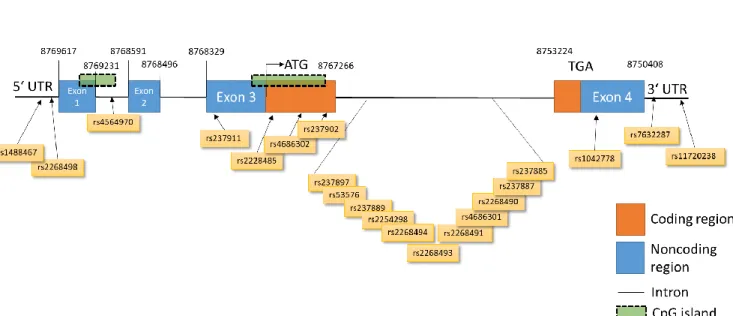
GWAS have revealed several genetic variants of the OTR that have been associated with various traits and behaviors in humans, psychosocial disorders or pathological conditions (International HapMap et al., 2007) (Figure 2).
Figure 2. Overview of single nucleotide polymorphisms (SNPs) in the human oxytocin receptor (OTR) gene on chromosome 3 consisting of four exons and three introns. SNP IDs are given in orange boxes below and chromosomal positions indicated in numbers above. Only SNPs that have been associated with psychological or psychiatric traits have been included. Data extracted from ensembl data base. Adapted from (Jurek and Neumann, 2018).
The associated features comprise social behavior and communication, cognition, parental care,
empathy, and symptoms that are allocated e.g. to attention deficit hyperactivity disorder,
depression or schizophrenia (Walum et al., 2012; Klahr et al., 2015; Skuse et al., 2014; Weisman
et al., 2015). Most of the associations in the OTR gene however, have been related to ASD.
- 9 -
Therefore, besides dysregulated plasma OT concentrations, OTR SNPs have been promoted as possible biomarkers for social impairments in ASD (Parker et al., 2017).
However, although intensely studied, the association between ASD and SNPs in the OTR gene remains controversially discussed. Several studies show contradictory results about the presence of a specific SNP and its contribution to the etiology of a given disorder.
Several associations of SNPs could not be replicated and often disappeared after correction for multiple comparisons (Campbell et al., 2011; Tansey et al., 2010; Wermter et al., 2010). An example repeatedly reported in this context is rs237887, where the association was found to be dependent on the behavioral tests performed. While few studies found the ‘A’ allele of rs237887 was not associated with ASD diagnosis, others confirmed an association with e.g. reduced face- recognition memory in families with an autistic child or impaired altruism in two separate behavioral tasks (Skuse et al., 2014; Verhallen et al., 2017).
It is not known whether disruptions in oxytocinergic signaling contribute to a risk for ASD or are associated with variability in social deficiency in ASD.
Since intranasally applied OT is considered to act through the OTR, the efficacy and inconsistent findings on symptom improvement might depend on SNPs in the OTR gene that might cause some loss or gain of functions. However, there are just very few studies about the functional significance of SNPs (Fueg et al., 2019; Furman et al., 2011). Therefore, studies regarding the molecular effects of SNPs, their role and possible combinatory effects are greatly needed to bridge the gap between genetic association and function.
1.5 Aim of the thesis
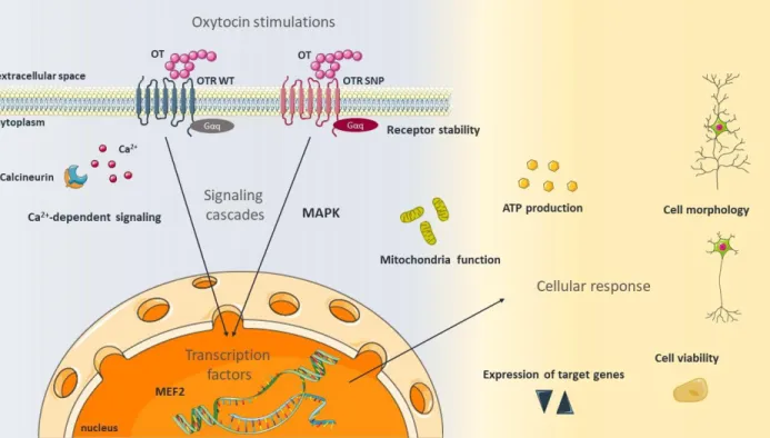
The aim of my thesis was, to study the molecular mechanisms and effects following OTR activation in order to understand the cellular changes in the context of psychosocial disorders and emotional dysfunctions. During the course of my PhD, I studied the OT system in vitro focusing on various intracellular aspects. After stimulating cells with OT, I identified activated signaling pathways and their consequences on cellular processes like neuronal morphology, neuronal connectivity, cell viability, mitochondrial function, and ATP production (Figure 3).
Further, considering common genetic aberrations in the OTR gene leading to different OTR
- 10 -
protein variants, I assessed how this affects the receptor stability, a selection of already known coupled signaling cascades and the expression of downstream target genes.
Figure 3. Aim of the thesis. This thesis aimed to assess the oxytocin (OT) -induced effects on signaling cascades including the mitogen-activated protein kinase (MAPK) pathway as well as calcium (Ca2+) signaling and downstream activated transcription factors like the myocyte enhancer factor (MEF) 2. Additionally, the aim was to assess the consequences for various cellular parameters such as mitochondria function, ATP production, the expression of target genes, cellular morphology and viability as well receptor stability, also with regard to the activated oxytocin receptor (OTR) variant. The schematic art pieces used in this figure were provided by Servier Medical art (http://servier.com/Powerpoint‐image‐bank). Servier Medical Art by Servier is licensed under a Creative Commons Attribution 3.0 Unported License.
- 11 -
2 Oxytocin alters the morphology of hypothalamic neurons via the transcription factor myocyte enhancer factor 2A (MEF2A)
2.1 Abstract
OT has gained attention not only as anxiolytic drug and as potential treatment option for autistic children; it also acts as a growth and differentiation factor in neuronal cells. While behavioral effects of OT have been studied in detail, knowledge about the cellular effects of OT is relatively sparse. In this study, we present evidence for three hypotheses: 1) OT leads to neurite retraction in hypothalamic neurons via the OTR 2) The transcription factor MEF2A is a central regulator of OT-induced neurite retraction, and 3) The MAPK pathway is critical for OT-induced MEF2A activation. Incubation of rat hypothalamic H32 cells with 10 nM to 1 μM OT, vasopressin, and the specific OTR agonist TGOT, over the course of 12 h resulted in a time-dependent, significant retraction of neurites. In addition, the size of the nuclear compartment increased, whereas the overall cell size remained unchanged. OT treatment for 10 h increased the cellular viability significantly, and this effect could be blocked by a specific OTR antagonist, providing evidence for a specific and pro-active effect of OT on neurite retraction, and not as an unspecific side effect of apoptosis. The molecular mechanism that controls OT-induced neurite retraction includes a reduced phosphorylation of the transcription factor MEF2A at S408. This dephosphorylation is under the control of the OTR-coupled MAPK pathway, as blocking MEK1/2 by U0126 inhibited MEF2A activation and subsequent neurite retraction. The siRNA-mediated knockdown of MEF2A prevented the OT-induced neurite retraction, providing direct evidence for a role of MEF2A in morphological alterations induced by OT treatment. In summary, the present study reveals a previously unknown OTR-coupled MAPK-MEF2A pathway, which is responsible for OT-induced neurite retraction of hypothalamic neurons.
2.2 Introduction
The neuropeptide OT is a central modulator of complex socio-emotional behavior, such as anxiety, affective behavior, and aggression (Jurek and Neumann, 2018; Kosfeld et al., 2005;
Kumsta and Heinrichs, 2013; Neumann and Landgraf, 2012). The molecular underpinnings of
these behavioral effects are largely unknown. The OTR is almost ubiquitously expressed in the
- 12 -
central nervous system (Grinevich et al., 2016; Jurek and Neumann, 2018), most notably in the hypothalamus, the site of OT synthesis. The OTR is a G protein coupled receptor, whose activation by its ligand leads to promiscuous coupling to various Gα-proteins (Busnelli and Chini, 2018), Ca
2+release from intracellular stores (Tobin et al., 2011) and subsequent influx of Ca
2+from the extracellular space through TrpV2 channels (van den Burg et al., 2015). These G proteins in combination with the Ca
2+influx activate several signaling cascades, most prominently the MAP kinase pathway, with its main kinases MEK1/2 and ERK1/2 (Blume et al., 2008; Jurek et al., 2012).
MAP kinase pathways have been associated with neurite outgrowth (Won et al., 2015; Kang et
al., 2011; Xu et al., 2015), anxiety (Blume et al., 2008; Jurek et al., 2012; Borges et al., 2015), and
memory formation (Tomizawa et al., 2003). Downstream effectors of ERK1/2 are the
transcription factors CREB (Tomizawa et al., 2003; Jurek et al., 2015) and MEF2 (Devost et al.,
2008). MEF2 was originally defined as a muscle-specific factor that binds an A/T-rich element in
the promoter of target genes (Gossett et al., 1989), and was later found to be ubiquitously
expressed in all types of tissue, especially in the brain (Dietrich, 2013). One central function of
MEF2 is the regulation of neuronal morphology, i.e. neurite outgrowth (Flavell et al., 2006), either
via gene inhibition or activation. Among the MEF2-regulated genes are immediate-early genes
such as c-JUN and NUR77, as well as regulators of neuronal activity, such as ARC, SYNGAP,
HOMER1A, and BDNF (Flavell et al., 2006). Consequently, MEF2 represents a central factor for
the regulation of neurite outgrowth, synapse formation, and, if dysregulated, was found to play
a key role in the development of ASD (Potthoff and Olson, 2007). Here, the functions between
OT and MEF2 may overlap, as both are implicated in ASD (Parker et al., 2014; Bales et al., 2013)
and neurite outgrowth (Lestanova et al., 2016; Lestanova et al., 2017). However, the literature
describes inconsistent effects of OT on cellular morphology. For instance, OT bursts during milk
letdown lead to retraction of glial cells in rat dams (Langle et al., 2002; Theodosis, 2002), and in
vitro application of synthetic OT leads to neurite retraction in hippocampal neurons (Ripamonti
et al., 2017); however, when tested in human SH-SY5Y neuroblastoma cells and glial U-87MG cell
lines, OT leads to neurite outgrowth (Lestanova et al., 2016; Lestanova et al., 2017). This
outgrowth seems to be executed by the rearrangement of actin filaments to the apical part of
neuronal cones and paralleled by changes in expression of the intermediate filament protein
- 13 -
nestin, which is implicated in axon growth (Bakos et al., 2012). It is unknown so far, whether the differential effects of OT on neuronal and glial cells, i.e. neurite retraction (Ripamonti et al., 2017) or neurite outgrowth (Lestanova et al., 2016), are caused by the use of different doses of OT, by the use of different cell types (primary neurons or cell lines), different species (mouse, rat, human cells), or even the sex of the cell donor (Li et al., 2016). Therefore, it is of paramount importance to choose a physiological relevant model to investigate the effects of OT on cell morphology. As OT and the OTR are expressed in the hypothalamus of the brain (Jurek et al., 2015; Freund- Mercier et al., 1994; Dabrowska et al., 2011), rat hypothalamic neurons (H32 cells) provide a suitable in vitro model to provide a full characterization of the cellular response to OT. A full characterization of the cellular response is pivotal, especially since the OT molecule is known to bind the receptor of the related nonapeptide vasopressin as well at higher concentrations, with the result of mixed OTR and vasopressin receptor-mediated cellular effects. In turn, vasopressin can also bind the OTR, which could result in a similar effect of both peptides on cellular morphology. In order to circumvent any non-OTR-mediated effects we made use of specific antagonists and agonists of the OTR such as Thr
4, Gly
7-OT (TGOT), an OT agonist with a 16.000 times higher affinity for the rat OTR than for the rat vasopressin receptor (Chini et al., 2008;
Manning et al., 2008); however, this OTR-specificity is lost in cells of human origin (Chini et al., 2008). The advantage of OTR-specificity of TGOT in rat cells, in combination with the hypothalamic origin, convinced us to conduct comprehensive morphological studies regarding OT's cellular effects in H32 cells. In summary, we aim to contribute to the clarification of the effects of OT on cellular morphology and identify the intracellular signaling cascades that control the observed morphological alterations. In order to do so, we tested the hypothesis that the transcription factor MEF2 is a key regulator of neurite retraction in hypothalamic neurons, and that its activation is dependent on the OTR-coupled MEK1/2-ERK1/2 pathway.
2.3 Materials and methods 2.3.1 Cell culture
Rat hypothalamic H32 cells (passages 15–30) were cultured in 1:1 Dulbecco's Minimum Essential
Medium/Ham F12 Medium (growth medium). The growth medium was supplemented with 10 %
heat inactivated fetal bovine serum (Capricorn, Germany), 0.1 % nonessential amino acids, 100
- 14 -
U/ml gentamycin (both Invitrogen, Germany) in humidified atmosphere containing 5 % CO
2at 37
°C. Passaging was performed at least once a week by gentle trypsination.
2.3.2 Cell viability assay
Cellular viability was tested using the PrestoBlue Cell Viability Assay (A13261, Invitrogen) according to manufacturer's protocol. Briefly, 20 × 10
3cells per well were seeded the day before the test in a 96-well-plate in growth medium. The volume of the treatment and serum-free medium (DMEM/F12 + 0.1 % BSA, sterile filtered) for the stimulation was calculated to a total of 90 μl per well. 10 μl of PrestoBlue Reagent were added directly to the cells, incubated for 30 min to 2 h, before reading the fluorescence intensity with a FluoStar Plate reader (BMG). Optimal DMSO concentrations as solvent for the U0126 MAPK inhibitor (Sigma Aldrich) were determined by a separate dose response experiment.
2.3.3 Protein isolation
For the extraction of proteins from adherent H32 cells, medium was removed and cells were washed with PBS supplemented with Protease and Phosphatase inhibitors (PI, A32959, Thermo Fisher). Cells were scraped, centrifuged, and the cell pellet was resuspended in 100 μl RIPA lysis buffer (R0278, Sigma Aldrich) with HALT Inhibitor and EDTA (78444, Thermo Fisher) to extract whole cell lysate.
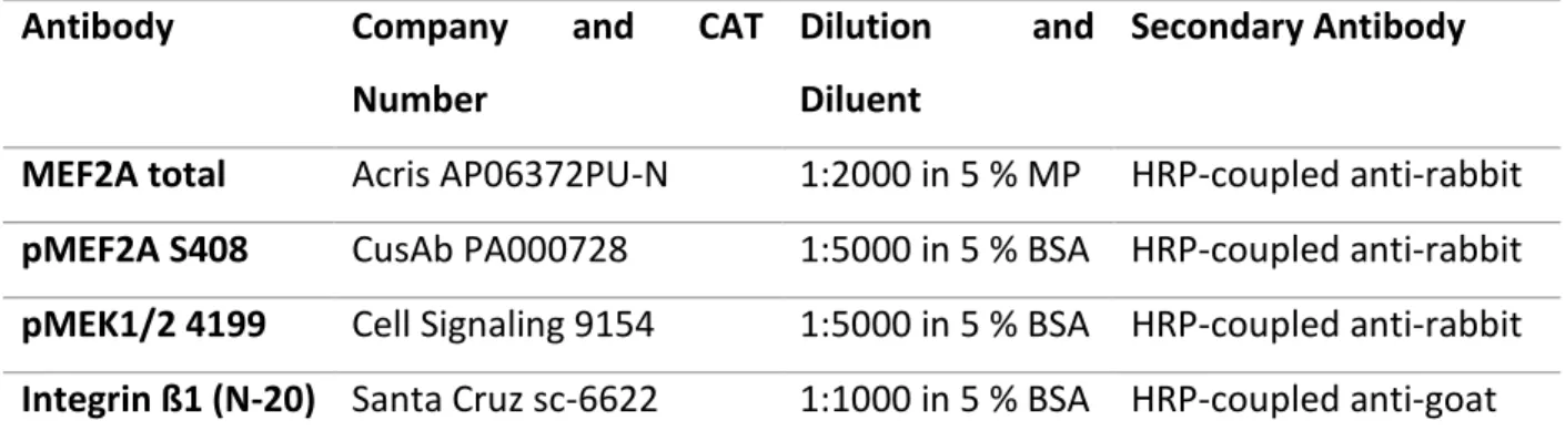
2.3.4 Western blot
Between 5 and 30 μg of whole cell extract and an equivalent volume of 4× loading buffer (2.4 ml
TRIS, 0.8 g SDS, 4 ml 100 % Glycerol (AppliChem), 0.01 % Bromphenol Blue (Sigma Aldrich), 1 ml
Mercaptoethanol and 2.8 ml H
2O) were applied to 12 % Mini and Midi PROTEAN or Criterion TGX
Stain-free gels (456–8044 and 5678045, BioRad). Western blot analysis was performed using the
Stain-free total protein method (BioRad) as loading control, according to the manufacturer's
protocol. Stain-free gels contain trihalo compounds that covalently bind to tryptophan residues
in proteins when exposed to UV light. By that, the proteins are made fluorescent directly in the
gel, allowing the immediate visualization of proteins at any point during electrophoresis or
blotting and permitting the user to normalize bands to total protein in each lane. This technology
circumvents the problematic use of housekeeping proteins as loading controls on western
- 15 -
blots. In order to detect activated MEF2, a phospho-specific MEF2A S408 (CSB-PA000728, Flarebio Biotech) antibody was used at 1:5000 in 5 % BSA concentration. A MEF2A total antibody (#TA307807, OriGene, 1:5000 in 5 % BSA) was used to detect total MEF2A levels. Changes in expression levels were analyzed measuring grey density in a semi-automatic manner by Image Lab Software (Version 6.0, BioRad).
2.3.5 Cell stimulations
Cells were grown in the presence or absence of 10 nM, 100 nM, or 1 μM of OT (Bachem, Germany) for 0, 2, 4, 8, 10, and 12 h in cell culture dishes or 3-part chamber slides (BD Falcon, Germany). A separate experiment was performed using cells incubated with [Thr
4, Gly
7]-oxytocin (TGOT, 100 nM, Bachem) or vasopressin (100 nM, Bachem). When inhibitors (U0126, Sigma Aldrich) or OTR antagonist des-Gly-NH
2d(CH
2)
5[Tyr(Me)
2Thr
4]-OVT (kindly provided by M.
Manning) were used, cells were incubated in serum free medium for 1 h, pretreated with the
inhibitor or VEH (H
2O or DMSO) for 10 min, and stimulated with the according treatment. To
assess the role of MEF2A, H32 cells were transfected with small interfering RNA (siRNA). 3 unique
27mer MEF2A siRNA duplexes (Origene, SR504191; for sequences see Table 1) and Lipofectamine
RNAiMAX Reagent (Invitrogen), diluted in OptiMEM were added for 72 h in a concentration of 1
nM siRNA. To verify the specificity of the knockdown, additional wells were transfected with a
scrambled siRNA CONTROL (siCTRL, SR30004). Effects of OT were assessed via co-stimulation
with 100 nM OT for 24 h.
- 16 -
Table 1. MEF2A siRNA duplex sequences. All three duplexes were mixed 1:1 and added for transfection.
siRNA Duplex sequence
SR504191A rGrArArCrUrUrUrCrUrGrCrArArGrGrArUrArArArArUrATT
SR504191B rGrArUrArUrUrGrArArGrArArArArUrUrArGrCrUrUrCrUGA
SR504191C rCrUrUrArArArUrUrGrGrUrGrArArUrArArGrGrArCrArUGA
2.3.6 Morphological assessments
The cells grown in chamber slides were washed with 1 ml PBS and subsequently fixed by gently adding an equal amount of 4 % paraformaldehyde (PFA, Sigma-Aldrich, Germany), resulting in 2
% PFA on the cells. After 2 min, this solution was discarded and replaced by 4 % PFA for 15 min
at room temperature. The fixed cells were washed three times with PBS and permeabilized by
500 μl PBS containing 0.1 % Triton X-100 (Sigma-Aldrich, Germany) for 5min. Unspecific binding
sites were blocked by 30 min in PBS +0.1 % Triton and 10 % NGS. The actin filaments were stained
by addition of 20 μl of 3.8 μM phalloidin tetramethylrhodamine B isothiocyanate (Sigma-Aldrich,
Germany) directly on the cells without access to light for 20min. Phospho-MEF2A S408 specific
antibody was diluted 1:500 in 5 % BSA/PBS-T and incubated on the cells overnight at 4 °C and
gentle rocking. Nuclei were stained by addition of Gold anti-fade mounting medium containing
4,6-diamidino-2-phenylindole (DAPI; Life Technologies, Germany). Cells were observed using the
confocal microscope Leica SP8. Images were taken from four random fields per chamber. Neurite
outgrowth was determined by manually tracing the length of the longest neurite per cell (using
Fiji 1.51k) for all cells in a field that had an identifiable neurite and for which the entire neurite
arbor could be visualized. Length of the neurite was measured from the edge of nucleus to the
apical end of the projection. Four independent members of the team evaluated neurite length in
a blinded manner. Hereby the MAX intensity Z-Projection was analyzed. DAPI images were used
for nucleus size measurements and as MASK for mean intensity analysis of MEF2A S408
phosphorylation exclusively in the nucleus. To assess the size of the cells, whole area
measurements were performed by outlining the Phalloidin stained area. During all the
measurements, grayscale images were processed. To control for staining artifacts with Phalloidin
or fixation artifacts we employed a second staining approach using the ZOE Fluorescence
- 17 -
microscope (Bio-Rad) and live cells in cell culture dishes. For nucleus staining, Hoechst 33342 stain (Thermo Scientific, H1399) was added to the stimulation medium and incubated for 30 min at 37 °C. Hoechst staining is non-toxic and can therefore be used in living cells. After 20 min, 1×
Plasmagreen membrane stain (Thermo Scientific, C37608) was added to the medium and co- incubated for at least 10 min at 37 °C to label the cell plasma. This method circumvents any potential artifacts that could arise by the use of a specific Actin staining like Phalloidin. Cells were washed two times with 1× PBS before imaging of the blue (nuclei) and green (cell plasma) channel. All Pictures were analyzed using ImageJ Fiji software.
2.3.7 Statistical analysis
Data were pooled and analyzed using SigmaPlot 13. Parametric data was analyzed by t-test or one-way analysis of variance (ANOVA), followed by Holm Sidak post hoc test. Non-parametric data was analyzed by the Kruskal-Wallis ANOVA on ranks and the Tukey post hoc test, or in case of different group sizes, Dunn's post hoc test. Statistical differences were accepted at p < 0.05.
For morphology analysis experiments with large n-numbers, analysis of effect size was performed. As indicator of effect size, either Cohen's f
2-value where f
2< 0.02 is considered as small effect size, f
2= 0.15 is considered as median f
2> 0.35 as strong effect or eta squared (η
2), where η
2< 0.039 is considered as small effect, η
2< 0.06–0.110 as intermediate, and η
2> 0.14 as strong effect, was calculated. Data are presented as means ± or + standard error of the mean (SEM), as indicated in the figure legend.
2.4 Results
2.4.1 OTR activation mediates OT-induced changes in neurite length and nucleus size
Increasing concentrations (10 nM, 100 nM, and 1 μM) and treatment duration (0, 2, 4, 6, and 8
h) of OT were tested with rat hypothalamic H32 cells, in order to reveal dose- and time-
dependent effects of OT treatment on neurite outgrowth (Figure 4A). VEH-treated cells elongate
their neurites over a time course of 10 h significantly from about 37 μm up to 45 μm (# p < 0.05
10 h vs 2 h). In contrast, OT treated H32 cells retracted their neurites from about 37 μm to about
27 μm over time and in a dose-dependent manner. In detail, 100 nM OT significantly reduced
- 18 -
neurite length (* p < 0.05 vs VEH, intermediate effect f
2= 0.123) after 2 h, with further retraction of neurites until 10 h of treatment. 1 μM of OT induced a slower onset of neurite retraction (significant at 4 h), but with a more prominent effect size (* p < 0.05 vs VEH, strong effect f
2= 0.477). After 8 and 10 h of cell stimulation, all treatment groups showed a reduced neurite length (* p < 0.05 vs VEH, median effect f
2= 0.267 after 8 h, very strong effect f
2= 0.77 after 10 h). As H32 cells also express the vasopressin receptor V1a, we tested whether the observed neurite retraction is mediated by the OTR, V1a, or a combination of both. Therefore, we incubated the cells with vasopressin or the specific OTR agonist TGOT. Similar to the OT-induced effect, vasopressin and TGOT (both 100 nM) significantly reduced neurite length over time (Figure 4B; * p < 0.05 vs VEH). However, vasopressin showed a slower onset of action compared to OT and TGOT, as after 2 h neurite length was not altered by vasopressin, but significantly reduced by OT and TGOT (100 nM; * p < 0.05 vs VEH, intermediate effect f
2= 0.123). Despite the slow onset of vasopressin action, after 4, 8, and 10 h, neurite length was significantly reduced in all treatment groups (* p < 0.05 vs VEH). To further dissect the morphological changes described in Figure 4A and B, we analyzed the OT-induced neurite length, whole cell size, and nucleus size separately in presence or absence of an OTR antagonist. We found that the OT-induced neurite retraction can be blocked by the specific OTR antagonist des-Gly-NH
2d(CH
2)
5[Tyr(Me)
2Thr
4]-OVT (* p < 0.05 vs VEH, # p < 0.05 vs OT, Figure 4E), indicating that OT acts via the OTR on cellular morphology.
There was no effect of OT or the antagonist on the whole cell size (i.e. total area covered by
perikaryon, nucleus, and neurites), indicating a specific effect on cell shape, rather than growth
or shrinking (Figure 4F). However, the size of the area stained by DAPI (a rough indicator of
transcriptional activity) was increased by OT, and this effect was not reversed by the OTR
antagonist, indicating OTR-independent transcriptional activity (* p < 0.05 vs VEH, Figure 4G).
- 19 -
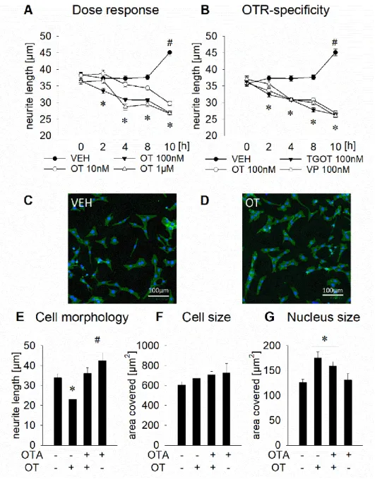
Figure 4. Effects of an OTR activation on neuronal morphology in rat hypothalamic H32 cells. (A) Mean neurite length of H32 cells determined at increasing OT concentrations from 0 h to 10 h. Neurite length decreases significantly from about 37 μm to about 27 μm over the course of 10 h of OT stimulation at 10 nM, 100 nM, or 1 μM; while VEH treated cells show increased neurite length after 10 h. * p < 0.05 vs VEH, #<0.05 vs previous timepoint. n = 314–628 cells.
(B) Incubation of H32 cells with OT (100 nM), the OTR agonist TGOT (100 nM), or vasopressin (VP, 100 nM) over 10 h leads to neurite retraction to a similar extent as seen in Figure 4A. n = 314–801 cells. (C) and (D) Representative pictures of non-treated H32 cells (C) and 100 nM OT-treated cells (D) for 4 h; cytoplasm, including neurites stained with CellMask plasma membrane stain (green) and nuclei stained blue with Hoechst 33342. Scale bar represents 100 μm. (E) Neurite retraction of OT-treated H32 cells after 12 h of stimulation is reversed when incubated with the OT antagonist (OTA, des-Gly-NH2d(CH2)5[Tyr(Me)2Thr4]-OVT). The OTA alone has no effect on neurite length. * p < 0.05 vs VEH, # < 0.05 vs OT-treated cells. (F) Whole cell size analyzed as mean of total area covered by individual cells (i.e.
perikaryon, nucleus, and neurites). No effect of the treatment was determined. (G) Overnight treatment of H32 cells with 100 nM OT led to an increase of area stained by DAPI, indicative of the size of the nuclear compartment. The OTA reduced the increase of size of the nuclear compartment, without reaching basal levels. * p < 0.05 vs VEH. Data represents mean + SEM. n = 21–74 cells.
- 20 -
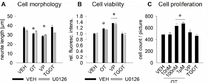
2.4.2 OT leads to a MAPK-dependent neurite retraction as well as increased cell viability, whereas vasopressin affects neurite length MAPK-independently
In order to exclude technical artifacts of the CellMask staining protocol in living cells used in Figure 4, we successfully reproduced our initial finding of reduced neurite outgrowth by 100 nM OT treatment with a different staining protocol (fixated cells, actin filament staining with Phalloidin, and evaluation by confocal microscopy; Figure 5A, * p < 0.05, intermediate effect size η
2= 0.078). When pretreated with the MAPK inhibitor U0126 (10 μM) the effect of OT was blocked, suggesting a central role for the MAPK pathway in the OT-induced neurite retraction.
Surprisingly, vasopressin also reduced neurite length, in presence or absence of U0126, indicating that neurite retraction by vasopressin is coupled to a separate, yet unknown pathway (* p < 0.05, intermediate effect size η
2= 0.078). However, a partial role of the OTR cannot be excluded, as vasopressin also binds the OTR with a comparable affinity (Manning et al., 2008). Overnight stimulation with the specific agonist TGOT did not result in significantly altered neurite length.
As stimulation with TGOT for only 10 h decreased neurite length from 37 to 27 μm (Figure 4A), but after prolonged (overnight) OT stimulation morphology changes were reversed to basal, we hypothesize that the exclusive OTR response is short lived (max. 10 h) and requires the additional activation of the vasopressin receptor for a prolonged response. Consequently, Figure 4B and 5A indicate that, in contrast to the fast and short-lived OTR response, vasopressin has a slow onset of action on neurite retraction (no effect after 2 h, Figure 4B), but bigger effect after prolonged incubation (overnight, mean difference of the neurite length compared to the non-treated group:
−8.98 μm for VP vs. −6.47 μm for OT, Figure 5A). Morphological assessments of neuronal cells
should only be conducted under optimal culturing conditions, which led us to control our
experimental setup for general cell viability (i.e. ability to reduce resazurin to resafurin) and cell
proliferation (cell number). Both OT and vasopressin treatment increased cellular viability, as
reflected by the increased amount of resafurin, suggesting a pro-active, and not apoptosis-
induced retraction of neurites. In addition, the OT-induced increase in cell viability is MAPK-
dependent, as pretreatment with U0126 blocked this effect (Figure 5B). Comparable to the
MAPK-independent effect of vasopressin on neurite length (Figure 5A), cell viability of
vasopressin-treated cells was increased, independent of MAPK inhibition, thereby again
- 21 -
excluding a role for the MAPK pathway in vasopressin-mediated effects. Moreover, in support of our hypothesis of a short-lived nature of the OTR response, neurite outgrowth (Figure 5A) and cell viability (Figure 5B) returned to baseline after an overnight incubation with TGOT. This finding was further supported by the lack of effect of TGOT on cell proliferation, in contrast to the dose dependent increase of proliferation by 100 nM or 1 μM OT overnight treatment (* p < 0.05, Figure 5C).
Figure 5. Effects of OTR activation on cell morphology, viability, and proliferation in rat hypothalamic H32 cells. (A) Effect of overnight incubation with 100 nM OT, vasopressin (VP), or TGOT, in presence or absence of the MEK1/2 inhibitor U0126 (10 μM) on neurite length in H32 cells. OT reduces neurite length significantly and in dependence of the MAPK pathway, whereas VP leads to neurite retraction independently of the MAPK pathway. TGOT has no significant effect. n = 65–200. (B) The effect on neurite length is accompanied by increased cell viability, which is blocked by U0126 in the OT-, but not in the vasopressin-treated cells. n = 18–48 wells. (C) Dose dependent increasing proliferation of cells treated with 10 nM (no effect), 100 nM (+∼100 cells) and 1 μM OT (+∼120 cells), but lack of effect with 100 nM VP and TGOT. n = 4–5 pictures. * p < 0.05 vs respective VEH. Data represent mean + SEM.
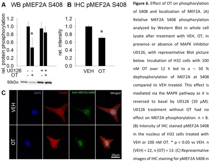
2.4.3 OT increases transcriptional activity of MEF2A
As increased nucleus size is a rough indicator for open chromatin, i.e. increased transcriptional activity, we tested the activation status of the transcription factor MEF2A by assessing its phosphorylation at S408. At this residue, dephosphorylation leads to transcriptional disinhibition.
Western Blot analysis revealed that incubation with 100 nM OT led to a reduced level of phospho-
MEF2A S408 in whole cell lysate in a MAPK-dependent manner, indicating increased
transcriptional activity (Figure 6A). The observed changes were validated by
- 22 -
immunohistochemical stainings, which revealed reduced intensity of pMEF2A S408 staining in the nuclear compartment, while staining in the cytoplasm was virtually absent (Figure 6B and 6C), indicating a dephosphorylation event in the nucleus and not translocation of pMEF2A S408 to the cytoplasm.
Figure 6. Effect of OT on phosphorylation of S408 and localization of MEF2A. (A) Relative MEF2A S408 phosphorylation analyzed by Western Blot in whole cell lysate after treatment with VEH, OT, in presence or absence of MAPK inhibitor U0126, with representative Blot picture below. Incubation of H32 cells with 100 nM OT over 12 h led to a ∼ 50 % dephosphorylation of MEF2A at S408 compared to VEH treated. This effect is mediated via the MAPK pathway as it is reversed to basal by U0126 (10 μM).
U0126 treatment without OT had no effect on MEF2A phosphorylation. n = 8.
(B) Intensity of IHC stained pMEF2A S408 in the nucleus of H32 cells treated with VEH or 100 nM OT. * p < 0.05 vs VEH. n (VEH) = 22, n (OT) = 13. (C) Representative images of IHC staining for pMEF2A S408 in H32 cells. Images were sequentially recorded for chromatin (DAPI, blue, first column), F-actin (Phalloidin, red, second column), and anti-pMEF2A S408 (green, third column). Merged images are shown for each row in the fourth column.
Labels left to the columns indicate the respective treatment for 12 h. Scale bar represents 20 μm. Data represent mean + SEM.
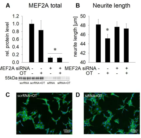
2.4.4 OT-induced neurite retraction is reversed by MEF2A knock down
To provide a causal link between the activation of MEF2A and the neurite retraction induced by
OT, we specifically knocked down MEF2A in H32 cells by siRNA, and assessed the effects of OT
on neurite length 72 h after transfection, a time point where the knock down was most effective.
- 23 -
MEF2A protein levels were highly effectively reduced by 85 % of the baseline level, and OT treatment did not affect this knock down (Figure 7A). Scrambled siRNA (scrRNA) served as transfection control and did not alter MEF2A protein levels significantly compared to non- transfected cells. Neurite length in cells that were transfected with scrambled or siRNA averaged around 47 μm (as compared to ∼ 37 μm of non-transfected, 12 h cultured cells in Figure 4E), as a result of prolonged culturing protocol, necessitated by the preceding transfection procedure.
Statistical analysis by one way ANOVA did not reveal a significant difference. However, separate statistical analysis by direct comparison of the control (-MEF2A siRNA/+OT) versus the MEF2A knock down (+MEF2A siRNA/+OT) groups confirmed the observed OT-induced reduction in neurite length whereas, in contrast, this effect was prevented by MEF2A knock down (Figure 7B).
The lack of effect of OT on neurite length when MEF2A is knocked down reveals the central role of MEF2A in the regulation of cellular morphology by OT.
Figure 7. Effect of MEF2A
knockdown on neuronal
morphology. (A) Relative MEF2A protein expression in whole cell lysate after treatment with 1 nM scrRNA or 1 nM MEF2A siRNA for 72 h and overnight stimulation with 100 nM OT. Total MEF2A protein is reduced after treatment with MEF2A siRNA compared to scrRNA- treated cells, which served as control. Representative Blot below the graph. n = 6. (B) Changes in the neurite length of H32 cells treated with either scrRNA or MEF2A siRNA in presence or absence of 100 nM OT. n = 339–336 cells. * p < 0.05 vs scrRNA control. Data represent mean + SEM. (C) and (D) Representative pictures of H32 cells treated with scrRNA + OT (C) or siRNA + OT (D); cytoplasm, including neurites stained with CellMask plasma membrane stain (green) and nuclei stained blue with Hoechst 33342. Scale bar represents 100 μm.