Analysis of the interaction of the LuxR-type
transcription factors BglJ and RcsB and antagonism of H-NS mediated silencing in Escherichia coli
I n a u g u r a l - D i s s e r t a t i o n
zur
Erlangung des Doktorgrades
der Mathematisch-Naturwissenschaftlichen Fakultät
der Universität zu Köln
vorgelegt von
Raja Venkatesh Ganesan aus Tamil Nadu, Indien
Köln, März 2010
Berichterstatter/in: Prof. Dr. Karin Schnetz Prof. Dr. Jürgen Dohmen
Tag der letzten mündlichen Prüfung: 26 April 2010
Acknowledgements Acknowledgements
I take this opportunity to thank all the people who have helped, encouraged and inspired me during my graduation and made this thesis possible. First and foremost, I would like to express my deepest gratitude to my PhD supervisor Prof. Karin Schnetz for providing me the opportunity to work in her group and sharing her knowledge for the betterment of my life. I am very thankful for her valuable advice, support and teaching me the word “organization”. Thanks for the pushes and constructive criticism which helped me to make this happen.
I would like to thank Prof. Dr. Siegfried Roth and Prof. Dr. Jürgen Dohmen for being part of my thesis committee. I thank Dr. Kevin Johnson for agreeing to be part of my thesis committee.
I am deeply grateful to Dr. Sabari and Dr. Palani for their immeasurable help and support professionally and personally. I thank Dr. Jain Nayar for his wise words during tougher times. I also thank him for the lighter moments and great movie sessions.
Many thanks to Dr. Antonio Jose Marque for his help with FPLC for the protein purification and also for the invitation for volleyball games.
I feel extremely fortunate having enjoyed my time with wonderful colleagues. I thank all my past lab members Vel, Madhu, Andreas, Kathleen, Birgit, Idriss for their help. I thank my present lab members, Kostya, Thomas, Lilia and Katrin for being very helpful and very supportive. I enjoyed our movie sessions, funny jokes, visit to the zoo, football talks, parties at Lilia`s house. Everything was really great and fun. It was pleasure working with you all.
I am very thankful to Brigitte Wilcken-Bergmann for her countless helps from IKEA to AOK and for taking care of us like her own child. I also thank Isabell Witt for all her help and time during our presentations and retreats. Thanks to Inge Götz-Krichi for making the administrative work easier.
My graduate school coordinators deserve a loud mention: Jo and Kathy, You two are awesome. I bet we would not see many better people than you two. I thank you both for all your help.
My special thanks to Prof. Dohmen`s group members Palani, Leo, Daniela, Toze, Marion, Maria, Annie, Ganapathy, Kerstin for treating me like one of them and inviting me for all occasions.
Special thanks to Daniela for being my Deutsch-Englisches Wörterbuch and helping me translating many things with patience. Thanks for being supportive and sharing
“The Good, the bad and the ugly” things in the PhD life.
I like to thank all my friends Bhagi, Vivek, Sam, Venky, Rajesh, Charles, Chenna, Ravi, Jain, Sudhir, Racchappa, Karthik, Ram, Vinodh, Zahid, Johnny, Numan, Giri, Arun, Saba, Naga, Andrews, Devender, for all their help and making Sunday more
Acknowledgements fun filled with cricket and arguments. I am also thankful for the delicious dishes
provided by Viji, Kayal and Preeti during special occasions.
I thank my wonderful lab neighbors Dr. Mirka, Nils, Eva, Sabrina, Dr. Martina, Dr.
Renjith, Dr, Sanjita, Dr. Gritt, Katarina, Julia, and Cornelia, for being nice and helpful. I also thank them for effectively engaging in Friday beer sessions and grilling in summer and extending fun beyond science.
I am lucky enough to come across many good people during this time like Ines, Fabian, Florian, Osmon, Carsten, Tanja, and Anne. I would like to thank Sascha Dargazanli and Nina Leuschen for their help in getting the chemicals with a smiling face even at their busiest times. Big thanks to our lovely kitchen staffs Elfie Hansen and Karin Küllenberg for the friendly smiles and help. I also thank my institute Hausmeister Ramdane and Ute for their help and funny chit chats.
I should also thank the people for building this Institute closer to life saving places like Uni Mensa, Backwerk, and Happy Döner. I thank Frau. Gotzmann for her extended help in filling the application form.
I thank Leo and Luis for helping me to adjust to the new life in Köln in the early days and special thanks to Jinu for her hospitality and treating me like a family member.
I thank my first German friend and roommate Sebastian, for saving my life in the very first days by cooking Fischstäbchen. I am indebted to him for introducing me to the great “Kölsch”. I also thank my Deutsch lehrerin Frau.Petra for her efforts to teach me some Deutsch.
I thank my soccer team members Tobi, Benny, Fabiano, Florian, Sascha, Max, Malou, Richie, Frank, Yinka, Marcel, Anni, Heike and Michael for the fun times on and off the ground.
Verena deserves a special mention for her unconditional care and support all the time.
I am thankful to my other family Alex, Jenny, Mara, Lina, Thomas, Wiebke, Arne, Kurt, Edith, Renate, Wolfgang, Michaela, Marion and Romina for their love and care.
My deepest gratitude goes to my father and mother for their support and love. This stage would have been simply impossible without them. I also thank my brother for being very supportive and helpful.
Finally my appreciation also goes to the city Köln, which has an extraordinary diversity of culture and life style. This city teaches you how to cherish the good things in your life. Everything is fun, from Zülpicher to Schildergasse, Live Music Hall to Rhein-Energie-Stadion and Carnival to Christopher Street Day. Ich liebe Köln!
Da simmer dabei, dat is prima, VIVA COLONIA!
Contents i Contents
Abbreviations
I Zusammenfassung 01
I Summary 03
II Introduction 04
1. H-NS, a nucleoid associated protein 04
2. H-NS mediated repression 05
3. Anti-silencing of H-NS repression 06
4. Bgl operon 07
5. Rcs two-component system 09
6. yjjQ-bglJ operon 10
7. Aim of the thesis 11
III Results 12
1. Interaction of RcsB with BglJ and YjjQ 12
1.1 Expression of epitope tagged proteins 12
1.2 Phenotype analysis 16
1.3 Interaction of RcsB with BglJ 17
1.4 RcsB interaction with YjjQ 20
2. RcsB-BglJ heterodimer binding to the bgl regulatory region 23 2.1 Linker insertion mutants of a putative RcsB/BglJ site in
bgl regulatory region
23
2.2 Bacterial one hybrid system for DNA-binding specificity 25
2.2.1 Testing bacterial one-hybrid system 27
2.2.2 Analysis of putative binding site 28
2.3 Protein purification and Electrophoretic Mobility Shift Assay (EMSA) 32 2.3.1 Expression of epitope tagged proteins 33
2.3.2 Strep-tagged protein purification 37
2.3.4 Electrophoretic Mobility Shift Assay (EMSA) 40
Contents ii
3. BglJ, a protease target in E. coli 46
3.1 BglJ protein stability 46
4. Microarray 48
IV Discussion 52
1. RcsB interaction with BglJ and YjjQ 52
2. De-repression of bgl operon by BglJ/RcsB might require a co-factor 54
3. Lon protease targets BglJ 56
4. Model 57
V Materials and methods 58
1. Media and agar plates 58
2. Antibiotics, sugars and amino acids 60
3. Buffers 60
4. Standard molecular biology techniques 61
5. Bacterial strains 62
6. Plasmids 62
7. DNA sequencing 65
8. Deletion of genes according to Datsenko and Wanner 65
9. Transduction with phage T4GT7 66
10. Integration of plasmids into the attB site of the E. coli chromosome 67
11. β-galactosidase assay 67
12. Preparation of competent cells and transformation 68
13. SDS-PAGE and Immunoblotting 69
14. Co-immunoprecipitation 69
15. Strep-tag protein purification 71
16. BglJ protein purification 72
17. Electrophoretic Mobility Shift Assay (EMSA) 73
VI Bibliography 74
Erklärung 85
Lebenslauf Curriculum Vitae
86 87
Abbreviations iii Abbreviations
BCA bicinchoninic acid
bp base pair
BTB bromothymol blue plates
CRP catabolite gene activator protein DTT dithiothreitol
EDTA ethylene diamine tetra acetic acid
HABA 4-hydroxy azobenzene-2-carboxylic acid H-NS histone-like nucleoid structuring protein
IgG immunoglobulinG
IPTG Isopropyl β-D-1-thiogalactopyranoside KDa kilo dalton
KCl potassium chloride LB Luria Bertani NaCl sodium chloride OD optical density
ONPG o-nitrophenyl- β-D-galactopyranoside PCR polymerase chain reaction
PMSF phenylmethyl sulphonyl fluoride rpm revolutions per minute
SDS-PAGE sodium dodecyl sulfate polyacrylamide gel electrophoresis TCS two-component system
wt wild type
Zusammenfassung
1 I. Zusammenfassung
Das nukleoid-strukturierende Protein H-NS ist eines der häufigsten DNA bindenden Proteine in Enterobacteriaceae. H-NS spielt eine Schlüsselrolle in der Organisation des bakteriellen Chromosoms und in der Regulation von Genaktivität. Als Repressor der Transkription reguliert H-NS in Escherichia coli die Aktivität von etwa 5 % des Genoms. Dabei reguliert es Genexpression in Antwort auf verschiedene Umweltsignale. H-NS wirkt dabei als globaler Repressor, indem es an spezifische DNA-Sequenzen bindet, woraufhin sich stabile Nukleoproteinkomplexe und H- NS-DNA-H-NS-Brücken ausbilden, welche die Transkriptionsinitiation verhindern können. Der H-NS-vermittelten Repression kann durch spezifische Transkriptionsfaktoren entgegengewirkt werden. Dieses Prinzip wurde bereits für mehrere H-NS-reprimierte Genloci gezeigt, z. B. für das bgl-Operon in E. coli, das als modellhaft für den Mechanismus der Repression durch H-NS gilt. Am bgl-Operon kann der LuxR-Typ-Transkriptionsfaktor BglJ die Repression durch H-NS aufheben, wobei der Antirepressoreffekt von BglJ abhängig ist von RcsB. RcsB ist der Antwortregulator des Rcs-Signaltransduktionssystems, das durch Membranstress aktiviert wird und zahlreiche zelluläre Prozesse in Enterobacteriaceae reguliert.
BglJ ist ein Trasnkriptionsfaktor vom LuxR-Typ und ist gemeinsam mit YjjQ in einem Operon kodiert. Frühere Analysen in einem Two-Hybrid-System ließen darauf schließen, dass RcsB sowohl mit BglJ als auch mit YjjQ interagieren kann. In der vorliegenden Studie wurde die Interaktion von RcsB mit BglJ und YjjQ mittels Co- Immunopräzipitation-Experimenten untersucht, die zeigten, dass RcsB eindeutig mit BglJ und weniger eindeutig mit YjjQ interagiert. Eine Analyse der Proteinstabilität von BglJ legte die Vermutung nahe, dass BglJ ein Substrat der Protease Lon und einer zweiten, unbekannten Protease ist. Die Vermutung, dass das Heterodimer BglJ-RcsB an eine Bindestelle in der Promotorregion des bgl-Operons bindet, wurde durch eine Analyse von Linker-Insertions-Mutanten der vermuteten Bindestelle unterstützt.
Bindestudien mithilfe von DNA-Gelretardationsversuchen zeigten, dass die Bindung von RcsB an ein bekanntes Zielgen (osmC) durch die Anwesenheit von H-NS stimuliert wurde, während eine Bindung von BglJ-RcsB an die vermutete Zielsequenz am bgl-Operon in diesen Ansätzen nicht bestätigt werden konnte, da die Aufreinigung von BglJ-Protein misslang.
Zusammenfassung
2 Desweiteren zeigten DNA-Microarray-Experimente, dass konstitutive Expression von BglJ zu einer signifikant höheren Transkription von Genen führte, die an zellulären Prozessen wie Säureresistenz, Stressantwort und Eisentransport beteiligt sind, sowie von Genen für Proteine der inneren und der äußeren Zellmembran. Diese Ergebnisse deuten auf eine globale Rolle von BglJ hin.
Zusammengefasst zeigen die Ergebnisse dieser Studie, dass BglJ und RcsB Heterodimere bilden können, welche die Repression des bgl-Operons durch H-NS aufheben können. Darüber hinaus wird deutlich, dass BglJ-RcsB eine wichtige Rolle als globaler Genregulator spielt.
Summary
3 I. Summary
The nucleoid-structuring protein H-NS is one of the most abundant DNA-binding protein in Enterobacteriaceae. H-NS plays a crucial role in the organization of the bacterial chromosome and in gene regulation. In E. coli H-NS regulates approximately 5% of genome by mostly acting as a transcriptional repressor. H-NS regulates gene expression in response to various environmental stimuli. H-NS act as a global transcriptional repressor by binding to specific sites followed by forming nucleoprotein complexes and DNA-H-NS-DNA bridges which prevent transcription initiation. H-NS mediated repression can be antagonized by specific transcription factors. This was shown for several H-NS repressed loci including the silent bgl operon in E. coli, which is a model system for studying H-NS mediated repression.
Silencing of bgl is relieved by the LuxR-type transcription factor BglJ. This anti- silencing effect of BglJ is RcsB dependent. RcsB is a response regulator of a membrane stress induced Rcs signal transduction system which regulates various cellular processes in Enterobacteriaceae.
BglJ is a LuxR-type transcriptional regulator encoded in an operon with YjjQ.
Previous two-hybrid analysis suggested that RcsB interacts with BglJ and also with YjjQ. In this present study the interaction of RcsB with BglJ and YjjQ was analyzed by Co-immunoprecipitation studies, which showed that RcsB interacts well with BglJ and weaker with YjjQ. Analysis of BglJ protein stability suggests that it is a target of Lon protease target and a second protease. Binding of BglJ-RcsB to a proposed putative RcsB/BglJ heterodimer binding site in the bgl regulatory region was supported by linker insertion mutant analysis in the putative binding site. Binding analyses by DNA shift assays indicate that binding of RcsB to a known target gene (osmC) at which act as homodimer is stimulated by H-NS, while binding analyses of BglJ-RcsB to its putative binding site in bgl could not yet be confirmed by DNA shift assays, as BglJ protein purification failed. In addition, DNA microarray experiments showed that constitutive expression of BglJ causes a significant increase in genes involved in acid resistance, stress response, iron transport, inner and outer membrane proteins suggesting a wide spread role of BglJ.
Taken together, the data support the model that RcsB forms a heterodimer with BglJ, that the RcsB-BglJ heterodimer antagonizes H-NS in bgl, and that it RcsB-BglJ plays an important role in global gene regulation.
Introduction 4
II. Introduction
The genomic DNA in bacteria is associated with a class of DNA binding proteins referred as “Nucleoid-associated proteins” (NAP`s). The nucleoid-associated proteins play a crucial architectural role in the organization and compaction of the bacterial chromatin. E. coli consists of at least 12 distinct types of such proteins and each has its own characteristic expression pattern and DNA- binding preferences (Azam and Ishihama, 1999). The bacterial histone-like nucleoid structuring protein (H-NS) is one of the most abundant nucleoid-associated proteins found approximately 20000 copies per genome equivalent (Falconi et al., 1988). H-NS is highly pleiotropic and best characterized as a global repressor of transcription in Gram-negative bacteria (Dorman, 2009). Many transcriptional regulators have been shown to act as H-NS antagonists by disrupting the nucleoprotein complex. BglJ is one of such transcriptional regulator which counteracts H-NS mediated repression. Here in this present study, we showed the interaction of the BglJ with another transcription factor RcsB and the anti-repression of RcsB/BglJ heterodimer in H-NS repressed bgl operon.
1. H-NS, a nucleoid-associated protein
H-NS is a small basic protein of approximately of 15.6 kDa size, which is widespread in gram negative bacteria (Bertin et al., 2001; Tendeng and Bertin, 2003; Azam T.A et al., 1999; Falconi et al., 1988). It was first identified as a heat stable, low molecular weight DNA-binding factor in E. coli (Cukier-Kahn et al., 1972; Jacquet et al., 1971).
Genomic and proteomic studies have shown that H-NS affects approximately 5% of the E. coli genes (Hommais et al., 2001). H-NS regulates gene expression in response to various environmental stimuli like temperature, osmolarity, acidic conditions and growth phase (White-Ziegler and Davis, 2009; Corbett et al., 2007; Amit et al., 2003;
Dorman, 2009; Atlung and Ingmer, 1997). It was discovered recently that H-NS plays an important role in silencing horizontally acquired genes, including Pathogenicity Island encoding important virulence factors in Escherichia coli and Salmonella enterica (Grainger et al., 2006; Lucchini et al., 2006; Navarre et al., 2006; Navarre et al., 2007; Oshima et al., 2006; Wade et al., 2007). Increasing evidences also shows
Introduction 5
that H-NS is involved in the regulation of bacterial biofilm formation (Gerstel and Romling, 2003; Vallet et al., 2004; Belik et al., 2008; Dalai et al., 2009).
2. H-NS mediated repression
H-NS is a pleiotropic regulator and mostly acts as a transcriptional repressor (White- Ziegler and Davis, 2009; Dame, 2005; Corbett et al., 2007) and (Dorman, 2007). The H-NS protein is 137 amino acids in length and has three domains (Fig. 1a). The amino terminal domain extending up to residue 65 is required for dimerization of H-NS; the carboxy terminal domain beginning at the residue 90 of the proteins has DNA binding activity. Both, the C and N terminal domains are connected by a highly flexible linker domain. This linker is required in formation of higher order oligomers of the protein.
Dimerization, oligomerization and nucleic acid binding are crucial to the biological activity of H-NS (Dorman et al., 1999; Badaut et al., 2002; Esposito et al., 2002;
Bloch et al., 2003).
H-NS preferentially binds to AT-rich and intrinsically curved sequences, which are commonly associated with promoters (Yamada et al., 1990; Yamada et al., 1991;
Bracco et al., 1989; Jauregui et al., 2003). DNA foot printing and chip-on-chip studies have shown that H-NS binds to the AT-rich portions of the genomes of Salmonella typhimurium (Lucchini et al., 2006; Navarre et al., 2006) and E. coli (Grainger et al., 2006; Oshima et al., 2006). Only recently, a consensus sequence for H-NS was characterized (Bouffartigues et al., 2007; Lang et al., 2007; Dole et al., 2004;
Nagarajavel et al., 2007). Repression of transcription by H-NS has been studied in detail at few promoters. These studies let to the following model; repression by H-NS is mediated by specific binding of the H-NS dimer to consensus sequence motifs which are also called nucleation sites. Then H-NS oligomerizes along the DNA and also forms DNA-H-NS-DNA bridges (Dorman and Kane, 2009). Formation of such complexes by H-NS results in DNA looping in which H-NA zips the two double strands that flank the promoter together. H-NS nucleoprotein complex formation may prevent binding of RNA polymerase or trap RNA polymerase at the promoter (Oshima et al., 2006; Noom et al., 2007; Dorman, 2007; Dame et al., 2006; Shin et al., 2005). The latter was shown in case of the P1 promoter of rrnB (rRNA encoding operon) and the hdeAB promoter (Shin et al., 2005).
Introduction 6
For this promoter it was shown that H-NS binds to high affinity ‘nucleation sites’ and then interaction of H-NS dimers creates a nucleoprotein complex trapping RNA polymerase at the promoter (Fig 1b) (Rimsky et al., 2001; Rimsky, 2004; Dorman, 2004; Bouffartigues et al., 2007; Dame et al., 2006).
Fig 1. H-NS repression and antagonism of a DNA–H-NS–DNA bridge. a) The domain structure of H-NS is shown schematically. The numbers indicate the amino acid residues. N and C refer to N and C-terminal end of H-NS. The dimerization, linker and nucleic acid binding domains are indicated. (b) Transcription repression and anti-silencing. In the upper portion, the H-NS protein is shown cross linking two segments of DNA to form a repression loop at a bacterial promoter. RNA polymerase is trapped at the promoter and is unable to transcribe the gene that is the target of repression. In the lower part of the figure, the VirB regulatory protein binds to its recognition site, represented by the Box 1 and Box 2 motifs. DNA wrapping around the VirB dimer undermines the DNA–H-NS–DNA bridge and displaces H-NS. This liberates RNA polymerase from the repression complex and transcription of the target gene can commence (Dorman and Kane, 2009).
3. Anti-silencing of H-NS repression
H-NS mediated silencing can be relieved by the binding of specific transcription factors that disrupt or changethe structure of the repressing nucleoprotein complex.
Temperature-dependent alteration of the DNA structure and other changes in the physiological conditions that affect the DNA structure and DNA supercoiling at
Introduction 7
specific loci can also relieve silencing by H-NS (Dorman, 2004; Navarre et al., 2007;
Schroder and Wagner, 2002). Furthermore, repression by binding of H-NS within transcription units can be affected by the transcription activity (Nagarajavel et al., 2007). Many types of DNA-binding proteins can counter-act H-NS mediated silencing, as shown recently (Fang and Rimsky, 2008; Stoebel et al., 2008). The MarR family of transcriptional regulator SlyA counters H-NS mediated repression of the hemolysin gene hlyE in E. coli (Westermark et al., 2000; Lithgow et al., 2007). The LysR family regulator LeuO counteractsH-NS-mediated repression of specific loci in Salmonella enterica and inE. coli (Chen et al., 2005; Fernandez-Mora et al., 2004;
Madhusudan et al., 2005; Stratmann et al., 2008). The regulatory protein TraJ counter- acts H-NS repression of the tra genes in the F-plasmid of E. coli (Will and Frost, 2006),. Ler, a homolog of H-NS encoded by LEE (Locus of Enterocyte Effacement) pathogenicity island in EHEC (enterohemorrhagic E. coli) EPEC (Enteropathogenic E.coli) antagonizes the H-NS repression (Torres et al., 2008; Williamson and Free, 2005). ToxT, an AraC like transcription factor antagonizes H-NS mediated silencing at the ctx and tcpA promoters in Vibrio cholarae (Nye et al., 2000; Yu and DiRita, 2002). Other nucleoid-associated protein can also antagonize H-NS repression, the heat unstable nucleoid protein (HU) can compete with H-NS for the same binding sites in the promoter region of the test DNA (pSFV1) (van et al., 2004). Similarly, Fis protein (factor for inversion stimulation) has been reported to antagonize H-NS repression at rRNA promoter (Schneider et al., 2003). Our group is more focused on studying the mechanism of H-NS mediated repression using bgl operon as a model.
4. bgl operon
The bgl operon encodes gene products necessary for the uptake and fermentation of aryl D-glucosides like arbutin and salicin. The bgl operon consists of six genes namely bglG, bglF, bglB, bglH, bglI and bglK (Fig 2). Two Rho-independent transcriptional terminators, t1 and t2, flank the first gene of the operon, which encodes an antiterminator, BglG (Prasad and Schaefler, 1974; Schaefler and Maas, 1967;
Mahadevan and Wright, 1987; Schnetz et al., 1987; Schnetz and Rak, 1988).
Introduction 8
Figure 2. The E. coli bgl operon. Scheme showing the bgl operon with the promoter (Pbgl), the CRP binding site (CRP), the Rho independent terminators (t1 and t2) and the structural genes bglG,B,F,H,I and K (bglG, transcriptional antiterminator, BglB, phospho β-glucosidase, bglF, EII permease, bglH, outer membrane porin, bglI xylanase, bglK, isomerase). The H-NS binding sites in URE (upstream regulatory element) and DRE (downstream reualtory element) are indicated with vertical hatched bars.
The bgl operon is present in three of the four phylogenetic groups of E. coli including commensals and pathogens, and in all strains examined the bgl operon is silenced by H-NS (Sankar et al., 2009). Interestingly, in uropathogenic and septicemic isolates silencing of bgl is less strict. In E. coli K12 wild type, the bgl operon is transcriptionally repressed (~100-fold) by the histone-like nucleoid associated protein.
So far, no laboratory conditions has been established for the activation of bgl operon (Schnetz, 1995; Higgins et al., 1988; Dole et al., 2004; Nagarajavel et al., 2007). H- NS binds to the upstream regulatory element (URE) located immediately upstream of the cAMP receptor protein (CRP)-dependent promoter and within a downstream regulatory element (DRE) +600 to +700 bp downstream of the transcription start site (Schnetz, 1995), (Dole et al., 2004) and mediates the silencing of the operon. H-NS mediated repression is relieved by the transcriptional regulators LeuO and BglJ, both of which presumably bind to the URE and counteract H-NS mediated repression (Ueguchi et al., 1998; Madhusudan et al., 2005). Recently, it was discovered in the lab that anti-silencing of the bgl operon by BglJ requires RcsB (Paukner, 2007). RcsB is the response regulator of the Rcs signaling system (see below). Two-hybrid analysis suggest that BglJ and RcsB form heterodimers (unpublished data of the lab).
In addition to BglJ and LeuO, various spontaneous mutations, which map close to the CRP-dependent promoter, including the deletion of an AT-rich regulatory region upstream of the promoter, integration of insertion elements, and point mutations that improve the CRP-binding site relieve silencing, presumably by disrupting the repressing nucleoprotein complex formed by H-NS (Schnetz and Rak, 1992; Mukerji and Mahadevan, 1997). Once activated, transcription from the bgl promoter initiates constitutively. However, the operon is still regulated substrate specifically. In the absence of inducer, the majority of transcripts terminate prematurely at one of the two rho-independent terminators within the operon, whereas, in the presence of substrate,
Introduction 9
transcription proceeds through both terminators to the ends of the operon (Schnetz and Rak, 1988; Amster-Choder, 2005).
5. Rcs two-component system
In prokaryotes, two-component systems (TCS) are widespread signal transduction devices that enable the bacteria to elicit an adaptive response to environmental stimuli, mainly through changes in gene expression. The Rcs phosphorelay is a complex, two-component system originally identified in E. coli as a regulator of the expression of cps operon, encoding the proteins required for the production of capsular polysaccharide colonic acid (Gottesman et al., 1985). The Rcs system is a membrane stress response signalling device exclusively found in enterobacteriaceae family (Conter et al., 2002; Erickson and Detweiler, 2006; Kaldalu et al., 2004; Sailer et al., 2003; Ebel et al., 1997; Ize et al., 2004; Parker et al., 1992; Shiba et al., 2004;
Laubacher and Ades, 2008). Unlike other TCS, the Rcs system is composed of three proteins (Fig. 1). RcsC is an inner membrane located hybrid sensor kinase with a conserved histidine kinase domain (H) and a receiver domain (D). The second protein, RcsD, consists of a histidine phosphotransfer domain (Hpt) but lacking a histidine domain (Fig 3). RcsB, the response regulator consists of a conserved N-terminal receiver domain and a C-terminal DNA binding helix-turn helix domain.
Upon sensing of an environmental signal, the conserved histidine in the kinase domain (H) of RcsC is phosphorylated by an autophosphorylation event and the phosphoryl group is transferred to the RcsC receiver domain (D). The phosphoryl group is then transferred to the conserved histidine in the Hpt domain of RcsD and finally to the receiver domain of RcsB. The phosphorylated RcsB regulates the target genes as a homodimer and also can form heterodimer with an auxiliary protein, RcsA, and regulate target genes. Transcriptome analyses suggest that up to 2.5% of the E.
coli genome might be regulated by the Rcs system (Ferrieres and Clarke, 2003);
Hagiwara et al., 2003). The Rcs system regulates the transcription of wide range of genes, including those encode the exopolysacharide synthesis operon (cps and yjbEFGH (Gottesman et al., 1985; Ferrieres et al., 2007), the cell division genes (ftsA and ftsZ, (Carballes et al., 1999) osmoregulated genes (osmB, (Boulanger et al., 2005); osmC, (Davalos-Garcia et al., 2001), flagellar biosynthesis genes (flhDC, (Francez-Charlot et al., 2003), stress response sigma factor σS ( rprA, (Majdalani et
Introduction 10
al., 2002), curli synthesis operon (csgDEFG, (Vianney et al., 2005), and genes involved in biofilm formation (bdm, (Francez-Charlot et al., 2005).
Fig 3. Model of signal transduction pathway for Rcs signal transduction system. The Rcs proteins RcsB,C,D with RcsF is shown. The kinase domain with conserved histidine (H) and receiver domain (D) with conserved aspartate and the phosphorylation process is shown. The jagged arrows indicate signals coming from outside the cell. The lipid biosynthesis protein (Rfa) is shown. The RcsB homodimer target genes and RcsB/RcsA heterominer target genes are shown. The + and – signs indicate positive and negative regulations respectively. P stands for phosphorylated form of the particular protein, in this case RcsB. The RcsB binding partners RcsA, BglJ and YjjQ are shown. The helix-turn helix motif is also mentioned. The black dots represent the H-NS regulated genes.
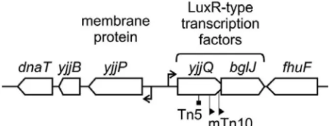
6. yjjQ-bglJ operon
The yjjP-yjjQ-bglJ operon is present in the enterobacterial species E. coli (including the Shigella spp.) and S. enterica. The yjjQ and bglJ genes are arranged in tandem with overlapping open reading frames. They belong to LuxR-type family of transcription factors with a typical DNA-binding helix-turn-helix (HTH) motif in the C-terminal domain (Fig 4). The This operon is repressed by the global regulator H-NS and the repression is counteracted by the LsyR-type transcriptional regulator LeuO (Chen et al., 2005; Madhusudan et al., 2005; Stratmann et al., 2008). The disruption if yjjQ by a transposon insertion resulted in attenuation of virulence in avian pathogenic E. coli (APEC) (Li et al., 2005).
Introduction 11
Fig 4. Organization of the yjjP-yjjQ-bglJ locus. The operon mapping at 99 min of the E. coli K-12 genome in between yjjB (encoding a conserved inner membrane protein) and fhuF (encoding a ferric iron reductase protein). The yjjQ and bglJ genes encode LuxR-type transcription factors. A yjjQ::Tn5 insertion mutation attenuates the virulence of APEC (Li et al., 2005), while mini-Tn10 insertions upstream of bglJ, causing the constitutive expression of bglJ, relieve the silencing of the bgl operon by H-NS in E. coli K-12 (Giel et al., 1996; Madhusudan et al., 2005). The yjjP gene encodes a membrane protein of unknown function (Daley et al., 2005). (Figure from Stratmann et al., 2008).
The yjjQ mutants were negatively selected in a genome-wide screen for Salmonella genes required for long-term systemic infection of the mouse (Lawley et al., 2006). A constitutively expressing bglJ (due to mini-Tn10 insertion) in E. coli K-12 have shown to de-repress H-NS regulated bgl operon (Giel et al., 1996). BglJ forms heterodimer with another transcriptional regulator RcsB and de-represses H-NS regulated bgl operon in E. coli (This study and unpublished data). The RcsB/BglJ heterodimer also counteract the repression of H-NS and activate the leuO (Schnetz K, unpublished data).
7. Aim of the thesis
The aim of the study is to understand the mechanism of anti-repression of H-NS regulated bgl operon silencing by the transcriptional factor BglJ and its dependence on RcsB. Firstly, heterodimerization of BglJ and RcsB was analyzed by Co- immunoprecipitation. These experiments included analyses of YjjQ, which is encoded with BglJ in one operon. Here we showed that the newly identified Lux-R type transcriptional regulators, BglJ and YjjQ interact with RcsB, the response regulator of Rcs signaling system. Secondly, the binding of RcsB-BglJ to the bgl URE was analyzed and a putative binding site for RcsB-BglJ was mapped. I also showed the binding of RcsB-BglJ may require H-NS as a necessary factor.
Results 12 III. Results
1. Interaction of RcsB with BglJ and YjjQ
Two-hybrid analyses had demonstrated that RcsB and BglJ interact. The first part of the present study focused on showing the interaction of RcsB with BglJ and YjjQ, respectively through biochemical analysis. To confirm the interaction I performed Co-immunoprecipitation assays. In a first step the expression of C-terminally epitope tagged BglJ and RcsB variants were tested. Then BglJ-Flag and RcsB-HA were co- expressed and the interaction was analyzed by immunoprecipitation with an HA specific antibody. Similarly, the interaction of RcsB and YjjQ was analyzed.
1.1 Expression of epitope tagged proteins
For the expression of epitope tagged BglJ protein, the bglJ gene was cloned into a set of plasmids which carry the inducible Ptac promoter followed by a multiple cloning site and a sequence encoding either a Myc-tag, a FLAG-tag, or a HA-tag (Fig 5). For efficient translation the plasmids carry the extended Shine-Dalgarno sequence SDgene10 derived from phage T7 gene 10 to which the bglJ and yjjQ genes were fused.
Upstream of the tac promoter maps the lacIq gene. Low and high copy variants of these vectors were used for cloning of the bglJ and yjjQ gene with a fusion of a tag sequences at the 3’ end (Fig 5).
Results 13
Fig 5. Plasmids for expression of C-terminal Flag/Myc/HA tagged BglJ and YjjQ proteins and HA-tagged RcsB protein. Schematic representation of plasmids for expression of C-terminally tagged BglJ, YjjQ, and RcsB variants (a,b). The plasmids carry a lacIq gene followed by the IPTG inducible tac promoter, a strong phage T7 gene Shine-Dalgarno sequence and a multiple cloning site for cloning of bglJ and yjjQ fusions with a tag (Flag/Myc or HA) at the 3`end. The low copy plasmids (a) carry a p15A origin and a kanamycin resistance gene. The high copy plasmids (b) carry a pMB1 origin and ampicillin resistance marker. (a) The low copy plasmids pKES169, pKES183, and pKES182 were used for cloning and pKES169 was used as empty control vector in expression studies. Similarly, high copy plasmids pKES171, pKES184, and pKES185 were used for cloning of the high copy vectors for expression of tagged bglJ and yjjQ. Plasmid pKES171 was used as control plasmid in expression studies. For expression of RcsB and its mutants high copy number similar plasmids were used which carry rcsB with its native Shine-Dalgarno fused to a HA-epitope encoding sequence at their 3’ end (Paukner A, 2007). The plasmid numbers and encoded genes are schematically represented and the cloning is documented in laboratory database.
The expression and solubility of the BglJ-HA, BglJ-FLAG, and BglJ-Myc from these plasmids was tested by Western blots (Fig 6). The expression of the BglJ-FLAG and BglJ-Myc tagged fusion proteins was marginally higher when directed by the high copy plasmid variant than low copy variants (Fig 6a and c). However, in the soluble
Results 14 fraction of a cell free protein extract BglJ-FLAG and BglJ-Myc tagged protein levels showed no significant difference between the low and high copy plasmid variants than the low copy variants (Fig 6b and d). The expression of the HA tagged BglJ fusion protein was weaker than that of the FLAG or Myc tagged BglJ protein (Fig 6e and f). Similarly, The expression of YjjQ-FLAG, Myc and HA tagged fusion proteins was higher in the high copy plasmid variants (Fig 6a, b and c). The soluble fraction of a cell free protein extract YjjQ-FLAG and YjjQ-Myc tagged protein levels were high and showed no significant difference between the low and high copy plasmid variants. The YjjQ-HA tagged protein levels were weaker in low and high copy variants than FLAG and Myc tagged variants (Fig. 6b, d and f).
Results 15
Fig 6. Expression of FLAG/Myc/HA tagged BglJ/YjjQ and HA-tagged RcsB proteins. For expression analyses of plasmids encoding the tagged variants of BglJ, YjjQ, and HA were transformed into E. coli strain S3377 carrying deletions of the yjjQ-bglJ and rcsB genes. Transformants were grown at 37°C in LB with antibiotics to OD600=0.3 and protein expression was induced for 2 hours with 1mM IPTG. For analysis of induction samples from uninduced (U) and induced (I) cultures were resolved on 12% SDS-PAGE and analyzed by Western blotting. Rat-anti HA and anti-rat alexaflour®680 antibodies were used for HA tagged proteins. Mouse-anti FLAG and mouse-anti-Myc antibodies with anti-mouse alexaflour®680 antibodies were used for FLAG and Myc tag proteins. For analysis of the solubility of the proteins a cell free protein lysate (L) was loaded next to the samples of induced cultures (I). Each lane was loaded with 0.05OD600 cells. The blots were visualized on an Odyssey infrared imaging scanner. Low copy plasmids used were BglJ-FLAG (pKERV10), BglJ-Myc (pKERV13), BglJ-HA (pKERV9), YjjQ-FLAG (pKERV6), YjjQ-Myc (pKERV8), and YjjQ-HA (pKES179.), High copy plasmids used were BglJ-FLAG (pKERV14), BglJ-Myc (pKERV15), BglJ- HA (pKERV12), YjjQ-FLAG (pKERV2), YjjQ-Myc (pKERV4), YjjQ-HA (pKES181) RcsB-HA (pKEAP38), RcsBD56N-HA (pKEAP44), and RcsBD56E-HA (pKEAP43) (See Materials and methods V.4). The ~27 kDa BglJ and 28 kDa YjjQ proteins with the respective epitope tags are indicated by an arrow.
Results 16 For expression of HA tagged RcsB and its mutants RcsBD56E and RcsBD56N high copy plasmids of similar structure were used, which carry rcsB and its mutants with their native Shine-Dalgarno sequence (Fig 5). Upon induction the expression of RcsB and its mutants RcsBD56E and RcsBD56N was similar (Fig 6g). Western analysis of the soluble fraction of cell free extract showed the wild type RcsB levels were higher than the levels of its mutants (Fig 6 h).
Based on the observed protein yields in the lysates, I decided to use low copy plasmids expressing BglJ-FLAG (pKERV10) and YjjQ-FLAG (pKERV06). For co- expression of RcsB, I used the high copy plasmid pKEAP38, encoding C-terminal HA tagged RcsB, and plasmids pKEAP43 and pKEAP44 encoding rcsBD56E -HA and rcsBD56N -HA, respectively.
Fig 6. Expression of FLAG/Myc/HA tagged BglJ/YjjQ and HA-tagged RcsB proteins continued.
The ~25 kDa HA-tagged RcsB and mutant proteins are indicated by an arrow.
1.2 Phenotype analysis
The functionality of the C-terminal FLAG/HA/Myc tagged BglJ and HA-tagged RcsB and their mutant was checked with a phenotype assay. In E. coli K12 utilization of β-glucosides like arbutin and salicin requires the expression of the bgl operon.
Wild type E. coli are phenotypically Bgl- due to H-NS silencing. An E. coli strain S2828 which carries a mini transposon insertion within the yjjQ-bglJ operon causing constitutive expression of BglJ (S2822) in addition to a rcsB gene deletion (Paukner, 2007) is transformed with RcsB and the mutants and plated on a BTB salicin plates.
Similarly, the C-terminal FLAG/HA/Myc tagged BglJ was tested in E. coli S524, which is phenotypically Bgl-. The plates were incubated at 37ºC, for β-glucoside
Results 17 utilization. The strain carrying plasmid control remained Bgl- and the strain carrying RcsB and mutants showed Bgl+ phenotype after 1 day of incubation (Fig 7).
Fig 7. Bgl phenotype assay with BTB salicin plates for β-glucoside utilization. E. coli strain S2828 (S524 rcsB::mTn10tet yjjQ/bglJ-Y6:: mTn10cm) was complemented with RcsB-HA (pKEAP38), RcsBD56E-HA (pKEAP43), RcsBD56N-HA (pKEAP44) and a vector control (pKEAP22). The plasmids coding for C-terminal HA/FLAG/Myc tagged BglJ (pKERV09, pKERV10, pKERV13) and vector control (pKES169) were transformed into E. coli S524 strain and plated on a BTB salicin plate. The plates were incubated at 37ºC for one day. The Bgl- phenotype was shown in blue background and the Bgl+ phenotype was shown in yellow background.
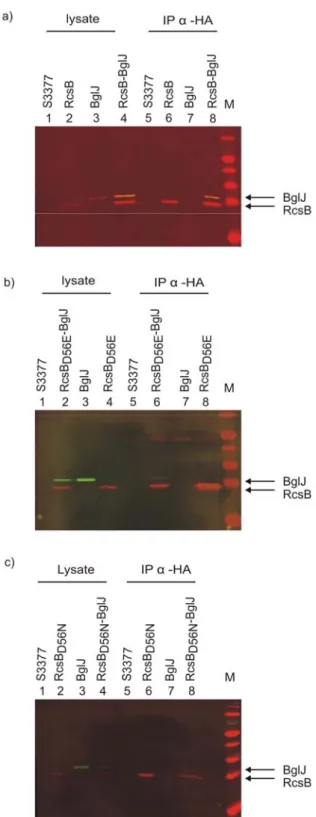
1.3 Interaction of RcsB with BglJ
The interaction of RcsB with BglJ was carried out by co-immunoprecipitation. An E.
coli strain S3377 (ΔrcsB::SpecR Δ(yjjP-bglJ)::KD3cmR) carrying deletions of the yjjQ-bglJ and rcsB genes was transformed with the two plasmids encoding RcsB-HA (pKEAP38) and BglJ-FLAG (pKERV10). As controls, single transformants with only one of the plasmids and the non-transformed strain were used. Cells were grown to early exponential phase (OD600=0.3) and then protein expression was induced with 1mM IPTG for 2 hours. The cell lysate was prepared by sonication and 200µg of the total protein was used for the immunoprecipitation assay.
The interaction between RcsB-HA and BglJ-FLAG was determined by co- immunoprecipitation of cell lysates with rabbit anti-HA IgG antibody in conjunction with protein-A sepharose (see Materials and Methods.14). The lysates and the
strain plasmids genotype/description phenotype S2822 pKEAP22 yjjQ/bglJ-Y6::mTn10cm + S2828 pKEAP22 S2822 rcsB::mTn10tet -
S2828 pKEAP22 Vector control -
S2828 pKEAP38 RcsB-HA +
S2828 pKEAP43 RcsBD56E-HA +
S2828 pKEAP44 RcsBD56N-HA +
S524 pKES169 Vector control -
S524 pKERV09 bglJ-HA +
S524 pKERV10 bglJ-FLAG +
S524 pKERV13 bglJ-MYC +
Results 18 precipitates were separated on 12% SDS-gels and analyzed by a Western blot. The Western blot was developed using rat-anti HA and anti-rat alexaflour@680 antibodies for HA tagged proteins. Mouse-anti FLAG and anti-mouse alexaflour@800 antibodies were used for FLAG tagged proteins. The lysate (expression control) showed that all the proteins were expressed (Fig 8a, lane 1-4). The co-immunoprecipitate with rabbit anti-HA IgG showed the presence of BglJ-FLAG with RcsB-HA when both proteins were co-expressed (Fig 8a, lane 8). BglJ-FLAG was not detected in the precipitate, when it was expressed alone (Fig 8a, lane 7). The controls were as expected, no bands were visible in the lysate and precipitate of the non-transformed bacteria (Fig 8a, lane 1 and 5), and RcsB-HA was expressed and immunoprecipitated when it was expressed alone (Fig 8a, lanes 2 and 6). These data show that RcsB-HA protein and BglJ-FLAG interact specifically. The protein ratio of RcsB-HA and BglJ-FLAG in the lysate and the immunoprecipitate remained the same, indicating that the interaction is efficient.
Similarly, I performed co-immunoprecipitation of BglJ-FLAG with a mutant RcsBD56E-HA protein (Fig 5). The multi-component Rcs phosphorelay signaling pathway absolutely requires its response regulator RcsB to regulate all its target genes. The mutant variant RcsBD56E, with the conserved aspartate residue at position 56 replaced by a glutamate residue, was isolated during a mutation analysis (Gupte et al., 1997). The mutated aspartate residue is conserved within the receiver domain of the family of bacterial response regulator proteins (Parkinson and Kofoid, 1992) and has been shown to be the site of phosphorylation in several response regulators (Keener and Kustu, 1988; Klose et al., 1993). The RcsBD56E variant mimics the phosphorylated form of the protein and activates the transcription of capsular polysaccharide (cps) genes constitutively (Stout, 1994). This mutant variant showed similar activity like the wild type when tested for heterodimer formation with BglJ using a bacterial two-hybrid system (unpublished lab data). So, we were interested to test the relevance of the variant in the in vitro analysis. In the co-immunoprecipitation assay with rabbit anti-HA IgG antibody BglJ-FLAG protein was co-precipitated with RcsBD56E-HA demonstrating interaction of RcsBD56E and BglJ proteins. Comparison of the protein ratio of RcsBD56E and BglJ in the lysate and precipitate indicates that the co-immunoprecipitation of BglJ with RcsBD56E was less efficient than with wild- type RcsB. However, this experiment was performed only once.
Results 19 In addition, I performed a co-immunoprecipitation assay of BglJ-FLAG with RcsBD56N (Fig 5). RcsBD56N is a mutant in which the conserved aspartate residue at position 56 was replaced by an aspargine residue. This mutation mimics the non- phosphorylated form of RcsB protein which might be inactive as transcriptional regulator (Gupte et al., 1997). No co-precipitation of BglJ-FLAG protein with RcsBD56N-HA was observed when precipitated with rabbit anti-HA IgG antibody.
This indicates a lower stability of the BglJ-FLAG and RcsBD56N interaction.
However, this experiment was performed only once and expression of the protein was poor. Also, the results contradict results of two-hybrid analyses performed previously in the lab (unpublished lab data).
Results 20 1.4 RcsB interaction with YjjQ
As for interaction of RcsB with YjjQ by co-immunoprecipitation, the same protocol was used. E. coli strain S3377 (ΔyjjP-yjjQ-bglJ), ΔrcsB) was transformed with two plasmids encoding RcsB-HA (pKEAP38) and YjjQ-FLAG (pKERV6). As controls,
Fig 8. Interaction of RcsB, RcsBD56E, and RcsBD56N with BglJ analyzed by co-immunoprecipitation.
Transformants of E .coli strain S3377 (ΔrcsB, Δ(yjjP-yjjQ-bglJ) with plasmids expressing C-terminal HA tagged RcsB and BglJ-FLAG. Cell lysates were prepared after induction of protein expression with 1mM IPTG for 2hrs at 37°C. 200µg of the total protein from the lysates expressing neither of the proteins (S3377), RcsB-HA or BglJ- FLAG alone, are both proteins were precipitated with rabbit-anti-HA IgG antibody in conjunction with protein-A sepharose. The precipitates and the lysate were separated on a 12% SDS gel and blotted for Western analysis. The Western blot was analyzed using rat-anti HA and anti-rat alexaflour@680 antibodies (red color) for HA tagged proteins. Mouse-anti-FLAG and anti- mouse alexaflour@800 antibodies (green color) were used for FLAG tagged proteins. The blot was developed using an Odyssey scanner at 700 and 800nm channels. (a) immunoprecipitation of RcsB-HA (pKEAP38, high copy) with BglJ-FLAG (pKERV10, low copy), (b) RcsBD56E-HA (pKEAP43, high copy) with BglJ-FLAG, (c) RcsBD56N-HA (pKEAP44, high copy) with BglJ-FLAG (see Materials and methods. 6). RcsB and BglJ protein are indicated by an arrow.
Results 21 single transformants with only one of the plasmids and the non-transformed strain were used. The cell lysates were immunoprecipitated with Rabbit anti-HA IgG antibody in conjunction with protein-A sepharose and the lysates and precipitates were analyzed by western blotting using tag specific antibodies (see Materials and Methods14). The lysate (expression control) showed that all the proteins were expressed (Fig 9a, lanes 1-4). The co-immunoprecipitate with Rabbit-anti-HA IgG showed the presence of YjjQ-FLAG with RcsB-HA when both the proteins were co- expressed (Fig 9a, lane 8). As expected, YjjQ-FLAG was not detectable in the precipitate, when it was expressed alone (Fig 9a, lane 7). Similarly, in the lysate and precipitates of the non-transformed strain no proteins were visible (Fig 9a.5). RcsB- HA was expressed and immunoprecipitated when it was expressed alone (Fig 9a, lanes 2 and 6). These data show that RcsB-HA protein and YjjQ-FLAG interact specifically. A 3.2 fold higher precipitation of BglJ protein than YjjQ indicates that BglJ interacts stronger than YjjQ with RcsB (The BglJ and YjjQ values obtained from band intensities which were normalized to RcsB). This result also supports the bacterial-two hybrid data which suggested that the interaction of RcsB with BglJ is stronger than with YjjQ (unpublished data of the group).
In addition, I performed co-immunoprecipitation of YjjQ-FLAG with the mutant proteins RcsBD56E-HA and RcsBD56N-HA. YjjQ-FLAG co-precipitated with the D56E mutant and the protein ratio was similar to the wild type indicating that the mutation has no significant effect in interaction with YjjQ protein. In contrast, no precipitation of YjjQ-FLAG with RcsBD56N-HA with rabbit anti-HA IgG antibody was observed.
This indicates that the affinity of YjjQ-FLAG to the mutant RcsBD56N which mimics the non-phosphorylated (i.e inactive) form of RcsB is lower. The co- immunoprecipitation experiments of YjjQ-FLAG with wild-type and mutant RcsB- HA protein was performed only once, but the results are in agreement with two- hybrid analyses performed earlier. In the two-hybrid analysis interaction of YjjQ with wild-type RcsB or with RcsBD56E was significantly more efficient than with RcsBD56N
(unpublished lab data).
Results 22
Fig 9. Interaction of YjjQ with RcsB, RcsBD56E, and RcsBD56N analyzed by co- immunoprecipitation. For co- immunoprecipitation cell lysates were prepared of transformants of E .coli strain S3377 (ΔrcsB, ΔyjjP-yjjQ-bglJ) with plasmids expressing C-terminal tagged RcsB-HA and YjjQ-FLAG. Tested were the non-transformed strain expressing neither of the proteins and transformants expressing RcsB-HA or YjjQ alone, are both proteins. Cultures were grown in LB with antibiotics and protein expression was induced with 1mM IPTG for 2hrs at 37°C. 200µg of the total protein from the lysates were immunoprecipitated with Rabbit-anti-HA IgG antibody in conjunction with protein-A sepharose.
The precipitates and the lysate were separated on a 12% SDS gel and blotted for Western analysis. The Western blot was analyzed using rat-anti HA and anti- rat alexaflour@680 antibodies (red color) for HA tagged proteins. Mouse-anti- FLAG and anti-mouse alexaflour@800 antibodies (green color) were used for FLAG tagged proteins. The blot was developed using an Odyssey scanner at 700 and 800nm channels. Co- Immunoprecipitation of YjjQ-FLAG (pKERV6) with (a) RcsB-HA (pKEAP38), (b) RcsBD56E-HA (pKEAP43) and (c) RcsBD56N-HA (pKEAP44).RcsB and YjjQ protein are indicated by an arrow.
Results 23 2. RcsB-BglJ heterodimer binding to the bgl regulatory region
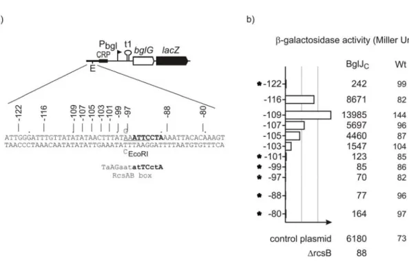
A transposon-mutagenesis screen for identifying factors which are required for activation of the bgl operon by BglJ identified the response regulator RcsB as a cofactor (Paukner, 2007). Further it was shown by two-hybrid analysis that RcsB and BglJ form heterodimers (unpublished data of the lab). Heterodimerization of RcsB and BglJ was substantiated in this thesis by co-immunoprecipitation analysis (Results chapter 1). As BglJ requires RcsB for activation (or rather de-repression) of the bgl operon, RcsB/BglJ heterodimers may bind to the bgl regulatory region and prevents repression of bgl by H-NS. This hypothesis is further supported by the identification of sequence motif which is very similar to one half-site of the consensus binding sequence of RcsA/RcsB heterodimers (Paukner, 2007) (and see below). The aim of the experiments described in this chapter was to characterize whether RcsB/BglJ heterodimers bind to the bgl regulatory region.
2.1 Linker insertion mutants of a putative RcsB/BglJ binding site in the bgl regulatory region
The response regulator RcsB regulates transcription by binding as a homodimer or as a RcsB-RcsA heterodimer (Stout and Gottesman, 1990) and see introduction).
Consensus binding sequences for RcsB homodimers and RcsB-RcsA heterodimers were determined from various mutagenesis experiments in enterobacteriaceae family members (Ebel et al., 1997; Wehland et al., 1999). The RcsAB heterodimer binds to a specific sequence “TaAGaatatTCctA” called RcsAB box is located 70 to 100 base pairs upstream to the transcriptional start site. The RcsB homodimer binding sequence “GAAgaAtAACctgC” is located immediately upstream to the -35 regions, requiring interaction with RNA polymerase to stabilize the binding (Wehland and Bernhard, 2000; Sturny et al., 2003). Interestingly, Paukner, A. identified a sequence motif in the upstream of bgl regulatory region matching half of the RcsAB consensus sequence. The motif maps between positions -90 to -96bp upstream of transcription start site (Fig 10). This motif may represent a binding site of RcsB-BglJ heterodimer in the bgl regulatory region.
Results 24
Fig 10. Putative RcsB/BglJ heterodimer binding site in bgl regulatory region. Schematic representation of bgl promoter region. The bgl promoter -10 and -35 sequence motifs are underlined.
The CAP binding site is marked in bold. The transcription start site is indicated by an arrow with +1 number. Inverted arrows denote bgl operon transcriptional terminator t1 and inverted repeat pho-IR, respectively. The half matching consensus sequence (putative RcsB/BglJ box) is marked in bold. The RcsAB box consensus sequence (Wehland M et.al. 2000) is given below the putative RcsB-BglJ binding site.
The relevance of this putative RcsB-BglJ binding site was tested using a collection of plasmids carrying linker insertions in the upstream bgl regulatory region. With this collection of plasmids it was tested which linker insertions within the bgl upstream regulatory region abrogates activation of the bgl promoter by RcsB-BglJ. To measure the bgl promoter activity, the plasmids carry a lacZ reporter gene fused 3’ to the first gene of the operon, bglG (Caramel and Schnetz, 1998). All plasmids with linker insertions carry in addition a single base-pair exchange within the regulatory region (creating an EcoRI-site used for construction of the linker insertion mutants). This mutation does not affect repression of bgl by H-NS (Caramel and Schnetz, 1998), and see below). The 6 bp MunI linker insertion map at different positions within the putative binding site, and also between the putative binding site and the promoter region, and upstream of the putative binding site (Fig 11). The bgl promoter activity was analyzed by β-galactosidase assay in strain S541 (Δbgl ΔlacZ) and a derivative of a strain which constitutively expresses BglJ (strain S3910). As expected, the β- galactosidase activity of the control plasmid (pFMAC20) was low in the wild-type, and the activity was approximately 88 fold higher in the strain which constitutively expresses BglJ (Fig11). In the rcsB (strain S3912) background constitutive expression of BglJ caused no activation confirming that activation of bgl by BglJ requires RcsB (Fig11). The β-galactosidase activity directed by plasmids with MunI linker insertion was similar to the control plasmid when analyzed in the wild type strain (Fig11). In strain S3910 constitutively expressing BglJ, β-galactosidase expression directed by