• Keine Ergebnisse gefunden

Transcriptional control of the H‐NS antagonists LeuO and RcsB‐BglJ in Escherichia coli

N/A
N/A
Protected

Academic year: 2021

Aktie "Transcriptional control of the H‐NS antagonists LeuO and RcsB‐BglJ in Escherichia coli"

Copied!
101
0
0

Wird geladen.... (Jetzt Volltext ansehen)

Volltext

(1)


Transcriptional control of the H‐NS antagonists  LeuO and RcsB‐BglJ in Escherichia coli 


I n a u g u r a l ‐ D i s s e r t a t i o n   zur 

Erlangung des Doktorgrades 

der Mathematisch‐Naturwissenschaftlichen Fakultät  der Universität zu Köln 

    vorgelegt von  Thomas Stratmann 

geb. in Warstein 


Köln, den 23. Februar 2012   

(2)


Berichterstatter:  Prof. Dr. Karin Schnetz        Prof. Dr. Jürgen Dohmen   

Tag der mündlichen Prüfung: 17. April 2012   

(3)

Zusammenfassung  ...  1 

Abstract ...  2 

1. Introduction  ...  3 

1.1 H‐NS is a nucleoid‐associated protein and global repressor of transcription  ...  3 

1.2 LeuO is a pleiotropic regulator and H‐NS antagonist ...  8 

1.3 RcsB‐BglJ is an antagonist of H‐NS‐mediated repression of the bgl operon  ...  10 

1.4 Aims of this thesis ...  12 

2. Results ...  13 

2.1 Analysis of LeuO and RcsB‐BglJ target loci ...  13 

2.1.1 RcsB‐BglJ activates transcription of several targets ...  13 

2.1.2 LeuO regulates transcription of several targets ...  15 

2.1.3 RcsB‐BglJ binds to regulatory region of bgl ...  16 

2.1.4 LeuO activates the cas operon ...  21 

2.2 Transcriptional control of leuO ...  23 

2.2.1 Transcription of leuO is activated by RcsB‐BglJ ...  23 

2.2.2 leuO is activated by an RcsB‐BglJ heterodimer independently of the Rcs phosphorelay  ...  24 

2.2.3 Transcription of leuO is repressed by H‐NS and StpA  ...  25 

2.2.4 RcsB‐BglJ binds to regulatory region of leuO ...  25 

2.2.5 RcsB‐BglJ activates one of two H‐NS‐StpA repressed leuO promoters in vivo ...  28 

2.2.6 Transcription of leuO is negatively autoregulated  ...  31 

2.2.7 In vitro mapping of promoters in the leuO‐leu operon intergenic region ...  35 

2.3 Screen for additional factors and upstream signals of leuO and bglJ transcription  ...  41 

2.3.1 Transposon insertions activating the leuO promoter or the yjjQ‐bglJ promoter ...  41 

2.3.2 Genomic library clones activating the yjjQ‐bglJ promoter ...  43 

3. Discussion ...  47 

3.1 What are the functions of RcsB‐BglJ and of LeuO? ...  47 

3.2 How do LeuO and RcsB‐BglJ regulate transcription? ...  49 

3.3 How does the feedback control of bglJ and leuO work?  ...  52 

4. Material and Methods  ...  55 

4.1 Material  ...  55 

4.1.1 Bacterial strains, plasmids and oligonucleotides  ...  55 

4.1.2 Media and antibiotics ...  62 

4.1.3 Enzymes, kits and chemicals  ...  63 

4.2 Methods ...  64 

4.2.1 Standard molecular techniques  ...  64 

4.2.2 Site‐specific mutagenesis by combined chain reaction (CCR) ...  65 

4.2.3 CaCl

2

‐competent cells and transformation  ...  65 

4.2.4 Electrocompetent cells and electroporation ...  65 

4.2.5 Gene deletion by cointegrate formation ...  66 

(4)

4.2.8 Gene deletion by λ‐Red mediated recombination  ... 68 

4.2.9 β‐galactosidase assay  ... 69 

4.2.10 Genomic library screen ... 69 

4.2.11 Transposon mutagenesis ... 70 

4.2.12 RNA isolation  ... 71 

4.2.13 Urea PAGE  ... 71 

4.2.14 cDNA synthesis  ... 71 

4.2.15 Microarray analysis  ... 72 

4.2.16 RT‐qPCR analysis ... 72 

4.2.17 5’ RACE (rapid amplification of cDNA ends)  ... 73 

4.2.18 Purification of LeuO

His6

  ... 74 

4.2.19 DNase I footprint analysis ... 74 

4.2.20 KMnO

4

 footprint analysis  ... 75 

4.2.21 In vitro transcription ... 75 

5. Appendix  ... 77 

5.1 Microarray analysis of RcsB‐BglJ targets ... 77 

5.2 Microarray analysis of LeuO targets  ... 81 

6. References ... 85 

Abbreviations  ... 93 

Figure Index ... 94 

Table Index ... 95 

Danksagung ... 96 

Erklärung  ... 97  Lebenslauf ... Fehler! Textmarke nicht definiert. 


(5)


Das  Nukleoid‐assoziierte  Protein  H‐NS  ist  an  der  Organisation  und  Kompaktierung  des  bakteriellen  Chromatins  beteiligt  und  fungiert  als  globaler  Repressor.  Dabei  reprimiert  es  vornehmlich die Transkription  von solchen  Genen,  die in Zusammenhang mit Stressantwort  und Pathogenität bekannt sind. Die Bindung von H‐NS an die DNA und die Ausbildung eines  Nukleoproteinkomplexes  an  Promotorregionen  führt  dabei  zur  Repression.  Der  Repression  durch H‐NS können jedoch genspezifische Transkriptionsfaktoren entgegengewirken, welche  die  Transkription  reprimierter  Gene  dadurch  aktivieren,  dass  sie  mit  H‐NS  um  die  DNA‐

Bindestelle konkurrieren oder die Ausbildung des Nukleoproteinkomplexes behindern (H‐NS‐

Antagonisten). Zwei Beispiele für H‐NS‐Antagonisten sind  der LysR‐Typ Transkriptionsfaktor  LeuO  und  der  FixJ/NarL‐Typ  heterodimere  Transkriptionsfaktor  RcsB‐BglJ.  LeuO  ist  ein  pleiotroper  Regulator  von  Stressanworten  und  Virulenzfaktoren.  RcsB‐BglJ  aktiviert  die  Transkription des H‐NS‐reprimierten bgl‐ (aryl‐β,D‐Glukosid) Operons. 

In  der  vorliegenden  Arbeit  wurden  neue  Zielgene  von  RcsB‐BglJ  in  Escherichia  coli  mittels  einer Microarray‐Analyse identifiziert. Die Ergebnisse dieser Analyse zeigen, dass die meisten  Zielgene durch  RcsB‐BglJ Heterodimere aktiviert werden. Unter  den Zielgenen befindet sich  neben  zahlreichen  Gene  Membran‐assoziierter  und  unbekannter  Funktion  auch  das  Gen  leuO.  Eine  detaillierte  Analyse  der  Transkriptionsregulation  von  leuO  zeigte,  dass  die  Transkription  von  leuO  stark  durch  RcsB‐BglJ  aktiviert  wird,  indem  RcsB‐BglJ  in  der  Nähe  eines neu kartierten Promotors bindet. RcsB‐BglJ wirkt der Repression von leuO durch H‐NS  und das H‐NS‐ähnliche Protein StpA entgegen. Weitere Daten zeigen, dass LeuO seine eigene  Expression negativ autoreguliert und die Aktivierung von leuO durch RcsB‐BglJ hemmt. 

Die Regulation des leuO‐Gens durch RcsB‐BglJ sowie die Autoregulation durch LeuO, wie hier  gezeigt,  und  die  Aktivierung  von  bglJ  durch  LeuO,  wie  zuvor  publiziert,  deuten  auf  einen  feedback  loop‐Kontrollmechanismus  hin,  der  die  Transkription  von  bglJ  und  leuO  koppelt. 

Dieser  loop könnte  für  das  gegenseitige  Anschalten  der  beiden  H‐NS‐Antagonisten  und  die 

Regulation  ihrer  Zielgene  als  Antwort  auf  bestimmte  Umweltbedingungen  von  Bedeutung 

sein.  Screens  lieferten  mögliche  weitere  Faktoren,  die  an  der  Regulation  des  bglJ‐leuO 

feedback loops beteiligt sein könnten. 

(6)


The  bacterial  nucleoid‐associated  protein  (NAP)  H‐NS  is  involved  in  the  organization  and  compaction of the bacterial  chromatin and acts as a global respressor, mainly of  genes  that  have been acquired by horizontal gene transfer and that are related to stress responses and  pathogenicity.  Binding  of  H‐NS  to  the  DNA  and  formation  of  a  nucleoprotein  complex  at  promoter  regions  leads  to  repression.  This  repressor  effect of  H‐NS  can  be antagonized  by  gene‐specific  transcription  factors  (H‐NS  antagonists)  that  activate  transcription  of  H‐NS‐

repressed  genes  by  competing  with  H‐NS  for  binding  or  by  disturbing  formation  of  the  nucleoprotein  complex.  Two  examples  of  such  H‐NS  antagonists  are  the  LysR‐type  transcription factor LeuO and the FixJ/NarL‐type transcription factor heterodimer RcsB‐BglJ. 

LeuO  is  a  pleiotropic  regulator  of  stress  responses  and  virulence  determinants.  RcsB‐BglJ  activates transcription of the H‐NS‐repressed bgl (aryl‐β,D‐glucoside) operon. 

In  this  work,  novel  targets  of  RcsB‐BglJ  were  identified  in  Escherichia  coli  by  microarray  analyses.  The  results  suggest  that  heterodimerization  of  RcsB  and  BglJ  is  essential  for  regulation.  Further,  in  addition  to  genes  related  to  unknown  or  predicted  function  in  the  membrane the leuO gene was identified as a target gene. Detailed analysis of transcriptional  regulation  of  leuO  demonstrated  that  RcsB‐BglJ  strongly  activates  transcription  of  leuO  by  binding proximal to a newly mapped  leuO  promoter. Thus RcsB‐BglJ antagonizes repression  of  leuO by  H‐NS and the  H‐NS‐like  protein StpA.  Additional  data presented  here  show  that  LeuO  negatively  autoregulates  its  own  expression  and  inhibits  activation  of  leuO  by  RcsB‐

BglJ. 

Regulation  of  leuO  by  RcsB‐BglJ  and  autoregulation  by  LeuO,  as  shown  here,  as  well  as  activation of bglJ by LeuO, as published previously, indicates a feedback control  mechanism  of  two  global  transcriptional regulators and H‐NS  antagonists.This  feedback  regulation may  ensure turn on of  their expression  in response to specific environmental  signals. Screens to  search for novel regulators or upstream signals were performed by transposon mutagenesis  and by using a genomic expression library. These screens indicate that additional factors may  be involved in the regulation of this leuO‐bglJ feedback loop. 


(7)

1. Introduction 

Bacteria face the need to structure their genetic material in a way that makes it compact and  storable, but at the same time accessible for replication and for transcription of the encoded  genes.  Similar to  eukaryotic chromatin,  there  is  a  bacterial  chromatin  that  consists  of DNA  and  different  kinds  of  nucleoid‐associated  proteins  (NAPs)  (Dame,  2005,  Dillon  &  Dorman,  2010).  These  NAPs  usually  exert  a  dual  function:  by  binding  to  DNA  they  confer  to  compaction of  the nucleoid  and  also to regulation  of gene expression. Nucleoid‐structuring  and  gene‐regulating  functions are  linked  to  the structural  properties  of the NAPs and  their  binding  of  DNA  target  sequences.  One  of  these  NAPs  is  the  heat‐stable  (or  histone‐like)  nucleoid‐structuring protein and pleiotropic repressor H‐NS. Repression mediated by H‐NS is  often modulated by specific regulators (H‐NS antagonists). In this work the role of LeuO and  RcsB‐BglJ, two examples of such H‐NS antagonists, and the transcriptional regulation of leuO  was analyzed. 


1.1 H‐NS is a nucleoid‐associated protein and global repressor of transcription 

The  H‐NS  protein  is  highly  conserved  in  Gram‐negative  bacteria,  and  functionally  and  structurally  related  H‐NS‐like  proteins  have  been  found  in  many  bacterial  species.  In  Eschericha  coli  H‐NS  is  present  at  approximately  20,000  copies  per  cell  during  the  exponential  growth  phase  in  which  the  transcriptional  activity  in  the  cells  is  high  (Azam  & 

Ishihama,  1999,  Ali  et  al.,  1999).  Large‐scale  binding  studies  have  revealed  that  H‐NS  is  bound to approximately 250 to 375 loci spread throughout the E. coli chromosome and that  it structures the  nucleoid into domains of  11 kb on average (Noom  et al., 2007, Grainger et  al., 2006, Oshima et al., 2006, Uyar et al., 2009). Despite the distribution of the H‐NS‐bound  sites all over the chromosome, these sites are spatially clustered into two distinct foci within  the cell as recently revealed by fluorescent live imaging (Wang et al., 2011). 

Besides  its  role  in  structuring  of  the  chromosome,  H‐NS  is  mainly  known  as  a  global 

repressor  affecting  the  transcription  of  5  %  of  the  E.  coli  genes  (Hommais  et  al.,  2001, 

Dorman,  2004).  The  repressor  function  of  H‐NS  has  been  best  studied  in  E. coli  and 

Salmonella  spp.  and  includes  the  control  of  pathogenicity  determinants  and  stress 

responses,  as  follows.  A  well‐studied  example  of  the  regulation  of  pathogenicity‐relevant 

(8)

processes  is  the  Salmonella pathogenicity  island  2  (SPI2),  which is required  for  infection  of  the  Salmonella  host’s  gut  cells.  This  SPI2  encodes  genes  for  the  secretion  of  virulence  effector proteins into the host cell. Expression of these effectors is tightly repressed by H‐NS  (Fass  &  Groisman,  2009,  Stoebel  et  al.,  2008).  Repression  by  H‐NS  of  SPI2  and  of  other  virulence  genes  involved  is  antagonized  and  fine‐tuned  by  a  complex  network  of  two‐

component  systems  (EnvZ/OmpR,  PhoPQ,  and  SpiR/SsrB),  additional  nucleoid‐associated  proteins (IHF, Fis), and transcription factors (SlyA) that respond to specific signals in the host  environment.  In  Escherichia  coli,  H‐NS  represses  transcription  at  the  locus  of  enterocyte  effacement  (LEE)  in  enterohemorrhagic  strains  (EHEC)  (Barba  et  al.,  2005,  Laaberki  et  al.,  2006).  This  pathogenicity‐relevant  locus  is  required  for  successful  colonization  of  the  host  tissue. The H‐NS‐like protein Ler antagonizes H‐NS at this locus. One of many examples of H‐

NS‐repressed  stress  response  systems  is  regulation  of  the  proU  (proVWX)  operon  which  encodes  transporters  for  osmoprotectants  (Owen‐Hughes  et  al.,  1992,  Lucht  et  al.,  1994). 

Other  classical  examples  of  H‐NS‐mediated  repression  of  stress  response  systems  are  the  three  acid  resistance  systems  in  E.  coli  (glutamate‐,  arginine‐,  and  lysine‐dependent  acid  resistance) for which H‐NS is the master repressor (Krin et al., 2010). H‐NS also represses the  rrn  (rRNA‐encoding)  operons  (Schneider  et al.,  2003).  Despite its  mainly  negative effect  on  transcription, H‐NS positively influences transcription of the flagellar regulator operon flhDC  and  thus  motility  of  the  bacteria  (Bertin  et  al.,  1994).  However,  H‐NS  does  not  activate  transcription of flhDC directly but downregulates its repressor HdfR (Ko & Park, 2000). Thus,  the absence of H‐NS leads to elevated expression of the HdfR repressor, repression of flhDC  and flagellar genes and decreased motility. 

It  has  further  been  shown  that  many  of  the  H‐NS‐repressed  genes  have  been  acquired  by  horizontal gene transfer (i. e. by phage infection, transformation or conjugation) (Navarre et  al.,  2007,  Navarre  et  al.,  2006,  Lucchini  et  al.,  2006).  This  lead  to  the  conclusion  that  selective  repression  (or  ‘silencing’) of  such xenogeneic material by  H‐NS  may  protect a  cell  from  expression of  potentially  deleterious  genes  which  may,  however,  provide  advantages  under  certain environmental conditions  (Oshima  et  al.,  2006, Navarre  et  al.,  2006, Lucchini  et al., 2006, Navarre et al., 2007, Dorman, 2007). 

Repression  by  H‐NS  is  mediated  through  binding  of  H‐NS  to  the  DNA  and  the  consecutive 

formation  of  an  H‐NS‐DNA  complex  (nucleoprotein  complex).  Numerous  studies  have 

(9)

addressed  the  mechanism  of  DNA  binding,  site‐specific  recognition  and  transcriptional  regulation  by  H‐NS  as  follows.  The  H‐NS  protein  has  a  molecular  weight  of  15.6  kDa  and  consists  of 137  amino  acids. Its  N‐terminal domain mediates dimerization  and  higher order  oligomerization, and the C‐terminal domain contains a winged helix‐turn‐helix motif (wHTH)  that  mediates  DNA  binding.  N‐  and  C‐terminus  are  flexibly  connected  by  a  linker  domain  which is  required for  higher  order  oligomerization of  H‐NS  (Figure 1A) (Rimsky, 2004, Stella  et  al.,  2005).  H‐NS  presumably  binds  as  dimer  to  a  high  affinity  site  on  the  DNA  (the  nucleation  site)  within  AT‐rich  DNA  sites  (Bouffartigues  et  al.,  2007).  Such  AT‐rich  sites  are  generally  associated  with  horizontally  acquired  genes  and  with  promoter  regions.  The  structural feature of bent DNA often found at AT‐rich sequences has been regarded as basis  for binding site recognition by H‐NS (Owen‐Hughes  et al., 1992, Dame et al., 2002). For this  initial binding the degenerate consensus DNA motif ‘TCGATAAATT’ has been proposed (Lang  et  al.,  2007).  However,  initial  binding  is  probably  not  based  on  sequence‐specific  protein‐

DNA interactions but rather on an indirect read‐out mechanism involving weak interaction of  arginine  residues in the wHTH  DNA  binding  domain  of H‐NS  with  the DNA minor groove  at  the nucleation site (Gordon et al., 2011, Cordeiro et al., 2011). Binding to the nucleation site  then  leads  to  the  formation  of  an  extended  nucleoprotein  complex  by  higher  order  oligomerization  of  H‐NS  dimers  along  the  AT‐rich  DNA  (defined  as  ‘coating’  or  ‘stiffening’,  Figure  1B)  and  by  building  DNA‐H‐NS‐DNA  bridges  (‘bridging’)  (Bouffartigues  et  al.,  2007,  Lang  et  al.,  2007, Dame et  al.,  2006).  Changes  in  the  intracellular  levels  of  divalent  cations 

such as  Mg

2+

  drive switching between bridging and stiffening  (Liu  et  al.,  2010).  A structural 

study  has  revealed  that  the  nucleoprotein  complex  can  be  modelled  as  a  superhelical 

structure that may explain the bridging and stiffening models (Figure 1C) (Arold et al., 2010). 

(10)


In  E. coli, the  H‐NS‐like  protein  StpA can contribute to gene regulation and silencing (Zhang  et  al.,  1996,  Wolf  et  al.,  2006,  Doyle  et  al.,  2007,  Muller  et  al.,  2006).  StpA  and  H‐NS  are  highly  similar,  showing  58 %  sequence  identity  on  the  amino  acid  level  and  can  form  heterodimers (Muller et al., 2010). It is believed that StpA, which is more prominent than H‐

NS  in  the  stationary  growth  phase  and  under  growth  at  high  temperature,  functions  as  a  molecular backup of H‐NS (Zhang et al.,  1996, Sonden &  Uhlin, 1996). However,  a  genome‐

wide  comparison  of  H‐NS  binding  sites  and StpA  binding sites  in hns and  stpA  mutants  has  revealed that deletion of hns diminishes binding of StpA at many sites with only one third of  H‐NS binding sites bound by StpA independently of H‐NS (Uyar et al., 2009). Binding of H‐NS  and StpA is similar in that both proteins can coat the DNA (stiffening) and form protein‐DNA  bridges.  Unlike  H‐NS,  StpA  is  able  to  form  such  bridges  also  under  conditions  of  low  magnesium concentration (Lim et al., 2011). 

The  formation  of  the  H‐NS‐DNA  nucleoprotein  complex  next  to  promoters  mediates  repression of transcription. Repression can be caused by occluding RNA polymerase from the  promoter  which  is  masked  by  the  nucleoprotein  complex  or,  as  shown  in  some  cases,  by  trapping  bound  RNA  polymerase  at  the  promoter  as  demonstrated  for  rrn  and  hdeAB 

Figure  1:  H‐NS  structure  and  DNA binding 

(A)  Schematic  model  of  H‐NS  monomer  consisting  of  an  N‐

terminal dimerization domain,  a  linker  domain  involved  in  oligoerization  (both  orange),  and a  C‐terminal  DNA binding  domain  (yellow).  (B)  Binding  of  H‐NS  dimers  to  DNA  (grey  line)  in  stiffening and  bridging  mode.  (C)  Model  of  H‐NS  superhelix  (yellow:  putative  position  of  the  wHTH  domains;  orange:  oligomer  of  the  H‐NS  N‐terminus;  blue: 

amino  acids  R15,  R19,  and 

K32  involved  in  DNA  binding) 

accommodating  two  DNA 

double  helices  (grey).  Panel  C 

modified  from  (Arold  et  al., 

2010). 

(11)

Additionally,  repression  by  binding  of  H‐NS  within  the  transcription  unit  has  been  demonstrated for some loci, such as the proU and bgl  operons, respectively (Nagarajavel et  al.,  2007).  Repression  by  H‐NS  is  most  commonly  relieved  by  the  binding  of  locus‐specific  transcriptional  regulators  which  compete  with  H‐NS  for  binding  or  restructure  the  H‐NS‐

nucleoprotein complex (Figure 2D) (Stoebel et al., 2008). Besides, alterations in H‐NS activity  by basic physiological conditions of the cells such as osmolarity, temperature, and pH (Liu et  al.,  2010),  locus‐specific  changes  of  the  DNA  structure  (bending)  (Falconi  et  al.,  1998),  or  enhancement of the transcription rate (Nagarajavel et al., 2007), can relieve H‐NS‐mediated  repression. Two  examples  of  transcriptional regulators that  act as H‐NS antagonists are the  LysR‐type  transcription  factor  LeuO  and  the  heterodimeric  FixJ/NarL‐type  transcription  factor  RcsB‐BglJ.  The  role  and  regulation  of  these  regulators,  LeuO  and  RcsB‐BglJ,  was  analyzed in this work. 


Figure 2: Schematic model of repression by H‐NS and StpA.

(A)  The  RNA  polymerase  holoenzyme  (RNAP)  requires access 

to  the  promoter  to  initiate  transcription.  (B)  H‐NS  represses 

transcription by initial binding  to nucleation sites followed by 

spreading  of  nucleoprotein  complex  of  H‐NS  and  DNA.  The 

nucleoprotein  complex  may  spread  into  a  promoter  and 

render  the  promoter  inaccessible  for  RNAP.  Alternatively, 

RNAP may  be trapped bound to  a promoter.  (C) H‐NS can be 

partially  or  fully  substituted  by  StpA.  (D)  Repression  by 

H‐NS/StpA  can  be  antagonized  by  a  specific  transcription 

factor  (TF).  The  transcription  factor  may  hinder  spreading  of 

the  repressing  nucleoprotein  complex  and/or  recruit  RNA 

polymerase to the promoter. 

(12)

1.2 LeuO is a pleiotropic regulator and H‐NS antagonist 

LeuO  is  a  LysR‐type  transcription  factor  and  a  master  regulator  of  multiple  loci  including  genes related to stress response and pathogenicity of Enterobacteriaceae. The LysR family is  the most abundant group of  transcription factors  in prokaryotes  (Schell, 1993). Initially, the  LysR family was described on the basis  of a conserved  wHTH motif in  their N‐terminal DNA  binding  domain  (Henikoff  et  al.,  1988).  A  typical  feature  of  LysR‐type  proteins  is  the  composition  of  a  central  domain  and  the  C‐terminal  domain.  Though  very  diverse  among  LysR‐family  members,  the  C‐terminal  domain  usually  contains  a  binding  pocket  for  a  small  co‐effector molecule that binds and alters the regulatory activity of the protein by causing a  conformational  change  of  the  active  tetrameric  form  of  the  protein  (Maddocks  &  Oyston,  2008). 

LeuO  has been  studied  mostly in Escherichia  coli and Salmonella  enterica, but  also in  other  bacteria  including  Yersinia  enterocolitica,  and  Vibrio  cholerae  (Rodriguez‐Morales  et  al.,  2006, Moorthy & Watnick, 2005, Lawrenz & Miller, 2007). Among other targets, LeuO acts as  a  transcriptional  activator  and  H‐NS  antagonist  at  the  bgl  operon  (Ueguchi  et  al.,  1998),  encoding  proteins  for  the  uptake  and  utilization  of  aryl‐β,D‐glucosides.  LeuO  further  activates  transcription  of  the  H‐NS‐repressed  outer  membrane  porin  genes  ompS1  and  ompS2  in  Salmonella  enterica  (Flores‐Valdez  et  al.,  2003,  Fernandez‐Mora  et  al.,  2004,  Rodriguez‐Morales et al., 2006).  LeuO regulates  transcription  of the yjjP gene  and activates  the  divergent  yjjQ‐bglJ  operon  encoding  FixJ/NarL‐type  transcription  factors  (Stratmann  et  al., 2008). At this locus, LeuO binding regions flank the core promoter of the yjjQ‐bglJ operon  and may compete with H‐NS for binding. It has further been demonstrated that at least two  out  of  three  different  LeuO  binding  sites  are  required  for  full  activation  the  yjjQ‐bglJ  promoter,  and  it  has  been  proposed  that  synergistic  binding  to  these  two  sites  may  restructure  the  nucleoprotein  complex  and  relieve  repression  (Stratmann  et  al.,  2008).  In  addition, LeuO antagonizes H‐NS at the CRISPR‐associated cas operon (Shimada et al., 2009,  Hernandez‐Lucas et al., 2008, Westra et al., 2010, Medina‐Aparicio et al., 2011). The CRISPR‐

cas  system  encodes  a  prokaryotic  defense  system  against  foreign  DNA  that  enters  the  cell 

during  bacteriophage  infections  and  plasmid  conjugation  (Brouns  et  al.,  2008).  Although 

mainly known as an activator, LeuO represses transcription of the acid stress regulator CadC 

(Shi  &  Bennett,  1995)  and  the  small  RNA  dsrA  (Repoila  &  Gottesman,  2001).  A  recent 

(13)

genome‐wide study of LeuO binding revealed that LeuO may regulate many targets involved  in structuring the cell surface such as fimbrial genes (Shimada et al., 2011). Moreover, most  of  the  LeuO  targets  are  repressed  by  H‐NS,  and  thus  LeuO  emerged  as  important  H‐NS  antagonist and global regulator as it has been shown that 90 % of LeuO‐bound loci also show  binding  of  H‐NS  (Stoebel  et  al.,  2008,  Hernandez‐Lucas  et  al.,  2008,  Shimada  et  al.,  2009,  Shimada et al., 2011). 

Remarkably,  expression  of  the  leuO  gene  is  itself  repressed  by  H‐NS  under  standard  laboratory growth conditions (Figure 3) (Klauck et al., 1997, Chen et al., 2001). As LeuO is an  H‐NS  antagonist  with  pleiotropic  function,  activation  of  leuO  expression  should  occur  in  response  to  specific  environmental  cues.  Indeed,  moderately  increased  expression  of  leuO  has  been detected upon amino acid  starvation  and  in the stationary  growth phase (Fang et  al., 2000, Majumder et al., 2001, Shimada et al., 2011). The promoter and regulatory region  of leuO gene is highly AT‐rich (Haughn et al., 1986), typical of H‐NS repressed loci (Navarre et  al.,  2007),  and  H‐NS  and  LeuO  binding  sites  have  been  mapped  to  this region  (Chen  et  al.,  2001, Chen et al., 2003, Chen et al., 2005, Chen & Wu, 2005). 


The leuO gene is  located inbetween the divergent leuLABCD (leu) and the downstream ilvIH  operons  encoding  enzymes  for  branched‐chain  amino  acid  synthesis  (Figure  3).  This  leu‐

leuO‐ilvIH  gene  cluster  is  a  paradigm  for  the  role  of  transcription‐induced  local  changes  of  DNA  supercoiling  in  promoter  regulation.  Initially,  the  leu‐500  promoter  mutant  of  S. 

Figure 3: Organization of the leuO locus. 

Organization of the leuO locus of the E. coli K‐12 chromosome. The leuO gene (NC_000913: 84,368 to  85,312)  is  located  between  the  leuLABCD  operon  encoding  genes  for  leucine  snthesis  and  the  ilvIH  operon  involved  in  synthesis  of  branched‐chain  amino  acids.  Transcription  of  leuO  is  repressed  by  H‐NS  and  activated  by  supercoiling  coupled  to  transcription  of  the  downstream  ilvIH  operon  (Wu  & 

Fang, 2003). Postive autoregulation has been suggested (Chen & Wu, 2005).

(14)

enterica  serovar  Typhimurium  that  carries a  point  mutation  rendering  the promoter of  the  leu  operon  inactive  was  found  to  be  repressed  in  topA  (topoisomerase  I)  mutants  (Graf  & 

Burns, 1973, Margolin et al., 1985). It was then  shown that the mutant leu‐500 promoter is  supercoiling‐sensitive  and  responds  to  transcription‐induced  local  changes  of  DNA  supercoiling  as  tested with plasmids (Pruss  &  Drlica,  1985, Richardson et  al.,  1988,  Lilley & 

Higgins,  1991,  Chen  et  al.,  1992,  Chen  et  al.,  1994,  Tan  et  al.,  1994).  Further  experiments  with  plasmids  carrying  the  natural context  of  the  leu‐leuO‐ilvIH gene  cluster  demonstrated  that  activation  of  the  leu‐500  promoter  (and  also  the  wild‐type  leu  promoter)  depends  on  transcription of the divergent ilvIH promoter and the leuO gene both in S. Typhimurium and  in E. coli.  This suggested  that topological  coupling operates  over  1.9 kb in the chromosome  (Wu  et  al.,  1995,  Fang  &  Wu,  1998a,  Fang  &  Wu,  1998b).  Taken  together  with  the  finding  that expression  of leuO and ilvIH  is elevated  in  the  late  stationary phase, these results lead  to  the  proposal  of  a  supercoiling  relay  mechanism  in  which  activation  of  ilvIH  results  in  activation of leuO and finally the leu promoter (Fang & Wu, 1998a, Fang & Wu, 1998b, Wu & 

Fang,  2003).  Furthermore,  the  presence  of  LeuO  binding  sites  in  the  leu‐leuO  intergenic  region  and  the  capability  of  LeuO  to  delimit  spreading  of  an  H‐NS‐DNA  nucleoprotein  complex  was  taken  as  indication  that  LeuO  acts  as  a  positive  autoregulator  in  the  natural  context  (Chen  &  Wu,  2005).  Besides  this,  activation  of  H‐NS‐repressed  loci  by  LeuO  is  presumably  achieved  by  competition  of LeuO  with  H‐NS  for  DNA  binding  (De  la Cruz  et al.,  2007, Shimada et al., 2011).  


1.3 RcsB‐BglJ is an antagonist of H‐NS‐mediated repression of the bgl operon 

The heterodimeric  transcription factor RcsB‐BglJ  has  been  identified  as  antagonist  of H‐NS‐

mediated  repression  of  the  bgl  operon.  RcsB  and  BglJ  belong  to  the  FixJ/NarL  family 

characterized  by  a  conserved  LuxR‐type  helix‐turn‐helix  (HTH)  motif  in  the  C‐terminal  DNA 

binding domain (Gao et al., 2007, Henikoff et al., 1990). RcsB is the response regulator of the 

Rcs (regulation of capsule synthesis) two‐component phosphorelay system (TCS) that senses 

perturbations of the outer membrane and the peptidoglycan layer (Majdalani & Gottesman, 

2005, Farris et al., 2010, Laubacher & Ades, 2008). RcsB is involved in the control of motility, 

cell  division,  outer  membrane  protein  expression,  capsule  synthesis,  acid  stress  response, 

and  the  regulatory  non‐coding  RNA  RprA (Castanie‐Cornet et  al.,  2010,  Huang  et al.,  2006, 

(15)

Majdalani & Gottesman, 2005). RcsB is a unique bacterial response regulator in that it acts as  homodimer  but  also  as  heterodimer  with  RcsA  (regulation  of  capsule  synthesis),  GadE  (regulation  of  acid  stress  response),  and  BglJ  (activation  of  the  bgl  operon),  respectively  (Majdalani & Gottesman,  2005,  Castanie‐Cornet et al.,  2010, Venkatesh et al., 2010).  These  three proteins likewise belong to the FixJ/NarL family of transcription factors (Henikoff et al.,  1990,  Giel  et  al.,  1996).  In  Salmonella  Typhi,  interaction  of  RcsB  with  TviA  was  found  to  control Vi antigen  synthesis  (Winter  et  al.,  2009).  The activity  of the RcsB‐RcsB  homodimer  and  the  RcsB‐RcsA  heterodimer  depends  on  phosphorylation  of  RcsB  (Majdalani  & 

Gottesman,  2005).  However,  the  RcsB‐BglJ  heterodimer  activates  transcription  of  the  only  known  target  locus  so  far,  the  bgl  operon,  independently  of  RcsB  phosphorylation  (Venkatesh et al., 2010). Since the rcsB gene is constitutively expressed, regulation of bgl by  RcsB‐BglJ  is  controlled  by  the  presence  or  absence  of  BglJ.  Interestingly,  the  three  genes  rcsA, gadE,  and bglJ,  respectively, encoding  the three RcsB heterodimerization  partners are  all repressed by H‐NS (Majdalani & Gottesman, 2005, Stratmann et al., 2008, Sayed & Foster,  2009). 

The  RcsB  heterodimerization  partner  BglJ  is  encoded  in  an  operon  together  with  YjjQ,  another FixJ/NarL‐type transcription factor (Figure 4) (Stratmann et al., 2008). YjjQ has been  proposed  to  be  important  for  pathogenicity  of  avian  pathogenic  E. coli  (APEC)  since  a  Tn5  transposon insertion in the 3’ end of yjjQ lead to attenuation of virulence of the mutant in an  animal infection model (Li et al., 2008). However, this insertion may also affect expression of  the downstream‐encoded bglJ  gene.  Further  transposon  insertions mapped  to  the  3’end  of  yjjQ  render  expression  of  the  bglJ  gene  constitutive  which  leads  to  activation  of  the  only  known  target  of  BglJ,  the  bgl  operon  (Giel  et  al.,  1996,  Madhusudan  et  al.,  2005). 

Transcriptional  regulation  of  the  yjjQ‐bglJ  operon  has  been  described  (Stratmann  et  al.,  2008). Transcription directed by the yjjQ‐bglJ promoter is repressed by H‐NS which binds to  the  AT‐rich  555‐bp  intergenic  region  of  yjjQ  and  the  divergent  yjjP  gene.  Binding  sites  upstream and downstream of the yjjQ‐bglJ promoter are required for efficient repression by  H‐NS.  The  core  promoter  region,  however,  is  not  bound  by  H‐NS.  Interestingly,  LeuO  was  found  to  antagonize  H‐NS‐mediated  repression  of  the  yjjQ‐bglJ  promoter,  as  described  in  chapter 1.2. 


(16)


1.4 Aims of this thesis 

Thus  far,  the  only  known  regulatory target  of  RcsB‐BglJ is  the  bgl operon.  LeuO  is  a  global  regulator  involved  in  the  regulation  of  stress  responses  and  pathogenicity  determinants. 

Moreover, LeuO activates transcription of the bglJ gene. Both transcription factors, RcsB‐BglJ  and LeuO, antagonize H‐NS‐mediated repression. The aims of this thesis were: 


 To identify novel  putative targets  of RcsB‐BglJ and LeuO by  microarray  analyses and  to confirm the obtained microarray data for selected targets, 

 to study regulation of the newly identified RcsB‐BglJ target leuO in detail, and 

 to  identify  additional  factors  that  may  affect  the  feedback  loop  regulation  of  RcsB‐

BglJ and LeuO. 


Figure 4: Organization of the yjjP‐yjjQ‐bglJ locus.

Organization of the yjjP‐yjjQ‐bglJ locus located between  yjjB (encoding  a conserved  inner membrane  protein)  and  fhuF  (encoding  a  ferric  iron  reductase  protein).  The  yjjQ‐bglJ  operon  (NC_000913: 

4,601,500  to  4,602,860)  encodes  FixJ/NarL‐type  transcription  factors.  Transcription  of  the  operon  is 

repressed  by  H‐NS  and  activated  by  LeuO  (Stratmann  et  al.,  2008).  A  yjjQ::Tn5  insertion  mutation 

attenuates the virulence of APEC  (Li et al., 2008) while mini‐Tn10 insertions upstream of bglJ, causing 

the  constitutive  expression  of  bglJ,  relieve  the  silencing  of  the  bgl  operon  (Giel  et  al.,  1996, 

Madhusudan et al., 2005). The yjjP gene encodes a membrane protein of unknown function. 

(17)

2. Results 

2.1 Analysis of LeuO and RcsB‐BglJ target loci 

The heterodimer RcsB‐BglJ activates the bgl operon, its only known regulatory target to date  (Venkatesh  et  al.,  2010).  LeuO  is  a  pleiotropic  regulator  which  regulates  transcription  at  several  loci  (Shimada  et  al.,  2011).  To  identify  novel  target  loci  of  RcsB‐BglJ  and  LeuO,  I  performed  DNA  microarray  analyses  and  overexpressed  BglJ  or  LeuO  in  different  genetic  backgrounds to measure transcriptome‐wide changes in response to expression of RcsB‐BglJ  and of LeuO, repectively (Figure 5). 


2.1.1 RcsB‐BglJ activates transcription of several targets 

For  the  microarray  analysis  of  RcsB‐BglJ  targets,  BglJ  was  expressed  from  low‐copy  vector  pKETS1 in E. coli strain T75 carrying a deletion of the yjjP‐yjjQ‐bglJ locus (termed ΔyjjPQ‐bglJ  in  the  following,  Table  3)  and  expression  levels  were  compared  to  T75  harboring  empty  vector  pKESK22  as  control  (Figure  5A). To  this  end, transformants of  the  respective  strains  were  grown  to  mid‐exponential  growth  phase  and  used  for  RNA  isolation  and  analysis  on  Affymetrix  GeneChip®  E.  coli  Genome  2.0  microarrays.  The  microarray  data  revealed  that 

Figure 5: Microarray analysis of RcsB‐BglJ and LeuO targets.

(A) In presence of bglJ (bglJ

+

), rcsB (rcsB

+

), and leuO (leuO

+

) all putative targets of RcsB‐BglJ or LeuO either 

upregulated  (grey)  or  downregulated (black)  could  be  identified  by  microarray analysis. For  this, BglJ was 

expressed  from  plasmid  pKETS1  in  strain  T75  (ΔyjjPQ‐bglJ)  and  expression  levels  were  compared  to  T75 

carrying control vector pKESK22. Chromosomal rcsB is constitutively expressed in this strain. (B) Deletion of 

rcsB  (ΔrcsB)  abolished  regulation  of  RcsB‐BglJ  targets  (white).  For  this,  BglJ  was  expressed  from  plasmid 

pKETS1  in  strain  T175  (ΔyjjPQ‐bglJ  ΔrcsB)  and  expression  levels  were  compared  to  T175  carrying  control 

vector pKESK22. In this strain leuO is repressed by H‐NS. (C) Analysis was performed in ΔleuO strain T177 to 

exclude targets of LeuO from the analysis. For this, BglJ was expressed from plasmid pKETS1 in strain T177 

(ΔyjjPQ‐bglJ  ΔleuO)  and  expression  levels  were  compared  to  T177  carrying  control  vector  pKESK22.  (D) 

Expression  of  LeuO  (leuO

+

)  from  plasmid  pKEDR13  in  ΔyjjPQ‐bglJ  strain  T75  revealed  bglJ‐independent 

targets of LeuO. 

(18)

overexpression of  BglJ lead  to  a  significant  (> 4‐fold, p value  < 0.05) upregulation of  39 loci  (summarized in Table 1, detailed list in appendix Table 7). 


Table 1: Microarray analysis of BglJ target loci. 

Number of loci  39 

RcsB‐dependent  38 

LeuO‐dependent  5 

co‐regulated  2 

H‐NS binding  22


Gene function known  leuO, bgl, chiA,   cas, setA, sfsB, csrB,  rhsA, rhsB, rhsC, rhsE, 

elfA, osmB, btuB, blc   


Gene function unknown/predicted  24  inner/outer membrane 

and cell surface  13 


Strikingly,  the  leuO  gene  was  one  of  the  genes  most  strongly  affected  by  BglJ  expression,  showing  a  45‐fold  upregulation.  The  known  target  of  RcsB‐BglJ,  the  bgl  operon,  was  also  strongly activated (29‐fold for bglG). To 22 of these 38 loci, binding of H‐NS has been shown  (Uyar  et  al.,  2009).  Moreover,  microarray  analysis  in  ΔrcsB  strain  T175  revealed  that  activation of  leuO, of  bgl, and  of  all other target genes was completely dependent  on RcsB  (Figure  5B,  Table  1,  Table  7).  This  suggests  that  heterodimerization  of  BglJ  and  RcsB  is  in  general essential for activation of gene expression. BglJ caused down‐regulation of only one  locus,  csrB, encoding  the  CsrA  regulating  non‐coding  RNA  CsrB.  Beside  targets  with  known  function  such  as  bgl,  leuO,  chiA (periplamic  endochitinase), and  setA  (sugar  efflux  system),  24  RcsB‐BglJ  targets  were  genes  of  unknown  or  predicted  function,  13  of  which  are  predicted  to  be  encoding  membrane  proteins,  such  as  the  RhsA,  RhsB, and  RhsC  elements  (encoding  hydrophilic  proteins  with  repetitive sequence  elements)  or  cell  surface  proteins. 

However, since  LeuO is  a  pleiotropic regulator of  transcription,  I  assumed that  some  of the 

genes which were upregulated by RcsB‐BglJ were targets of LeuO and only indirectly affected 

by  activation  of  leuO  through  RcsB‐BglJ.  Therefore,  I  repeated  the  microarray  analysis  and 

overexpressed BglJ in ΔleuO strain T177 to exclude indirect  targets (Figure 5C). Surprisingly, 

(19)

(encoding a fimbrial‐like adhesion protein), the predicted fimbrial‐like adhesin protein YadN,  the  RhsE cluster,  and the predicted protein  YhjX.  All other loci were  activated by  RcsB‐BglJ  independently of leuO ( Table 1 Table 7 ) 


2.1.2 LeuO regulates transcription of several targets 

To  identify  LeuO  targets  which  are  regulated  independently  of  RcsB‐BglJ,  LeuO  was  expressed from low‐copy vector pKEDR13 in E. coli strain T75 (Figure 5D). In accordance with  previously  published  studies,  LeuO  showed  pleiotropic  function  (Shimada  et  al.,  2011). 

Results of the LeuO microarray are summarized in Table 2, and a detailed list of LeuO targets  is given in the appendix, Table 8. Overexpression of LeuO lead to the significant (> 4‐fold, p  value < 0.05) upregulation of 119 genes, whereas 61 genes were downregulated. For the 88  of  the  upregulated  genes,  binding  of  LeuO  to  the  regulatory  regions  of  the  respective  transcription  units  has  been  shown,  as  demonstrated  by  a  comparison  of  the  LeuO  microarray  results  with  the  published  genome‐wide  binding  data  of  LeuO  (Shimada  et  al.,  2011)  (Table  2,  Table  8).  Of  the  61  downregulated  genes,  only  19  showed  also  binding  of  LeuO.  All  targets  which  showed  binding  of  LeuO  are  also  bound  by  H‐NS  as  evident  from  comparison with the study by Shimada et al. (2011). Most interestingly, the regulatory sRNA  MicC was upregulated almost 200‐fold. Other strongly upregulated targets were the CRISPR‐

associated  cas  operon,  chiA  (periplasmic  endochitinase),  and  the  bgl  operon  as  known 

targets,  among others.  Please note that in strain  T75 that  was  used for  the  microarray, the 

yjjP‐yjjQ‐bglJ  region  was  deleted, so that these  genes were  not  detected  in  the  microarray 

results. A downregulation was observed for the known LeuO targets dsrA as well as the acid 

stress system cadAB. The  five  LeuO‐dependend targets  detected in the RcsB‐BglJ micoarray 

were  also found to be  clearly  regulated by LeuO  (these were casA,  elfA, yadN, RhsE  cluster, 

yhjX). In general, the notion of LeuO as regulator of efflux systems and cell surface structures 

as  suggested  before  (Shimada  et  al.,  2009,  Shimada  et  al.,  2011)  was  confirmed  by  the 

results. 

(20)


Table 2: Microarray analysis of RcsB‐BglJ‐independent LeuO targets. 

Number of genes (LeuO binding shown) 180 (101)  upregulated  119 (88)  downregulated 61 (13)   

Previously described LeuO targets bgl, chiA, cas, rhsE, dsrA  acrEF, sdsRQP 

Gene function unknown/predicted 83 

fimbria/pili 8 

membrane 24 

intergenic 16 


To  sum  up,  a  comparison  with  the  RcsB‐BglJ  microarray  results  reveals  that  most  of  the  LeuO‐regulated  loci  were  not  affected  indirectly  by  BglJ  expression.  Interestingly,  only  two  loci,  bgl  and  chiA  (encoding  enzymes  for  ‐glucoside  utilization  and  a  periplasmic  endochitinase, respectively), are activated by RcsB‐BglJ as well as by LeuO independently of  each  other.  In  the  following,  binding  of  RcsB‐BglJ  to  the  bgl  regulatory  region  was  shown. 

Additionally,  activation  of  the  CRISPR‐cas  operon  by  LeuO  was  analyzed.  Moreover,  transcriptional regulation of leuO was analyzed in detail as described in chapter 2.2. 


2.1.3 RcsB‐BglJ binds to regulatory region of bgl 

Recently  it  has  been  shown  that  BglJ  requires  RcsB  as  a  heterodimerization  partner  to 

activate  bgl  transcription  (Figure  6)  (Venkatesh  et  al.,  2010).  The  finding  that  RcsB‐BglJ 

heterodimers counteract repression of bgl by H‐NS indicates binding of RcsB‐BglJ next to the 

bgl promoter. However, the location of a presumptive binding site so far was unknown. For 

RcsA‐RcsB  heterodimers,  a  consensus  sequence  of  the  binding  sites  has  been  proposed 

(Majdalani & Gottesman, 2005, Wehland & Bernhard, 2000). The RcsAB consensus sequence 

(also  RcsAB  box)  is  non‐palindromic  (Figure  7A)  and  presumably  recognized  by  binding  of 

RcsB  to  one  half‐site  and  by  binding  of  RcsA  to  the  other  half‐site  (Francez‐Charlot  et  al., 

2003). Interestingly, within  the bgl regulatory region a  perfect match to  one  half‐site of the 

RcsAB box is located at position ‐88 to ‐95 (relative to the transcription start site). Assuming 

(21)

subunit of the RcsB‐BglJ heterodimer may bind to this motif and that BglJ contacts adjacent  base pairs. 


To  test  the  relevance  of  this  presumptive  RcsB‐BglJ  binding  site  for  derepression  of  bgl  by  RcsB‐BglJ, site‐specific mutations were introduced in the most conserved bases matching the  right half site  of the RcsAB box (mutant  1 in Figure 7A). In addition, the left half‐site  of the  presumptive RcsB‐BglJ box was mutated (mutant 2 in Figure 7A), and mutations in both half‐

sites were combined (mutant 3 in Figure 7A). The effect of these mutations on derepression  of  bgl  by  RcsB‐BglJ  was  tested  using  a  bgl‐lacZ  reporter  fusion,  which  carries  all  elements  required  for  repression  by  H‐NS  (Figure  7B).  However,  expression  of  this  reporter  is  independent  of  regulatory  elements  for  β‐glucoside‐specific  regulation  as  it  carries  a  mutation  of  terminator  t1  (Nagarajavel  et  al.,  2007,  Radde  et  al.,  2008).  Note  that  sugar‐

specific regulation of the bgl operon is  promoter‐independent and mediated by the specific  transcriptional  antiterminator  BglG,  whose  activity  is  regulated  by  phosphorylation  dependent  on  the  availability  of  the  substrate  and  other  sugars.  BglG  allows  transcription  read‐through  at  terminator  t1  in  the  leader  of  the  operon  and  another  terminator  located  within the operon (not present in the lacZ reporter gene fusion) (Görke, 2003).  

For  expression  analyses,  the  bgl‐lacZ  reporter  fusion  with  the  putative  wild‐type  and  mutated  RcsB‐BglJ binding  sites,  respectively, were integrated  at  the chromosomal phage λ  attB site of strain T314 (ΔlacZ ΔyjjPQ‐bglJ ΔleuO) (Figure 7B).  This strain carries deletions  of  the lacZ gene, of the yjjP‐yjjQ‐bglJ locus, and of the leuO gene (remember that LeuO also de‐

represses the bgl operon). To analyze derepression of bgl by RcsB‐BglJ, BglJ was provided in  trans  using  low‐copy  plasmid  pKETS1  carrying  bglJ  under  control  of  the  inducible  tac  promoter.  Empty  vector  pKESK22  was  used  as  control  (absence  of  BglJ).  β‐galactosidase  expression  levels  directed  by  the  bgl‐lacZ  fusion  were  determined  of  exponential  cultures 

Figure 6: Activation of the bgl operon is dependent on rcsB. 

Bgl phenotype on BTB salicin  plates  of wild‐type strain T28 (Bgl

), 

of wild‐type  T28  expressing BglJ  from  plasmid  pKETS1  (Bgl

+

)  and 

of  ΔrcsB  strain  T30  expressing  BglJ  from  pKETS1  (Bgl

).  Strains 

were  grown  on  BTB  salicin  plates  supplemented  with  0.2  mM 

IPTG. 

(22)

grown in LB supplemented with kanamycin to an OD

600

 of 0.5. Please note that there is some  ambiguity  about  the  translation  start  codon  of  bglJ.  Plasmid  pKETS1  includes  the  most  N‐

terminal  AUG  which  maps  within  yjjQ.  This  plasmid  directs  the  expression  of  active  BglJ  protein  while  plasmids  pKETS9  and  pKETS10,  which  include  only  the  second  or  third  start  codon,  respectively,  provide  no  functional  BglJ  (data  not  shown).  This  suggests  that  translation of the bglJ gene begins at the very first start codon of the open reading frame.  


The  bgl‐lacZ  reporter  fusion  with  the  presumptive  wild‐type  RcsB‐BglJ  box  directed  low  levels of β‐galactosidase activity, as expected (12 units of β‐galactosidase activity, Figure 7C). 

When  BglJ was provided  in trans (using plasmid pKETS1 with bglJ under control of the lacI

q

  tac  promoter  cassette)  expression  of  the  bgl‐lacZ  reporter  increased  57‐fold  to  690  units  (Figure 7C). In a ΔrcsB mutant, expression was low (9 units) and expression remained low (8 

Figure 7: Mapping of RcsB‐BglJ binding site at the bgl regulatory region.

The  expression  level  directed  by  bgl‐lacZ  fusions  (schematically  shown  in  B)  with  wild‐type  and  mutant 

RcsB‐BglJ  binding  sites  (B)  was  determined  of  exponential  cultures  grown  in  LB  (with  appropriate 

antibiotics and 1 mM IPTG) (C). The bgl‐lacZ fusions were integrated at the phage λ attB site (strains listed 

in  Table  3).  Expression  levels  were  determined  in  the  ‘wild‐type’  strain  T314  (ΔyjjPQ‐bglJ  ΔleuO) 

transformed with the empty vector pKESK22 (‐, black bars), with plasmid pKETS1 for expression  of BglJ in 

trans (+BglJ), or with plasmid pKEDR13 for expression of LeuO (+LeuO). In addition, the expression level of 

the  bgl‐lacZ fusions was  analyzed in transformants  of  Δhns,  ΔrcsB, and  Δhns stpA mutant  derivatives,  as 

indicated. 

(23)

units)  when  BglJ  was  provided  in  trans  confirming  again  that  derepression  of  bgl  by  BglJ  requires RcsB (Figure 7C). 

Next, the expression levels directed by the RcsB‐BglJ binding‐site mutants 1 to 3 (Figure 7A)  were tested in the absence or presence of BglJ. In case of mutants 1 and 3, which both carry  exchanges corresponding to the conserved bases of the right half‐site, induction of plasmid‐

encoded  BglJ  had  no  effect  (10  to  13  units  in  all  cases,  Figure  7C).  This  demonstrates  that  mutations  in  the  presumptive  RcsB‐BglJ  binding  site  abrogate  derepression  of  bgl by  RcsB‐

BglJ. Interestingly, also binding site mutant 2 affected derepression of the bgl promoter lacZ  fusion by RcsB‐BglJ, as the expression level increased merely 8‐fold from 12 to 92 units when  BglJ  was  expressed  (Figure  7C).  Mutant  2  carries  mutations  in  the  left  half  of  the  putative  RcsB‐BglJ  box  which  is  presumably  contacted  by  the  BglJ  subunit  of  the  RcsB‐BglJ  heterodimer (Figure 7A). Taken together these data demonstrate that the putative RcsB‐BglJ  motif is important for de‐repression of bgl by RcsB‐BglJ heterodimers.  

As  further  control,  expression  of  the  bgl‐lacZ  reporter  constructs  with  the  wild‐type  RcsB‐

BglJ box and its mutants was also tested in isogenic Δhns strains. Expression levels were high 

(290  to  360  units, Figure  7C),  as expected,  as H‐NS represses  the bgl  promoter. Further, in 

the  Δhns  mutant  the  activity  was  similarly  high,  irrespective  of  whether  the  RcsB‐BglJ  box 

was  mutated  or  not,  demonstrating  that  the  site‐specific  mutations  do  not  affect  the 

promoter  activity  or  repression  by  H‐NS  (Figure  7C).  Interestingly,  the  expression  level 

directed by the bgl‐lacZ fusion was lower in the Δhns mutant (290 units) than when plasmid 

encoded BglJ was provided in the wild‐type (690 units). This indicated that the bgl promoter 

is  not  fully  active  in  the  hns  mutant.  In  agreement  with  previous  studies,  which 

demonstrated that StpA  partially represses bgl  in  hns  mutants  (Free  et  al.,  2001,  Muller  et 

al.,  2010,  Wolf  et  al.,  2006),  the  expression  level  directed  by  the  bgl‐lacZ  fusion  was  715 

units in the Δhns stpA double mutant and thus similarly high as upon de‐repression of bgl by 

RcsB‐BglJ.  However,  growth  of  the  Δhns  stpA  double  mutant  was  significantly  slower  than 

that  of  the  Δhns  mutant.  Similarly,  expression  of  plasmidic  BglJ  resulted  in  significantly 

slower  growth  in  the Δhns  mutant  and  caused  a  severe growth  reduction  in the Δhns  stpA 

double  mutant  (data  not  shown).  Therefore,  I  could  not  test  whether  RcsB‐BglJ  further 

enhances bgl promoter activity in the  absence of H‐NS and StpA. However, taken together, 

the  data  suggest  that  the  RcsB‐BglJ  heterodimer  binds  to  the  mapped  site  within  the 

(24)

upstream regulatory region of bgl (Figure 8) and antagonizes repression of bgl by H‐NS, and  also by StpA. 

Since the bgl operon has been shown to be regulated by RcsB‐BglJ and by LeuO, I also tested  whether  de‐repression  of bgl  by LeuO is independent of BglJ, whose expression is activated  by  LeuO,  and  whether  the  mutations  in  the  RcsB‐BglJ  binding  site  interfere  with  derepression of bgl by LeuO, expression levels were in addition tested with LeuO provided in  trans. For  this, plasmid  pKEDR13  carrying leuO under control of  the  inducible  tac promoter  was  used.  Induction  of  plasmid  encoded  LeuO  caused  derepression  of  the  bgl‐lacZ  fusion  (directing 200 units of β‐galactosidase activity, Figure 7C) demonstrating that LeuO activates  bgl independently  of RcsB‐BglJ (remember that the strain in  which expression was tested  is  ΔyjjPQ‐bglJ ΔleuO). 

Similarly  high  expression  levels  were  directed  by  the  RcsB‐BglJ  binding‐site  mutants  when  LeuO was present (200 units for the wild type compared to 215 to 265 units for the mutants)  (Figure  7C). This  demonstrates  that the mutations of the presumptive RcsB‐BglJ box do  not  interfere  with  derepression  of  bgl  by  LeuO.  However,  LeuO  did  not  cause  full  activation  (approximately 200 units in the presence of LeuO compared to 690 units in the presence of  BglJ).  Nonetheless,  the  data  suggest  that  LeuO  de‐represses  bgl  by  directly  binding  to  the  upstream  regulatory  region  of  the  bgl  promoter.  Importantly,  these  data  suggest  that  mutations in the presumptive RcsB‐BglJ binding site do not abolish LeuO binding. Moreover,  the position of the LeuO binding site at the bgl promoter has been mapped in our laboratory  (Venkatesh  et  al.,  2010).  The  LeuO  footprint  showed  protection  of  approximately  60  bp,  extending  from  positions −101 to −160 rela ve to the  bgl transcription  start site. Thus, the  LeuO  binding  site  maps  just  adjacent  to  the  RcsB‐BglJ  site  (Figure  8).  Such  extended  footprints are typical of LeuO (De  la Cruz et al., 2007,  Hernandez‐Lucas et al., 2008,  Westra  et al., 2010) and other LysR‐type transcription factors (Maddocks & Oyston, 2008). However,  it  remains  unknown  whether  LeuO  and  RcsB‐BglJ  can  bind  simultaneously  to  the  bgl  regulatory  region.  The results  presented  in  this chapter have  been published in (Venkatesh  et al., 2010). 


(25)


2.1.4 LeuO activates the cas operon 

Among other targets, the microarray analysis revealed that overexpression of LeuO led to an  upregulation  the  CRISPR‐associated  cas  operon  encoding  genes  for  the  bacterial  immunity  system  against  foreign  DNA  (chapter  2.1.2,  appendix  Table  8).  RNA  samples  isolated  from 

ΔyjjPQ‐bglJ  strain  T75 containing  LeuO  expression vector  pKEDR13 were  compared to  RNA 

isolated  from  T75  containing  the  empty  vector  pKESK22.  Comparison  of  cas  gene  transcription  levels  between  the  LeuO‐expressing  strain  and  the  control  strain  in  the  microarray experiment revealed a significant upregulation of transcription of casABCDE, cas1  and cas2, showing a gradual  decrease from casA (65‐fold) to  cas2 (5‐fold) (Figure 9B,  Table  8).  No change  in  the  transcription  level  of  cas3  was  detected.  These  results  are  consistent  with  a  polycistronic  transcription  of  the  casABCDE  and  possibly  the  cas1  and  cas2  genes,  with polar effects for the transcription of the more downstream genes. To confirm the effect  of  LeuO on  cas  gene expression detected in the  microarray experiment, transcript  levels  of  the  E.  coli  K12  cas  genes  in  exponential  growth  phase  were  examined  using  reverse  transcription  followed  by  quantitative  PCR  (RT‐qPCR).  For  this,  RNA  was  isolated  from  the  same  strains  as  described  for  the  microarray  analysis  and  was  reverse‐transcribed  using  random  hexameric  oligonucleotides  as  primers.  The  expression  of  the  cas  operon  was  measured  by  qPCR  with  primers  specific  for  either  casA,  casC,  cas2,  or  cas3,  respectively  (Figure 9C). The measured activation was 31.6‐fold for casA, 16.7‐fold for casC, and 3.3‐fold 

Figure 8: Sequence of the bgl promoter and upstream regulatory region.

Indicated  are  the  ‐35,  ‐10,  and  transcription  start  sites  of  the  bgl  promoter;  a  CRP  binding  site  (boxed) 

(Reynolds et al., 1986); and a Fis binding sites (dotted lines) (Caramel & Schnetz, 2000), as well as the LeuO 

(dashed lines) and RcsB‐BglJ (solid lines) binding sites. H‐NS binds to the upstream regulatory element and 

the  promoter,  but  the  H‐NS  nucleation  sites  have  so  far not  been  mapped.  The  stop  codon  of  the  phoU 

gene located upstream of  bgl  is underlined, and  the inverted  arrows indicate an inverted repeat that may 

represent a transcriptional terminator. Figure from (Venkatesh et al., 2010).  

(26)

for  cas2.  Transcription  of  cas3  was  not  activated.  Thus,  the  RT‐qPCR  confirmed  the  microarray  data  (Figure  9C).  A  detailed  functional  analysis  of  cas  regulation  has  been  published in (Westra et al., 2010). 


Figure  9:  LeuO  activates  transcription  of  cas genes. 

(A)  Schematic  illustration  of  the  CRISPR/Cas  locus  in  E.  coli  K12  (pos.  in  NC_000913:  2,885,241  to  2,875,640)  that  consists  of  the  eight  genes  cas3,  casABCDE12  and  a  downstream  CRISPR  locus  containing  12  spacers  and  13  repeats  (CRISPR  I,  black  rectangles).  The  cas3,  cas  and  CRISPR  promoters  are  indicated  with  bent  arrows.  Small  arrows below the genes show the map positions of  primer pairs used for RT‐qPCR. (B) The fold change  of  cas  expression  was  determined  by  microarray  analysis.  RNA  was  harvested  from  cultures  of 

ΔyjjPQ‐bglJ  strain  T75  harboring  either  LeuO 

expression vector pKEDR13 (+, grey bars) or empty  control  pKESK22  (‐)  from  exponential  growth  phase  in  LB  supplemented  with  kanamycin  and  1  mM  IPTG.  RNA  was  used  for  hybridization  on  Affymetrix  E.  coli  2.0  array.  Expression  level  is  given  as  fold‐change  compared  to  the  control  T75/pKESK22.  (C)  Analysis  of  expression  of  cas2,  casC,  casA,  and  cas3  by  RT‐qPCR  to  confirm  microarray data.  RNA  was  isolated  as described  in  (B).  After  reverse  transcription,  qPCR  was  performed  using  dilutions  of  cDNA  and  the  following  primer  pairs:  T411/T412  for  cas2,  T413/T414  for  casC,  T415/T416  for  casA,  and  T417/T418  for  cas3.  C

t

  values  were  normalized  to  rpoD  expression  determined  with  primers  T247  and  T248. Expression level  is  given  as  fold‐change  compared to the control T75/pKESK22. 


(27)

2.2 Transcriptional control of leuO 

The microarray data showed that leuO was one of the genes that is most strongly activated  by RcsB‐BglJ. Since LeuO is regarded as a pleiotropic regulator of stress responses and as an  important  H‐NS  antagonist,  transcriptional  control  of  leuO  was  analyzed  in  detail.  Results  presented in this chapter have been published in Stratmann et al. (2012). 


2.2.1 Transcription of leuO is activated by RcsB‐BglJ 

To study the regulatory effect  of RcsB‐BglJ on transcription of leuO,  I used a leuO promoter  lacZ reporter fusion and integrated it into the chromosomal phage λ attachment site attB of  isogenic  derivatives  of  ΔlacZ  strain  S4197  (Table  3).  The  lacZ  fusion  encompasses  the  complete 659 bp intergenic region between the leu operon and leuO (Figure 10). In the wild‐

type  strain  (T28)  this  leuO  promoter  lacZ  fusion  directed  only  3  Miller  units  of  β‐

galactosidase activity  (Figure  10B). Low  expression is  in agreement with repression of  leuO  by  H‐NS (Chen  et  al.,  2001).  However,  when  BglJ  was  expressed from  plasmid  pKETS1,  the  expression  level  increased  to  338  Miller  units  (Figure  10B).  Note  that  in  this  strain  the  chromosomal copy of bglJ is repressed by H‐NS (Stratmann et al., 2008). Similar results were  obtained  in  the  ΔyjjPQ‐bglJ ΔleuO  double  mutant  T308.  In  this  strain  background,  BglJ  caused  an  increase  in  expression  from  3  units  to  406  units  (Figure  1B).  Accordingly,  the  expression  analyses  demonstrate  a  more  than  100‐fold  activation  of  leuO  by  expression of  BglJ, confirming the microarray data. 


Abbildung

Figure  1:  H‐NS  structure  and  DNA binding 
Figure 2: Schematic model of repression by H‐NS and StpA.
Figure 3: Organization of the leuO locus. 
Figure 4: Organization of the yjjP‐yjjQ‐bglJ locus.
+7

Referenzen

ÄHNLICHE DOKUMENTE

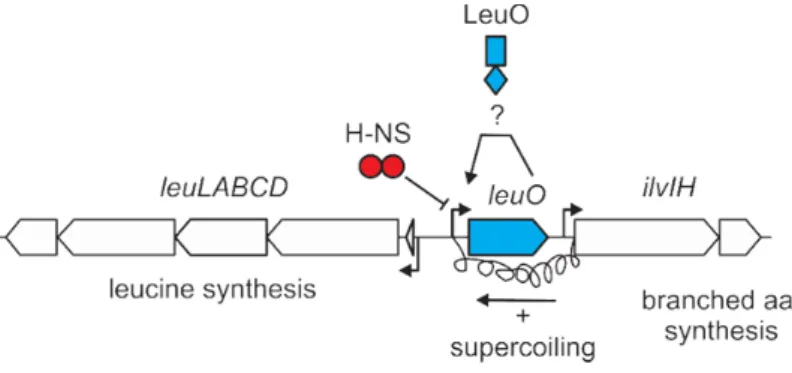
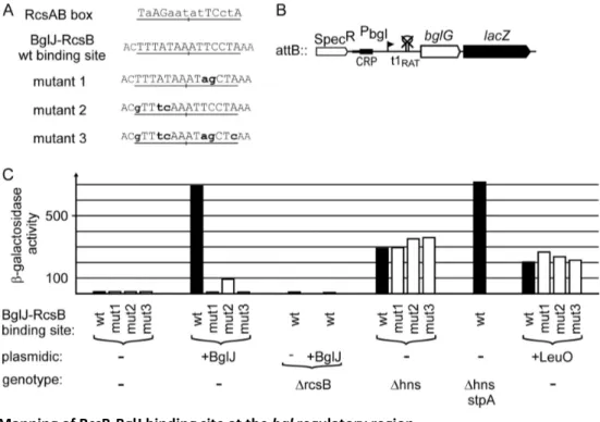
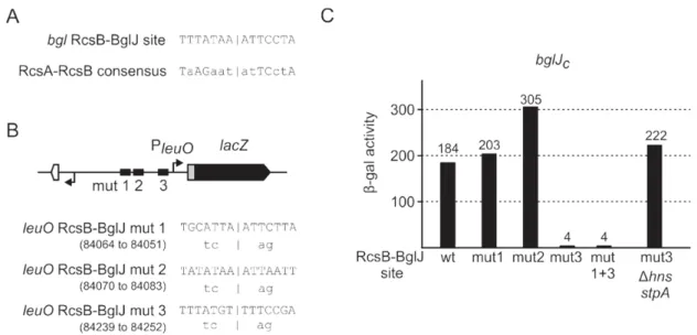
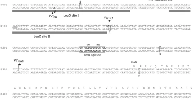
Previous data suggest, that leuO regulation is tightly controlled and involves a double-positive feedback loop, since the leuO gene is activated by BglJ-RcsB,

An increase in the promoter activity with the increase in osmolarity in case of proU, and by the action of specific transcription factors in bgl leading to an increase in

• Simulation of the phase equilibria: Carrying out GEMC-NPT and NVT sim- ulations using the four developed ab initio 5-site intermolecular potentials of hydrogen and fluorine for

One cyano group of the counter anion completes the coordina- tion environment of the caesium atom (Cs-N 3.420(8) ˚ A), while the second one is involved in hydrogen bonding. *

Auf Proteinebene konnten wir nachweisen, dass der C-terminale Knock down von Nesprin-2 zu einer verminderten Expression von Histon H1.2 in HaCaT Zellen

Synthetic promoter sequences did not show as high variation in their expression levels across cells and sites for specific transcription factors were not predicted. To evolve

Large SIMs occur infrequently in randomized networks (Table 1, P &lt; 0.01), because there is a low probability that a large number of operons controlled by a single