Analysis of transcriptional regulation by RcsB homo-‐
and heterodimers in Escherichia coli
I n a u g u r a l -‐ D i s s e r t a t i o n zur
Erlangung des Doktorgrades
der Mathematisch-‐Naturwissenschaftlichen Fakultät der Universität zu Köln
vorgelegt von Hans Derk Pannen
aus Geldern
Berichterstatter:
(Gutachter) Prof. Dr. Karin Schnetz
Prof. Dr. Jürgen Dohmen Tag der mündlichen Prüfung: 27. Oktober 2015
Zusammenfassung ... 1
Abstract ... 2
1. Introduction ... 3
1.1. The Rcs phosphorelay and its cognate response regulator RcsB ... 4
1.2. Auxiliary regulators of RcsB belong to the FixJ/NarL-‐family ... 7
1.3. Aims of this thesis ... 11
2. Results ... 12
2.1. Homo-‐ and heterodimer formation of RcsB ... 12
2.2. Establishment of reporter systems ... 14
2.2.1. RcsB-‐RcsB activates PrprA in a phosphorylation dependent manner ... 15
2.2.2. RcsA-‐RcsB activates Pwza in a phosphorylation dependent manner ... 18
2.2.3. BglJ-‐RcsB reporter system ... 21
2.2.4. MatA-‐RcsB activates Pmat
CFT073in a phosphorylation independent manner ... 21
2.2.5. Search for potential DctR targets ... 26
2.2.6. Triple reporter system ... 29
2.3. Relevance of particular residues of RcsB for specific RcsB dimers ... 31
2.3.1. Identification of relevant amino acids within RcsB ... 31
2.3.2. The relevance of particular residues of RcsB varies with its interaction partner ... 33
2.4. Expression and stability of RcsB mutants ... 38
2.5. Protein-‐protein interaction of RcsB mutants ... 40
2.6. Mechanism of transcriptional activation ... 42
2.6.1. Interaction studies of RcsB with RNA polymerase by a bacterial two-‐hybrid system ... 42
2.6.2. Overexpression of the RNA polymerase α subunit ... 45
3. Discussion ... 52
3.1. Model ... 53
3.2. Homo-‐ and heterodimerization of RcsB regulates a variety of cellular functions ... 55
3.3. RcsB regulates targets dependent and independent of phosphorylation ... 56
3.4. Residues within and close to active site of RcsB play a role for phosphorylation dependent activation ... 57
3.5. The mechanism of transcriptional activation by BglJ-‐RcsB depends on the promoter ... 58
4. Materials and Methods ... 61
4.1. Material ... 61
4.1.1. Bacterial strains, plasmids and oligonucleotides ... 61
4.1.2. Media, buffers and antibiotics ... 74
4.1.3. Enzymes, kits and chemicals ... 75
4.2. Methods ... 76
4.2.1. Standard molecular techniques ... 76
4.2.2. CaCl
2competent cells and transformation ... 76
4.2.3. Electro-‐competent cells and electroporation ... 77
4.2.4. Mutagenesis PCR ... 77
4.2.5. Random mutagenesis screen ... 78
4.2.6. Promoter replacement and gene deletion ... 78
4.2.7. Preparation of lysate and transduction ... 79
4.2.8. Chromosomal integration ... 79
4.2.9. RcsB structure prediction ... 80
4.2.10. β-‐galactosidase assay ... 81
4.2.11. Motility assays ... 81
4.2.12. RNA isolation ... 81
4.2.13. Urea PAGE ... 81
4.2.14. cDNA synthesis ... 82
4.2.15. Microarray analysis ... 82
4.2.16. qPCR analysis ... 82
4.2.17. SDS PAGE and western blotting ... 83
4.2.18. Strep-‐protein interaction experiment (SPINE) ... 85
5. References ... 87
Abbreviations ... 95
Figure Index ... 96
Table Index ... 97
Danksagung ... 98
Erklärung ... 99
Zusammenfassung
Der FixJ/NarL-‐typ Transkriptionsfaktor RcsB ist der Response-‐Regulator des Rcs-‐
Phosphorelays, einem komplexen Signaltransduktionssystem, welches die Integrität der bakteriellen Zellhülle überwacht. RcsB reguliert die Expression zahlreicher Loci die mit Motilität, Biofilmbildung und verschiedenen Stressantworten assoziiert sind. Dabei wird die Aktivität von RcsB durch zwei Mechanismen gesteuert: Erstens durch das Rcs-‐System, welches einen konservierten Aspartatrest in der Empfängerdomäne von RcsB phosphoryliert. Zweitens wird die Aktivität von RcsB durch Interaktion mit verschiedenen Hilfsproteinen gesteuert. Solche sind RcsA (Regulation der Kapselsynthese), BglJ (pleiotropischer Regulator, aktiviert bgl und leuO) sowie GadE (Säurestress). Diese Hilfsproteine gehören ebenfalls zur Familie der FixJ/NarL-‐Transkriptionsfaktoren. Zudem wurden zwei weitere Transkriptionsfaktoren der FixJ/NarL-‐Familie identifiziert, welche mit RcsB interagieren: MatA (kontrolliert Synthese der Mat-‐Pili) und DctR (in einem Acid-‐Stress-‐
Island kodiert).
In dieser Arbeit wurden die Voraussetzungen der transkriptionellen Aktivierung durch RcsB-‐Homo-‐ und Heterodimere analysiert. Zu diesem Zweck wurden geeignete Reportersysteme für RcsB-‐Homodimere und RcsB-‐Heterodimere mit RcsA und MatA entwickelt und getestet. Die Ergebnisse zeigen, dass MatA RcsB als Dimerisierungpartner benötigt um den mat-‐Promoter des UPEC-‐Stammes CFT073 zu aktivieren. Die Aktivierung erfolgt hierbei unabhängig von einer RcsB-‐Phosphorylierung. Zudem wurde gezeigt, dass MatA-‐RcsB die Motilität in E. coli K-‐12 hemmt. Des Weiteren bestätigen die Daten, dass die transkriptionelle Aktivierung durch RcsA-‐RcsB und RcsB-‐RcsB von einer RcsB-‐
Phosphorylierung abhängt. In dieser Arbeit wurden außerdem bestimmte Aminosäuren der Empfängerdomäne von RcsB identifiziert, welche für die Aktivierung durch spezifische Dimere notwendig sind. Hierbei hat sich gezeigt, dass RcsB-‐Homodimere und RcsA-‐RcsB-‐
Heterodimere -‐solche, deren Aktivität phosphorylierungsabhängig ist-‐ die größte Ähnlichkeit aufweisen. Alle relevanten Aminosäuren sind innerhalb oder nahe des aktiven Zentrums lokalisiert und tragen vermutlich zur Strukturveränderung bei, welche durch Phosphorylierung induziert wird. Weitere Ergebnisse zum Mechanismus der transkriptionellen Aktivierung deuten daraufhin, dass einige Promotoren durch Interaktion von BglJ-‐RcsB und RNA-‐Polymerase in einem Pre-‐Recruitment-‐Mechanismus aktiviert
werden.
Abstract
The FixJ/NarL-‐type transcription factor RcsB is the response regulator of the Rcs phosphorelay, a complex signal transduction system that senses perturbations of the bacterial cell envelope. RcsB regulates expression of multiple loci related to motility, biofilm formation, and various stress responses. The activity of RcsB is controlled by two mechanisms. First, the Rcs phosphorelay controls RcsB activity by phosphorylating a conserved aspartate residue within its receiver domain. Second, RcsB activity is modulated by interaction with auxiliary proteins, such as RcsA (regulation of capsule synthesis), BglJ (pleiotropic regulator, activating bgl and leuO), and GadE (acid stress response). These auxiliary regulators likewise belong to the FixJ/NarL transcription factor family and their activity depends on RcsB. Previously, RcsB was demonstrated to interact with two additional transcriptional regulators of the FixJ/NarL-‐family, MatA (control of the Mat pili expression) and DctR (encoded in the acid stress island).
In this work, determinants for transcriptional activation by RcsB homo-‐ and heterodimers were analyzed. To this end, suitable reporter systems for RcsB homodimers and RcsB heterodimers with RcsA, and MatA were established. The results show that MatA requires RcsB as a dimerization partner for activating the matA promoter of UPEC strain CFT073 and that activation is independent of RcsB phosphorylation. In addition, it was shown that MatA-‐
RcsB is able to repress the motility of E. coli K-‐12. Moreover, the results confirmed that transcriptional activation by RcsA-‐RcsB and RcsB-‐RcsB is phosphorylation dependent. This work also identified particular residues of the RcsB receiver domain being relevant for transcriptional activation by a specific dimer where RcsB homodimers and RcsA-‐RcsB heterodimers that are depending on RcsB phosphorylation possess the most similar properties. All relevant amino acids are located close to the active site, suggesting an important role for the structural change that is elicited by phosphorylation. Finally, data respective the mechanism of transcriptional activation, suggest that at some promoters BglJ-‐
RcsB activates transcription by direct contacts to the RNA polymerase in a pre-‐recruitment
mechanism.
1. Introduction
Bacteria are highly adaptive organisms with a large number of genes and pathways that allow exploiting a plenty of different environments. The central element of adaptation to a particular environment is the ability to modulate the expression of a gene subset in response to specific signals (Stephenson et al., 2000). This response requires signal transduction systems for recognition and interpretation of signals, and the conversion of these signals resulting in a specific transcriptional regulation. For this purpose bacteria possess two-‐
component signal transduction systems (TCS) that sense environmental stimuli and adjust the expression of specific genes appropriately. Two-‐component systems can also be found in eukaryotic organisms (Stock et al., 2000). A typical two-‐component system consists of a sensor histidine kinase (HK) with an input and phosphorylation domain as well as a response regulator (RR) with an N-‐terminal receiver domain and a C-‐terminal output domain. Signal sensing by the input domain of the sensor histidine kinase triggers its autophosphorylation at a conserved histidine residue. From this histidine residue the phosphoryl group is transferred to a conserved aspartate residue within the receiver domain of its cognate response regulator, whose phosphorylation affects its output function (Stock et al., 2000).
Response regulators exist presumably in equilibrium between active and inactive state, while phosphorylation favors the active conformation and shifts the balance (Stock et al., 2000). Beside prototypical two-‐component systems, a variety of bacteria possess more complex two-‐component systems. One more complex two-‐component system is the Rcs phosphorelay (Rcs = Regulator of capsule synthesis) in Escherichia coli and other members of the Enterobacteriaceae, whose response regulator is RcsB. RcsB is a versatile transcription factor possessing the rare feature of homo-‐ and heterodimerization with auxiliary regulators.
RcsB is involved in many regulatory networks related to motility, cell division and various stress responses (Majdalani & Gottesman, 2005). In this work the determinants of transcriptional activation by RcsB are analyzed.
1.1. The Rcs phosphorelay and its cognate response regulator RcsB
The Rcs phosphorelay (Figure 1) is a complex two-‐component signal transduction system that was originally identified as a regulatory system of colanic acid capsule biosynthesis in Escherichia coli (Gottesman et al., 1985). Nowadays the Rcs system is recognized as a key regulator of motility, biofilm formation, and various stress responses in Enterobacteriaceae and plays an important role in the control of lifestyle transition from a planktonic cell to a sessile cell in a biofilm (Evans et al., 2013, Farris et al., 2010, Laubacher & Ades, 2008, Majdalani & Gottesman, 2005). In many bacterial pathogens the Rcs phosphorelay is involved in controlling expression of virulence determinants. In enterohemorrhagic E. coli, for example, the Rcs system controls expression of genes at the locus of enterocyte effacement (LEE) that promote attachment to host cells (Tobe et al., 2005). In Salmonella enterica serovar Typhimurium the Rcs system regulates the Salmonella pathogenicity island 2 (SPI-‐2) encoding a type III secretion system to transfer effector proteins into host cells (Wang et al., 2007). In addition, the Rcs system has been implicated in pathogenicity in Yersinia enterocolitica, Erwinia amylovora and other pathogens (Hinchliffe et al., 2008, Wang et al., 2009, Majdalani & Gottesman, 2005). The Rcs phosphorelay consists of the inner membrane-‐bound histidine kinase RcsC and the response regulator RcsB (Figure 1). In addition to these two components, the Rcs system possesses the inner membrane-‐bound phosphotransfer protein RcsD, the outer-‐membrane lipoprotein RcsF and the inner membrane protein IgaA (YrfF) (Figure 1) (Cho et al., 2014, Castanie-‐Cornet et al., 2006, Majdalani & Gottesman, 2005). The core proteins RcsB, RcsC and RcsD are encoded at a single locus of the E. coli genome. The genes rcsD and rcsB form an operon which is immediately adjacent to rcsC but in a convergent orientation (Majdalani & Gottesman, 2005). Rcs-‐inducing conditions are, predominantly, perturbations of the cell wall and outer membrane by impaired lipopolysaccharide (LPS) synthesis through deletion of rfaD (Parker et al., 1992) and galU (Girgis et al., 2007), outer membrane protein (OMP) misfolding through deletion of surA (Castanie-‐Cornet et al., 2006), and antibiotic-‐mediated peptidoglycan stress by β-‐lactam antibiotics such as amdinocillin (Laubacher & Ades, 2008).
The lipoprotein RcsF is involved in sensing these perturbations and activating signaling, while
IgaA inhibits the signaling cascade (Cho et al., 2014, Farris et al., 2010).
Figure 1. Model of activation and phosphotransfer of the Rcs phosphorelay
The lipoprotein RcsF is translocated from the inner membrane (IM) to the outer membrane (OM) by LolA (dashed arrows). At the OM RcsF binds BamA which funnels it to OmpA (dashed arrows). OmpA displays RcsF to the cell surface. By defects of the Lol system or the Bam machinery, RcsF interacts with IgaA inducing the autophosphorylation activity of the histidine kinase domain (H) of RcsC. The phosphotransfer of the active system is indicated by curved black arrows. From the histidine kinase domain, the phosphoryl group is transferred to the receiver domain (D) of RcsC, to the histidine phosphotransfer protein RcsD and in turn to the receiver domain of the response regulator RcsB.
RcsB can also be phosphorylated unspecifically by acetyl phosphate (AcP) which is removed by the phosphatase activity of the uninduced Rcs system in reversible phosphotransfer direction (dashed curved arrow).
Both RcsF and IgaA are acting upstream of RcsC and RcsD (Figure 1) (Laubacher & Ades, 2008, Evans et al., 2013, Cho et al., 2014). On a molecular level, RcsF monitors lipoprotein transport through the periplasm and the β-‐barrel assembly. The chaperone LolA escorts lipoproteins including RcsF from the inner to the outer membrane (Figure 1). Defective LolA-‐
mediated transport causes RcsF accumulation at the inner membrane what activates the Rcs
system through IgaA which in turn induces lolA expression (Tao et al., 2012, Cho et al.,
2014). At the outer membrane, RcsF monitors the β-‐barrel assembly machinery (Bam) by
interacting with BamA. Active BamA funnels RcsF to the outer membrane porin OmpA that
exposes RcsF to the cell surface (Figure 1) (Cho et al., 2014). Disruption of this machinery
results in RcsF accumulation in the periplasm where RcsF can activate the Rcs system by
interaction with IgaA (Cho et al., 2014). Both RcsC and RcsD are anchored to the inner
membrane and exhibit a cytoplasmic part (Figure 1). The cytoplasmic part of RcsC includes a
sensor kinase domain which is autophosphorylated at a conserved histidine residue upon
activation of the signaling cascade. From this histidine, the phosphate is transferred to a
conserved aspartate residue within the receiver domain of RcsC. In the next step of the phosphorelay cascade the phosphate is transferred to a conserved histidine residue in the histidine-‐phosphotransfer domain of RcsD, and from there to the receiver domain of RcsB (Majdalani & Gottesman, 2005). Notably, in the absence of a stimulus, RcsB can be phosphorylated by acetyl-‐phosphate as a phosphoryl group donor (Figure 1) (Hu et al., 2013). RcsC and RcsD have a phosphatase activity to remove the phosphate from RcsB in reversible phosphotransfer reactions (Figure 1) (Majdalani et al., 2002, Majdalani et al., 2005).
The complexity of the output that is generated by the Rcs phosphorelay via the response regulator RcsB is likewise high and involves additional protein components. RcsB is a 216 amino acid protein with an N-‐terminal receiver domain containing the phosphorylation site (D56) and a C-‐terminal DNA binding domain (Henikoff et al., 1990, Gao et al., 2007). The receiver domain comprises residues 5 to 124 and the DNA-‐binding domain residues 144 to 209 with a FixJ/NarL-‐type typical helix-‐turn-‐helix-‐DNA-‐binding motif (HTH) from residue 151 to 194 (Majdalani & Gottesman, 2005, Pristovsek et al., 2003). As canonical bacterial response regulators, RcsB can regulate target genes as a homodimer (Majdalani &
Gottesman, 2005). Dependent on phosphorylation, RcsB activates transcription of multiple loci including rprA, encoding the small regulatory RNA RprA, the cell division genes ftsZ and ftsA, bdm coding for a protein involved in biofilm formation, as well as osmB and osmC, encoding an osmotically inducible lipoprotein and peroxidase (Majdalani & Gottesman, 2005, Francez-‐Charlot et al., 2005). RcsB binding sites, that were mapped for osmC and bdm locate just upstream of the -‐35 promoter region, probably requiring interaction with RNA polymerase for activation (Sturny et al., 2003, Francez-‐Charlot et al., 2005, Majdalani &
Gottesman, 2005).
Strikingly, in addition to forming homodimers, RcsB interacts with auxiliary
transcriptional regulators including RcsA, GadE, BglJ, MatA, and DctR that likewise exhibit a
DNA-‐binding domain of the FixJ/NarL-‐type (Majdalani & Gottesman, 2005, Castanie-‐Cornet
et al., 2010, Venkatesh et al., 2010, Fabisch, 2008). The interactions of RcsB with the
auxiliary partners alter the DNA-‐binding specificity (Venkatesh et al., 2010, Castanie-‐Cornet
et al., 2010, Wehland & Bernhard, 2000) and thus extend the regulatory repertoire of the
Rcs system to the control of multiple loci related to motility and biofilm formation, various
stress responses, cell surface components, and additional functions (Majdalani &
Gottesman, 2005, Clarke, 2010). Although heterodimerization of response regulators is a common feature in Eukaryotes, it is very rare in bacteria. To date, the only described example for heterodimerization of bacterial response regulators beside RcsB are BldM and WhiI in the filamentous bacteria Streptomyces (Al-‐Bassam et al., 2014). Notably, BldM and WhiI likewise belong to the FixJ/NarL family of transcriptional regulators.
1.2. Auxiliary regulators of RcsB belong to the FixJ/NarL-‐family
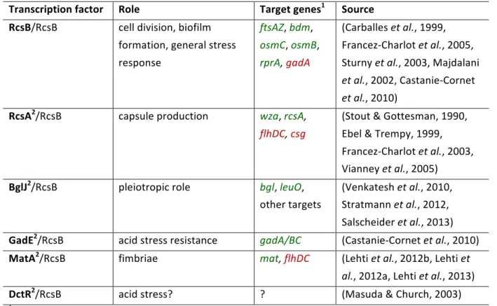
E. coli has 18 proteins with a FixJ/NarL-‐type typical HTH-‐motif, of which RcsA, GadE, BglJ, MatA and DctR were shown to interact with RcsB (Fabisch, 2008). Their targets and cellular roles are summarized in Table 1 and their relations in Figure 2. RcsA is a 207 amino acids long protein whose intracellular concentration is generally low. The amount of the RcsA protein is limited by its rapid degradation by Lon, an ATP-‐dependent protease (Stout et al., 1991). Moreover, the expression of rcsA is repressed by HNS (Sledjeski & Gottesman, 1995).
However, RcsA was reported to activate its own expression together with RcsB (Ebel &
Trempy, 1999, Wehland & Bernhard, 2000). The small RNA DsrA was found to activate rcsA expression by anti-‐silencing HNS repression (Sledjeski & Gottesman, 1995). Together with RcsB, RcsA activates expression of loci such as the cps/wza gene cluster and yjb operon for exopolysaccharide production, or rcsA expression itself (Stout et al., 1991, Ferrieres et al., 2007). The flhDC operon encoding the flagella master regulator is repressed by RcsA-‐RcsB (Soutourina & Bertin, 2003, Francez-‐Charlot et al., 2003). RcsA-‐RcsB heterodimers regulate their targets by binding a specific DNA sequence, called RcsAB box, which is located around 100 bp upstream of the transcription start site of wza as well as rcsA and downstream of the promoter of flhDC (Wehland & Bernhard, 2000, Francez-‐Charlot et al., 2003). The activity of the RcsA-‐RcsB heterodimer depends on phosphorylation of RcsB (Majdalani & Gottesman, 2005).
Table 1. Cellular roles of RcsB homo-‐ and heterodimers
Transcription factor Role Target genes
1Source
RcsB/RcsB cell division, biofilm formation, general stress response
ftsAZ, bdm, osmC, osmB, rprA, gadA
(Carballes et al., 1999, Francez-‐Charlot et al., 2005, Sturny et al., 2003, Majdalani et al., 2002, Castanie-‐Cornet et al., 2010)
RcsA
2/RcsB capsule production
wza, rcsA,flhDC, csg
(Stout & Gottesman, 1990, Ebel & Trempy, 1999, Francez-‐Charlot et al., 2003, Vianney et al., 2005)
BglJ
2/RcsB pleiotropic role
bgl, leuO,other targets
(Venkatesh et al., 2010, Stratmann et al., 2012, Salscheider et al., 2013) GadE
2/RcsB acid stress resistance
gadA/BC(Castanie-‐Cornet et al., 2010)
MatA
2/RcsB fimbriae
mat, flhDC(Lehti et al., 2012b, Lehti et
al., 2012a, Lehti et al., 2013)
DctR
2/RcsB acid stress? ? (Masuda & Church, 2003)
1activated targets in green, repressed targets in red
2HNS repressed
BglJ is a 225 amino acid transcriptional regulator that is encoded in an operon together with YjjQ, another transcription factor belonging to the FixJ/NarL-‐family (Stratmann et al., 2008).
Notably, YjjQ was also identified as a transcriptional repressor of the flhDC operon and other
targets (Wiebe et al., 2015). Expression of yjjQ-‐bglJ is repressed by HNS and activated by the
LysR-‐type transcription factor LeuO that antagonizes HNS-‐mediated repression (Stratmann
et al., 2008). Interestingly, BglJ-‐RcsB heterodimers in turn activate leuO expression and
hence BglJ and LeuO form a small regulatory network (Stratmann et al., 2012). Furthermore,
BglJ-‐RcsB activates expression of the bgl operon, encoding proteins for the uptake and
utilization of aryl-‐β,D-‐glucosides and more than 10 additional loci (Venkatesh et al., 2010,
Stratmann et al., 2012, Salscheider et al., 2013). For regulation of expression, BglJ-‐RcsB was
proposed to act in the context of the promoter as a class I activator interacting with RNA
polymerase or as an HNS antagonist. At the bgl promoter BglJ-‐RcsB acts synergistically with
CRP (Salscheider et al., 2013). Moreover, a consensus DNA-‐binding motif was defined that
suggested a DNA phasing-‐ and orientation-‐dependent positioning of the BglJ-‐RcsB
heterodimer in relation to the transcription start site which varies with the promoter
(Salscheider et al., 2013). Unlike RcsA-‐RcsB, the activity of BglJ-‐RcsB is independent of RcsB phosphorylation (Venkatesh et al., 2010, Stratmann et al., 2012).
Figure 2. Targets of RcsB homo-‐ and heterodimers in E. coli.
RcsB homodimers activate ftsAZ, bdm, osmC, osmB, and rprA. RcsA-‐RcsB activates wza, rcsA and represses csg and flhDC. MatA-‐RcsB activates mat in NMEC and represses flhDC. BglJ-‐RcsB activates several loci including bgl and leuO. GadE-‐RcsB activates gadA and gadBC. Direct targets of DctR-‐RcsB are unknown.
GadE, the central activator of glutamate-‐dependent acid resistance, is encoded by the gadE-‐
mdtEF operon located within the acid fitness island. GadE’s expression is negatively regulated by HNS (Tucker et al., 2002, Tramonti et al., 2008, Sayed & Foster, 2009). Together with RcsB, GadE activates the expression of the gadA/BC genes in a phosphorylation independent manner by binding the GAD box located around -‐60 bp upstream of the transcription start. The genes gadA and gadB encode glutamate decarboxylases conferring resistance to extreme acidic conditions. Interestingly, phosphorylated RcsB represses expression of gadA as a homodimer by binding to a site upstream of the -‐10 promoter element (Castanie-‐Cornet et al., 2010).
In a systematic approach, interaction of RcsB with all other 18 FixJ/NarL-‐type proteins
in E. coli K-‐12 was previously investigated (Fabisch, 2008). These heterodimerization studies
using the bacterial LexA-‐based two-‐hybrid system (Dmitrova et al., 1998) revealed MatA and
DctR as further interaction partners of RcsB (Fabisch, 2008). Notably, all FixJ/NarL-‐type
proteins (except GadE and EvgA) that form heterodimers with RcsB do not form homodimers
and vice versa (Fabisch, 2008). The one/two-‐ hybrid analyses for EvgA showed
homodimerization as well as heterodimerization with RcsB. For GadE neither homo-‐ nor
heterodimerization was detected under standard conditions, possibly due to acid-‐stress
dependent dimerization behavior (Fabisch, 2008).
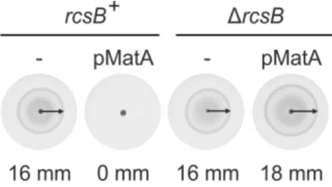
MatA (also termed EcpR) is a 196 amino acids long transcriptional regulator encoded as the first gene of the common mat fimbria operon (Lehti et al., 2012a). Mat fimbriae are a common colonization factor promoting biofilm formation as well as bacterial adherence to epithelial cells (Lehti et al., 2010, Lehti et al., 2012b). MatA is the key activator of this operon in newborn meningitis-‐associated E. coli (NMEC) and enterohemorrhagic E. coli (EHEC) although the mat operon remains cryptic in the non-‐pathogenic E. coli K-‐12 and strains belonging to the lineages A and B1 (Martinez-‐Santos et al., 2012, Lehti et al., 2012a, Lehti et al., 2012b, Lehti et al., 2013). In NMEC and other E. coli strains belonging to the lineages B2, D, and E, MatA forms a positive autoregulatory circuit (Lehti et al., 2013). However, also the MatA protein of E. coli K-‐12 strain MG1655 is fully functional (Lehti et al., 2013). In addition to MatA, RcsB is required to activate mat expression in NMEC (Lehti et al., 2012a, Lehti et al., 2012b). Notably, the mat operon is repressed by HNS (Lehti et al., 2012b, Lehti et al., 2013).
The flhDC operon is under negative control of MatA and hence the importance of MatA in the transition from planktonic to adhesive lifestyle is discussed (Lehti et al., 2012a).
DctR is a 176 amino acids long predicted transcriptional regulator encoded in the slp-‐
dctR operon whose expression is likewise repressed by HNS (Krin et al., 2010). The gene dctR is a member of the E. coli acid fitness island (AFI) and its expression is activated by YdeO (Mates et al., 2007, Masuda & Church, 2003). Deletion of slp-‐dctR abolishes YdeO-‐induced acid resistance (Masuda & Church, 2003). Slp and DctR were implicated in protection against metabolic end products under acidic conditions, however a direct target of DctR as a transcriptional regulator is not known (Mates et al., 2007, Yamanaka et al., 2014).
1.3. Aims of this thesis
For the viability of bacteria it is essential to fine-‐tune gene expression according to the environmental conditions. RcsB has the unique feature to interact with several auxiliary co-‐
regulators and is able to activate and repress targets dependent on interaction partner and phosphorylation state. These different modes of regulation by RcsB imply specific recognition mechanisms between RcsB and auxiliary regulators. Thus, RcsB is a versatile transcription factor being involved in many regulatory networks that contribute to adjust gene expression in response to environmental conditions. So far the specific determinants for transcriptional activation by RcsB are poorly understood. The aims of this work were:
• To study a potential heterodimer formation of FixJ/NarL-‐type transcription factors
• To further characterize the RcsB regulon by identification of putative targets of MatA-‐RcsB and DctR-‐RcsB heterodimers
• To establish reporter systems to analyze transcriptional regulation by RcsB homo-‐
and heterodimers
• To study the phosphorylation dependence of RcsB for activity of specific heterodimers
• To identify particular amino acid residues within RcsB which are important for the activity of RcsB as well as residues which are crucial for specific interaction with only one or a subset of the partners
• To study the mechanism of transcriptional activation by BglJ-‐RcsB
2. Results
To address the aims of this work, I applied following approaches:
• Analysis of heterodimer formation by bacterial LexA-‐based two-‐hybrid system
• Search for MatA and DctR targets by microarray analysis
• Analysis of MatA-‐RcsB effect on motility on soft agar plates
• Reporter construction by fusion of known target promoters to lacZ
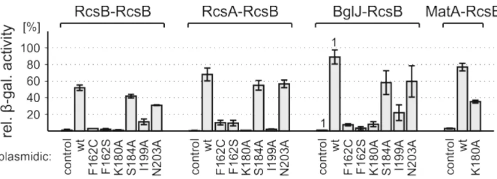
• Analysis of the activity of RcsB mutants as homo-‐ and heterodimers by β-‐galactosidase assays using the established reporter systems
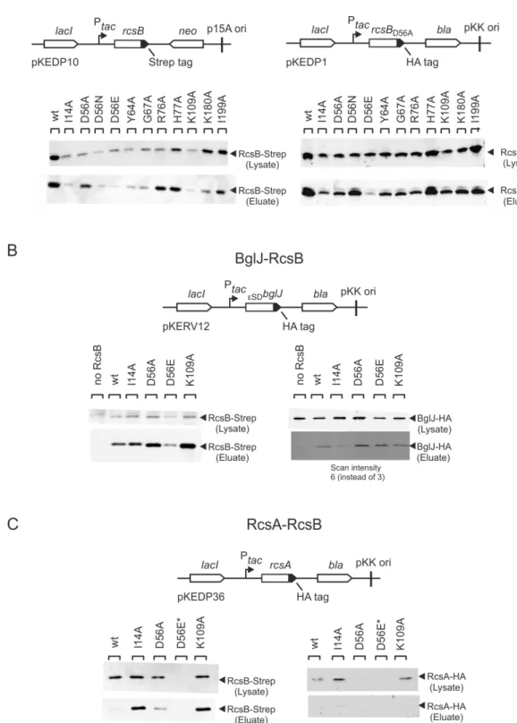
• Stability test of RcsB mutants by western blotting
• Interaction analysis of RcsB mutants by strep-‐protein interaction experiment
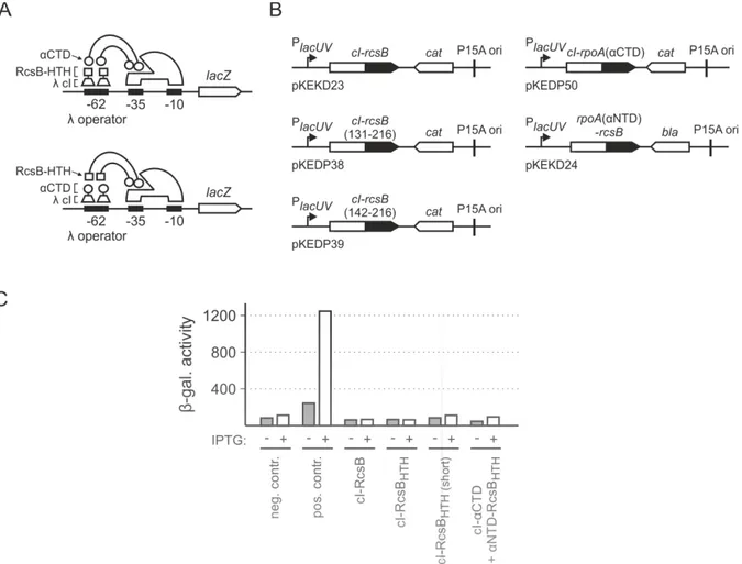
• BglJ-‐RcsB-‐RNA polymerase interaction studies by a bacterial two-‐hybrid system as well as overexpression of the RNA polymerase α subunit + mutants
2.1. Homo-‐ and heterodimer formation of RcsB
The FixJ/NarL-‐type transcriptional regulator RcsB is known to interact with auxiliary regulators such as RcsA, BglJ and GadE which also belong to the FixJ/NarL family (Majdalani
& Gottesman, 2005, Castanie-‐Cornet et al., 2010, Venkatesh et al., 2010). In previous studies, homo-‐ and heterodimerization of RcsB was analyzed using the bacterial LexA-‐based one/two-‐hybrid system (Dmitrova et al., 1998). The one-‐hybrid reporter for examining homodimer formation consists of the native sulA promoter (+/+) fused to lacZ (Figure 3A).
Only homodimers of proteins fused to the wild-‐type DNA-‐binding domain of the LexA
repressor
are able to bind to the lexA operator and repress PsulA lacZ expression. For analyzing heterodimer formation, the sulA promoter carries a hybrid lexA operator (408/+) with a mutation in one half-‐site (Figure 3B). Only heterodimers in which one partner is fused to the LexA
wild-‐type DNA-‐binding domain and the other partner is fused to the LexA
408mutant DNA-‐binding domain
are able to bind the hybrid operator and repress PsulA lacZ
expression (Dmitrova et al., 1998).
Figure 3. Homo-‐ and heterodimer formation by RcsB, BglJ, RcsA, MatA and DctR.
(A) In the LexA-‐based one/two-‐hybrid system, the sulA promoter lacZ fusion with the wild-‐type LexA +/+ operator was used to analyze homodimerization. For analysis of homodimerization, a fusion of the respective protein to the wild-‐type LexA DNA-‐binding domain was expressed from a plasmid under the control of the IPTG-‐inducible lac
UV5promoter (P
UV5). (B) The sulA promoter lacZ reporter fusion with a hybrid lexA 408/+ operator served as a reporter for heterodimerization. For heterodimerization analysis, fusions of one protein to the wild-‐type LexA DNA-‐binding domain and the potential interaction partner fused to the LexA
408mutant DNA-‐binding domain were co-‐
expressed from compatible plasmids. (C) Summary of the results for heterodimer formation by RcsB, BglJ. The fold repression of the lexAop
408/+sulA promoter lacZ fusion is a measure of heterodimerization, and was calculated by dividing the expression levels (values given in smaller font size) of the PsulA lacZ reporter that were obtained without and with induction of the LexA-‐
fusion proteins. For analyzing heterodimer formation of RcsA, MatA, and DctR with each other, strains S3440 (ΔrcsB) was co-‐transformed with plasmids encoding for LexA
WT-‐X and LexA
408-‐Y fusions, respectively. The following plasmids were used: pKEMK4 (LexA
WT-‐MatA), pKEMK1 (LexA
WT-‐
DctR), as well as pKEDP59 (LexA
408-‐MatA) and pKEDP60 (LexA
408-‐RcsA). The cultures were grown in LB supplemented with antibiotics in the presence and absence of IPTG to an OD
600of 0.5. (D) Summary of homodimer formation of RcsB, BglJ, RcsA, MatA and DctR. The fold repression of the sulA promoter lacZ fusion with the lexA operator (lexAop
+/+) is a measure of homodimerization.
Values indicated with
aare taken from (Venkatesh et al., 2010). Unpublished laboratory results are indicated with
bfrom (Fabisch, 2008) and
cfrom (Dreck, 2013).
Previous analyses using the LexA-‐based two-‐hybrid system (Dmitrova et al., 1998) demonstrated an interaction of RcsB also with MatA, and DctR, respectively (Figure 3C) (Fabisch, 2008). Analyses using the LexA-‐based one-‐hybrid system demonstrated that these interaction partners of RcsB partners do not form homodimers (Figure 3D). Since RcsB interacts with RcsA, BglJ, MatA, and DctR, these proteins may form heterodimers in other combinations as well. In previous experiments, heterodimer formation between BglJ with RcsA, MatA, or DctR, respectively, was not observed (Figure 3C) (Dreck, 2013). Here I tested heterodimer formation between RcsA and MatA, or DctR, respectively, and between MatA and DctR using the bacterial LexA-‐based two hybrid system.
The two-‐hybrid assays for heterodimer formation of RcsA, MatA and DctR were conducted in a ΔrcsB background strain carrying the sulA 408/+ hybrid promoter fused to lacZ. This strain (S3440) was co-‐transformed with a plasmid harboring RcsA or MatA fused to the LexA
408DNA-‐binding domain together with a plasmid harboring one of the other FixJ/NarL-‐type proteins MatA or DctR fused to the wild-‐type LexA
WTDNA-‐binding domain.
Neither the co-‐induction of LexA
408-‐RcsA with LexA
WT-‐MatA or LexA
WT-‐DctR, respectively, nor the co-‐induction of LexA
408-‐MatA with LexA
WT-‐DctR resulted in a repression, suggesting that RcsA, MatA and DctR do not form heterodimers with each other (Figure 3C). Taken together, these data combined with the previous findings show that RcsB forms heterodimers with RcsA, BglJ, MatA, and DctR, and that these interaction partners neither form homodimers nor heterodimers with each other.
2.2. Establishment of reporter systems
To study the regulatory effect of RcsB homo-‐ and heterodimers, appropriate reporter
systems for RcsB-‐RcsB, RcsA-‐RcsB, MatA-‐RcsB and DctR-‐RcsB remained to be established. To
this end, the promoter regions of specific targets were fused to lacZ, integrated, and their
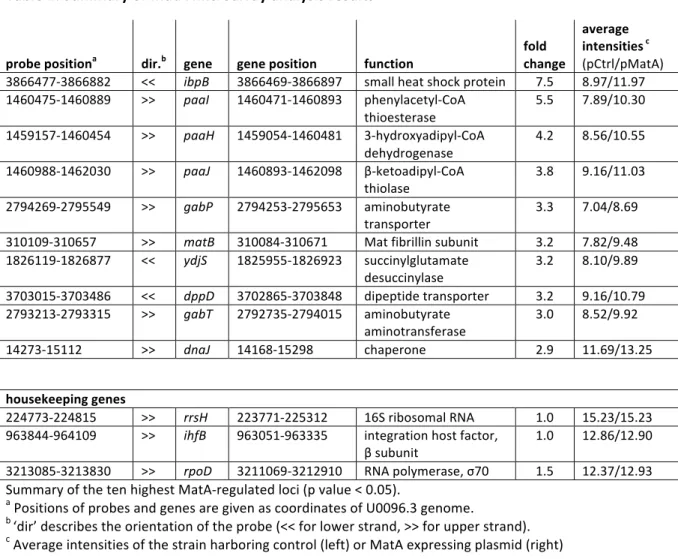
activation tested in different strain backgrounds. Given that for MatA and DctR no targets
are known in E. coli K-‐12, I performed a Microarray analysis. Furthermore, I investigated
whether transcriptional activation by RcsB homodimers or heterodimers with RcsA, BglJ, and
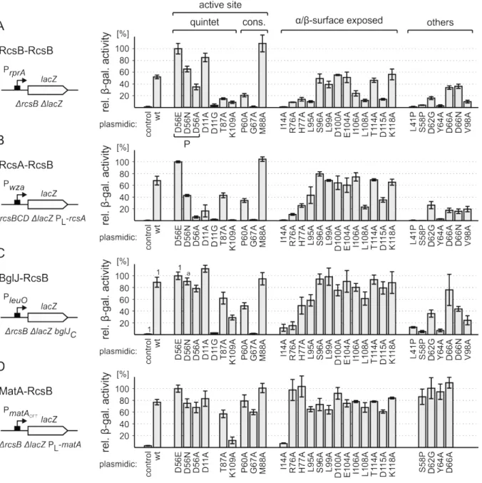
MatA depends on RcsB phosphorylation. For this, I expressed wild-‐type RcsB or mutants
D56E, D56N, and D56A in the appropriate reporter strains. RcsB-‐D56E mimics
phosphorylated RcsB, and RcsB-‐D56N and -‐D56A mimic non-‐phosphorylated inactive RcsB
2.2.1. RcsB-‐RcsB activates PrprA in a phosphorylation dependent manner
For analyzing the RcsB homodimer activity, first a PftsA lacZ fusion was tested as a reporter.
This fusion, which was constructed before, comprises the promoter region (-‐70 to +30) of the cell division gene ftsA that is activated by RcsB (Carballes et al., 1999), fused to the reporter gene lacZ (constructed by Öztürk, Figure 4A). Previously, this reporter was tested in ΔrcsB ΔlacZ strain T818 (Öztürk, 2010). This strain was transformed with empty plasmid pKESK22 or plasmids expressing wild-‐type RcsB and mutants D56E, D56N, D56A, or M88A (pKETS6, pKETS7, pKETS8, pKES235 and pKES232) and β-‐galactosidase activities were determined. RcsB-‐D56E mimics phosphorylated RcsB, and RcsB-‐D56N and -‐D56A mimic non-‐
phosphorylated inactive RcsB (Scharf, 2010). In RcsB mutant M88A, methionine at position 88 is replaced by alanine. At this position, response regulators except for RcsB carry usually a conserved small residue such as alanine or glycine (Bourret, 2010). Compared to the empty vector control, the plasmidically expressed wild-‐type rcsB did not activate the ftsA promotor (Öztürk, Figure 4B). The finding may be due to non-‐induced Rcs signaling resulting in an equilibrium shift to unphosphorylated RcsB in the cytoplasm. Accordingly, the RcsB mutant D56A, mimicking inactive RcsB, did not activate the promoter and RcsB-‐D56N only slightly.
RcsB-‐D56E, mimicking phosphorylated RcsB, as well as RcsB-‐M88A, activated the promoter
around 5-‐fold compared to the control plasmid (Öztürk, Figure 4B).
Figure 4. Analysis of the PftsA lacZ reporter in different strain backgrounds
(A) The ftsA promoter region from -‐70 to +30 relative to the transcription start site was fused to lacZ on plasmid pKES243 and integrated into the attB site of the chromosome of different strains. These reporter strains were transformed with empty plasmid pKESK22 (pCtrl), wild-‐type RcsB (pKETS6) or RcsB-‐mutants D56E, D56N, D56A and M88A (pKETS7, pKETS8, pKES235, and pKES232) expressing plasmids and reporter expression levels were determined. (B) PftsA lacZ expression levels in ΔrcsB ΔlacZ strain T818, ΔrcsBCD ΔlacZ strain T868 and ΔrcsB ΔlacZ rfaD::mTn-‐cat strain T866. Cultures for β-‐galactosidase assays were grown in LB medium to an OD
600of 0.5, supplemented with 1 mM IPTG and 25 µg/ml of kanamycin. Values obtained for ΔrcsB strain (indicated with
1) are unpublished laboratory results from Öztürk, 2010.
Based on these previous results, I constructed two PftsA lacZ reporter strains to potentially increase RcsB phosphorylation. The first reporter strain ΔrcsBCD ΔlacZ (T868) lacks both the sensor kinase RcsC and the phosphotransfer protein RcsD to avoid RcsB dephosphorylation by the Rcs system. In the second reporter strain ΔrcsB ΔlacZ rfaD::mTn10-‐cat (T866), rfaD is mutated by transposon insertion. Mutation of rfaD which is involved in lipopolysaccharide (LPS) synthesis (Pegues et al., 1990) was reported to activate Rcs signaling (Parker et al., 1992). Each reporter strain was transformed with an empty plasmid or plasmids expressing wild-‐type RcsB and mutants D56E, D56N, D56A, or M88A and β-‐galactosidase activities were determined. In the ΔrcsBCD ΔlacZ strain, wild-‐type RcsB activated expression approximately 2-‐fold to 61 units compared to the empty vector control with 27 units (Figure 4B). RcsB mutants D56E and M88A activated PftsA lacZ up to 155 units and 291 units, respectively (Figure 4B). RcsB mutant D56N activated expression only slightly with 46 units and RcsB-‐
D56A did not activate PftsA lacZ expression (Figure 4B). In the ΔrcsB ΔlacZ rfaD::mTn10-‐cat
strain wild-‐type RcsB only slightly activated expression from 28 units for the empty vector
176 units and 189 units, respectively. RcsB mutant D56N activated expression only slightly with 40 units and RcsB-‐D56A did not activate PftsA lacZ expression (Figure 4B). In none of the three reporter strain backgrounds the PftsA lacZ expression was more than approximately 2-‐fold upregulated by wild-‐type RcsB and around 6-‐fold by RcsB-‐D56E compared to the control. Thus the PftsA lacZ fusion did not prove to be optimal for further analyses and a reporter was needed which is strongly activated by wild-‐type RcsB that allows distinguishing smaller differences.
Therefore the promoter of rprA (-‐124 to +4) encoding the small RNA RprA whose expression is activated by RcsB (Majdalani et al., 2002) was fused to the lacZ reporter gene and this construct was integrated into the chromosome of different strain backgrounds (Figure 5A).
Figure 5. PrprA lacZ as a reporter for studying activation by RcsB
(A) The rprA promoter region from -‐142 to +4 relative to the transcription start site was fused to lacZ on plasmid pKES299 and integrated into the attB site of the chromosome. (B) The expression levels of the PrprA lacZ reporter were determined in rcsB
+ΔlacZ strain T2023, rcsB
+ΔgalU ΔlacZ strain T2041, and ΔrcsB ΔlacZ strain T1052. For complementation, rcsB was expressed from plasmid pKETS6. RcsB derivatives D56E, D56N, D56A and M88A from plasmids pKETS7, pKETS8, pKES235, and pKES232, respectively. Empty cloning vector pKESK22 served as control (pCtrl). Cultures for β-‐
galactosidase assays were grown in LB medium to an OD
600of 0.5, if transformed supplemented with 1 mM IPTG and 25 µg/ml of kanamycin.
In the rcsB
+strain T2023 the PrprA lacZ expression was poorly activated (7 units, Figure 5B).
In a ΔgalU strain that cannot produce UDP-‐D-‐glucose, the Rcs system is constitutively
activated (Girgis et al., 2007). Deletion of galU in the rcsB
+background (T2041) activated
PrprA lacZ expression up to 39 units (Figure 5B). In the ΔrcsB strain (T1052) PrprA lacZ
expression is very low (2 units, Figure 5B) and when transformed with control plasmid
pKESK22 completely off (<1 unit, Figure 5B). Complementation of ΔrcsB strain with wild-‐type
rcsB (pKETS6) induced PrprA lacZ expression up to 52 units (Figure 5B). RcsB mutants D56E
and M88A activated the promoter up to 100 units and 109 units, respectively, and RcsB
derivative D56N and D56A activated the rprA promoter up to 65 and 35 units, respectively.
This lower activity for the inactive RcsB-‐D56A mutant confirms an at least partial phosphorylation dependent activation. Although activation of PrprA by RcsB homodimers is phosphorylation dependent, the complementation with wild-‐type rcsB partially overcomes the requirement for induction of the Rcs signaling cascade. In the ΔrcsB strain, plasmidically expressed wild-‐type RcsB activates the PrprA more than 50-‐fold and RcsB-‐D56E more than 100-‐fold compared to the control plasmid, allowing to distinguish also small differences.
Taken together, the PrprA lacZ reporter in the ΔrcsB strain proved to be appropriate for the analysis of transcriptional activation by RcsB and mutants. The results also confirmed a phosphorylation dependent activation by RcsB homodimers.
2.2.2. RcsA-‐RcsB activates Pwza in a phosphorylation dependent manner