Regulation of polyamine biosynthesis in Saccharomyces cerevisiae
Inaugural-Dissertation
zur
Erlangung des Doktorgrades
der Mathematisch-Naturwissenschaftlichen Fakultät
der Universität zu Köln
vorgelegt von
Palani Murugan Rangasamy
aus Tamilnadu, Indien
Köln, März 2005
1. Berichterstatter:
Prof. Dr. R. Jürgen Dohmen
2. Berichterstatter:
Prof. Dr. Thomas Langer
3. Berichterstatter:
Prof. Dr. Martin Scheffner
Tag der mündlichen Prüfung: 27 Mai 2005
1 Zusammenfassung...1
2 Abstract...3
3 Introduction...4
3.1 Polyamines and their cellular functions... 5
3.2 Metabolism of polyamines... 6
3.3 Regulation of polyamine biosynthesis... 8
3.3.1 Ornithine decarboxylase (ODC) ... 9
3.3.2 ODC antizyme ... 11
3.3.3 Programmed ribosomal frameshifting ... 12
3.3.4 Ubiquitin-proteasome system (UPS) ... 14
3.3.5 The peptide aptamers ... 19
3.4 Aims of the current study...25
4 Materials and Methods...26
4.1 Materials ...26
4.2 Methods ...37
5 Results...53
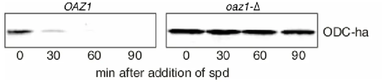
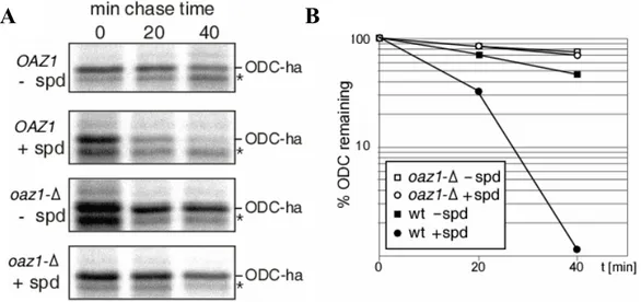
5.1 Identification of ornithine decarboxylase antizyme (Oaz1) in S. cerevisiae. 53 5.2 Oaz1 mediates degradation of ornithine decarboxylase by the proteasome ... 57
5.3 ODC degradation requires a functional proteasome... 60
5.4 Oaz1 physically interacts with ODC ... 61
5.5 Yeast ODC N-terminus contains the degradation signal ... 62
5.6 The ODC N-terminus interacts with 19S lid components of the proteasome ... 69
translation of OAZ1 mRNA ... 70
5.8 Cis regulatory elements associated with +1 ribosomal frameshifting
during decoding of yeast OAZ1 mRNA ... 74
5.9 Control of Oaz1 levels involves its ubiquitin-mediated
degradation by the proteasome ... 77
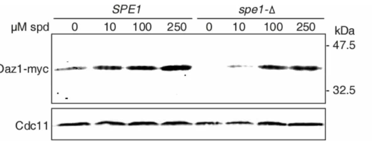
5.10 Polyamines block degradation of Oaz1 ... 80
5.11 Isolation of Peptide aptamers inhibiting ubiquitin dependent protein degradation in S. cerevisiae... 84
6 Discussion...90
6.1 Conservation of ODC antizyme function and the
regulation of polyamine biosynthesis ... 91
6.1.1 ODC is the target for regulating polyamine biosynthesis... 91 6.1.2 Oaz1 is the S. cerevisiae orthologue of mammalian antizyme ... 91 6.1.3 Regulation of polyamine biosynthesis and
+1 ribosomal frameshifting... 97
6.2 Regulation of antizyme degradation by polyamines... 101
6.3 Genetic screen to isolate peptide inhibitors of
ubiquitin proteasome system ... 103
7 References...105
Appendix
Acknowledgements
Eidesstattliche Erklärung
Lebenslauf
1 Zusammenfassung
Polyamine sind essentielle organische Kationen mit vielfältigen zellulären Funktionen.
Ihre Synthese wird durch eine „Feedback“-Regulation kontrolliert, deren hauptsächliches Ziel die Ornithindecarboxylase (ODC) ist. ODC ist das geschwindigkeitsbestimmende Enzym in der Polyaminbiosynthese. Es war bekannt, dass ODC in Säugerzellen durch ODC-Antizym inhibiert und der Ubiquitin- unabhängigen Proteolyse zugeführt wird. Die Synthese von Antizym in Säugerzellen beinhalten einen Polyamin-induzierten ribosomalen Leserastersprung. Hohe Polyamin- Konzentrationen inhibieren somit die de novo Synthese von Polyaminen, indem sie den Abbau von ODC induzieren. In dieser Arbeit wurde eine zuvor nicht bekannte Sequenz im Genom der Hefe Saccharomyces cerevisiae identifiziert, die für ein Antizym (Oaz1) in dieser Hefe kodiert. Sequenzelemente mit der Bezeichnung OFRE (OAZ1 Frameshifting Repressor Element) und OPRE (OAZ1 Polyamine Responsive Element), die für die Polyamine-Regulation des Leserastersprungs verantwortlich sind, wurden in der OAZ1 mRNA kartiert. Der Abbau von ODC der Hefe durch das Proteasom benötigt Oaz1. Oaz1 vermittelt den Abbau von ODC, in dem es an dasselbe bindet, wodurch ein Abbausignal (ODS für „ODC degradation signal“) am N-Terminus exponiert wird, dass in dieser Arbeit identifiziert wurde. Mit Hilfe von Testproteinen, die dieses neue ODS- Abbausignal trugen, konnte eine Rolle des Transportfaktors Rad23 entdeckt werden. Es konnte außerdem gezeigt werden, dass ODS in vivo mit verschiedenen Untereinheiten des „Lid“-Komplexes des 19S-Aktivatorkomplexes des 26S-Proteasoms interagiert.
Mithilfe des neuen Hefemodellsystems für das Studium der Polyaminregulation konnte ein neuer Kontrollmechanismus identifiziert werden. Oaz1 wird selbst durch Ubiquitin- abhängige Proteolyse durch das Proteasom reguliert. Der Abbau von Oaz1 wird durch Polyamine inhibiert. Diese Beobachtungen führten zu einem Modell, in dem Polyamine ihre Synthese durch zwei Mechanismen hemmen. Zum einen stimulieren sie die Synthese durch Erhöhung der Häufigkeit des Leserastersprungs bei der Translation der OAZ1-mRNA, zum anderen inhibieren sie den Abbau von Oaz1.
Im zweiten Teil dieser Arbeit wurden sogenannte Peptid-Aptamere isoliert, die den Ubiquitin-vermittelten Abbau von Testsubstraten über das Proteasom inhibieren.
Diese Aptamere inhibieren entweder die Ubiquitylierung oder direkt das Proteasom und
Zukunft eingesetzt werden, um weitere potentiell klinisch relevante Peptidinhibitoren zu identifizieren, die spezifisch die Funktion des Proteasoms stören.
Polyamines are essential organic cations with multiple cellular functions. Their synthesis is controlled by a feedback regulation whose main target is ornithine decarboxylase (ODC), the rate-limiting enzyme in polyamine biosynthesis. In mammals, ODC has been shown to be inhibited and targeted for ubiquitin-independent degradation by ODC antizyme. The synthesis of mammalian antizyme was reported to involve a polyamine-induced ribosomal frameshifting mechanism. High levels of polyamine therefore inhibit new synthesis of polyamines by inducing ODC degradation.
In this work, a previously unrecognized sequence in the genome of Saccharomyces cerevisiae encoding an orthologue of mammalian antizyme was identified. Synthesis of yeast antizyme (Oaz1) involves polyamine-regulated frameshifting. New elements, termed OFRE (OAZ1 frameshifting repressor element) and OPRE (OAZ1 polyamine responsive element) that are necessary for the polyamines to regulate frameshifting were mapped in the OAZ1 mRNA. Degradation of yeast ODC by the proteasome depends on Oaz1. Oaz1 mediates the degradation by binding to ODC thereby exposing a degradation signal at the N-terminus of ODC. Using the novel transplantable yeast ODC degradation signal (ODS) identified in this work a new possible role of the shuttle factor Rad23 in ODC degradation was identified. In addition, ODS is shown to interact with multiple 19S lid subunits in the proteasome. Using this novel model system for polyamine regulation another level of its control was discovered. Oaz1 itself is subject to ubiquitin-mediated proteolysis by the proteasome. Degradation of Oaz1, however, is efficiently inhibited by polyamines. I propose a model, in which polyamines inhibit their ODC-mediated biosynthesis by two mechanisms, the control of Oaz1 synthesis and inhibition of its degradation.
In a second part of the work, peptide aptamers were isolated that inhibit the ubiquitin-dependent turnover of test substrates of the proteasome. These aptamers appear to either inhibit ubiquitylation or the proteasome and thereby lead to a stabilization of test substrates. I Propose that ODS due to its ubiquitin-independent mode of degradation can be used as a tool in aptamer screens that are aimed at identifying additional peptide inhibitors of the proteasome with potential clinical relevance.
3 Introduction
Small molecules are very important for the existence of life. Examples are nucleotides, which makes up the genetic material, and the amino acids that are the building blocks of proteins. However there are many small molecules that are essential for life without having to attain a macromolecular form. To illustrate the later type of molecule, the polyamines are one of the best examples. Polyamines are essential for life and they are associated with all life forms ranging from the most primitive to the most elaborate ones. Many chemists have long back discovered polyamines, however their association with life became clear only in 1687 after Antonie Van Lewenhoeck’s observation that the seminal fluid contains a slowly crystallizing substance that was later identified as the polyamine spermine phosphate (Lewenhoeck, 1678). Thereafter many studies have contributed to understanding the role of polyamines in the evolution and the existence of life (Cohen, 1998). Although it is far from clear what all the functions of polyamines are, certainly the most important ones are their association with DNA and RNA (Coffino, 2001b). This association stabilizes the structure of DNA and RNA.
Polyamines neutralize the negative charges in DNA thereby allowing it to be condensed into the chromosomes. On the other side, abnormal levels of polyamines are also associated with many diseases including cancer. Many genetic studies in mammals have concluded that altering either synthesis or catabolism of polyamines leads to abnormalities which are described in later sections. Therefore understanding the molecular mechanisms regulating the polyamines level is critical in order to understand the patho-physiology of the polyamine associated abnormalities.
In the present work, I focused on understanding the important role of selective proteolysis in controlling a metabolic process in the unicellular fungus Saccharomyces cerevisiae.
3.1 Polyamines and their cellular functions
Polyamines are essential, organic, aliphatic polycations that are present in all the life forms ranging form prokaryotes to higher eukaryotes. There are many modified forms of polyamines detected in the cell, but putrescine, spermidine and spermine are the three most abundant and important natural polyamines (Figure 1). Polyamines have multiple functions. A substantial amount of polyamines are secreted into human and rat milk and also into the seminal fluid. Polyamines are also known to act as odors and to modulate taste (Cohen, 1998). Polyamines are essential for normal cell growth and viability (Coffino, 2001b). Although identifying the molecular functions of polyamines is not easy because of their importance in normal cell physiology, many studies have recognized their direct role in stabilization of chromatin and the cytoskeleton, as well as in processes ranging from DNA replication, transcription and translation, ion transport, to the regulation of cell growth and apoptosis (Childs et al., 2003) (Coffino, 2001b) (Wallace et al., 2003). Polyamines alter protein-protein interactions as well as proteins interactions with DNA and RNA (Childs et al., 2003).
a Fig. 1. Natural polyamines
(a) Putrescine
b (b) Spermidine
(c) Spermine c
Aside of the processes mentioned above, polyamines are associated with two rare physiological processes. The first one is hypusination, a rare posttranslational protein modification with an unusual amino acid hypusine (Shiba et al., 1971). In hypusination, the polyamine spermidine serves as the donor of an aminobutyl moiety which is attached to the lysine residue of eukaryotic translation initiation factor eIF-5A. Later the amino butyl group is converted to hypusine (Park et al., 1993). EIF-5A which is involved in RNA processing (Peebles et al., 1983) is the only known substrate for hypusination. Even though it is a rare modification, inhibition of hypusination is lethal in the yeast S. cerevisiae (Park et al., 1993). In a second process, polyamines control the
frameshifting is an important event in the auto-regulatory circuit of polyamine biosynthesis (Matsufuji et al., 1995). In addition, polyamines can inhibit eukaryotic transposable element (Ty1) propagation by altering the +1 frameshifting efficiency during the decoding of its RNA (Balasundaram et al., 1994).
3.2 Metabolism of polyamines
Polyamines can be synthesized as well as broken down or modified within the cell.
Polyamine biosynthesis involves several enzymatic processes and they are highly conserved between higher and lower eukaryotes. Polyamine catabolism is also composed of several enzymatic processes and requires subcellular compartments for detoxification of toxic intermediates. Moreover polyamine catabolism also helps to interconvert the higher polyamines spermine and spermidine to the lower polyamine putrescine. Figure 2 depicts the general scheme of polyamine metabolism as it occurs in prokaryotes as well as in eukaryotes. Polyamines are synthesised from amino acid precursors in several enzymatic steps. Ornithine decarboxylase (ODC) is the rate- limiting enzyme in the biosynthesis of the polyamines spermine and spermidine (Coffino, 2001b) (Wallace et al., 2003). It catalyses the conversion of ornithine derived from arginine into the diamine putrescine. By addition of an aminopropyl group, putrescine is subsequently converted to spermidine by spermidine synthase. Spermine is derived from spermidine by the action of spermine synthase (Wallace et al., 2003).
Some plants and bacteria derive putrescine via an additional pathway that requires arginine decarboxylase (ADC) and agmatinase. The existence of this pathway in eukaryotes such as yeast and mammals, however, is not clear (Coffino, 2001b). In addition, polyamines are either interconverted or catabolized by the enzyme pair spermidine/spermine acetyl transferase (SSAT) and polyamine oxidase (PAO) (Wallace et al., 2003). Polyamine levels are furthermore controlled by their uptake or excretion into the environment (Cohen, 1998).
N-Acetylspermine N-Acetylspermidine
PAO Arginine
Ornithine Agmatine
S-Adenosylmethionine
Decarboxy-S-
Adenosylmethionine
Putrescine
Spermidine
Spermine ODC
(animals/eukaryotes)
SpmS
Agmatinase
SamDC
SpdS
SSAT SSAT
PAO ADC (plants/bacteria) Arginase
PAO
Fig. 2. Polyamine metabolism Polyamines are synthesized from the amino acid precursor arginine. In some plants and bacteria, arginine decarboxylase (ADC) converts arginine to agmatine, which is converted to the diamine putrescine by agmatinase. But in animals and other eukaryotes arginase initiates polyamine synthesis by producing ornithine from arginine, and ornithine decarboxylase (ODC) converts ornithine to putrescine.
Afterwards the higher order
polyamine spermidine is synthesized by addition of an aminopropyl group to putrescine by spermidine
synthase (SpdS). Accordingly spermine synthase (SpmS) catalyses the addition of a second
aminopropyl group to spermidine leading to spermine. The
aminopropyl group is derived from S-adenosylmethionine by S- adenosylmethionine decarboxylase (SamDC). Polyamines are either interconverted or destroyed by spermidine/spermine acetylase (SSAT) and polyamine oxidase (PAO). ODC, SamDC and SSAT are three key rate-limiting enzymes in controlling the synthesis and catabolism of polyamines.
These processes are collectively important for polyamine homeostasis in the cell.
Although polyamine transport has been well studied in bacteria, polyamine transport across the plasma membrane in eukaryotes is less clear (Igarashi and Kashiwagi, 1999).
In S. cerevisiae, however, polyamine transporters have been identified in the vacuolar membrane which may be important for either the storage or detoxification of polyamines (Tomitori et al., 1999). Although experimental evidences strongly suggest that polyamines are transported across the plasma membrane, there are no specific plasma membrane transporters known to be involved in polyamine transport. A report that described Tpo1 in S. cerevisiae as a plasma membrane polyamine transporter is controversial (Igarashi and Kashiwagi, 1999) (Albertsen et al., 2003) (Uemura et al.,
3.3 Regulation of polyamine biosynthesis
The intracellular concentration of polyamine is controlled at several steps including their uptake, catabolism and their biosynthesis. In eukaryotes the regulation of biosynthesis is mainly achieved by controlling the cellular ODC activity via an unusual mechanism involving ODC antizyme (AZ) (Hayashi et al., 1996) (Coffino, 2001a).
Higher levels of polyamine induce +1 ribosomal frameshifting during the decoding of AZ mRNA (Matsufuji et al., 1995). This in turn increases AZ levels in the cell. If present, AZ breaks ODC/ODC homodimers and forms ODC/AZ heterodimers. In the heterodimer, an ODC degradation signal is exposed which is recognised by the proteasome leading to loss of ODC and recycling of AZ (Figure 3).
Fig. 3. Regulation of polyamine biosynthesis
Polyamine biosynthesis is mainly regulated by the degradation of ornithine decarboxylase (ODC) which is the rate-limiting enzyme in polyamine biosynthesis. 1.) ODC catalyses the conversion of ornithine to putrescine, which in turn
increases the polyamine levels. 2.) Higher levels of polyamines induce the +1 ribosomal frameshifting thereby increasing the cellular antizyme (AZ) level. 3.) AZ disrupts ODC homodimers and inhibits ODC activity. 4 &5.) Antizyme forms ODC/AZ heterodimers, resulting in the degradation of ODC by the proteasome leaving the antizyme for recycling.
1 2
3
4
5
ODC
Polyamine
Proteasome
AZ
ODC/AZ
Proteasomal degradation usually requires ubiquitylation of substrates. ODC, however, is the only example where ubiquitin is not strictly required for its degradation (Murakami et al., 1992). Although the mechanism of regulation of polyamine biosynthesis is highly conserved, this complex regulation is mainly studied in mammals. A large number of biochemical and in vivo studies in mammals have revealed the importance of the components involved in regulating polyamine biosynthesis for normal cell physiology.
In the coming subsections, I will introduce the state of knowledge on ODC, ODC antizyme, ribosomal frameshifting and the ubiquitin proteasome system.
3.3.1 Ornithine decarboxylase (ODC)
Animals solely depend on ODC for polyamine biosynthesis, unlike plants and bacteria which have an additional pathway for synthesizing polyamines (Figure 2). The unicellular fungus S. cerevisiae also uses ODC for its polyamine biosynthesis and it is likely to remain the only pathway dedicated for polyamine biosynthesis since strains lacking the ODC encoding gene (SPE1) fail to grow when they are transferred to polyamine-free media (Schwartz et al., 1995). The active ODC enzyme is a homodimer and the crystal structures of C-terminally truncated mouse ODC (Kern et al., 1999) and of human ODC (Almrud et al., 2000) have been solved. Because ODC is the rate- limiting enzyme in polyamine biosynthesis, it was the target of many studies aimed to understand the role of polyamines. Initially an irreversible inhibitor of ODC, DFMO (DL-alpha-Difluoromethylornithine), has been used extensively for this purpose. Later genetic studies targeting ODC have shown that it is an important gene that is not only essential for cell physiology but also has a role during the development of multicellular organisms (Pendeville et al., 2001) . Firstly, the overexpression of ODC in mouse testis causes male sterility which resembles the ‘Sertoli cells-only syndrome’ that causes infertility in man (Halmekyto et al., 1991). Overexpression in the skin increases the spontaneous skin tumor development which can be prevented by using ODC inhibitor DFMO (Ahmad et al., 2001). Aside of tumor development this mouse also showed a very complex skin phenotype including complete and irreversible loss of hair, excessive skin wrinkling and enhanced nail growth (Megosh et al., 1995). Other studies have shown that overexpression also affects the central nervous system and the cardiac system (Shantz et al., 2001). On the other side, deletion of the ODC gene in mouse is embryonic lethal with the embryos not developing beyond day 3.5 (Pendeville et al., 2001). Interestingly, mammals have an ODC homolog called antizyme inhibitor, which is enzymatically inactive. However antizyme inhibitor has higher affinity towards antizyme than ODC. Therefore antizyme inhibitor is widely believed to inhibit antizyme
biosynthesis to take place (Bercovich and Kahana, 2004). The mechanism controlling the level and the activity of the antizyme inhibitor, however, is not clear. In lower eukaryotes, a homolog of antizyme inhibitor has not been identified suggesting that other mechanisms govern the regulation of excess antizyme in those organisms. Since many studies in the transgenic mouse model suggest a direct role of ODC in tumor formation, this enzyme has become a potential target for tumor prevention especially in the skin where such treatment has been proven to be very effective (Feith et al., 2001).
An interesting property of ODC is that it is degraded by the proteasome when it binds to ODC antizyme. Characterization of the 461 residue mouse ODC showed that 37 residues at the C-terminus contain the degradation signal that is exposed upon association with antizyme (Ghoda et al., 1989). Further characterisation revealed that residues between 117 and 140 are important for ODC association with antizyme (Kern et al., 1999).
The effects of lowering and increasing the intracellular production of polyamines have been extensively studied in transgenic models. In addition, abnormalities associated with abnormal polyamines levels are known to occur in humans (Janne et al., 2004). Inactivation of the ODC gene in mice e.g. led to embryonic lethality (Pendeville et al., 2001). In S. cerevisiae mutants lacking the SPE1 gene encoding ODC are viable but cease to grow and become morphologically abnormal upon transfer to polyamine- free media (Schwartz et al., 1995). On the other side, elevated polyamine levels are usually linked to cancer. Consistent with that notion, mutations associated with hepatomas in humans have been mapped to the ODC degradation signal, which result in stabilization of ODC (Tamori et al., 1995). Lowering polyamine levels by the overexpression of SSAT (spermidine/spermine N-acetyl transferase) in the mouse led to hairlessness (Pietila et al., 1997) and female infertility (Min et al., 2002). Moreover, in humans the X-linked syndrome keratosis follicularis spinulosa decalvans (KFSD) affecting skin and eye is related to polyamines. The genetic alterations leading to this syndrome apparently include a duplication of a region on the X-chromosome that contains the SSAT gene (Gimelli et al., 2002). The relevant mutation in the human X- linked mental disorder Snyder-Robinson syndrome has recently been mapped to the spermine synthase gene (Cason et al., 2003) that is involved in the synthesis of the higher polyamine spermine.
3.3.2 ODC antizyme
The ODC anti-enzyme (antizyme) was first identified in mammals (Murakami et al., 1985), where it is now known to exist in several isoforms (AZ1, AZ2, AZ3, AZ4) (Heller et al., 1976) (Ivanov et al., 1998) (Ivanov et al., 2000b). Among those, AZ1 disrupts enzymatically active ODC homodimers by forming ODC/AZ heterodimers (Li and Coffino, 1992) (Mitchell and Chen, 1990). AZ1 binding moreover mediates ODC degradation by the 26S proteasome (Elias et al., 1995) (Li and Coffino, 1992) (Murakami et al., 1992). In contrast, binding of AZ2 does not result in degradation of ODC (Zhu et al., 1999). The molecular functions of AZ3, whose expression appears to be limited to the testis, and of AZ4 have not been analysed in detail (Ivanov et al., 2000b) (Coffino, 2001b). AZ1-dependent degradation of ODC was shown both in vitro and in vivo not to require its ubiquitylation (Rosenberg-Hasson et al., 1989) (Murakami et al., 1992). It was reported that instead a C-terminal degradation signal in ODC is exposed upon AZ1 binding that mediates binding to a ubiquitin recognition site in the 19S cap of the proteasome (Zhang et al., 2003). AZ levels increase with rising intracellular polyamine concentration. Polyamine-induction of AZ thus constitutes a feedback control in polyamine homeostasis. In mammals, it is shown that polyamines induce AZ levels by promoting +1 ribosomal frameshifting during the decoding of antizyme mRNA (Matsufuji et al., 1995). Although the function of ODC antizyme is conserved, its orthologues in mammals and S. pombe share very little sequence similarity (~10%) (Ivanov et al., 2000a). A few studies have concluded that mammalian antizyme is involved in other cellular process aside of its well known function of targeting ODC for ubiquitin-independent proteasomal degradation. Antizyme is known to be translocated into the nucleus during embryonic development (Gritli-Linde et al., 2001). Moreover antizyme forms a ternary complex with the transcription factor Smad1 and the proteasomal subunit HsN3 that is translocated into nucleus in response to bone morphogenetic protein receptor activation (Gruendler et al., 2001). In addition, a recent report showed a role of antizyme in targeting cyclin D1 for proteasomal degradation (Newman et al., 2004).
A mouse line overexpressing antizyme was shown to be less susceptible to skin tumorigenesis (Feith et al., 2001). Overexpression of antizyme in the fore-stomach also significantly reduced tumor formation and multiplicity (Fong et al., 2003). Based on these studies the authors have proposed that antizyme may represent a general tumor repressor gene. The carboxy-terminus of antizyme is involved in ODC interaction, but the amino-terminus of antizyme is required for inducing the degradation of ODC via an unknown mechanism (Li and Coffino, 1994). Many years after the identification of antizyme and after the discovery of mechanisms controlling its synthesis, it is still unclear how antizyme is removed from cells after ODC degradation.
AZ1 degradation was reported to be inhibited by proteasome inhibitors in cell lines and the degradation also appears to require a functional ubiquitin activating enzyme (E1) suggesting that antizyme requires ubiquitin and the proteasome for its degradation (Gandre et al., 2002). However this study has not shown that ubiquitylation of antizyme is directly involved in its degradation by the proteasome leaving this process still unclear.
3.3.3 Programmed ribosomal frameshifting
Translational recoding such as hopping, programmed ribosomal frameshifting and termination codon suppression are the exceptions to the general dogma of protein synthesis (Namy et al., 2004). These poorly understood phenomenons in turn make the definition of the genetic code an incomplete process. Several genes in prokaryotes and eukaryotes have been reported to use translational recoding as a biological regulatory mechanism. Programmed ribosomal frameshifting is the translation recoding mechanism that is used as the molecular switch in regulating polyamine homeostasis (Matsufuji et al., 1995). During this event, the ribosome either moves forward (+1) or backward (-1) without the detachment of peptidyl tRNA thereby changing the coding frame. The former process is called the +1 programmed ribosomal frameshifting and the later is called -1 ribosomal frameshifting. The decoding of antizyme mRNA involves +1 ribosomal frameshifting and the decoding signal (5’ UCC UGA U 3’) is highly conserved in almost all known antizyme genes (Ivanov et al., 2004). In all species the antizyme is encoded by two overlapping ORFs (Figure 4A). ORF1 is short but contains
an AUG codon that initiates translation, whereas ORF2, which encodes a larger part of the antizyme lacks the proper initiation codon. The ribosome slips to the +1 frame during the decoding of antizyme mRNA in the “slippery site” or “frameshifting site”
and continues then translation in ORF2 resulting in the functional antizyme.
Interestingly, this process is induced by free polyamines via an unknown mechanism (Matsufuji et al., 1995). In vertebrates, all the known antizymes (AZ1, AZ2, AZ3 and AZ4) require +1 ribosomal frameshifting during their decoding and AZ1 has been widely used for the studies on the frameshifting mechanism. But despite major efforts the mechanism is still largely unknown (Namy et al., 2004). These studies, however, revealed that there are three elements in AZ1 mRNA that are required for +1 ribosomal frameshifting depicted in Figure 4B. The most important element required for the frameshifting is the UGA stop codon and its surrounding sequences of ORF1 which is the main part of the frameshifting signal.
Fig. 4. +1ribosomal frameshifting in antizyme mRNA decoding
Decoding of ODC antizyme mRNA requires +1ribosomal frameshifting and this event is induced by free polyamines when present in excess. A. Antizyme is encoded by two ORFs (ORF1, ORF2) present in the same mRNA. Translation initiates from the AUG of ORF1, then the ribosome slips forward (+1) at the +1 frameshifting signal (5’UCC UGA UGC 3’). Thereby the translation continues into ORF2 without the detachment of the ribosome and peptidyl tRNA. +1 ribosomal frameshifting is induced by polyamines, which constitutes the auto-regulatory loop of regulating polyamine biosynthesis. B. AZ1 mRNA has been shown to have three important elements required for frameshifting, 1.) +1 ribosomal frameshifting signal that contains the UGA stop codon of ORF1 where the actual frameshifting occurs.
2.) A stem loop structure 3’ to the frameshifting signal that forms a pseudoknot important for enhanced 5’
3’
ORF1 STOP
1
2
3
3’
A. B.
Antizyme mRNA
+1frameshift ORF1
UGA
….UCC UGC………….
5’
ORF2
+ Free Polyamines
UGA
Antizyme
An RNA pseudoknot that is 3’ to the UGA stop codon of ORF1 is the second element which enhances the frameshifting efficiency of AZ1 by an unknown mechanism (Matsufuji et al., 1995). This element was identified in the vertebrate antizyme mRNA.
But a recent study identified a new putative RNA pseudoknot in lower eukaryotes that enhances the frameshifting efficiency in these organisms as well (Ivanov et al., 2004).
The third element, which is 3’ to the pseudoknot, is also known to influence the frameshifting (Coffino, 2001b). Importantly none of these elements are known to be influenced by polyamines in order to induce the frameshifting. The mechanism underlying polyamine-mediated induction of +1 ribosomal frameshifting is therefore completely unknown as of yet.
3.3.4 Ubiquitin-proteasome system (UPS)
Proteolysis by the 26S proteasome is the main pathway for ATP-dependent non- lysosomal degradation of intracellular proteins in eukaryotes (Hershko and Ciechanover, 1998). Usually, the substrates destined for degradation by this multi- subunit protease are marked by the attachment of poly-ubiquitin chains, which are recognised by binding sites in the proteasome (Verma et al., 2004). Several exceptions to this principle of ubiquitin-dependent degradation by the proteasome have been reported. The first described and best-studied substrate of ubiquitin-independent degradation by the proteasome is ornithine decarboxylase (ODC) (Coffino, 2001b).
Other examples are c-jun in mammals and Rpn4 in S. cerevisiae (Jariel-Encontre et al., 1995) (Xie and Varshavsky, 2001). In the latter two cases, however, ubiquitylation has been shown to be relevant to proteolysis in vivo (Ju and Xie, 2004) (Treier et al., 1994).
The ubiquitin proteasome system therefore remains to be the major player in the selective proteolysis in eukaryotes.
3.3.4.1 The proteasome
The 26S proteasome is a ~2.0 MDa multi-subunit protease present in all eukaryotes, which has been studied and reviewed extensively (Heinemeyer et al., 2004) (Pickart and Cohen, 2004). The proteasomal protein turnover is a highly specific and regulated process. Unlike other proteases, the proteasome usually requires the substrate proteins to be modified by the attachment of ubiquitin. In eukaryotes, the proteasome is the most
important protease in the degradation of proteins that occur in the cytosol and in the nucleus (Hershko and Ciechanover, 1998). Protein degradation in general is an important mode of regulation of temporal processes such as the cell cycle. One specific feature of protein degradation is that it ensures that factors controlling such processes are eliminated irreversibly thereby allowing for a progression until a new cycle is reached (Peters, 2002). The proteasome is in addition important for protein quality control (Hampton, 2002). The proteasome is a fine complex molecular machine, which is composed of two particles, namely the 20S catalytic core and the 19S cap. The 20S proteasome (20S core) is a dimer of 14 different subunits. The 19S cap or regulatory particle is composed of 18 different subunits. The 20S particle is the catalytically active part, which degrades substrates that enters to its catalytic centre. The 19S caps in the 26S proteasome (Figure 5) are important for substrate recognition, unfolding and delivery to the catalytic centre, which is in the 20S core particle (Adams, 2003).
19S Cap
20S Core
Fig. 5. The 26S proteasome
The proteasome is a multisubunit protease.
The atomic electron micrograph (Glickman, et al, 1998) of S. cerevisiae proteasome shows that it contains a single 20S core with two 19S caps attached to both of its end. The 20S core contains two α and two β rings. The 19S cap contains a lid and a base. The base is attached to an α ring of the 20S core particle.
The 19S cap regulates the entry of substrate to the proteasome and the 20S particle is responsible for degradation of the selected substrate.
3.3.4.1.1 The 20S proteasome
The 26S proteasome which is derived by the addition of two 19S regulators to the 20S proteasome, appears to be present only in eukaryotes. The 20S proteasome (20S core) in contrast, has been found in all phylae of life (Volker and Lupas, 2002). Unlike in eukaryotes, studies on the eubacterium Mycobacterium smegmatis and the archaeon Thermoplasma acidophilum showed that the proteasome is not essential for their viability (Knipfer and Shrader, 1997) (Ruepp et al., 1998). The precise function of the proteasome in those species is not clear. 20S proteasomes are composed of two types of subunits, α and β. In prokaryotes, proteasomes are either formed from one of two types
of α and β subunits (Heinemeyer et al., 2004). In contrast, eukaryotic 20S proteasome is composed of seven distinct α and seven distinct β subunits. The prokaryotic proteasome contains fourteen active sites compared to only six present in the β subunits of eukaryotic proteasome. Higher eukaryotes such as mammals, in addition, have a new type of proteasome that is called immunoproteasome. The immunoproteasome has three β subunits which are distinct from the normal proteasome. It has been implicated in antigen processing (Pamer and Cresswell, 1998) (Rock and Goldberg, 1999). 20S proteasomes are defined by a characteristic architecture, a stack of four heptameric rings with two outer α subunit rings and two central β subunit rings, which posses the catalytic centres. Eukaryotic proteasomes in comparison to prokaryotic ones have a high subunit complexity. The crystal structures of the yeast and the bovine particles have been solved, which confirmed that the topology is conserved from the yeast to the mammals (Bochtler et al., 1997; Unno et al., 2002). The crystal structure of yeast proteasome also revealed the specific subunits that possess peptidase activity. The β subunits β1/Pre3 mediates the post-acidic activity, β2/Pup1 has the trypsin-like activity and β5/Pre2 is responsible for chymotrypsin-like activity (Arendt and Hochstrasser, 1997; Heinemeyer et al., 1997).
The 20S proteasome biogenesis is fairly well studied in prokaryotes. Because, bacterial proteasomes lack the subunit complexity, there assembly and maturation is relatively simple. In contrast, proteasome biogenesis is very complex in eukaryotes because of its large number of distinct subunits. The assembly and maturation of 20S proteasome in the eukaryotes has been shown to involve a dedicated proteasome maturation factor termed Ump1 (Ramos et al., 1998). The Figure 6 shows the current model of 20S proteasome assembly and maturation in the yeast S. cerevisiae. First, the individual subunits assembled into a complex with Ump1 which is called 15S complex or half-proteasome. Then two half-proteasomes dimerise and form an intermediate with the Ump1 trapped within the complex. Conformational changes that coincide with dimerisation and that depend on Ump1 activate the catalytic centres, which in turn degrade Ump1 leading to the mature 20S proteasome. 19S activator complexes bind to 20S proteasomes in an ATP-dependent, poorly understood process that leads to the formation of 26S proteasome. In eukaryotes, it is also not clear if the 20S proteasome without the 19S cap plays a role in protein degradation. As only the 26S proteasome is
capable of degrading ubiquitylated proteins this form of the protease is essential for eukaryotic cells (Heinemeyer et al., 2004).
15S 20S
15S
.
Fig. 6. 20S proteasome assembly and maturation in S. cerevisiae
The 20S proteasome assembly and maturation is a complex process. Individual α and β subunits form a 15S complex with the proteasome assembly and maturation factor Ump1. Later two 15S complexes associate to form an intermediate complex which contains Ump1. Finally conformational changes induce the activation of catalytic centers that in turn degrade Ump1.
(modified from Heinemeyer et al, 2004)
3.3.4.1.2 The 19S regulatory particle
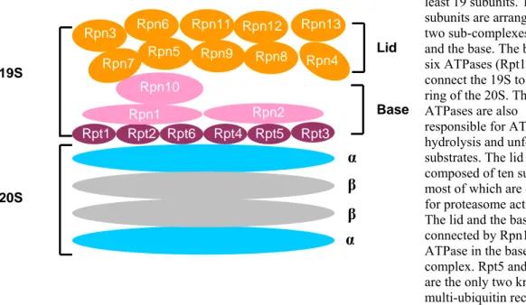
In eukaryotes, the 19S caps in the 26S proteasome are essential in controlling protein degradation. The 19S caps perform most of the sequential actions that are required for efficient protein turnover by the proteasome. They include recognition of ubiquitylated substrates, de-ubiquitylation, unfolding and delivery to the 20S catalytic centre (Pickart and Cohen, 2004). In the yeast S. cerevisiae, the 19S cap is composed of at least 19 different subunits, which are assembled into two sub-complexes. The base sub-complex contains six ATPases, and three non-ATPase subunits, the lid sub-complex contains ten non-ATPase subunits. The lid and the base are connected by a non-ATPase subunit Rpn10 (Regulatory Particle Non-ATPase). The base is attached to an α ring of the 20S core particle via its Rpt (Regulatory Particle ATPase) subunits (Glickman et al., 1998).
Figure 7 is the schematic representation of the 19S cap subunits and their arrangements in the lid and base sub-complexes. One of the main functions of the 19S cap in the proteasome is to identify the polyubiquitylated proteins. This is thought to be achieved by the ability of the 19S cap subunits Rpn10 and Rpt5 to bind to the polyubiquitin chain with four or more ubiquitin molecules (Deveraux et al., 1994; Lam et al., 2002). The S. cerevisiae 19S cap interacts with several proteins which are not stoichiometric parts of the 26S proteasome including ubiquitin ligase (E3) such as Hul5 and the de- ubiquitylating enzyme Ubp6 (Leggett et al., 2002) (Verma et al., 2000). Another 19S lid
subunit, Rpn11, which contains a JAMM (or MPN+) domain has a Zn2+ dependent metalloprotease activity. Mutation of its active site residues leads to accumulation of polyubiquitin conjugates and causes cell death illustrating the essential role of de- ubiquitylation in proteasomal protein degradation (Maytal-Kivity et al., 2002; Verma et al., 2002; Yao and Cohen, 2002).
Fig. 7. 19S regulatory particle
The 19S cap contains at least 19 subunits. The subunits are arranged in two sub-complexes, the lid and the base. The base has six ATPases (Rpt1-6) that connect the 19S to the α ring of the 20S. These ATPases are also responsible for ATP hydrolysis and unfolding of substrates. The lid is composed of ten subunits, most of which are essential for proteasome activity.
The lid and the base are connected by Rpn10, a non- ATPase in the base complex. Rpt5 and Rpn10 are the only two known multi-ubiquitin receptors in the proteasome.
Lid
Base
α
α β β
19S
20S
Rpt1 Rpt2 Rpt6 Rpt4 Rpt5 Rpt3
Rpn1 Rpn2
Rpn10 Rpn3
Rpn4 Rpn5
Rpn6
Rpn7 Rpn8
Rpn11 Rpn12 Rpn13 Rpn9
The cooperation among these different factors in performing the sequential actions that are important for substrate delivery and the release of the polyubiquitin chains from the substrates is poorly understood. Another important function of the 19S base sub- complex is the unfolding and translocation of proteins to the 20S core active site chamber. This is an energy-dependent process that requires hydrolysis of ATP by the base sub-complex ATPases (Pickart and Cohen, 2004).
Several organic molecules are widely used as proteasome inhibitors in in vitro and in vivo studies. In humans, the proteasome has been shown to be an important target for treatment of diseases such as cancer (Adams, 2004). Molecules such as aclacinomycin A (Figueiredo-Pereira et al., 1996), bortezomib (PS-341) (Adams et al., 1998), benzamide (Lum et al., 1998), calpain inhibitors I and II (Orlowski et al., 1993), eponemycin (Meng et al., 1999), epoxomycin (Kim et al., 1999), lactacystin (Almond and Cohen, 2002) and MG132 (Elliott et al., 2003) have been shown to inhibit
proteasomal activity and are undergoing preclinical trails as drugs against several diseases (Adams, 2004). Among those, bortezomib (Valcade) is currently being used for treatment of refractory multiple myeloma in humans (Adams et al., 1998). The identification of novel proteasome inhibitors such as peptides that interfere with various aspects of its function (see below) could therefore potentially lead to a drug that can be used for treatment of several diseases in humans.
3.3.5 The peptide aptamers
Aptamers (‘to fit’ in greek) are novel biological tools that are based on biological macromolecules such as DNA, RNA or proteins. Peptide aptamers are selected from random peptides. The concept of peptide aptamers basically comes from antibodies. The antibodies have the ability to bind specifically to a large array of antigens despite having most of its structure unchanged. This specificity of the antibody binding to the antigen is due to the sequence variation in the VL and VH regions of the antibody that allows different conformations in that particular domain. Equally, peptide aptamers have been generated by the insertion of random peptide sequences into conformationaly stable proteins such as E.coli thioredoxin (Colas et al., 1996), green fluorescent protein (Abedi et al., 1998), retinol binding protein or the bilin binding protein (Beste et al., 1999;
Skerra, 2000). Figure 8 illustrates the thioredoxin-based aptamer developed by John McCoy and Roger Brent. It contains a C-region that is the E.coli thioredoxin protein and the V-region which has inserted random peptides of 20 animo acid residues. Based on that design they constructed a library that can be expressed in the yeast S. cerevisiae.
Using this library these authors were able to isolate peptide aptamers that specifically inhibit cyclin-dependent protein kinase 2 (Colas et al., 1996).
C-Region
Fig. 8. Thioredoxin peptide aptamer
Shown is a thioredoxin peptide aptamer composed of two regions.
The constant or C-region is the thioredoxin protein and the variable or V-region is the 20mer random peptide, which is inserted into
3.3.5.1 Ubiquitylation
Ubiquitin is a 76 amino acid globular protein present in all eukaryotes. The multi-step attachment of ubiquitin to other proteins is called ubiquitylation (Hershko and Ciechanover, 1998). The best characterised role of ubiquitylation is to render proteins susceptible to degradation by the 26S proteasome. This occurs as a consequence of modification of proteins with chains of four or more ubiquitins linked through lysine 48 of ubiquitin. Protein modification by K63-linked ubiquitylation has roles that are independent of proteasomal degradation e.g. in protein kinase activation or in DNA repair.(Cheng et al., 2004; Hoege et al., 2002; Pickart, 2002) (Wang et al., 2001). In general, ubiquitylation occurs as a result of the sequential action of three classes of enzymes, E1 or ubiquitin activating enzyme, E2 or ubiquitin conjugating enzyme, and E3 or ubiquitin ligase (Figure 9). E1, the first enzyme in the ubiquitylation pathway forms a thiol-ester bond between its active site cysteine and the carboxy-terminus glycine of ubiquitin.
Fig. 9. Ubiquitylation
E1 Ub
Ub-E2
RING E3
nUb+Substrate S S
ATP
E2
1 2
HECT E3
Ubiquitin activating enzyme (E1) activates free ubiquitin by forming a thiol-ester link with the C-terminal glycine of ubiquitin in the presence of ATP. Ubiquitin is then transferred to one of many ubiquitin conjugating enzymes (E2s). Later the Ub-E2 associates with substrate bound ubiquitin ligase (E3). 1.) The ubiquitin is directly transferred from E2 to the substrates in the case of RING E3-mediated ubiquitylation. 2.) Ubiquitin is transferred to the active-site cysteine residue of a HECT domain in case of HECT E3 before the ubiquitylation of the substrate. This process is
repeated several times until the multi-ubiquitin chain is formed.
The activated ubiquitin on E1 is subsequently transferred to the active site cysteine of E2 by transesterfication. E3 binds ubiquitin-charged E2 and substrate and facilitates formation of an iso-peptide linkage between carboxy-terminal glycine of the ubiquitin and the ε-amino group of an internal lysine residue of the substrate, or an ubiquitin already attached to the protein (Hershko and Ciechanover, 1998). In rare instances ubiquitin is attached to the free α-amino group of the substrate rather than to an internal lysine (Aviel et al., 2000; Reinstein et al., 2000) (Doolman et al., 2004).
3.3.5.1.1 Ubiquitin activating enzyme (E1)
In the multistep ubiquitylation process, activation of ubiquitin is the initial reaction which is catalysed by the E1 or ubiquitin activating enzyme. In the yeast S. cerevisiae, E1 is encoded by a single and essential gene. Mammals are also widely believed to have only a single E1 which governs the ubiquitylation. Mammals E1, however, comes in two isoforms E1a and E1b (Handley-Gearhart et al., 1994) that are the result of the utilization of two translation initiation sites. In order to activate ubiquitin, E1 binds to MgATP and subsequently to ubiquitin forming an ubiquitin adenylate that serves as the donor of ubiquitin to the active cysteine in E1 (Haas and Rose, 1982; Hershko et al., 1983). Each fully loaded E1 carries two molecules of ubiquitin, one as a thiol-ester and the other as an adenylate. The activated ubiquitin is then transferred to the active site cysteine in E2. The carboxyl-terminal glycine of ubiquitin is essential for its activation by E1. The evolutionary conservation in activation for ubiquitin and other ubiquitin-like (UBL) protein modifiers is exemplified by the presence of a carboxyl-terminal glycine in the active forms of most UBLs, such as SUMO/Pic-1/Sentrin, Nedd8/Rub1, ISG15/UCRP and FAT10 (Hochstrasser, 2000; Raasi et al., 2001). In the case of SUMO and Nedd8 the activating enzymes are heterodimers, with the subunits displaying homology to the amino or carboxyl portions of the ubiquitin E1 (Tanaka et al., 1998).
3.3.5.1.2 Ubiquitin conjugating enzyme (E2)
After activation, ubiquitin is transferred to the ubiquitin conjugating enzyme (UBC) or E2. By sequence homology there are 13 known members of E2 family in the yeast S. cerevisiae, which are termed Ubc1-Ubc13, and there are at least 25 members in mammals. One of the yeast E2s (Ubc9) is specific for conjugation of SUMO, another
E2s have a ~150 amino acid, highly conserved core (UBC) domain that includes an invariant cysteine that accepts ubiquitin from E1. Some E2s have carboxy- and amino- terminal extensions, some have insertions in the core domain (Pickart, 2001). These extensions are believed to either facilitate or preclude interactions with specific E3.
3.3.5.1.3 Ubiquitin Ligase (E3)
Ubiquitin ligase or E3 is the third type of enzymes involved in ubiquitylation. E3 interacts directly with the substrate and the E2 thereby facilitating the transfer of ubiquitin to the substrate. Among the three classes of enzymes, E3s have more specificity towards the substrate than E2s and the E1. There are two known families of ubiquitin ligases in the cell. E3s of the first family have a characteristic RING (Really Interesting Gene) finger domain (Joazeiro and Weissman, 2000), which is highly conserved in those family members. E3s of the second family are characterized by the presence of a HECT (Homology to E6-AP Carboxy Terminus) domain (Huibregtse et al., 1995). This domain bears a conserved cysteine residue that forms a thioester with ubiquitin obtained from its Ubc enzymes (Scheffner et al., 1995). This class of E3s is named after its prototype cellular E6-AP, that is recruited by papilloma virus E6 to target the tumor suppresser p53 for degradation (Scheffner et al., 1994).
3.3.5.1.3.1 RING E3s
The family of RING type E3 enzymes is the largest so far. The characteristic RING finger ranges from 40 to 100 amino acids. The RING finger is defined by eight conserved cysteine and histidine residues that together coordinate two zinc ions in a cross-braced fashion [CX2CX(9–39)CX(1–3)HX(2–3)C/HX2CX(4–48)CX2C] (Borden and Freemont, 1996). The role of the RING finger in ubiquitylation became apparent several years ago with the determination that a small RING finger protein Rbx1, is essential for multi-subunit SCF (Skp1-Cul-F-box) complex E3 activity (Kamura et al., 1999), and the demonstration that a number of unrelated RING finger proteins all mediate ubiquitylation (Lorick et al., 1999). Since then, numerous RING finger proteins have been shown to mediate ubiquitylation. There are, however, a few RING finger proteins that neither mediate E2-dependent ubiquitin transfer by themselves nor cooperate with other RING fingers proteins in ubiquitylation when evaluated for auto ubiquitylation in vitro with a range of E2s. Whether the RING finger in those proteins
has functions unrelated to ubiquitylation remains to be determined. Two other motifs related to the RING finger are now implicated in ubiquitylation, the PHD finger and the U-box. The PHD finger is a RING finger variant that includes a cysteine rather than a histidine in the fourth predicted coordinating position and an invariant tryptophan before the seventh zinc-binding residue (Capili et al., 2001). Several herpes virus encoded PHD finger proteins have been implicated in ubiquitylation of major histocompatibility complex (MHC) class I molecules and membrane proteins in the endoplasmic reticulum and at the cell surface (Boname and Stevenson, 2001; Coscoy and Ganem, 2003; Coscoy et al., 2001). PHD finger-dependent E3 activities have been demonstrated for mammalian proteins such as MEKK1, which not only activates MAP kinase but also mediates its ubiquitylation (Lu et al., 2002). The U-box is distantly related to the RING finger in sequence but has no conserved zinc coordinating residues.
The first U-box protein implicated in ubiquitylation was UFD2 (Koegl et al., 1999).
Subsequently, CHIP (Carboxyl-terminus of Hsc70 Interacting Protein) was shown to be involved in the degradation of unfolded proteins by functioning as an E3 for Hsp70- interacting proteins (Jiang et al., 2001). Sequence analysis led to the realization that these proteins and others share conserved charged and polar residues and have a predicted structure resembling of RING finger (Aravind and Koonin, 2000). A number of other U-box proteins have now been shown to mediate ubiquitylation in vitro in a manner similar to RING finger E3s (Hatakeyama et al., 2001).
3.3.5.1.3.2 HECT E3s
This E3 family is defined by the HECT domain. It was identified as a consequence of the seminal discovery of E6-AP (E6-Associated Protein), as the mediator of HPV E6- dependent ubiquitylation of p53 (Scheffner et al., 1994). It was subsequently recognized that substantial homology to the carboxyl-terminal half of this molecule exists in a number of otherwise unrelated proteins (Huibregtse et al., 1995). This highly conserved
~350 amino acid domain is invariably located in the carboxyl-terminal portion of HECT proteins. A cysteine positioned about 35 amino acids upstream of the carboxyl-terminus accepts ubiquitin from bound E2, which is subsequently transferred to the substrate (Scheffner et al., 1995). This E3 family is smaller than the RING-type E3 family. In
3.3.5.2 Substrate targeting to the proteasome
The effect of ubiquitylation on protein stability is well studied. Polyubiquitin chains appear to specifically target the designated protein to the proteasome. It is widely believed that the proteasome has receptors that can bind to the ubiquitin chain thereby selecting substrates for subsequent degradation. The subunits Rpn10 and Rpt5 in the base of the 19S activator complex of the proteasome have been shown to bind to polyubiquitin chains (Pickart and Cohen, 2004) and are believed to act as polyubiquitin receptors on the proteasome. In vivo targeting of many physiological substrates of the proteasome, however, is not affected when the Rpn10 subunit is deleted suggesting that other binding sites are either more important or can complement for the loss of Rpn10 (Verma et al., 2004). In addition to the ubiquitin receptors on the proteasome, shuttle factors such as Rad23, Dsk2 and Ddi1 are known to bind polyubiquitin chains and the proteasome via their characteristic UbL and UBA (Ubiquitin Like and Ubiquitin Associated) domains (Madura, 2004) (Elsasser et al., 2004) (Verma et al., 2004) (Kim et al., 2004). The shuttle factors directly bind to the polyubiquitin chains via their UBA domains and associate with the proteasome via their UbL domains thereby bringing the substrate in close proximity to the proteasome (Madura, 2004). Proteasomes not only degrade proteins which are marked with ubiquitin, but also degrade ODC without ubiquitin both in vivo and in vitro. It is therefore important to know the targeting mechanism involved in ODC degradation. Surprisingly, ODC in vitro appears to utilise the same binding sites that are used by ubiquitylated proteins. Degradation of ODC by the proteasome was inhibited by the addition of oligo-ubiquitin chains (Zhang et al., 2003).
3.4 Aims of the current study
Many cellular processes are first identified and studied in unicellular model organisms such as E.coli and S. cerevisiae. Mainly because they are relatively easy to handle and to genetically manipulate and yet have mechanisms that are well conserved during the evolution of multi-cellular organisms. Although well conserved, the regulation of polyamine biosynthesis has been largely studied in mammals. These studies revealed that the failure to control polyamine levels is associated with many abnormalities including cancer in mouse as well as in humans. Molecular studies in higher eukaryotes are particularly challenging in humans because of their complexity and the technical limitations going along with it. As a consequence, many important molecular mechanisms governing the regulation of polyamines are still unknown. It is known that higher levels of polyamine induce +1 ribosomal frameshifting during the decoding of antizyme mRNA. Antizyme targets ODC, the rate-limiting enzyme in polyamine biosynthesis, for ubiquitin-independent proteasomal degradation thereby inhibiting the synthesis of polyamines. Interestingly, antizyme and its involvement in the regulation of polyamine synthesis is highly conserved between the unicellular fungus Schizosaccharomyces pombe and mammals. Regulation of polyamine biosynthesis in the yeast S. cerevisiae, the most elaborate unicellular eukaryotic model system, however, was not well understood at the onset of this study. The latter was mainly due to the absence of an identified antizyme-like protein in S. cerevisiae. Previous biochemical as well as bioinformatics approaches to identify antizyme in S. cerevisiae were not successful. The specific aims of this study were,
1.) to identify an antizyme orthologue in the unicellular fungus S. cerevisiae, 2.) to dissect the molecular mechanisms underlying the regulation of polyamine biosynthesis in S. cerevisiae, and
3.) to establish a new genetic screen to isolate inhibitors of selective proteolysis in S. cerevisiae.