The evolution of silent β-glucoside systems in Escherichia coli
Inaugural-Dissertation
zur
Erlangung des Doktorgrades
der Mathematisch-Naturwissenschaftlichen Fakultät
der Universität zu Köln
vorgelegt von
Sabari Sankar Thirupathy aus Tamilnadu, Indien
Köln, Dez 2007
Berichterstatter/in: Prof. Dr. Karin Schnetz
PD. Dr. Röbbe Wünschiers
Tag der mündlichen Prüfung: 11 Feb 2008
Acknowledgements
First and foremost, I wholeheartedly thank Karin, for being a wonderful advisor, for her guidance, suggestions, criticisms, imparting organizational capabilities in me, showing me the computer-tricks and so on. I am indebted to her for all her encouragement and support.
I thank the Schnetz Group, it has been very nice to work in a friendly atmosphere. My special thanks to Vel, Madhu, Girish, Andreas, Kathleen, Frant and Tinka. Coffee time with Raja was quite refreshing. Thanks to Maria for all the help with translation.
I am grateful to Mark, for giving me the opportunity to carry out the MLST work in his lab and for all his scientific discussions, suggestions and criticisms. He has been a great source of inspiration.
I express my sincere thankfulness to Thomas Wiehe, for helping me with the computational analysis of Z locus, discussions with him has always been very fruitful.
I thank Röbbe very much, for reading my thesis, being my referee and offering helpful suggestions at the beginning of my thesis work.
A special mention to Prof. RJ and Prof. Munavar, whose thought provoking lectures drove my fascination towards E. coli genetics.
I thank Dr. Ludwig Eichinger, who helped me to spot my arrays.
I am pleased with Vartul and Chris for extending their genuine support in performing MLST and microarray.
My special gratitude to Brigitte, who was there always to favor me, from housing to administration. I also thank Inge for her kind administrative help. I thank the Graduate School for the fellowship.
All my friends have been very supportive, Ras, Bala, Senthil, Palani and Palani. Special thanks to Sam, for proofreading my thesis. Mensa time with Jayan has been very nice to discuss anything under the sun.
I am thankful forever to Amma, Naina, Andal, Mari and Priya. Neil and Daya are little inspirations.
Kayal, thank you!
Contents
I Zusammenfassung 1
I Summary 3
II. Introduction 5
1. Diversity of bacterial genomes 6
2. Bacterial genome evolution: the three facets 7
3. Escherichia coli: Phylogeny and population structure 9
4. Cryptic genes 12
5. Objectives of the current study 14
III. Results 15
1. Evolutionary genetic analysis of the bgl operon and Z locus in the E. coli population 15 1.1 Population structure of the E. coli collection 15 1.2 Genetic diversity of the bgl operon/ Z5211-5214 locus in E. coli natural isolates 20 1.3 Genetic variation in the core genome flanking the bgl operon/Z5211-5214 locus 23 1.4 The evolution of the bgl/Z5211-5214 locus is coupled to species evolution 26 1.5 Clonal evolution of the bgl operon/Z5211-5214 locus 28 1.6 Phylogenetic analysis of the complete bgl operon sequence 29
1.7 Functional analysis of bgl operon 32
1.8 Insertions/deletions in bgl /Z5211-5214 loci 34
1.9 The bgl operon is vertically inherited in Enterobactericiae 34 1.10 The bgl operon does not affect fitness in LB medium 38
1.11 Sequence evolution of the bgl locus 40
2. Population genetic analysis of the E. coli bgc locus 42
2.1 The second cryptic β-glucoside operon, bgc 42
2.2 Typing of the bgc operon in the E. coli 42
2.3 Correlation of the prevalence of the bgc operon with the E. coli phylogeny 45
3. Comparative genomics of E. coli 47
3.1 Evolution of bacterial genomes 47
3.2 E. coli oligonucleotide microarray 49
3.3 Microarray fabrication 50
3.4 Probe preparation and Hybridization 51
3.5 Data acquisition and analysis 53
3.6 Trial experiments with E. coli K12 MG1655 strain 55
IV Discussion 57
1. Origin and evolution of the bgl operon 58
2. Phylogeny and clonality of bgl 59
3. String of β-glucoside systems 60
4. Function and Selection 61
5. Conclusions 62
V Materials and Methods 64
1. Chemical, enzymes and other materials 64
2. Media and agar plates 64
3. Antibiotics 65
4. General methods 65
5. E. coli and other strains 65
6. Multilocus sequence typing (MLST) 67
7. Typing of bgl operon/Z5211-5214 locus and bgc operon 68
8. DNA sequencing 68
9. Phylogenetic analysis 68
10. BLAST survey 68
11. Gene deletion according to Datsenko and Wanner, 2000 69 12. Electrocompetant cells and electroporation 69
13. Transduction with phage T4GT7 70
14. Microarray-CGH protocols 71
VI Appendix 76
VII References 85
Erklärung 91
Lebenslauf 92
Curriculum vitae 93
1 Zusammenfassung
I Zusammenfassung
Der Aufbau bakterieller Genome ist sehr dynamisch. Beispielsweise ist bei Escherichia coli nur etwa 60 bis 70% des Genoms allen individuellen Isolaten gemeinsam. Der Rest des Genoms besteht aus einem flexiblen Pool von Genen, die nur in einigen Isolaten vorkommen. Diese Genom-Diversität basiert auf Genaufnahme durch horizontalen Transfer sowie auf Genverlust und der Mutation von Genen. In dieser Arbeit wurde die Evolution zweier kryptischer β-Glukosid Loci, die Teil des flexiblen Genpools sind, analysiert.
Das bgl-Operon ist in etwa 80% aller E. coli Isolate vorhanden, während der Rest der Isolate stattdessen einen Locus (Z5211-5214) mit Genen unbekannter Funktion trägt. Der bgc Locus ist in etwa 50% aller E. coli Isolate vorhanden. Zur Analyse der Evolution dieser Loci, wurde die Phylogenie einer repräsentativen Kollektion von 175 E. coli Isolaten mittels der Methode des
„multilocus sequence typing (MLST)“ etabliert. Parallel dazu wurden die bgl und bgc Loci in diesen Stämmen per PCR und Sequenzierung typisiert. Dies zeigte, dass beim bgl / Z Locus vier Gruppen unterschieden werden können, wobei drei davon bgl Varianten sind. Die Korrelation dieser bgl / Z Gruppen mit der Spezies-Phylogenie zeigte eine erstaunliche Deckung: unter den 4 phylogenetischen Gruppen von E. coli, ist bgl in den Gruppen A, B1, and B2 vorhanden, während der Z Locus (fast) ausschließlich in D Stämmen vorkommt. Diese strikte Korrelation belegt, dass die Evolution des bgl / Z Locus an die Evolution der Spezies E. coli gekoppelt sein muss. Weiterhin zeigte diese phylogenetische Analyse, dass die bgl und Z Loci nicht in drei E.
coli Isolaten vorhanden sind, die vermutlich Reste einer früheren E.coli Population darstellen. Auch fehlt der bgl / Z locus in der nah verwandten Art Escherichia albertii und in der Gattung Salmonella. Dies deutet auf einen horizontalen Transfer der bgl und Z Loci in Vorläufer der modernen E.coli Population hin. Im Widerspruch dazu zeigte eine BLAST-Suche, dass bgl Homologe in Erwinia, Klebsiella und Photorhabdus sp. vorhanden sind, wobei deren Phylogenie mit der 16S rRNA Phylogenie der Spezies übereinstimmt.
Dies ist ein guter Beleg für eine vertikale Vererbung des bgl Locus, wobei bgl vermutlich in einigen enterobakteriellen Linien verloren ging. In den E. coli
2 Zusammenfassung Isolaten der phylogenetischen Gruppe D, wurde das bgl-Operon vermutlich mit Entstehung dieser Subgruppe durch den Z-Locus ersetzt, möglicherweise durch horizontalen gentransfer. Weiterhin zeigte die Korrelation einer funktionalen Analyse des bgl-Operon mit der E.coli Phylogenie, dass das bgl- Operon in den meisten Stämmen der Gruppen A und B1 intakt aber stillgelegt ist. Interessanterweise, wird das bgl-Operon in ~50% der Stämme der Gruppe B2 schwach exprimiert. Diese Daten kombiniert mit dem Ergebnis eines
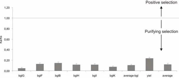
“nonsynonymous-to-synonymous substitution ratio test” (KA/KS Test) spricht dafür, dass das bgl-Operon einen unbekannten ökoligischen Selektionsvorteil bewirkt. Das zweite stumme β-Glukosid-System, bgc, kommt hauptsächlich in Isolaten der phylogenetischen Gruppen B1 and B2 vor. Es ist in Isolaten der Gruppe D zum Teil vorhanden und nur in einigen A-Isolaten nachweisbar.
Dieses Verteilungsmuster des bgc-Locus kann sowohl durch Genaufnahme als auch durch Genverlust erklärt werden. Zusammengefasst zeigt die vorliegende Arbeit, dass die Flexibilität des Genoms zusätzlich zur Aufnahme von Genen auch wesentlich durch Genverlust bestimmt wird, und dass eine sorgfältige Analyse einzelner Loci notwendig ist, um zwischen diesen beiden Mechanismen unterscheiden zu können.
3 Summary
I Summary
The genomes of bacterial species are very dynamic. For example in Escherichia coli, individual isolates may share as little as 60 to 70% of their genome with other isolates. The remainder of the genome consists of a flexible pool of genes, which are present only in some isolates. This genome diversity is manifested through gain of genes by horizontal transfer as well as by loss or mutations of genes. In this study the evolution of two silent β- glucoside loci belonging to the flexible gene pool of E. coli was traced. The bgl operon is present in ~80% of all E. coli isolates, while in the rest it is replaced by a locus (named Z5211-5214) of unknown function. The bgc locus is present in roughly 50% of E. coli isolates. To trace the evolution of these loci, the phylogeny of a representative collection of 175 E. coli isolates was established by multilocus sequence typing (MLST). In parallel, the bgl and bgc loci were typed by PCR and sequencing. This revealed four groups of the bgl / Z locus, including 3 bgl variants. Mapping of these groups demonstrated a striking correlation to the species phylogeny and population structure:
among the four phylogenetic groups of E. coli, bgl is present in the A, B1, and B2 groups, while the Z locus is present in D strains, which suggests a coupled evolution of the bgl / Z locus with the host. Further, three ancestral E. coli isolates and strains of the closely related species Escherichia albertii as well as the closely related genus Salmonella enterica, lack the bgl / Z locus, indicating horizontal transfer of the bgl and Z loci into the root of the modern E. coli. However, BLAST surveys revealed the presence of bgl homologs in Erwinia, Klebsiella and Photorhabdus species. The phylogeny of E. coli bgl and these homologs is concordant with the 16S rRNA phylogeny contradicting horizontal transfer. In conclusion, these results implicate vertical inheritance of bgl and its loss in some enterobacterial lineages. In E. coli isolates belonging to the phylogenetic group D, the bgl operon presumably was replaced by the Z locus, which may have been horizontally acquired. Further, correlating the data of a functional analysis of bgl with the species phylogeny demonstrated that bgl is functional although silent in the majority of strains in groups A and B1, while, interestingly, in more than 50% of B2 strains, bgl was not silent but
4 Summary weakly expressed. These data together with the results of nonsynonymous-to- synonymous substitution ratio test (the KA/KS test), suggest that bgl may confer an unknown ecological advantage. The second silent β-glucoside system bgc analyzed here, is predominant in the phylogenetic groups B1 and B2, it is present in D group isolates and rarely found in A strains. The widespread occurrence could be due to either gain or loss of bgc in evolution.
Cumulatively, the study suggests that in addition to gene gain, also gene loss may significantly contribute to the flexibility of the genome, and that a careful analysis is required for individual loci belonging to the flexible gene pool.
5 Introduction
II. Introduction
Bacterial evolution is very dynamic. Bacterial genomes are mosaic in nature consisting of a core pool of genes, which are shared by all individuals of a species, and a flexible pool of genes, which are present only in a subset of individuals of the species. This duality of the genome allows maintaining essential function and provides the flexibility to explore new niches (Feil, 2004). Evolution of bacterial genomes is brought about by three major mechanisms; the gain of genes through horizontal transfer, gene loss, and the modification of existing genes (Lawrence, 2005). The amount of genetic diversity seen within a species is remarkable. For example, the species Escherichia coli includes commensals and diverse pathogens. Their mosaic genomes can vary in size by up to one megabase (Bergthorsson and Ochman, 1998), and the core genes make up only 60 to 70% of individual genomes (Welch et al., 2002). The diversity of E. coli is due to genome rearrangements that occurred on a microevolutionary scale, as suggested by comparative genomic studies (Fukiya et al., 2004; Perna et al., 2001; Wei et al., 2003). Among the three major mechanisms, gene modification, gene loss, and gene gain, which work behind the observed diversity of bacteria, the latter has been extensively studied for more than a decade. Horizontal transfer of genes is considered a major force in shaping bacterial genome evolution (Gogarten and Townsend, 2005; Lawrence and Hendrickson, 2003). Gene loss is evident in the evolution of obligate parasites and symbionts (Mira et al., 2001). However, the relative role of gene gain and loss in the evolution of a species is not well known. For E. coli, this lack of knowledge is mainly due to the focus of research on horizontally acquired pathogenicity islands (Groisman and Ochman, 1996; Hacker and Carniel, 2001; Hacker and Kaper, 2000). In contrast, the focus of the current study was on understanding the mechanisms of genome evolution by tracing the evolutionary history of cryptic genes in the population of E. coli.
6 Introduction
1. Diversity of bacterial genomes
The textbook definition of ‘species’ is that individuals differ from others by minor but identifiable differences. However, in bacterial species the genomes display such a wide range of diversity that the definition of the bacterial species was questioned (Gevers et al., 2005). The genomes of individual isolates of bacterial ‘species’ can differ up to 50% in the case of Streptococcus (Marri et al., 2006), and 60-70% in E. coli as revealed by genome sequence comparison (Welch et al., 2002). Moreover, microarray based comparative genomic hybridization studies on 23 natural isolates of E.
coli showed that ~3000 genes belong to the genomic core and ~1000-1500 genes are variable (Dobrindt et al., 2003b). However, this diversity of the bacterial genome is based on the flexible gene content rather than on sequence variation throughout the genome. The sequence of the core genome, which is the part present in all strains of a given species, is highly conserved. Further, the comparison of core genome genes between close and more distantly related ‘species’ can be used to build robust phylogenetic trees, which reflect the evolution of the bacterial lineages. The core genome of each species differs significantly from the core genome of closely related species, and these differences reflect the phylogeny of the species. Furthermore, the analysis of core genome genes revealed that about 200 genes are common to the gamma-proteobacteria. Only 60 genes are shared by all cellular organisms; these genes are mainly important for translation (Koonin, 2003).
In contrast to the core genome, which is assumed to encode the essential functions for the species, the flexible gene pool is considered to confer a selective advantage under specific conditions. Genes that belong to the flexible gene pool include virulence factors, antibiotic resistance genes, genes for symbiosis among others. These genes are often part of genomic and pathogenicity islands, which are horizontally transferred into the genome (Hacker and Carniel, 2001). Considering the variability of the bacterial genome, recently, the term “pan-genome” was introduced in bacterial genomics to accomplish a broader definition of bacterial species. The “pan- genome” includes the core genes as well as all the genes of the flexible pool
7 Introduction found in different strains of one species (Medini et al., 2005). The size of the pan genome of a given bacterial species is anticipated to increase with the availability of genome sequences of individual strains. The diversity of bacterial genomes makes it an attractive case for the analysis of bacterial evolution.
2. Bacterial genome evolution: the three facets
As mentioned before, the principal driving forces that shape bacterial genomes are i) the modification of vertically transmitted genes, ii) gene loss, and iii) gene gain (Fig. 1). In eukaryotes, evolution occurs by the modification of existing genes (McDonald and Kreitman, 1991), whereas in prokaryotes there are countable instances showing such gene modifications. One such case is the increase of pathogenicity in Salmonella by the alteration of the pmrD gene encoding polymyxin B resistance to become regulated by the PhoPQ two-component regulatory system. Another example is that in Bordetella the expression of a toxin gene ptxA is enhanced by mutations (Parkhill et al., 2003a; Winfield and Groisman, 2004). Gene gain by horizontal gene-transfer (HGT) is considered a hallmark of bacterial evolution, especially of pathogens (Hacker and Kaper, 2000; Ochman et al., 2000). Horizontal gene transfer is mediated by three mechanisms: transformation (of plasmids or naked DNA), transduction (of genomic and pathogenicity islands), and conjugation. Horizontally transferred genes generally have different GC content and Codon usage when compared to the host genomes (Ochman et al., 2000). Examination of genomes based on DNA composition of commensals and pathogens for detecting foreign genes showed abundant signs of recent acquisitions ranging from 0% in Mycoplasma genitalium to 17% in Synechocystis. HGT has been well studied in E. coli. Based on GC content and Codon usage analyses the E. coli K12 MG1655 strain is estimated to contain about 18% of foreign DNA with a transfer rate of 16 kb/Myr since speciation (Lawrence and Ochman, 1998; Ochman et al., 2000).
Even in the presence of extensive transfer of DNA in E. coli, the chromosome size of different strains remains relatively constant around 5 megabases and in general prokaryotic genome sizes tend to remain constant. Thus, obviously gene acquisition must be balanced by loss of genes in order to reflect the
8 Introduction observed constant size of the genomes. Gene loss has been estimated to occur at a rate of two-three times higher as horizontal gene-transfer, when
~12,000 protein families were analyzed, but this balance need not necessarily operate on individual species (Kunin and Ouzounis, 2003). Gene loss has been the hallmark of evolution of pathogens and symbionts. Massive genome reduction is noticed in Mycobacterium leprae, Buchnera, and Rickettsia (Cole et al., 2001; Parkhill et al., 2003b; van Ham et al., 2003).
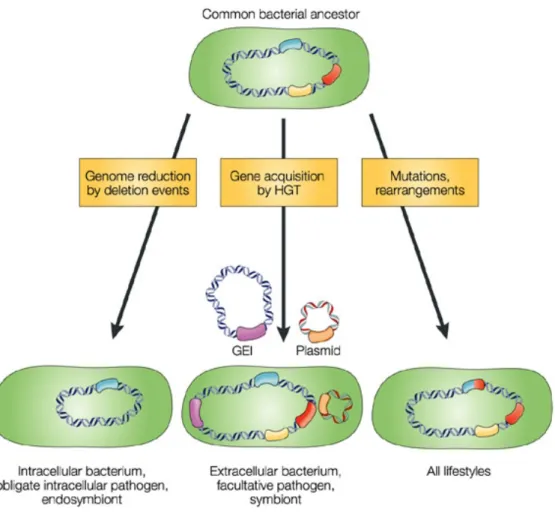
Fig. 1: Mechanisms that drive bacterial genome evolution. Three mechanisms are considered responsible for evolution of genome structures, which may reflect different bacterial lifestyles.
These are, firstly the modifications of existing genes by mutations and rearrangements, which suit all lifestyles. 2. Gene loss is the major force in genome reduction by deletion events, seen in host-dependent bacteria. 3. Gene gain by horizontal gene transfer (HGT) increases the adaptability of commensal and pathogenic bacteria by introducing genomic islands (GEI), pathogenic and symbiotic islands. Figure adopted and modified from (Dobrindt, 2004).
9 Introduction Further, gene loss occurs on special functions that may be detrimental to pathogenic lifestyle, creating “black holes” (deletions) in the genome. For example in Shigella, Cadaverine produced by the decarboxylation of lysine inhibits Shigella enterotoxic activity,and deletion of cadA encoding the lysine decarboxylase was demonstrated to enhance virulence (Maurelli et al., 1998).
Shigella are bacteria that belong to the species E. coli, which can be distinguished from other E. coli strains by specific markers (as Shiga-toxin expression) and whose name is maintained for medical historical reasons (Maurelli, 2007). Therefore, evolution of Shigella from E. coli is marked by gene gain of virulence traits and loss of biochemical functions that are adaptive to pathogenic lifestyle.
Detection of horizontally acquired genes is primarily performed by compositional analysis of sequences and by BLAST searches on related genomes. But it has been pointed out that gene loss can be interpreted as gene gain if one relies on BLAST searches for orthologs due to limitation of availability of genome sequences (Zhaxybayeva et al., 2007). Moreover, the quantification of gene gain and loss events has been performed on protein families rather than on individual genes. One example of a study on individual genes is the lac operon of E. coli. The lac operon was thought to be horizontally acquired in E. coli and therefore E. coli metabolizes lactose (Ochman et al., 2000). In contrast to this opinion, D.M. Stoebel (2006) showed that the lac operon is vertically transmitted in enterobactericiae and that the Lactose negative phenotype of some members including Salmonella, Shigella are due to loss of the operon. Hence, for a detailed study of loss and gain of genes in bacteria, rigorous phylogenetic methods need to be performed at the population level for individual genes.
3. Escherichia coli: Phylogeny and population structure
The versatile E. coli represents an excellent model to understand bacterial genome evolution owing to its well-established phylogenetic groups and population structure. Classic multilocus enzyme electrophoresis (MLEE) typing of 72 reference strains of E. coli, the ECOR collection, (Ochman and Selander, 1984) indicated the existence of four phylogenetic groups of E. coli,
10 Introduction which are designated A, B1, B2 and D. A minor group E has been neglected later, because of inconsistent clustering in subsequent analyses. Within this decade, the new molecular technique, multilocus sequence typing (MLST) was introduced for bacterial strain typing. MLST is conceptually similar to MLEE (multilocus enzyme electrophoresis) but characterizes each strain of a bacterial species by assigning alleles for seven housekeeping genes directly from the nucleotide sequence of internal fragments of genes, rather than indirectly from the electrophoretic mobilities of their gene products (Maiden, 2006). The genotype of strains characterized by MLST is defined by their allelic profiles. MLST has several advantages over MLEE. MLST is highly discriminative as it detects all the nucleotide polymorphisms within a gene rather than just those mutations that alter the electrophoretic mobility of the protein product. Bacterial strains harbor sufficient variation within the housekeeping loci that many different alleles can be resolved and by using seven genes, billions of genotypes can be obtained. A second advantage of MLST is the accuracy and portability of DNA sequence data, which can be rapidly and unambiguously compared with previously characterized strains by interrogation through a common web server (http://www.mlst.net/). MLST therefore provides a precise and unambiguous method for characterizing bacterial strains.
For E. coli three MLST schemes (Le et al., 2007; Reid et al., 2000) are established, one of them was designed by Wirth et al., (2006) who put forth a broader picture on the evolutionary history of E. coli. Briefly, MLST was used to assess the genetic relatedness of 406 natural isolates of E. coli, by analyzing the allelic profile of seven housekeeping genes distributed around the chromosome (Fig. 2a). Fragments of these seven genes are PCR amplified and sequenced on both strands using the PCR primers. Sequences are manually curated and each unique sequence of a gene is assigned an allele number. Thus, seven allele numbers are obtained for a strain at seven housekeeping genes. Combination of the seven numbers for a strain constitutes its allelic profile or Sequence Type (ST). In their study, Wirth et al., (2006) presented a star-like phylogeny depicting the rapid population expansion that resulted in the diversity of E. coli species (Fig. 2b). The four
11 Introduction groups A, B1, B2 and D comprise the modern E. coli strains and two divergent isolates, which are E. coli, are considered as remnants of ancestral diversity.
Moreover, they identified 278 sequence types (STs) at that time and currently 721 STs (as of 10.12.2007) are deposited at the web-based server for E. coli MLST (http://web.mpiib-berlin.mpg.de/mlst/dbs/Ecoli). Further, they suggested
that the clonal structure established by MLST would provide a better framework for studying the evolution of strains, compared to the use of classic phylogenetic groups whose boundaries are fluid. Recently, Weissman et al.
(2006) used MLST analysis on E. coli pathogens to deduce a clonal framework. In that work they identified their strains to belong to a sequence type complex ST95 and studied the evolution of fimbrial genes at the clonal complex level. From their work, Weissman et al., (2006) showed the horizontal transfer of fimbrial operons into ST95 complex strains and divergence of the genes after entry into the complex. This suggests that clonal level analysis gives a finer evolutionary framework, which when combined
Fig. 2: (A) Genomic locations of seven housekeeping genes used in E. coli MLST analysis. (B) The star-like phylogeny depicted by the neighbor-joining tree of 462 E. coli, three E. albertii and one Salmonella typhi (outgroup) based on the concatenated sequences from seven loci. The dark grey circle represents the main group of 460 isolates; light grey circle includes two divergent isolates. Figure adopted and modified from (Wirth et al., 2006)
B A
12 Introduction with phylogenetics, can be promising to trace the history of individual genes at a deeper resolution.
4. Cryptic genes
Genes that are not expressed under any tested condition are considered cryptic or silent. Silent genes are found in bacteria in many species including Lactobacillus, Bacillus, Escherichia, and Salmonella (Birge EA, 2006). The well-studied examples exist in E. coli. These are the bgl and asc operons, which are involved in β-glucosides metabolism (Fig. 3). Among these two, the bgl operon is well characterized, known to encode proteins for uptake and hydrolysis of aryl-β-D-glucosides such as salicin, arbutin (Schnetz et al., 1987). The asc (arbutin, salicin, cellobiose) locus of E. coli encodes a regulator, a permease and a β-glucosidase necessary for transport and hydrolysis of the β-glucosides. Previously another silent β-glucoside system called bgc (Fig. 3) was discovered in the laboratory (Neelakanta, 2005). bgc (β-glucoside and cellobiose) locus comprises an operon and a divergent regulator gene, needed for utilization of β-glucosides and cellobiose at low temperature. The bgl operon of E. coli is a paradigm of crypticity, as it is not expressed under any laboratory-tested conditions. Silencing of bgl operon is mediated by the histone-like nucleoid structuring protein (H-NS) (Dole et al., 2004a; Nagarajavel et al., 2007). Intuitively, silent genes should be undesirable, as selection will not favor their function, ultimately leading to their erosion. On the contrary, the bgl operon is present in the E. coli laboratory strain and surprisingly, in the uropathogenic CFT073 strain as well (Welch et al., 2002). Previous work in the laboratory by G. Neelakanta (2005) showed that the bgl operon is predominant in natural isolates of E. coli including commensals and pathogens (Neelakanta, 2005). In addition, it was found that in a subset of strains it was replaced by Z5211-5214 locus of unknown function, similar to the published genome sequences of E. coli O157 strains.
Noticeably, several types of the bgl locus were recognized in E. coli.
Downstream of the operon, two hypothetical ORFs yieJ and yieI are present in one group of strains while the yieI gene alone is present in another group. The strains in which bgl is replaced by the Z211-5214 locus, both yieJ and yieI
13 Introduction genes are absent. The prevalence of bgl and its variability is intriguing in the context of its evolution.
H-NS selectively represses horizontally transferred genes in E. coli and Salmonella (Lucchini et al., 2006; Navarre et al., 2007). Since bgl operon is a model system for the analysis of repression by H-NS, would it be a horizontally transferred operon is a question. Further, the presence of several silent systems leads to the question of the role of cryptic genes in bacterial evolution. Hall et al., (1983) proposed that under one set of conditions members with a cryptic gene are more fit than those members who express it, while under alternative conditions those members expressing the gene are at a selective advantage. This argues for the retention of silent genes, whose Fig. 3: Cryptic β-glucoside operons in E. coli. Genomic structure of three β-glucoside operons, bgl, bgc, asc and the Z locus replacing bgl in E. coli. Gene names and protein encoded are indicated. The bgl operon is the well-studied example of a cryptic operon, repressed by histone-like nucleoid structuring protein (H-NS) (Dole et al., 2004a). The bgc is another cryptic operon involved in utilization of β-glucosides and cellobiose at low termperature (Neelakanta and Schnetz, unpublished data). The asc operon is yet another cryptic system in E. coli encoding protein for metabolism of arbutin, salicin and cellobiose (Hall and Xu, 1992).
14 Introduction evolution is rather poorly understood. Therefore, a systematic approach to trace the evolution of bgl and bgc system will shed light on the evolution of bacterial genes, which are retained but with no known advantage.
5. Objectives of the current study
The objective of this study is to contribute to the understanding of the evolution of bacterial genomes by considering the evolution of silent β- glucoside systems as a model in E. coli. In the present study, a diverse population of E. coli was typed by MLST to establish their phylogenetic and clonal structure. This laid the framework to trace the evolution of the cryptic bgl operon and the bgc operon. The results revealed the dynamic evolution of the bgl operon. It is vertically inherited in enterobactericiae and deleted in some lineages. In E. coli, the vertical history of the operon is coupled to evolutionary history of the species, indicating a strong purpose for its retention. The prevalence of the second silent system, bgc was analyzed in the population, which revealed the possibilities of gain or loss of bgc in evolution. The implications of gene gain and loss on the evolution of individual operons from a genomic perspective as well as the fate of the paradoxically silent operons is discussed.
15 Results
III. Results
1. Evolutionary genetic analysis of the bgl operon and Z locus in the E. coli population
To investigate the evolution of the bgl operon, the prevalence of the operon within a collection of E. coli strains was analyzed before (Neelakanta, 2005).
However, based on this previous analysis no data were available, which allowed to correlate the evolution of the bgl operon and Z5211-5214 locus with the species phylogeny. To achieve such a correlation it is imperative to have a collection of E. coli strains which is representative and for which the phylogenetic and population structure is established. In addition, the phylogeny of the bgl locus needs to be analyzed at the sequence level.
1.1 Population structure of the E. coli collection
The E. coli collection used in this study includes strains from diverse sources.
These are 98 clinical human isolates, of which 52 are commensals and 46 are pathogens (Dr. G. Plum, Institut für Medizinische Mikrobiologie, Immunologie und Hygiene, Universität zu Köln). In addition, a septicemic E. coli strain i484 (Khan and Isaacson, 1998), two uropathogenic E. coli (UPEC) strains, J96 and 536 (Brzuszkiewicz et al., 2006), and the 72 strains of the ECOR reference collection (Ochman and Selander, 1984) were analyzed.
Furthermore, in the course of the analysis two divergent E. coli strains RL325/96 and Z205 which are presumably of an ancestral E. coli type (Wirth et al., 2006) and three strains of the closely related species Escherichia albertii were included. Thus, the entire collection includes 178 strains.
To determine the phylogeny and the population structure of the E. coli collection multilocus sequence typing (MLST) was performed, using the scheme established for E. coli by Wirth et al., (2006). Of the collection, 99 strains were of unknown phylogeny. These were subjected to MLST by sequencing fragments of the seven housekeeping genes adk, fumC, gyrB, icd,
16 Results mdh, purA and recA for each strain (Wirth et al., 2006). Sequences were curated manually using Bionumerics software (Version 4.0), which was used in the laboratory of M. Achtman, Max Planck Institute for Infection Biology, Berlin. Any ambiguities were resolved by re-sequencing a newly generated PCR fragment and with additional internal primers in case of adk and fumC genes.
The population structure of bacterial species can be studied by allele-based population genetic analysis. For each housekeeping gene, the different sequences present within a species are assigned distinct alleles (specified by numbers). Further, for each strain, the alleles corresponding to seven loci define the allelic profile or Sequence Type (ST). STs sharing 6 or more than 6 alleles define a clonal complex referred as Sequence Type complex (ST complex). For the allele-based analyses, the MLST data for E. coli established in the MLST database (Wirth et al., 2006) (http://web.mpiib- berlin.mpg.de/mlst/dbs/Ecoli) was used as reference. With the help of computational algorithms the following was determined for each strain: a) the allelic profile of 7 genes, i.e. the sequence type (ST), b) the clonal relationship of strains and the sequence type complexes (ST complex) and c) the ancestral group (Wirth et al., 2006). New allele numbers and STs were assigned to strains with novel allelic sequences. Following this 98 out of the 99 strains were entered into the public E. coli MLST database (http://web.mpiib-berlin.mpg.de/mlst/dbs/Ecoli). One strain, E466, was identical to E464 and hence omitted. The strain collection typed in this study represents 49 different sequence types (STs). Out of 98 strains 77 occurred in 25 different ST complexes and the remaining are not assigned to any ST complex and simply designated by their STs (Refer to supplementary Table S1 in appendix). Sequences and MLST information for the 72 ECOR strains, UPEC strains J96 and 536, E. coli RL325/96, Z205 and three E. albertii strains were downloaded from the publicly available E. coli MLST database.
Two ECOR strains 23 and 32 from the lab collection had ST different from that in the E. coli MLST database and hence these two strains were omitted from
17 Results this study. Taken together, the entire strain collection represented 92 STs and 115 out of 175 strains appeared in 25 different ST complexes. The MLST data including the individual alleles, STs and ST complexes are listed in Table S1.
The result of these population genetic based analyses is visualized on a minimal spanning tree, referred to as MSTREE (Fig. 4). Cumulatively, the analysis established the population structure of the collection of strains used, and it demonstrated that the collection is representative.
To establish the phylogenetic relationships, sequences of the housekeeping genes for each strain were concatenated (3423bp), and the concatenated sequence was used for phylogenetic reconstruction using the neighbor-joining (NJ) method included in the MEGA software V3.1 (Kumar et al., 2004). For convenience, ECOR strains were analyzed separately. The neighbor-joining tree from the sequence data of all strains resulted in four clades concordant with the classical ECOR groups A, B1, B2 and D (Fig. 5). As shown before, the two strains RL325/96 and Z205 are very divergent from the rest of the strains in four clades, and are closely related to three E. albertii strains (Wirth et al., 2006). Interestingly, strain E10083 isolated as a human commensal closely clustered with the two ancestral strains RL325/96 and Z205 (Fig. 5).
The latter were isolated from dog and parrot respectively (Wirth et al., 2006).
This suggests that the human isolate E10083 is probably another remnant of the ancestry of E. coli. These three divergent strains are referred to in this study as ancestral E. coli and the rest of the strains appearing in four phylogenetic clades, as modern group of E. coli as reported before (Wirth et al., 2006). The neighbor-joining tree of the concatenated sequences of housekeeping genes for the ECOR strains was also constructed. This tree was consistent with previous reports (Escobar-Paramo et al., 2004; Lecointre et al., 1998). Thus, the neighbor-joining tree (Fig. 5) represents the whole genome phylogeny of the strain collection used in this study and the strain collection is representative of all phylogenetic groups.
18 Results Fig. 4: Minimal spanning tree (MSTREE) depicting the Sequence Types (STs) of strain collection based on MLST analysis. Each circle represents one ST, denoted by its number on the circle. Size of the circle corresponds to number of strains, the smallest represents one strain. Black lines connecting pairs of STs indicate sharing of six alleles (thick lines), five (thin) or four (dotted) between them. Grey dotted lines of increasing length indicate sharing of three to one alleles respectively.
19 Results Fig. 5: Whole genome phylogeny. Neighbor-joining trees of concatenated seven MLST housekeeping genes from (a) Isolates (b) ECOR collection. Strains of different phylogenetic groups are color-coded: green-A, cyan-B1, red-B2, blue-D, magenta-AxB1, orange-ABD and grey-ancestral/E. albertii strains. Strains displayed in black are not assigned to any group and those indicated (•) are considered odd strains. Numbers on the nodes are bootstrap scores from 1000 replicates and scores above 50% are indicated.
20 Results Further, to discern the phylogenetic group of each strain, the information for strains of known ST was extracted from the MLST database. For strains with a new ST, the program STRUCTURE was used with the help of Vartul Sangal in M. Achtman’s group as described (Wirth et al., 2006). Wirth et al., (2006) had established a scheme to correlate the ST determined by MLST to the phylogenetic groups A, B1, B2 and D established for the ECOR collection by MLEE (Ochman and Selander, 1984). Further, it was shown that some strains represent hybrids created by recombination. The hybrid group AxB1 represents strains, which derive their ancestry largely from A and B1, and the hybrid group ABD derives its ancestry from all phylogenetic groups. The ancestral group of each modern isolate is listed in (Table S1, appendix). In total 48 strains belong to the phylogenetic group A, 17 strains belong to B1, 48 to B2, 21 to D, 17 strains to AxB1, and 13 strains to ABD respectively. For 5 strains the groups have not been assigned. The ancestry of the isolates as determined by STRUCTURE and graphically displayed using the program DISTRUCT (Rosenberg NA, 2004) (Fig. 6).
1.2 Genetic diversity of the bgl operon/ Z5211-5214 locus in E.
coli natural isolates
The bgl operon of E. coli is not expressed under laboratory conditions due to effective silencing by the histone-like nucleoid structuring protein H-NS (Defez and De, 1981; Schaefler and Maas, 1967 Higgins et al., 1988, Mahadevan and Wright, 1987, Schnetz, 1995, Dole et al., 2004b; Nagarajavel et al., 2007). Previous analysis in the laboratory revealed that bgl operon is highly prevalent in the population (Neelakanta, 2005). In order to study the genetic diversity of the bgl and Z5211-5214 loci, phylogenetic analysis was carried Fig. 6: Ancestry of E. coli isolates. Proportions of ancestry from groups A, B1, B2 and D as inferred by STRUCTURE and their assignments to six groups as displayed with DISTRUCT.
The plot shows one vertical line for each isolate indicating the proportions of ancestry from the four groups, which are color-coded as green (A); red (B1); yellow (B2) and blue (D). The groups are indicated at the bottom and ND refers to strains for which groups are not assigned.
21 Results out. Earlier analysis in typing bgl and Z5211-5214 also showed that sequences upstream and downstream of bgl are polymorphic. To analyze the genetic variation more systematically, fragments of DNA from the bgl and Z5211-5214 loci were sequenced (Fig. 7). Fragments were amplified by PCR and sequenced on both the strands. The PCR analysis was consistent with earlier results (Neelakanta, 2005), which revealed that 78% of isolates (136 of 175) carry bgl operon and 19% lacked bgl operon, but carried a different locus containing four open reading frames as annotated in the E. coli 0157:H7 EDL933 genome sequence. The prevalence of bgl operon and the phylogenetic groups showed a correlation, when both were related to each other. Noticeably bgl operon was present in strains of A, B1, B2 groups and totally absent in D group strains. Noticeably the A and B1 strains have the genes yieJ and yieI present downstream of bgl operon and the B2 strains lack the yieJ gene. The D strains have neither yieJ nor yieI gene (Fig. 8A).
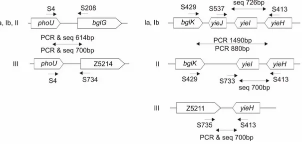
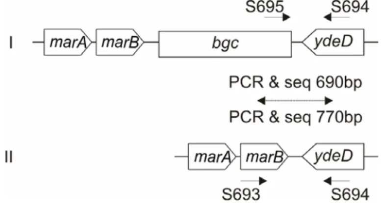
Fig. 7: PCR and sequence typing of bgl/Z loci. Primers and their mapping positions are indicated.
Groups of bgl/Z loci are given at the left of the genomic structure of the loci. Sizes of the PCR product sequenced on both the strands are given. Same primers were used for both PCR and sequencing, otherwise indicated in case of sequencing fragments downstream of bgl operon.
Multiplex PCR reaction consisting of primers S4, S208 and S734 was performed to distinguish between strains having bgl (group Ia, Ib and II) and Z5211-5214 locus (group III). For sequencing fragments downstream of bgl, PCR reaction with S429 and S413 was performed and the products were1490bp (group Ia, Ib) and 880bp (group II), which were sequenced with primers S537 and S733 respectively on the forward strand and S413 on the reverse strand. Z5211-5214 was PCR amplified and sequenced independently.
22 Results To phylogenetically reconstruct evolution of the bgl locus, fragments of sequences following the stop codon of the flanking genes, which are internal to a breakpoint (point from where polymorphism was observed in bgl locus), were concatenated. 811bp (537+277bp) of sequence of bgl locus for each isolate was obtained (Fig. 7B). Isolates in which the analyzed region of the bgl operon or the Z5211-5214 locus was disrupted by insertions and/or deletion
(see later) were omitted. A multiple alignment was generated using the concatenated 811bp sequence of bgl from each isolate, and this was used for phylogenetic reconstruction according to the neighbor-joining method (Fig.
9A). Again, a separate tree was built for the sequences derived from the Fig. 8: A) Genomic organization of the E. coli bgl/Z5211-5214 loci.
Gene names are indicated within genes and on the top for flanking genes and Z5211-5214 locus. E. coli phylogenetic groups are indicated on the left and the number of strains on the right. B2 strains lack the yieJ gene. Ancestral E. coli and E. albertii strains lack bgl operon/Z5211-5214 locus, while E. albertii strains carry yieJ gene. (B) Schematic illustration of sequencing strategy. Fragments of the bgl operon/Z5211-5214 locus (1& 2) and the flanking gene phoU of the core genome (core) were sequenced from the indicated breakpoints on the left and right.
Sequence lengths are given at the bottom.
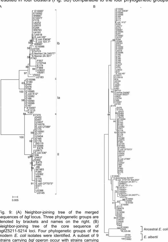
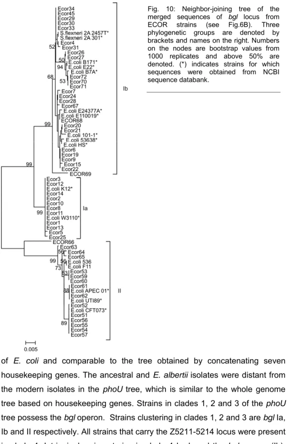
23 Results ECOR strains (Fig. 10). For further comparison, the respective bgl sequences from the published E. coli genome sequences were also included in the phylogenetic analysis. The tree established 3 clusters, which were named bgl group Ia, Ib, and II (Fig. 9A). This demonstrates the presence of three groups of the bgl locus in modern E. coli isolates.
The sequences of the Z5211-5214 locus was also phylogenetically analyzed (Fig. 26 appendix), but no groups were assigned as the tree showed several clusters, probably denoting rapid evolution of the locus (Fig. 26, Appendix).
Hence, the strains harboring this locus were arbitrarily assigned to group III (Fig. 9A). The ancestral E. coli isolates and the E. albertii strains lack both the bgl operon and the Z5211-5214 locus (Fig. 8A). The identified structure of the genome in these strains was assigned to groups IV and V respectively.
Strikingly similar results were obtained with ECOR strains (Fig. 10). These analyses demonstrate that three phylogenetic groups of the bgl operon exist among the modern group of E. coli and Z5211-5214 locus can be considered as the fourth group in which bgl is replaced. Intriguingly, the absence of bgl operon/ Z5211-5214 locus in the ancestral E. coli and the related E. albertii might indicate a probability of horizontal transfer of these two loci into modern E. coli isolates.
1.3 Genetic variation in the core genome flanking the bgl operon/Z5211-5214 locus
The sequences obtained from the regions flanking the bgl and Z5211-5214 loci, respectively were also phylogenetically analyzed. The phoU gene flanking the two loci is present in all E. coli isolates, including the ancestral strains and E. albertii. The phoU gene is essential for survival of E. coli when phosphate is limiting, a condition that is frequent in the natural habitats of E.
coli (Buckles et al., 2006; Steed and Wanner, 1993). Therefore, phoU belongs to the core genome of E. coli. Partial sequences of the phoU gene were obtained from all the isolates and 204bp of fragments of these sequences were used to construct a tree by the neighbor-joining method. The tree
24 Results resulted in four clusters (Fig. 9B) comparable to the four phylogenetic groups
Fig. 9: (A) Neighbor-joining tree of the merged sequences of bgl locus. Three phylogenetic groups are denoted by brackets and names on the right. (B) Neighbor-joining tree of the core sequence of bgl/Z5211-5214 loci. Four phylogenetic groups of the modern E. coli isolates were identified. A subset of 9 strains carrying bgl operon occur with strains carrying Z5211-5214 locus (←). Both in (A) and (B) numbers on the nodes are bootstrap scores (above 50%) from 1000 replicates. (*) denotes strains for which sequences were obtained from NCBI.
25 Results of E. coli and comparable to the tree obtained by concatenating seven housekeeping genes. The ancestral and E. albertii isolates were distant from the modern isolates in the phoU tree, which is similar to the whole genome tree based on housekeeping genes. Strains in clades 1, 2 and 3 of the phoU tree possess the bgl operon. Strains clustering in clades 1, 2 and 3 are bgl Ia, Ib and II respectively. All strains that carry the Z5211-5214 locus were present in clade 4. Intriguingly, nine strains in clade 4 harbored the bgl operon (Ib).
Fig. 10: Neighbor-joining tree of the merged sequences of bgl locus from ECOR strains (see Fig.6B). Three phylogenetic groups are denoted by brackets and names on the right. Numbers on the nodes are bootstrap values from 1000 replicates and above 50% are denoted. (*) indicates strains for which sequences were obtained from NCBI sequence databank.
26 Results Thus, for these strains the presence of bgl did not correlate to the phoU phylogenetic clades, which may indicate recombination. Thus, the diversity of bgl operon/ Z5211-5214 locus is reflected on the core genome flanking these loci and provides striking evidence for parallel evolution. The result may indicate a more recent recombination event for strains that carry the bgl group Ib operon.
1.4 The evolution of the bgl/Z5211-5214 locus is coupled to species evolution
The phylogeny of the bgl operon derived from the sequenced fragments is comparable to the phylogeny of the core gene phoU flanking the operon.
Further, to deduce an evolutionary relationship of the bgl operon/Z5211-5214 locus with the species, a one-to-one phylogenetic comparison was performed between their phylogenies. The bgl groups were marked on the whole genome phylogenetic tree (Fig. 11). The clustering of strains in the bgl operon phylogeny was consistent with the major clades or phylogenetic groups seen in the whole genome phylogeny generated from the concatenation of seven housekeeping genes. This reveals that the bgl operon shares the same evolutionary history as the species. The Z5211-5214 locus is exclusively present in D group strains. Owing to the absence of the Z5211-5214 locus in the other three phylogenetic groups (A, B1, B2) of E. coli, it is possible that the Z locus was horizontally transferred into the ancestor of D group strains. An interesting exception is strain F905, which carries the Z5211-5214 locus, but clusters with the A group strains. This incongruence suggests a recent transfer of Z5211-5214 locus into strain F905. Leaving out the single exception, the strong congruence implies that the loci have a shared evolutionary history with the species. Further, this evolutionary congruence is augmented by similar results from the ECOR strains (Fig. 11).
27 Results Fig. 11: bgl /Z5211-5214 loci groups and Bgl phenotype marked on the phylogenetic tree obtained from concatenation of seven housekeeping genes (represented earlier in Fig.5).
Groups are indicated by brackets on the right. Bgl phenotype next to strain name. “1”-Bgl- with papillae formation indicative of presence of a functional but repressed bgl operon; “0“ refers to Bgl- without papillation indicating absence of a functional bgl and “2” refers to a weak positive (or relaxed) phenotype after 3-5 days of incubation at 37°C. Refer to Fig.5 for the color codes of the phylogenetic groups of the strains.
28 Results
1.5 Clonal evolution of the bgl operon/Z5211-5214 locus
It is a controversial view that phylogenetic approaches are largely unsuitable for most modern E. coli and hence allele-based population genetic analyses of E. coli strains were considered more informative to discern the deep evolutionary relationships (Wirth et al., 2006). MLST data from the strains are used to identify clonal groups based on the sharing of the allelic profiles (see section 1.1). To deduce the clonal evolution of the bgl operon/Z5211-5214 locus, their groups obtained by sequencing and phylogenetic analysis were mapped on the MSTREE depicting the clonal structure of the strains used in this study. The groups of the bgl operon/Z5211-5214 were color coded and represented within the MSTREE (Fig. 12). In the MSTREE every circle representing an ST acquired a uniform color indicating that all the strains were of the same bgl operon or Z5211-5214 group. The bgl Ia group mapped exclusive to the ST10 complex which contains ST10 and related STs. bgl Ib appeared exclusively in several ST complexes such as ST23, ST10, ST86, ST155, totally in 13 different complexes. bgl II was largely restricted to ST73, ST95 and ST12 complexes. Z5211-5214 strains occurred in different ST complexes, like ST31, ST38, and ST59. Importantly, there is almost no intermingling of bgl-Z groups in a single ST or ST complex. Two exceptions were found contradicting this strong congruence. As previously noted in the phylogenetic analysis, strain F905 with ST10 (ST10 complex, A group) is the only strain lacking the bgl operon but harboring the Z5211-5214 locus. This indicates the possibility of horizontal transfer of Z5211-5214 locus into ST10 complex. Another likelihood of horizontal transfer of Z5211-5214 locus was noted in ST350 complex with two strains Ecor31 and E179 (STs, ST57 and 350 respectively). Ecor31 has bgl operon group Ia, whereas E179 has Z5211- 5214 locus, suggesting the introduction of Z locus in E179 by horizontal transfer. These results show that the prevalence of bgl and Z groups strongly fits the clonal structure of the species, which suggests that the bgl operon and Z5211-5214 locus clonally descended with E. coli.
29 Results Fig. 12: Groups of bgl/Z5211-5214 loci on MSTREE represented in Fig. 4. The groups are color codes: Ia (dark green); Ib (light green); II (dark red) and III (blue).
30 Results
1.6 Phylogenetic analysis of the complete bgl operon sequence The genetic diversity of the bgl operon, analyzed from fragments within the locus gave rise to three phylogenetic groups, which are strongly congruent with the phylogenetic groups and the clonal complexes of the species.
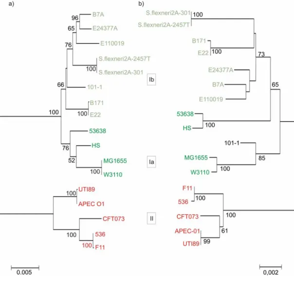
Further, to analyze the genetic diversity of the complete bgl operon, the sequence of the entire locus, which includes six genes of bgl operon (bglGFBHIK) and downstream yieJ/yieI genes, were obtained from the whole genome sequences of E. coli and Shigella strains available at NCBI Microbial genomes. Sequences were extracted from 17 strains (Fig. 13) and a multiple alignment was generated using ClustalW implemented in MEGA software V3.1. Insertion sequences distorting the multiple alignment were removed from strains having IS elements (Strains S. flexneri 2A-301, 2457T, E. coli E22, E110019, 53638). Neighbor-joining method was used to build the phylogeny of the bgl locus including yieJ/yieI genes. The tree likewise revealed three phylogenetic groups (Ia, Ib and II) very similar to the phylogenetic tree obtained from partial sequences of the locus (Fig. 13). Thus, indeed the bgl locus diverged into three groups within E. coli.
To see, if the phylogenetic relation obtained with the complete bgl operon together with downstream yieI gene also correlates to the species evolution, of the 17 E. coli and Shigella genome sequences, the sequences of the seven housekeeping genes used in MLST analysis were extracted. The sequences were concatenated as before (see section 1.1) and used for phylogenetic analysis. The resulting neighbor-joining tree of the housekeeping genes identified three phylogenetic groups of the strains. The tree of the housekeeping genes and the bgl operon tree showed strong congruence (Fig.
13). Incongruence was noted with strains HS and 53658, which are closely related to strains MG1655 and W3110 (group Ia) in the bgl locus tree, but distantly related in the housekeeping genes tree. The incongruence could indicate a putative recombination at the bgl operon in strains HS and 53638 with the bgl Ia strains. The phylogeny of the complete bgl locus is comparable to the phylogenetic relations obtained by the analysis of bgl sequence
31 Results fragments presented above. Yet again, these results suggest that the core genome and the bgl locus have a shared evolutionary history.
Fig. 13: Phylogenetic comparison of bgl locus and species trees. (a) Neighbor-joining tree of complete bgl locus from 17 strains obtained from NCBI sequence databank. (b) Strain phylogeny based on 7 MLST house-keeping genes. Boxed numbers in the middle refer to phylogenetic groups of bgl locus. Numbers on the nodes are bootstrap values from 1000 replicates (scores above 50 are indicated). Scale at the bottom depicts evolutionary distance.
32 Results
1.7 Functional analysis of bgl operon
The bgl operon encodes the proteins for utilization of β-glucosidic sugars arbutin and salicin. Due to repression of bgl by H-NS wild type E. coli K12 cells are phenotypically Bgl-. However, in E. coli K12 spontaneous Bgl+ mutants arise as papillae (Schaefler and Malamy, 1969). In previous work performed in the laboratory by G. Neelakanta (2005), the Bgl- phenotype of all strains of the E. coli collection was tested on BTB salicin indicator plates at 28°C and 37°C (Neelakanta, 2005) and three phenotypes were distinguished.
The phenotypes identified, were (i) Bgl-,without papillation indicating that no functional operon is present, (ii) Bgl- with papillation indicative of the presence of a functional but repressed operon, (iii) a weak positive (or ‘relaxed’) phenotype after 3 to 5 days of incubation at 37°C, and (iv) one strain (Ecor49) showed a Bgl+ phenotype.
In order to analyze a relationship between the bgl genotype, phenotype and the phylogenetic groups of E. coli, the phenotypes were mapped on the housekeeping genes tree (Fig. 11). To this end, the phenotypes were classified into types 0, 1 and 2, where ‘type 0’ was assigned to non papillating Bgl- strains, type 1 to papillating Bgl- strains, and ‘type 2’ to strains with a relaxed phenotype. These phenotypic types were marked on the housekeeping genes tree in which the bgl/Z groups were marked previously (Fig. 11). The marking of phenotypic groups on the tree revealed that majority of A, B1, hybrid AxB1 strains (bgl Ia or Ib group) showed Bgl+ papillation phenotype and a minority showed Bgl- phenotype. All but one D group strain, which carried Z5211-5214 locus exhibited Bgl- phenotype, as expected.
Ecor49 was the only D strain showing a weak Bgl+ phenotype on day2.
Noticeably more than half of the B2 strains corresponding to bgl II group exhibited the relaxed phenotype. The rest of the B2 strains showed Bgl- papillation phenotype except two B2 strains. Further, the phenotypic types 0, 1 and 2 were visualized on the MSTREE depicting the clonal complexes (Fig.14). This visualization resulted in similar correlation seen
33 Results Fig. 14: Groups assigned on Bgl phenotype mapped on the MSTREE presented earlier in Fig.4.
The groups are color-coded: type “0” - Bgl- non papillating (blue); “1” - Bgl- but papillating (dark green) and “II” - weakly Bgl+ (dark red).
34 Results above with the phylogenetic groups. Strains in ST10, ST23 complexes and of multiple STs corresponding to bgl Ia or Ib displayed 20% non-papillating Bgl- and 80% papillating Bgl- phenotypes. Strains in ST73, ST95, ST12 complexes exhibited 3% Bgl- , 40% papillating Bgl- and 57% relaxed phenotypes. This demonstrates that only strains that belong to the B2 phylogenetic group and concomitantly carrying a bgl of the group II may have a relaxed phenotype.
The mapping of the phenotypes on the MSTREE revealed that strains of the two clonal groups of ST73 and 12 show a high frequency of the relaxed phenotype. This relaxed phenotype and weak expression of bgl might be selected in these strains.
1.8 Insertions/deletions in bgl /Z5211-5214 loci
Typing of the bgl operon/Z5211-5214 locus not only revealed the presence/absence of the loci but also indicated the presence of insertion sequences or insertion associated deletions within the loci (Neelakanta, 2005). In the earlier work of G. Neelakanta (2005), there were discrepancies in the sequencing of the insertions identified in the strains. Those discrepancies were resolved in the current study by re-sequencing in 12 strains. In perceiving the correlations between occurrence of insertion/deletions and groups of bgl, it was notable that predominantly disruption of bgl operon was seen in bgl Ia strains and relatively less in bgl Ib and II strains.
1.9 The bgl operon is vertically inherited in Enterobactericiae The typing of the bgl operon/Z5211-5214 locus revealed that both loci are absent in the ancestral E. coli strains and in the closest species E. albertii.
This poses several possible scenarios. Firstly, the bgl operon and the Z5211- 5214 locus have been horizontally introduced into the modern group of E. coli.
Secondly, vertical inheritance of the bgl operon and horizontal transfer of Z5211-5214 into the ancestor of D strains and loss in ancestral and E. albertii strains is possible. To test the above possibilities, proteobacterial genomic sequences were searched using tblastn program of BLAST (Altschul et al.,
35 Results 1990) for orthologs of genes of the bgl and Z5211-5214 loci. The bgl sequence of E. coli K12 strain MG1655 was used as the query, and BLAST was performed for individual genes bglGFBHIK and yieJ, yieI genes. Similarly, the individual genes of the Z5211-5214 locus were used for BLAST to obtain orthologs using the E. coli 0157:H7 EDL933 sequence as query. Searching for bgl operon orthologs identified BLAST hits among members of enterobactericiae family, Klebsiella sp, Erwinia sp and Photorhabdus sp, with multiple hits above 30% identity in Erwinia and Klebsiella. In addition, BLAST yielded very week hits in Yersinia and other gamma-proteobacteria members like Vibrio. Protein sequences of the hits were obtained from NCBI sequence databank and used for phylogenetic analysis.
In order to determine whether the bgl operon is vertically transmitted, individual genealogies were constructed from protein sequences and compared to the phylogeny of the strains. Neighbor-joining trees were generated from individual protein sequences (Fig. 15). The species phylogeny for representative members of enterobactericiae was reconstructed with 16S rDNA sequences obtained from Ribosomal Database Project (RDP) hosted by Michigan State University. Independent phylogenetic analyses of bgl orthologs revealed a high level of congruence to that of 16S rDNA of the strains. This indicates that the bgl operon is vertically inherited in enterobactericiae.
Surprisingly BLAST results for yieJ/yieI genes yielded strong hits only within E. coli/Shigella and weak identity hits in few other bacteria. Neighbor-joining trees from the protein sequence were constructed (Fig. 15). In the yieJ phylogeny, the E. albertii yieJ gene, sequenced in this study was included.
The phylogenies of yieJ/yieI are highly inconsistent with that of 16S rDNA arguing against vertical inheritance of these genes. Therefore, yieJ and yieI are potentially, horizontally transferred genes into E. coli, consistent with the previous report (Lawrence and Ochman, 1998). Collectively, these results suggest that genes of the bgl operon are vertically inherited from a common ancestor of enterobactericiae.
36 Results Fig. 15: Neighbor-joining trees of genes of bgl locus constructed from the protein sequence of E.
coli K12 MG1655 and its orthologs obtained by BLAST; 16S rDNA sequences of representative members of enterobactericiae. Individual gene names are indicated on top of the tree. Numbers on the nodes are bootstrap values from 1000 replicates (scores above 50 are indicated). Scale at the bottom depicts evolutionary distance.
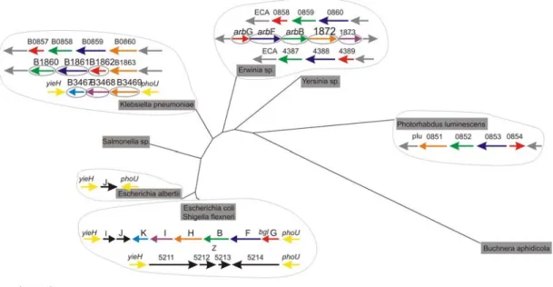
37 Results These genes were retained in E. coli (including Shigella), Erwinia, and Klebsiella species and lost in other enterobacteriaceae members analyzed in this study. Furthermore, it was interesting to observe the structure of bgl orthologs in the other bacteria. In Klebsiella sp, Erwinia sp, bgl genes are organized in a similar fashion at least the first three genes of the operon bglGFB as in E. coli but in a different chromosomal location (Fig. 16). In Klebsiella, the orthologs of bglHIK are present in the same chromosomal position as in E. coli.
In contrast, no orthologs were obtained for Z5211-5214 locus, even when the search was extended to the entire non-redundant database (nr) at the NCBI.
Furthermore, the compositional analysis showed that the GC content of Z5211-5214 (31%) locus was significantly lower than the E. coli genome average (50.4%) (data not shown), which suggests that Z5211 to Z5214 are horizontally acquired genes.
Fig. 16: Structure of bgl locus genes of E. coli K12 MG1655 and its orthologs obtained from BLAST in Enterobactericiae displayed on a neighbor-joining tree of 16S rDNA sequences of the indicated members. Gene names are indicated on top and colors refer to different genes of bgl locus keeping E. coli as reference.