proprioceptive reflex in a walking insect
I n a u g u r a l - D i s s e r t a t i o n
zur
Erlangung des Doktorgrades
der Mathematisch-Naturwissenschaftlichen Fakultät der Universität zu Köln
vorgelegt von
Katja Hellekes
aus Olpe
Köln 2012
Prof. Dr. Peter Kloppenburg
Tag der mündlichen Prüfung: 19.06.2012
Abstract v
Zusammenfassung vii
1 Introduction 1
2 Materials and Methods 11
2.1 Animals . . . 11
2.2 Preparations and experimental design . . . 11
2.3 Electrophysiology . . . 17
2.4 Data recording and evaluation . . . 18
3 Results 21 3.1 Forward and backward walking . . . 21
3.1.1 Influence of fCO signals on tibial MN activity in the front leg . . . 22
3.1.2 Influence of fCO signals on tibial MN activity in the middle leg . 25 3.1.3 Influence of fCO signals on the tibial MN activity of the hind leg . 27 3.1.4 Influence of ThC joint position on the processing of fCO signals in tibial MNs of the hind leg . . . 29
3.1.5 Influence of ThC joint position and CS ablation on the processing of fCO signals in tibial MNs of the hind leg . . . 32
3.1.6 Summary1: Influence of walking direction . . . 35
3.1.7 Summary2: Influence of segment specificity . . . 35
3.1.8 Front and hind leg stance phase kinematics during forward and backward walking . . . 38
3.1.9 Influence of walking direction on interjoint reflex response to fCO stimulation . . . 40
. . Influence of fCO signals on the tibial MN activity during curve walking . . . 45 3.2.2 Activity offlexor tibiaeMNs during fCO stimulation in curve walking 47 3.2.3 Activity of extensor tibiae MNs during fCO stimulation in curve
walking . . . 50 3.2.4 Influence of curve walking on the processing of fCO-mediated sig-
nals in nonspiking interneurons . . . 53 3.2.5 Summary: Influence of curve walking on the NSIs . . . 70
4 Discussion 71
4.1 Movement related reflex reversal in stick insect walking . . . 72 4.2 Task-dependent modulation of proprioceptive reflexes . . . 74
4.2.1 Influence of fCO signals on the tibial MN activity during forward and backward walking . . . 74 4.2.2 Influence of fCO signals on the tibial MN activity during curve
walking . . . 78 4.3 The femur-tibia control network . . . 80 4.3.1 Tibial motoneurons generating the reflex reversal . . . 81 4.3.2 Nonspiking interneurons involved in the generation of reflex reversal 82 4.4 Influence of other leg sense organs . . . 89 4.5 Intersegmental and descending control of local processing of propriocep-
tive signals . . . 91 4.6 Functions of reflex reversals: reinforcement of movement in the control of
walking . . . 93
Bibliography 95
List of Figures 113
Abbreviations 115
Danksagung 116
Teilpublikationen 118
The generation of task-dependent and goal-directed walking behaviour requires feed- back from leg sense organs for regulating and adapting the ongoing motor activity.
Sensory feedback from movement and force sensors influences the magnitude and the timing of neural activity generated in the neural networks driving individual joints of a leg. In many animals, the effects of sensory feedback on the generated motor out- put change between posture maintenance and locomotion. These changes can occur as reflex reversals in which sensory information, that usually counteract perturbations in posture control, instead reinforce movements in walking. In stick insects, for example, flexion of the femur-tibia joint is measured by the femoral chordotonal organ, which mediates reinforcement of the stance phase motor output of the femur-tibia joint when the locomotor system is active. Flexion signals promote flexor and inhibit extensor mo- toneuron activity. However, the mechanisms underlying these changes are only partially understood.
Therefore, the purpose of the present thesis was to investigate whether the process- ing of movement and position signals of the FTi joint is task-specifically modified in the generation of adaptive leg movements, which is required when locomotion is adapted to changes in walking direction or in turning movements. To study the role of these task-dependent changes in walking behaviour on the processing of local sensory sig- nals, the generation of reflex reversals mediated by the femoral chordotonal organ in the femur-tibia joint of the stick insectCarausius morosuswas measured in a semi-intact walking preparation. In several experimental conditions either in front, in one or both middle or in hind legs, the femoral chordotonal organ was mechanically displaced and the motoneuronal responses in the flexor and extensor tibia were monitored, while the remaining legs performed either forward, backward or curve walking on a slippery sur- face.
haviour executed. While in forward walking flexion signals from the front leg fCO regu- larly elicit reflex reversal in the tibial motoneurons, this cannot be observed in backward walking. Similarly, during optomotor-induced curve walking, reflex reversal occurred reliably in the middle leg on the inside of the turn, however not in the contralateral leg on the outside of the turn. Thus, the experiments revealed that the nervous system modulates proprioceptive reflexes in individual legs during task-specific walking adap- tation. Furthermore, I showed that nonspiking interneurons, known to be involved in the premotor network of the FTi joint, participate in reflex responses in both the inner and outer middle leg during curve walking. First results show that the reflex response in some interneuron types is altered between the inner and outer leg, while no differences were found in others.
Zielgerichtete und verhaltensabhängige Fortbewegung setzt die Anpassung rhythmisch alternierender motoneuronaler Aktivität mittels sensorischer Rückkopplung durch Pro- priozeptoren und weitere Sinnesorgane voraus. Diese Signale von Bewegungs- und Belastungssensoren beeinflussen die Stärke und zeitliche Abstimmung der, von rhyth- musgenerierenden Netzwerken erzeugten, neuronalen Aktivität. In vielen Tieren än- dern sich die Effekte der sensorischen Rückkopplung in Abhängigkeit vom Verhalt- enszustand. Diese Änderungen können als Reflexumkehrungen auftreten. Reflexe die zur Aufrechterhaltung der Positur dem sensorischen Eingang entgegenwirken, wirken im Falle der aktiven Bewegung, bei gleichem sensorischen Eingangssignal verstärkend auf den Bewegungsablauf. Dieser Mechanismus der Reflexumkehr tritt zum Beispiel im Femur-Tibia Gelenk der Stabheuschrecke auf. Während einer aktiven Beugung des Gelenks werden mittels eines propriozeptiven Sinnesorgans, dem femoralen Chordoto- nalorgan, Positions- und Bewegungssignale des Femur-Tibia Gelenks gemessen. Diese Beugungssignale führen dann, also bei aktiver Beugung, zur Verstärkung der Beugung und verhindern gleichzeitig die Streckung des Gelenks. Die Mechanismen die diesen Änderungen der Reflexantwort in der verhaltensabhängigen senso-motorischen Verar- beitung unterliegen, werden bisher nur teilweise verstanden.
In dieser Arbeit soll untersucht werden, ob und wie weit die verhaltensabhängige Ver- arbeitung sensorischer Bewegungs- und Positionssignale des femoralen Chordotonalor- gans, im speziellen beim Vorwärts-, Rückwärts- und Kurvenlaufen, moduliert wird.
Dazu wurde die Auftretenswahrscheinlichkeit der Reflexumkehr im Femur-Tibia Ge- lenk der Stabheuschrecke Carausius morosusin semi-intakten Präparationen untersucht.
In verschiedenen experimentellen Ansätzen wurde das femorale Chordotonalorgan im Vorderbein, in einem oder beiden Mittelbeinen oder im Hinterbein mechanisch stim- uliert und gleichzeitig die motoneuronale Aktivität des Femur-Tibia Gelenks gemessen, während die übrigen Beine auf einer rutschigen Oberfläche vorwärts, rückwärts oder
Es konnte gezeigt werden, dass es verhaltensabhängige Unterschiede in der Auftretens- wahrscheinlichkeit der Reflexumkehr gibt. Während des Vorwärtslaufens im Vorderbein wurde die Reflexumkehr regelmäßig ausgelöst, im Hinterbein hingegen nicht. Ähn- liches konnte während des optomotor-induzierten Kurvenlaufens gezeigt werden. Im Mittelbein, welches sich auf der Innenseite der Kurve befand, trat die Reflexumkehr sig- nifikant häufiger als im kontralateralen Außenbein auf. Somit konnte im laufenden Tier eine Modifikation der Reflexverarbeitung während zielgerichteter, verhaltensabhängiger Lokomotion nachgewiesen werden. Außerdem konnten erste Charakterisierungen Nicht- Spikender-Interneurone, welche sowohl an der Reflexantwort im ruhenden Tier, als auch im aktiven Tier beteiligt sind, durchgeführt werden. Es wurden Hinweise darauf gefun- den, dass in einigen Interneuronentypen die Reflexantwort, im Innenbein und Außen- bein unterschiedlich ist und in anderen gleich ausgeführt wird.
Task-dependent, active locomotion is of decisive importance for the survival of all ani- mal species. In the course of evolution different locomotor strategies and also various locomotion systems have developed along with the diversity of species and their habi- tats. A wide range of terrestrial invertebrates and vertebrates have evolved highly adap- tive walking gaits. Therefore, different numbers of limbs, ranging from two in humans up to750in myriapoda have to be adapted to different walking terrains, body postures and behavioural situations.
Rhythm generating networks, networks which mediate alternating leg muscle coordi- nation, and networks for inter-limb coordination, which are modified by sensory and neuromodulatory influences, underlie the generation of walking. Since the beginning of the 20th century, major advances have been made in the understanding of locomotion and the underlying processes that establish rhythmic motor patterns. Sir Charles Sher- rington suggested that reflexes, in particular flexion and extension reflexes in spinalised quadrupeds, are integrated in the generation and control of movement, while proprio- ceptive sensory signals mediate phase transitions (Sherrington,1910,1913). At the same time, the first suggestion of an intrinsic pattern-generating mechanism, termedhalf cen- ter, arose from experiments, in which alternating muscle activities were still generated in the absence of sensory information (Brown, 1911, 1914). The idea of these func- tional networks was additionally underpinned by experiments, in which locomotor-like activity seen as alternating flexor and extensor activity in spinal cats was elicited by in- travenous injection of L-dopa (L-3,4-dihydroxyphenylalanine) (Jankowska et al.,1967).
The generation of rhythmic motor patterns was extensively studied in a variety of motor systems in vertebrates and invertebrates leading to the commonly accepted concept of central pattern generators (CPGs), neuronal networks which generate motor rhythms in the absence of descending inputs from higher centers and sensory feedback (for reviews see Bässler,1986c; Delcomyn,1980; Grillner,1975,1981,1985; Grillner and Wallen,1985;
Grillner and Zangger, 1979; Pearson et al.,1993; Selverston and Moulins,1985).
Subsequently, motor patterns were found to execute different tasks with the same lo- comotor appendages, like walking, airstepping, scratching, and paw shake in the cat (Giuliani and Smith, 1985; Koshland and Smith, 1989; Pratt and Loeb, 1991). In chick- ens, the same interneuronal circuits were found to establish different behavioural tasks, like walking, scratching, and posture control (Berkinblit et al., 1978; Gelfand et al., 1988). Furthermore, in turtles (Berkowitz, 2002, 2005), similarities between scratching and swimming movements were found. Several studies in invertebrates reported a mul- titude of movements performed with the same consistent motor structures (stick insect:
searching, rocking, walking, grooming(Bässler and Wegner,1983); locust: jumping and kick- ing(Burrows,1995; Gynther and Pearson,1989; Hedwig and Burrows,1996; Heitler and Burrows, 1977a,b); cricket: flight and stridulation (Hennig,1990)). For example, Pflüger and Burrows (1978) demonstrated that the same motoneurons are involved in the move- ment generation of kicking, jumping and swimming in the locust. Interestingly, the three different motor outputs were generated similarly. The movements started with flexion of the FTi joint, followed by a co-contraction of flexor and extensor muscle and, finally, a rapid extension of the tibia. However, a major problem with the half-center organisation is that mixed-muscle synergies were found that are characterised by, at least, a partial co-activation of antagonistic muscles, which is not in conformity with the half-center model. Therefore, a more flexible modular concept was proposed, in which distinct behaviours were executed by units of a small number of interneurons or groups of functionally-related interneurons (Bässler and Büschges, 1998; Grillner, 1981; Stein and Smith, 1997). However, it is only partially understood which modules exist and what they are composed of.
Subsequently, researchers investigating such basic motor patterns found several mecha- nisms involved in the tuning motor patterns. First, afferent signals from the periphery are involved in the control of movement and posture, for example in chicks (Bekoff et al., 1989,1987), in the cat (Grillner and Rossignol,1978), in stick insects (Bässler,1986a,1988)
and co-workers ( ) in which removing of sensory feedback from the legs results in a difference of the motor output of walking and hatching. In particular, the intralimb movements became more similar. Second, descending control signals from the brain, which is becoming an increasingly important research area, for initiation, maintenance and modulation of locomotion was studied extensively in vertebrates (human: Capaday et al.,1999; Gerloff et al., 1998; Petersen et al.,1998,2001; Schubert et al.,1997;monkey:
Eidelberg et al.,1981; Fetz and Cheney, 1980; Kobayashi and Isa,2002; cat: Armstrong, 1986; Beloozerova and Sirota, 1993; Friel et al., 2007; Grillner, 1975; Lajoie and Drew, 2007; Shik and Orlovsky,1976;mouse: Hagglund et al., 2010; lamprey: Shaw et al.,2010; Smetana et al.,2010) and invertebrates (drosophila: Strausfeld,1999; Strauss,2002; Strauss and Heisenberg,1993; cockroach: Bender et al., 2010; Mu and Ritzmann, 2008a; Ridgel et al., 2007; Ritzmann et al., 2005; Schäfer and Ritzmann, 2001). For example, in cats, the medullary reticular formation is known to generate responses in limb extensors and flexors that are modulated during locomotion (Drew,1991). In addition, recent studies in cats suggest that the posterior parietal cortex is involved in the fine-tuning of visually guided locomotion (Lajoie and Drew,2007). Studies in insects further support the role of descending signals in the contol of locomotion. For example, in cockroaches, the central body complex (CBC) and the surrounding regions affect the control of turning (Ridgel et al.,2007). In this study, the researchers showed that lesions of the CBC or in regions immediately surrounding the CBC, results more likely in an abnormal turning behavior than lesions in other brain regions. Finally, neuromodulators play an important role in the shaping of rhythmic motor output (pyloric rhythm: Hooper and Marder,1987; feed- ing: Kupfermann and Weiss,1982; swimming: Sillar et al.,1998; locomotion: Brownstone et al.,1992; Wallen and Grillner,1987; Zagoraiou et al.,2009). In neonatal rats and mice, recent studies using spinal cord preparations, pharmacological, and genetic approaches identified a variety of neurotransmitters and neuromodulators and, accordingly, several types of receptors involved in the control of locomotion. In a recent study, Zagoraiou and co-workers (2009) found evidence that cholinergic premotor interneurons are a de- fined class of intrinsic neuromodulatory neurons, which modulate the mouse locomotor
activity. So far, however, these different sources of modulatory influences on the lo- comotor output that are known still fail to explain the generation of the tremendous flexibility in locomotor behaviour sufficiently.
The variability of the environment and the challenges animals have to overcome (such as foraging, avoidance of predators, and reproduction) requires flexibility and results in adaptive and goal-directed motor outputs that are modifiable with regard to walking speed, direction, and turning. Therefore, the output of pattern-generating networks, including the CPGs, has to be modified in the generation of different motor behaviours (reviewed in Büschges,2005; Grillner,1975; Marder and Bucher,2001; Marder and Cal- abrese, 1996; McCrea and Rybak,2008; Orlovsky et al., 1999; Pearson et al.,1993; Pear- son, 1995a,2004; Rossignol et al., 2006; Zehr and Duysens,2004). In several studies on stepping in human infants, where descending supraspinal control is still in ongoing de- velopment, the flexibility of the locomotor system was studied. For example, in infants walking in various directions, mechanical disturbances elicited specific reflex responses and a modulation in their interlimb coordination (Lamb and Yang2000; Pang and Yang, 2000, 2002; Pang and others 2003). Researchers have shown an increased interest in understanding the generation of task-dependent motor behaviour, such as forward and backward locomotion (human: Choi and Bastian,2007; Pang and Yang,2002;cat: Buford and Smith,1990; lamprey: Islam et al.,2006; salamander: Ashley-Ross and Lauder,1997; crayfish: Ayers and Davis,1977), turning (stick insect: Dürr,2005; Dürr and Ebeling,2005; Gruhn et al.,2009;cockroach: Mu and Ritzmann,2005;drosophila: Bender and Dickinson, 2006), gap-crossing (stick insect: Bläsing and Cruse,2004a,b;drosophila: Pick and Strauss, 2005) and obstacle climbing (cockroach: Watson and Ritzmann,2002; Watson et al.,2002).
For example, in lampreys, it is known that during forward swimming, periodic waves of lateral body flexion propagate from head to tail. In a recent study, by Islam and co-workers (2006) it was demonstrated that during backward swimming this wave is reversed, thus forming a wave in tail-to-head direction. Similarly, during curve walking, changes in leg kinematics, step length, directions of the legs, stepping frequencies and the interleg coordination were described (Dürr, 2005; Dürr and Ebeling, 2005; Jander,
focus of present neurophysiological research.
To date, several studies have provided first insights into the neuronal mechanisms un- derlying locomotor adaptation in vertebrates (Cheng et al., 1998; Gabriel et al., 2011; Gosgnach et al.,2006; Stein, 2005; Zagoraiou et al.,2009) and invertebrates (Akay et al., 2007; Bender et al., 2010; Briggman et al., 2005; Lockery and Kristan, 1990; Pick and Strauss, 2005; Ridgel et al., 2007; Ridgel and Ritzmann, 2005; Schäfer and Ritzmann, 2001). For example, in the control of locomotor speed, genetic and neurophysiological approaches show that V1inhibitory spinal interneurons are involved in the frequency regulation of central pattern generated rhythm (Gosgnach et al.,2006). Also, in cock- roaches, brain structures were identified that are involved in the control of locomotor speed (Bender et al.,2010). In this study, it was shown that neural activity in the cen- tral complex is correlated with the walking frequency of the cockroach and, further, that electrical stimulation in the same area could generate and alter walking (Bender et al., 2010). Another example of locomotor adaptation is a recent study of decision- making processes in the leech by Briggman and co-workers (2005). In this study it was reported that activity patterns of a small number of neurons are correlated with the leech’s behavioural choice to swim or crawl. The authors successfully identified one single neuron that affected this choice by injection of a hyperpolarising or depolarising current (Briggman et al.,2005). However, there is still insufficient data to fully explain adaptive motor behaviour. One major issue in the research on locomotion concerns the role of sensory information. Changes in sensory feedback are an important component of locomotor adaptation (for review Pearson et al.,1993). Several studies have identified the role of sensory feedback, for example, in the transition from stance to swing phase in decerebrate and spinal cats. Sensory signals from Ib afferents of the Golgi tendon organ (GTO) in the ankle extensor muscle and afferent signals, measuring hip extension, me- diate the transition from stance to swing at the end of the stance phase (Conway et al., 1987; Duysens and Pearson,1980; Gossard et al.,1994; Hiebert et al.,1996; Pearson et al., 1992; Whelan et al., 1995; Whelan and Pearson,1997). Further evidence, for phasic sen-
sory signals being involved in the timing of phase transition is shown for insect walking (Büschges,2005) and flight (Pearson and Ramirez,1997).
In a variety of systems, the effects of sensory input during walking differ from those seen in postural control. Commonly, these changes occur as reflex reversals (Duysens et al., 2000). For example, during posture control, the GTOs of vertebrates generate reflexes (Prochazka, 1996). During walking, however, these receptors tend to amplify muscle tension in the stance phase (Pearson,1993). Similar reflex reversals are known in other receptors of vertebrates (human: Duysens et al.,1990;cat: Forssberg et al.,1975;rat:
Fouad and Pearson, 1997) as well as invertebrates (crayfish:DiCaprio and Clarac,1981; Skorupski and Sillar, 1986; locust: Burrows and Pflüger, 1988; Theophilidis and Burns, 1990; Zill,1985;stick insect: Bässler,1976,1986b,1988). In some instances, the generation of reflex reversals depends on the phase of activity in rhythmic movements (e.g. Fouad and Pearson,1997; Pearson and Collins,1993; Skorupski and Sillar,1986). For example, in crayfish, the thoracocoxal muscle receptor organ mediates reflexes that are known to activate promotor MNs when active that are, however, inhibitory when the remotor MNs are active (Skorupski and Sillar, 1986). More frequently, however, the generation of reflex reversals depends on the behavioural state of the animal (e.g. Bässler, 1988; Zill,1985, for review see e.g. Büschges and El Manira,1998; Clarac et al.,2000; Pearson, 1993). It is further known that locomotion patterns are extensively modified with re- gard to direction of progression and during visually guided stepping (Pang and Yang, 2002). So far, there is no satisfactory explanation how such changes affect the occurrence of reflex reversals, although their regulation must be part of the adaptation of walking patterns (Pang and Yang,2002; Pearson et al.,1993).
The neural mechanisms underlying reflex reversal and the flexibility of adaptive loco- motion have been extensively studied in the stick insect’s walking system (for review see Bässler, 1983b; Bässler and Büschges, 1998; Büschges,2005, 2012; Büschges and Gruhn, 2008). First studies of the walking pattern and, particularly, the generation of rhythmic leg movements in the stick insect date from the early 20th century (von Buddenbrock,
ments and the underlying neuronal network architecture in stick insects is available.
The insect leg consists of five main segments: the coxa, the trochantero-femur (which is fused inCarausius morosus), the tibia, and the segmented tarsus. Leg movements are mainly controlled by muscles of the thorax-coxa (ThC) joint, the coxa-trochanter (CTr) joint and the femur-tibia (FTi) joint. The muscles of the ThC joint move the leg forwards by theprotractor coxae muscle and backwards by the retractor coxae muscle. The levator anddepressor trochanteris muscles elevate and depress the leg in the CTr joint and the flexion and extension of the FTi joint is mediated by theflexorandextensor tibiaemuscles (see Graham and Epstein, 1985. These antagonistic muscle pairs are alternately-active during the generation of a step, which can be divided into a stance and a swing phase.
The transition between stance and swing phase is controlled by signals from a variety of leg sense organs, like hair fields, the campaniform sensilla and the femoral chordotonal organ (for review see Büschges,2005,2012).
Movements of the leg segments are measured by hair plates (fields) on the leg joints, particularly of the CTr joint. These hair fields consist of groups of hair sensilla that measure position and movement of the joint. The ventral coxal hair plate (vcxHP) com- prises two groups of hair cells, group G1 and G2, which are located ventrally on the coxa and detect position and movement of the ThC joint (Büschges and Schmitz,1991; Cruse,1985b; Dean and Schmitz,1992). The trochanteral hair plate (trHP) is situated on the dorsal side of the trochanter and measures the CTr joint position (Schmitz,1986b).
In addition, the rhombic hair plate (rHP) on the ventral trochanter was described by Tartar (1976) and Schmitz (1986a,b). Furthermore, the levator receptor organ, an inter- nal sense organ, located inside the coxa parallel to thelevator trochanterismuscle, detects movements of the trochanter (Schmitz and Schöwerling, 1992). It measures the length change of thelevator trochanteris muscle and acts similar to strand receptors in the lo- cust (Bräunig and Hustert,1985a,b). Also, the campaniform sensilla (CS) are important leg sensors, which signal load information of the leg (Delcomyn, 1991; Hofmann and Bässler,1982) and cuticular stress (Hofmann and Bässler,1982; Pringle,1938). To date,
there are four CS groups known on the proximal leg joints. One is located on the proxi- mal femur (fCS- femoral campaniform sensilla) and three are located on the trochanter (trCS- trochanteral campaniform sensilla). Akay and co-workers (2001) found that sen- sory signals of the fCS influence the activity of FTi joint motoneurons. Several studies revealed that signals of the fCS and trCS modifies the motor output of the CTr and ThC joint (Akay et al., 2004, 2007; Schmitz, 1993). Recently, Zill et al. (2011) identified two groups of campaniform sensilla, group6A and6B located distally from the FTi joint.
In addition to these groups of exteroceptors, a further sensory proprioceptor, the femoral chordotonal organ (fCO), is important for the control of leg movement and position.
The fCO is located dorsally in the proximal part of the femur (Bässler, 1972; Bässler and Büschges,1998; Field and Matheson,1998; Kittmann and Schmitz,1992). It extends through the femur with a thin receptor tendon and its distal end attaches to the tibia. It consists of two parts, of which the dorsal part contains more than400sensory cells and the ventral part more than80(Füller and Ernst,1973). Füller and Ernst (1973) described the fine structure of the fCO and were able to show that every scolopidium comprises two sensory cells, one scolopale cell, a fiber cell, and at least one sheath cell. The sensory cells of the dorsal part of the fCO measure and control position, velocity, and accelera- tion of the FTi joint and combinations of these signals (Büschges, 1994a; Hofmann and Koch, 1985; Hofmann et al., 1985). In contrast, the fCO cells of the ventral part are not involved in the adjustment of the FTi joint (Field and Pflüger, 1989; Kittmann and Schmitz,1992). The influence of sensory signals from the fCO has been studied under various experimental conditions and is known to produce resistance reflexes in resting animals that function in postural compensation (Bässler et al., 1974; for summary see Bässler,1993). These reflexes have been shown to change when the animal generates ac- tive leg movements (Bässler,1976,1988). In such cases, afferents of the fCO that signal joint flexion also inhibit extensor firing and assist the generation of flexor activity. These changes represent a reflex reversal and occur as the first part of a sequence of muscle activities termed the active reaction (AR; Bässler, 1988). Together with inter-joint influ- ences of the fCO on motoneurons of the tarsus, the active reaction appears to assist the
also found that inter-joint reflexes were mediated by the fCO. It has been shown that sensory signals from the fCO in the inactive and active, but stationary, animal influence muscle activity of the adjacent CTr joint (Hess and Büschges,1997,1999).
Several studies have examined the underlying neural elements within the local pre- motor network that mediate both the resistance and assistance reflexes of the fCO in a distributed fashion (for review see Bässler and Büschges,1998; Büschges and El Manira, 1998; Büschges and Gruhn, 2008). However, no information is currently available on how the reflex reversal is initiated and regulatedin vivo during walking and, particu- larly, while performing task-dependent walking, such as curve or forward and backward walking when leg movement kinematics are specifically modified to the movement task to be executed. Several studies have demonstrated effects of descending input upon local reflexes in insects (Knop et al.,2001; Mu and Ritzmann,2008a; Ridgel et al.,2007).
However, these experiments were not performed in animals that were actually walking.
In the present thesis, I will present evidence for the task-specific modulation of a propri- oceptive reflex in walking stick insects as the result of neuronal adaptation processes to specific walking tasks. The results section of my thesis is divided into two main parts.
The first part addresses the modification of reflexes mediated by the fCO during for- ward and backward walking. The second part focuses on the adaptation during curve walking.
The first part of the results addresses the following questions:
1. Is there any difference in the segmental processing of fCO information during forward and backward walking, in particular between the front and hind leg?
2. How does sensory load and position information influence the adaptation pro- cesses in the FTi joint in forward and backward walking?
3. What are the kinematic differences in the front and hind legs of forward and backward walking stick insects?
4. Are the inter-joint reflexes from the fCO on the CTr joint affected by a change in walking direction (i.e., forward and backward walking)?
The second part of the results addressed two questions:
1. Is there any difference in the task-dependent processing of fCO information during curve walking?
2. How are these signals encoded in the premotor network of the FTi joint, in particu- lar, in the responses of the membrane potential of nonspiking interneurons caused by fCO stimulation during curve walking?
2.1 Animals
Experiments were performed on adult female stick insects of the speciesCarausius moro- sus, Brunner (Phasmatodea) at room temperature (20−22◦C) under reduced light con- ditions. Animals were obtained from a breeding colony maintained by the University of Cologne, Germany. The colony was kept under constant conditions at temperatures between 20◦C and 25◦C, high humidity (55−70%) and under an artificial 12 : 12 hours light/dark cycle. Animals were fed with blackberry leaves(Rubus fructiosus). The exper- imental procedures reported in the study comply with the German national and state regulations for animal welfare and animal experiments.
2.2 Preparations and experimental design
Positioning of the experimental animal for walking and sensory stimulation
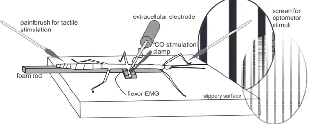
All experiments were performed on an air table (MICRO-g, TMC, Peabody, MA, USA) surrounded by a Faraday cage. Experimental animals were positioned above a plate (acrylic glass) at a height of about 8 - 12 mm to establish resting angles of the femur-tibia joints (FTi) in the middle and hind legs of roughly 90◦ (Fig.2.1, Epstein and Graham, 1983; Graham and Wendler, 1981; Gruhn et al., 2006, 2011). To ensure free stepping movements of the tethered stick insect and to reduce the mechanical coupling between legs via ground contact, the surface of the plate was made slippery by covering it with a glycerine/water mix (95%/ 5%). Animals were fixated dorsal-side-up on a foam-covered metal rod with dental cement (ProTempII,3M ESPE, Seefeld, Germany) applied to the meso- and metathorax (Fig.2.1). The leg that was to be investigated was glued to an extension of the rod. Coxa and femur of this leg were immobilized and the tibia pro- truded over the edge of the rod extension with a fixed FTi joint angle of approximately 110◦. Depending on the experimental setup, either the front, middle or hind leg was
slippery surface foam rod
flexor EMG
fCO stimulation clamp
paintbrush for tactile stimulation
extracellular electrode screen for
optomotor stimuli
Figure2.1:Preparation for studying reex reversals in stick insect walking. The stick insect is mounted on a foam-covered metal rod above a slippery surface. Mechanical stimuli that mimic femorotibial (FTi) joint exion are applied to the femoral chordotonal organ (fCO) of either the front, middle (shown here) or hind leg. Reex responses to fCO stimuli are monitored in recordings of the femoral nerve 2 (F2), which innervates the tibial extensor (tib ext) and in an electromyogram (EMG) of the tibial exor muscle. Forward or backward walking was induced by a mechanical stimulation on the antennae or the abdomen. Turning movements were induced by the display of optomotor stimuli. (Taken from Hellekes et al., 2012).
immobilised. When immobilised, the front leg was fixed at a position of 45◦ anterior with respect to the body axis, middle legs at 90◦ (Fig.2.1) and the hind leg at 45◦ either anterior or posterior. At the same time, all other legs were free to move. In some exper- iments, the trochanteral and the femoral campaniform sensilla (trCS, fCS) were ablated by pushing an insect pin through the cuticle at the location of the CS (Schmitz,1993). For better clarity, the different experimental conditions are displayed as simplified sketches in the results section, indicating the walking direction as well as the leg on which the fCO stimulation and the nerve recordings were performed.
Positioning of the experimental animal for walking and kinematic monitoring
In a further experimental study to understand the kinematics of the front and hind leg during forward and backward walking the animal was also fixated dorsal-side-up onto a foam-covered metal rod by means of dental cement and positioned above a slippery surface. Here, all legs were free to move (Fig.2.2). The animal’s body axis and the inspected leg were marked with dots of fluorescent dye. For this purpose, fluorescent pigments (Dr. Kremer Farbmühle, Aichstetten, Germany) were mixed with dental ce-
Figure 2.2: Preparation for studying stance phase kinematics of front and hind legs in stick insect forward and backward walking. The stick insect is mounted on a foam-covered metal rod above a slippery surface. Femur and tibia are marked with uorescent pigments for leg tracking. The exor and extensor tibiae muscle activity and the tarsal contact were measured. Forward or backward walking was induced by optomotor stimuli or mechanical stimulation on the antennae or the abdomen. (Taken, with permission, from Gruhn et al., 2011)
.
ment and applied on the distal tibia, the distal femur, the head, and the pro-, meso- and metathorax. An externally triggered high-speed video camera (Marlin F-033C, Al- lied Vision Technologies, Stadtroda, Germany) recorded the walking animal at 100 fps (frames per second) from above. For an additional sideward view, mirrors were placed in a 45◦ position, either in front of the front leg or behind the hind leg, depending on the observed leg. The fluorescent markers were illuminated with arrays of blue LEDs (30 - 50 V DC, luminance 24 cd, Electronics Workshop, Zoological Institute, University of Cologne). The experiments were performed under low-light conditions. A yellow filter in front of the camera lens filtered out short wavelengths to ensure a higher contrast for the video recordings. Furthermore, to monitor leg ground contact, a tarsal contact signal was recorded. For this purpose, a current flow was measured between the tarsus and the slippery surface plate. A small voltage (2.4 mV) was applied to the slippery surface by a pulse generator (Model MS501, Electronics Workshop, Zoological Institute, University of Cologne). An isolated copper wire (47µm diameter) was attached to the tibia and connected with a differential amplifier via an alligator clamp. The copper in-
sulation was stripped and electrode cream (Marquette Hellige, Freiburg, Germany) was used on the contact points to allow for proper current conduction. When the tarsus touched the plate the electric circuit was closed and the current flow indicated stance phase. In contrast, when the leg did not have any ground contact the circuit was opened and no current flow was measured indicating swing phase.
Induction of stepping in different directions
Forward walking was induced by tactile stimulation of the animal’s abdomen with a small paint brush (Fig.2.1). Backward walking was elicited by tactile stimulation of the head or by pulling manually on the antennae (Graham, 1985). Once stepping was initiated, tactile stimulation was stopped. An animal was considered to be walking forward or backward when the unrestrained legs showed forward or backward stepping movement. In many of the backward walking experiments, walking activity was also monitored by myographic recordings of the levator trochanteris and the retractor coxae muscles of the middle leg to distinguish the walking direction by the different phase relationships in forward and backward walking (Graham, 1985). Curve walking was induced by optomotor stimulation. To this end, a vertical black-and-white stripe pattern was projected on two screens, placed laterally in front of the stick insect (Scharstein, 1989). To elicit curve walking in the stick insects, the stripes on both screens moved either to the right or to the left (Fig.2.1, 2.2, Gruhn et al., 2011). Corresponding to the location of the leg, where the fCO stimulation was performed, and relative to the turning direction, the extremities are denoted as inner legs and outer legs throughout this thesis). In the kinematic studies, optomotor stimulation was also used to elicit forward walking, by progressive forward-directed stripe patterns.
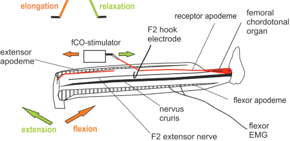
Preparation for fCO stimulation
A small opening was cut dorsally into the femoral cuticle, allowing for mechanical stim- ulation of the fCO and for extracellular recordings from tibial muscles and motor nerves (Fig.2.3, Büschges, 1989). The leg cavity was filled with saline (Weidler and Diecke, 1969), the apodeme of the fCO was cut and the distal ending attached to a moveable
flexor apodeme receptor apodeme
fCO-stimulator elongation
flexion
relaxation
extension
femoral chordotonal organ F2 hook
electrode extensor
apodeme
nervus cruris
F2 extensor nerve
flexor EMG
Figure2.3:Leg anatomy and preparation for fCO stimulation, nerve and muscle recordings. The leg was opened dorsally and the receptor apodeme of the fCO was attached to a moveable clamp controlled by a linear motor. Mechanical displacements of the apodeme, like an elongation (red arrow) corresponds to upward ramp movement (red) and simulates a leg exion (red arrow). Relaxation of the receptor apodeme (green arrow) corresponds to downward ramp stimuli (green) and mimics a leg extension (green arrow). Simultaneously, the extensor activity was measured by extracellular recording of the F2 extensor nerve with a hook electrode and exor tibiae muscle activity by a exor muscle electromyogram (EMG).
clamp controlled by a linear motor. Mechanical displacement of the apodeme parallel to the leg towards or away from the body were produced by applying voltages to the motor by a stimulus generator (Electronics Workshop, Zoological Institute, University of Cologne). The fCO was stimulated with ramp-and-hold stimuli which produced dis- placements of 300 - 400µm (sometimes also higher displacements of up to 670µm) from starting position. These displacements correspond to FTi joint angles (inner angle) from 110◦- 60◦or rather from 110◦- 80◦ (Weiland et al.,1986).
Preparation for extracellular and intracellular recordings
For experiments investigating the influence of walking direction on the inter-joint pro- cessing of fCO signals in the ThC joint, the coxa nerve branch1(C1), which innervates the levator trochanteris, was recorded extracellularly in the mesothorax. In additional experiments to investigate the processing of fCO signals during curve walking, intra- cellular recordings of different neurons in the mesothoracic ganglion were performed.
For both experiments, the animal was prepared as previously described. Additionally, the thorax was opened dorsally by a midline incision to gain access to the ganglion or to the C1 nerve (Fig.2.4). The thorax cavity was filled with stick insect saline (Weidler
and Diecke, 1969). The gut was placed intact besides the animal, and fat and connec- tive tissue was removed. For the intracellular recordings, the ganglion was placed on a wax-coated holder (see also Büschges, 1989, 1990. In order to prevent movement of the ganglion, it was fixed with small cactus needles (Nopalea dejecta). The surrounding tissue was removed in the area of the intracellular recording. Afterwards, this region was treated for 60 s with a proteolytic enzyme (Pronase E, Merck, Darmstadt, Germany) to facilitate electrode penetration through the ganglion sheath. Finally, the enzyme was washed out by repeated rinsing with saline (Weidler and Diecke, 1969) and the thorax cavity was filled with saline.
slippery surface rod
flexor EMG intracellular electrode
fCO stimulation
extracellular electrode screen for
optomotor stimuli
nervus cruris
connective
FETi
Ci1 SETi
extensor nerve recording
Figure2.4:Preparation for studying fCO-mediated reex responses in the FTi joint premotor network.
Shown are the used recording devices and typical time courses of the acquired signals. Additionally to the preparation for extracellular analyses (shown in Fig. 2.1), the mesothoracic thorax was opened and intracellular recordings were obtained in the mesothoracic ganglion ipsilateral to the fCO stimulation.
Extracellular recordings were performed by using myographic recordings (electromyogram, EMG) of the exor muscle and extracellular extensor nerve recordings. In the extensor nerve recording, two excitatory motoneurons, the fast (FETi) and slow (SETi) extensor tibiae MNs and one inhibitory MN, the common inhibitor 1 (CI1) are identiable.
2.3 Electrophysiology
Extracellular Recordings
Electromyograms (EMGs)
The activity of the flexor tibiae muscles in the fixated leg was monitored via an elec- tromyogram (EMG; Fig.2.1, 2.3, 2.4, e.g. Gruhn et al., 2011; Rosenbaum et al., 2010).
The tips of two twisted copper wires (Elektrisola, Eckernhagen, Germany; 47µm outer diameter), insulated except for the tip, were inserted into small holes of the cuticle (transfixed by insect minuten pins) in the ventral femur. In some experiments, flexor muscle activities were measured in the proximal and in the distal part of the ventral femur. The wires were fixated with small drops of dental cement. A silver wire was inserted into the abdomen to obtain a reference (indifferential) signal. Wires for EMG electrodes of theretractor coxaewere placed inside the thorax anterior to the leg and for thelevator trochanterisEMGs dorsally on the anterior side of the coxa.
Extracellular nerve recordings
The activity of the extensor tibiae motoneurons was recorded extracellularly from the femoral branch2(F2). The nerve was placed on a hook electrode (modified after Schmitz et al., 1988) and insulated with silicone oil (Baysilone-Paste mittelviskos, Bayer AG, Leverkusen, Germany). In further experiments, the motoneuronal activity of thelevator trochanteriswas recorded extracellularly via hook electrodes in the thorax from the coxa branch1(C1).
Intracellular recordings
Intracellular recordings were obtained from neurons involved in the femur tibia con- trol loop. In the present thesis, only recordings from nonspiking interneurons (NSI), as well as flexor and extensor motoneurons (MNs) are shown. The recordings were col- lected with thin-walled borosilicate glass microelectrodes (GB100-TF8P, Science Prod- ucts, Hofheim, Germany) from the neuropilar aborisations in the mesothoracic hemi- ganglion, ipsilaterally to the fCO stimulation (Fig.2.4). The microelectrodes were pulled
on a P-1000 filament puller (Flaming/ Brown Micropipette Puller, Sutter Instruments, Novato, USA) and filled with a solution of 3 M potassium acetate and 0.1 M potassium chloride (3 M KAc / 100 mM KCl) and had electrode resistances between 15-25 MΩ.
Signals were amplified with an SEC-05 amplifier (NPI Electronics, Tamm, Germany) in bridge mode (switching frequency 12-25 kHz). The extensor tibiae MNs were identified by a one-to-one relationship of intracellularly recorded spikes compared with spikes in the extracellular F2-nerve recording. Flexor tibiae MNs were identified by a one-to- one relationship between the intracellular spikes and the flexor muscle potentials in the flexor EMG. Interneurons were identified as nonspiking interneurons if they were in accordance with the following six criteria (see also Burrows, 1981; Büschges, 1990; Hengstenberg,1977; Siegler,1985; Wilson,1981: no generation of spike by1) fCO stim- ulation,2) unspecific tactile stimulation,3) change of the behavioural state of the animal (Bässler,1988),4) after a long and large hyperpolarisation,5) during depolarisation de- spite an increase of the amplitude of the EPSPs, and 6) also during graded effects on the activity of postsynaptic MNs. Furthermore, the identification of the different NSIs was accomplished by their characteristic responses to fCO stimulation and either their excitatory or inhibitory effect on extensor MNs activity (see also Akay, 2002; Büschges, 1990; Stein and Sauer,1998). Recordings without any stable resting membrane potential were discarded.
2.4 Data recording and evaluation
Both the intra- and the extracellularly recorded signals were amplified 100-fold by a pre-amplifier (Electronics Workshop, Zoological Institute, University of Cologne). Sub- sequently, the extracellular recordings were amplified 10-fold and band-pass filtered (nerve recordings 300 Hz - 4.5 kHz / EMG recordings 30 Hz - 2 kHz) (4-Channel Am- plifier / Signal Conditioner ModelMA102, Electronics Workshop, Zoological Institute, University of Cologne). The voltage output of the fCO stimulator and the electrophys- iological signals were digitised using an A/D converter (MICRO 1401k II, CED, Cam- bridge, UK) and recorded with a sampling rate between 6.25 kHz and 12.5 kHz with SPIKE2(data acquisition and analysis software; version7.01; Cambridge Electronic De-
sign, Cambridge, UK) on a personal computer (operating system: Microsoft Windows 7). Video files were analysed using motion-tracking software (WINanalyze, Vers.1.9, Mikromak Service, Berlin, Germany).
Analysis of extracellular recordings
To analyse extracellular nerve recordings, peristimulus time histograms (PSTHs) were generated. For this purpose, spikes were counted from 1 s before stimulus onset (fCO elongation) to 2 - 3 s after stimulus onset over a certain number of stimulations. To normalise the data, the counted spikes in each bin were divided by the total number of stimulus events. To evaluate the flexor muscle activities, EMG recordings were rectified and smoothed (time constant 1 ms) and averages of the waveforms were generated. The averages include the EMG signals also from 1 s before onset of fCO elongation up to 2 - 3 s after onset of the fCO elongation. With regard to differences between the location of the EMG, as well as differences of recording quality, the flexor activity was normalised between forward and backward walking in each animal and the relative change in the muscle activity is given by the here defined arbitrary units (a.u.).
Analysis of intracellular recordings
To examine the reflex responses induced by fCO stimulation on the level of the involved motoneurons and the premotor network, overdraws of the neuron membrane poten- tial were created for all stimulations in the inner and outer leg during curve walking.
Furthermore, the changes in membrane potential during reflex reversals were analysed by waveform averages. For averaging of the MN membrane potential, spikes were re- moved from the intracellular recordings and replaced by a straight line (maximal5 ms before and after the peak of the spike).
Statistical analysis
To determine the reliability of the frequency of reflex reversal, 95% confidence inter- vals for the different experimental situations were defined (Hayes, 1988). If the con- fidence intervals of the mean values do not overlap, the differences between the pro-
cessing of fCO signals in the compared data are statistically significant. For further evaluation also between different experimental conditions, the differences in the fre- quency of reflex reversal were tested with (2×2) contingency tables, conducting either a one-sided or two-sided analysis depending on the test hypothesis. The Pearson’s chi-squared test was used, if the sample size was > 60 with the expectancy value
> 5, otherwise the Fisher’s test was used (Agresti, 1992; Sachs, 1972). P-values of all combinations between the frequency of reflex reversal in different legs during for- ward as well as during backward walking were determined. Furthermore, the p-values for the differences in reflex reversal frequencies between forward and backward walk- ing in the same leg and between the inner and outer leg during curve walking were calculated. The statistical significance is indicated as follows: (n.s.) not significant p>0.05;(∗)p ≤0.05;(∗∗)p≤0.01;(∗∗∗)p≤0.001. Evaluation of the data and plotting of the graphs were performed with Matlab R2011b, Origin Pro8.5G and Corel Draw X4. In the text and figures, N refers to the number of animals, andn refers to the sample size (steps or stimuli).
3.1 Forward and backward walking
Converging evidence from a variety of animals suggests that reinforcement of movement is one important mechanism by which sensory feedback contributes to the generation of the motor output for walking (for reviews see Büschges, 2005; Clarac et al., 2000).
For example, in stick insects, flexion of the femur-tibia (FTi) joint is measured by the femoral chordotonal organ (fCO). The fCO is known to reinforce stance phase motor output of the FTi joint when the locomotor system is active (Bässler, 1988). In active stick insects, reinforcement of flexor activity reflects the reflex reversal of a strong re- sistance reflex (RR). During this resistance reflex fCO elongation (indicating FTi joint flexion) excites extensor MNs and inhibits flexor MNs in resting stick insects (Bässler, 1983a). In active animals, the same elongation of the fCO reverses this reflex, seen as excitation in theflexor tibiaemotoneurons (MNs) and as inhibition in the extensor tibiae MNs. This reflex reversal represents the first part of the so-called active reaction (AR) (Bässler,1988). When the chordotonal organ signals a certain flexed-joint position, the extensor tibiaeMNs are strongly excited and theflexor tibiaeactivity decreases (part II of the AR) (Bässler,1976,1983b, 1986a, 1988). The transition between part I and part II of the active reaction is independent of velocity, yet position-dependent and assumed to contribute to the stance-swing transition during walking. Furthermore, it is known that load signals from the femoral campaniform sensilla (fCS) inhibit extensor tibiae MNs, activateflexor tibiaeMNs, and also increase the occurrence of the AR (Akay et al.,2001; Akay and Büschges, 2006). Akay and co-workers (2007) have recently shown that the influence of the CS signals on the thorax-coxa (ThC) joint is reversed in backward versus forward walking, and thereby assists the generation of stance phase muscles activity in both walking directions. With regard to the walking direction, it is also important to mention that the individual legs, in particular front and hind legs, show different leg kinematics during forward and backward walking. Cruse and Bartling (1995) have de-
scribed that during forward walking, the FTi joint angle in the front leg decreases during stance phase and increases during swing phase. In the hind leg, a functional reversal was observed. This supports the assumption that stance and swing phase in the front and hind legs are mediated by different activities of leg muscles. One major objective of the present study was to investigate, if the processing of movement-related fCO sig- nals in the individual legs differs during forward and backward walking. Furthermore, this study attempts to determine, if the processing of fCO signals is segment-specific.
Therefore, the experiments were conducted separately in the front, middle and hind leg.
Finally, a better understanding of the kinematics during forward and backward walking is anticipated, to correlate the sensory processing to the actual motor behaviour. Accord- ingly, the kinematics of front and hind leg stance phases was studied for both walking directions.
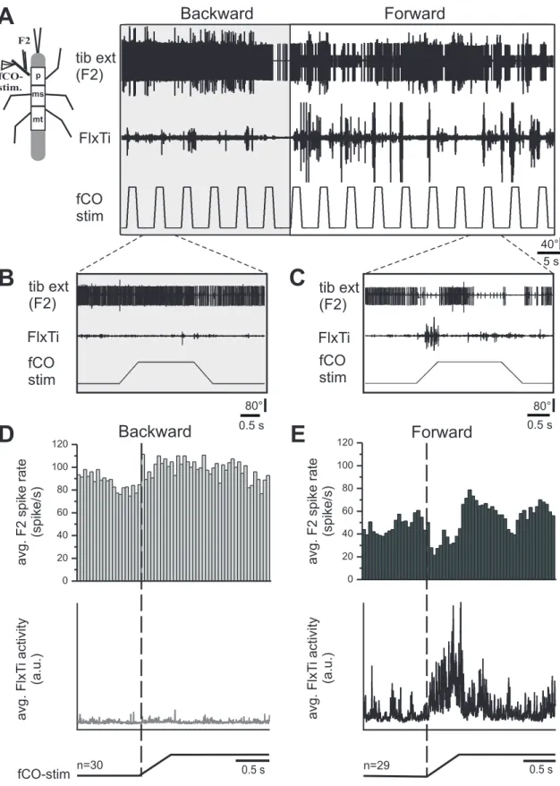
3.1.1 Influence of fCO signals on tibial MN activity in the front leg
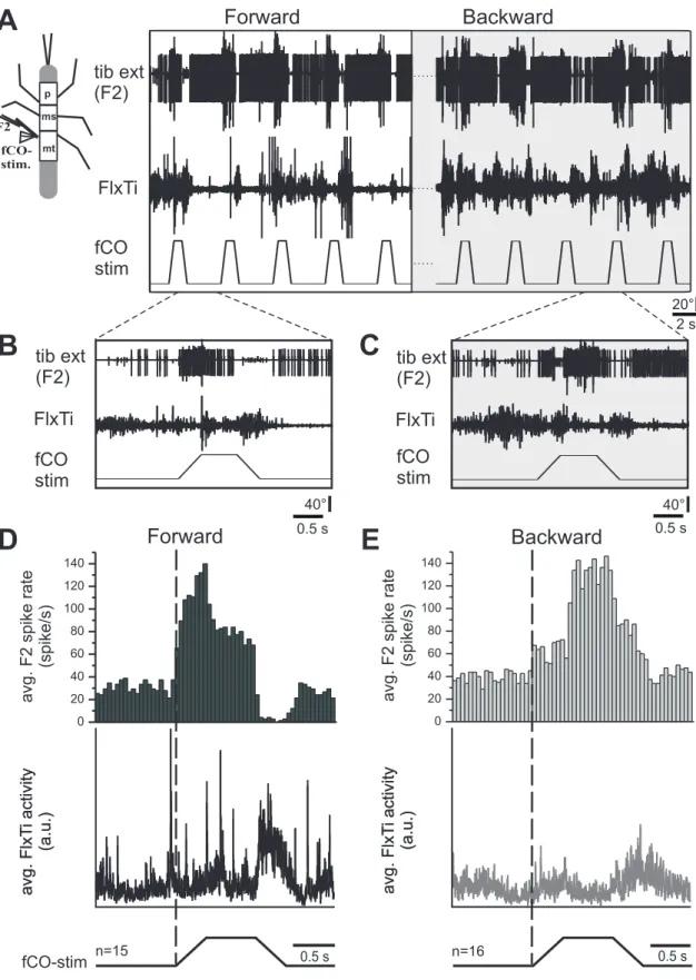
To test whether the generation of reflex reversal depends on the walking direction, an ex- perimental setup was used that allowed inducing animals to forward or backward walk- ing by applying mechanical stimuli to the abdomen or antennae, respectively (Rosen- baum, Wosnitza et al.,2010). In general, forward walking was elicited more readily than backward walking. Sometimes, a puff of breath or a slight touch on the abdomen was sufficient to elicit long-lasting forward walking periods. In contrast, in almost all cases of backward walking, a continuous stimulation of the antennae was necessary to maintain sustained stepping. In stick insects walking with five legs either forward or backward on the slippery surface, the front leg fCO was stimulated, while the mesothoracic tibial motoneuron activity was monitored (Fig.3.1). In general, backward walking was charac- terised by a higher ongoing activity in tibial extensor motoneurons compared to forward walking. At the same time, flexor tibiae activity was diminished (Fig.3.1 A, B). During forward walking, elongation of the prothoracic fCO elicited a reflex reversal: extensor motoneuron activity was terminated by imposed FTi joint flexion, while flexor motoneu- ron activity and common inhibitor 1 (CI1) activity was initiated (Fig.3.1A, C). On the contrary, reflex reversals were less often observed during backward walking (Fig.3.1A,
FlxTi tib ext (F2)
fCO stim
D
BackwardE
avg. F2 spike rate (spike/s)
F2
fCO- stim.
p ms
mt
A
FlxTi
fCO stim
fCO stim
B C
tib ext (F2)
Backward Forward
66
FlxTi tib ext (F2)
avg. F2 spike rate (spike/s)
5 s
0.5 s 80°
0.5 s 80°
Forward
0 20 40 60 80 100 120
0 20 40 60 80 100 120
avg. FlxTi activity (a.u.) avg. FlxTi activity (a.u.)
fCO-stim n=30 0.5 s n=29 0.5 s
11 33 66 77
88 99
40°
Figure3.1:(A) Inuence of fCO signals on the tibial MN activity in the front leg during forward and backward walking. The femoral chordotonal organ of the front leg was displaced (fCO stim) while monitoring the tibial extensor (tib ext (F2)) and exor muscle activities (Flx Ti) in that leg. In the shown sequence, the animal was walking backward and then forward. Reex reversals occurred during forward walking. B) and C) Expanded traces of responses during backward (B) and forward (C) walking.
D) and E) Peri-stimulus time histograms of the ring frequency of the tibial extensor motoneurons (top) and rectied waveform averages of the exor muscle activity (bottom) during fCO stimulation in the front leg during backward (D) and forward (E) walking (D: n=30; E: n=30, data from one animal).
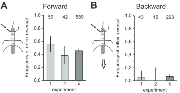
59 42 589
A Forward B Backward
1 2 x
0,0 0,2 0,4 0,6 0,8 1,0
experiment
Frequencyofreflexreversal
1 2 x
0,0 0,2 0,4 0,6 0,8 1,0
experiment Frequencyofreflexreversal 43 15 293
p m m
p m m
Figure 3.2:Reex reversals in the front leg depend on walking direction. Bar histograms show the frequency of reex reversals in the front leg's tibial muscles during displacement of the front leg fCO in animals that were walking forward (A) and backward (B). Each histogram shows two typical experiments (light bars) and the mean values of all experiments (dark bars; N=17). Reex reversals occurred during forward, however, not during backward walking. Arrows indicate walking direction; lines designate95%
condence intervals.
B). Stimuli applied to the fCO during backward walking did not show any reliable in- fluence on the extensor activity (Fig.3.1A, B). This difference in forward and backward walking becomes evident in the PST-histograms of the averaged F2 extensor nerve ac- tivity (Fig.3.1D, backward; Fig.3.1 E, forward). In backward walking stick insects the extensor firing frequency was, in general, enhanced and slightly increased caused by the fCO elongation (Fig.3.1 D, top). Simultaneously, the flexor activity remained low and no change in response to fCO stimulation was found (Fig.3.1D, bottom).
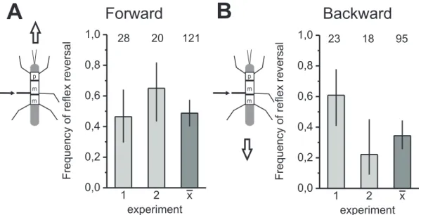
Additionally, to examine the differences in the processing of fCO signals in the front leg of forward and backward walking stick insects, the frequency of reflex reversals was quantified for both walking directions. Reflex reversals were generated in front legs of forward walking animals in 45.5% of trials (N=17, n=589; Fig.3.2A), compared to 6.7%
during backward walking (N=17; n=293; Fig.3.2B). In 16 of the 17 animals, a higher frequency of reflex reversals in forward walking than in backward walking was found (two typical experiments are displayed in Fig.3.2A, B). The frequency of occurrence of reflex reversals differed significantly between forward and backward walking in 10 of 17animals (Fisher’s exact test).
3.1.2 Influence of fCO signals on tibial MN activity in the middle leg
In order to investigate the segment specificity of the processing of fCO signals during forward and backward walking, in the following experiments the mesothoracic fCO was stimulated and the tibial motoneuronal activities were detected. In middle legs, the dif- ferent influences of forward and backward walking on the processing of fCO signals were unincisive. Both, in the forward walking and in the backward walking stick insect, reflex reversals were elicited (Fig.3.3 A, B, C). The enlarged view of one stimulus for each walking direction, highlights the reflex reversal during fCO elongation (Fig.3.3B, C). Although the inhibition in the F2 extensor nerve upon fCO elongation in this par- ticular animal becomes more obvious during forward walking (Fig.3.3 E, PSTH), the decrease in the averaged F2spike activity is also identifiable during backward walking (Fig.3.3D, PSTH). Moreover, during forward and backward walking, the averagedflexor tibiaemuscle activities increased during fCO elongation and, therefore, revealed the ac- tivity pattern of a reflex reversal (Fig.3.3D, E).
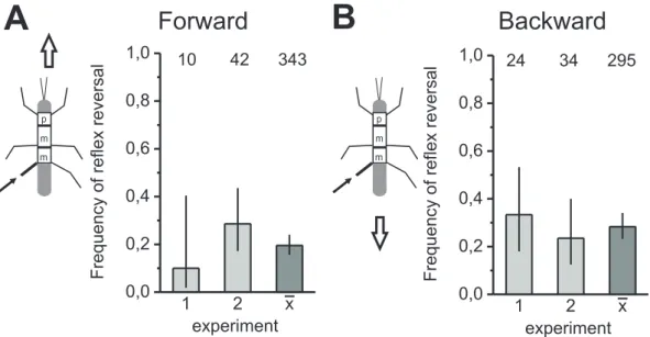
The frequency of reflex reversals in the bar histograms in exemplary animals reached a quite similar level in both walking directions (Fig.3.4 A, B). The mean frequency of reflex reversals in forward walking (48.7%) is approximately 10% higher than in back- ward walking (34.4%). However, the overlap of the 95% confidence intervals indicates that the observed numerical difference is, in fact, not statically significant. In total, only one of seven animals showed a significant difference in the frequency of reflex reversal between forward and backward walking. Five of seven animals showed a higher fre- quency of reflex reversals during forward walking and two during backward walking.
In summary, in the middle leg, only in rare cases an influence of walking direction on the probability of reflex reversals upon fCO stimulation was found.
0 20 40 60 80 100 120 140 160
0 20 40 60 80 100 120 140 160
D
BackwardE
avg. F2 spike rate (spike/s) avg. F2 spike rate (spike/s)
0.5 s 0.5 s
Forward
avg. FlxTi activity (a.u.) avg. FlxTi activity (a.u.)
fCO-stim n=22 n=14
0 20 40 60 80 100 120 140 160 F2
fCO- stim
A
FlxTi
fCO stim tib ext (F2)
Backward Forward
tib ext (F2)
5 s 40°
11
FlxTi tib ext (F2)
fCO stim
B C
fCO stim FlxTi tib ext (F2)
40° 40°
p ms
mt
0.5 s 0.5 s
Figure3.3:(A) Inuence of fCO signals on the tibial MN activity in the middle leg during forward and backward walking. The femoral chordotonal organ was displaced (fCO stim) in the middle leg while monitoring the tibial extensor (tib ext (F2)) and exor muscle activities (Flx Ti) in that leg. In this sequence, the animal was walking backward and then forward. Reex reversals occurred during forward and backward walking. B) and C) Expanded traces of responses during backward (B) and forward (C) walking. D) and E) Peri-stimulus time histograms of the ring frequency of the tibial extensor motoneurons (top) and rectied waveform averages of the exor muscle activity (bottom) during fCO stimulation in the middle leg during backward (D) and forward (E) walking (D: n=22; E: n=14, data from one animal).
1 2 x 0,0
0,2 0,4 0,6 0,8 1,0
experiment
Frequencyofreflexreversal
A Forward B Backward
1 2 x
0,0 0,2 0,4 0,6 0,8 1,0
experiment
Frequencyofreflexreversal 28 20 121 23 18 95
p m m p
m m
Figure3.4:Reex reversals in the middle leg do not depend on walking direction. Bar histograms show the frequency of reex reversals in the middle leg tibial muscles during displacement of the middle leg fCO in animals that were walking forward (A) and backward (B). Each histogram shows two exemplary experiments (light bars) and the mean values of all experiments (dark bars; N=7). Reex reversals occurred during forward and backward walking. Arrows indicate walking direction; lines designate95%
condence intervals.
3.1.3 Influence of fCO signals on the tibial MN activity of the hind leg
In order to determine the role of the processing of fCO signals in hind legs while the re- maining legs walked forward and backward, again, the activity of the F2extensor nerve and the flexor tibiae muscle activity were recorded and simultaneously ramp-and-hold stimuli were applied to the fCO. During forward, as well as during backward walking, elongation of the fCO could generate reflex reversals (Fig.3.5A, B, C). However, during forward walking, only in some cases fCO elongation elicited a reflex reversal (Fig.3.5 A). Mostly, however, the neuronal activity in the F2extensor nerve remained high and no inhibition during elongation was found. This is also shown in the PSTH of the av- eraged F2 extensor nerve activity, in which only in the backward walking condition a slight decrease of the activity, caused by the fCO elongation, becomes apparent (Fig.3.5 E). Nonetheless, averaged flexor muscle activity increased during fCO elongation in for- ward, as well as in backward walking (Fig.3.5D, E bottom).
Closer inspection of the frequency of reflex reversals during forward and backward walking revealed only a small difference in the metathoracic processing of fCO signals (Fig.3.6 A, B). During forward walking, the frequency of reflex reversals did not ex-
0 20 40 60 80 100 120 140 160 180
0 20 40 60 80 100 120 140 160
D
Forward 0.5 sE
180 Backward 0.5 sfCO-stim n=28 n=29
avg. F2 spike rate (spike/s)avg. FlxTi activity (a.u.) avg. F2 spike rate (spike/s)avg. FlxTi activity (a.u.)
F2 fCO- stim.
FlxTi tib ext (F2)
fCO stim
A
FlxTi
fCO stim
fCO stim
B C
tib ext (F2)
Forward Backward
FlxTi tib ext (F2)
2 s 30°
60° 60°
66
s
p ms
mt
0.5 s 0.5 s
Figure3.5:(A) Inuence of fCO signals on the tibial MN activity in the hind leg during forward and backward walking. The femoral chordotonal organ was displaced (fCO stim) in the hind leg while monitoring the tibial extensor (tib ext (F2)) and exor muscle activities (Flx Ti) in that leg. In this sequence, the animal was walking forward and then backward. Reex reversals occurred during forward and backward walking. B) and C) Expanded traces of responses during forward (B) and backward (C) walking. D) and E) Peri-stimulus time histograms of the ring frequency of the tibial extensor motoneurons (top) and rectied waveform averages of the exor muscle activity (bottom) during fCO stimulation in the hind leg during forward (D) and backward (E) walking (D: n=28; E:n=29, data from one animal).
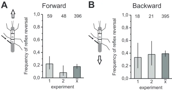
A Forward B Backward
10 42 343 24 34 295
1 2 x
0,0 0,2 0,4 0,6 0,8 1,0
experiment
Frequencyofreflexreversal
1 2 x
0,0 0,2 0,4 0,6 0,8 1,0
experiment
Frequencyofreflexreversal
p m m p
m m
Figure 3.6: Reex reversals in the hind leg depend on walking direction. Bar histograms show the frequency of reex reversals in the hind leg's tibial muscles during displacement of the hind leg fCO in animals that were walking forward (A) and backward (B). Each histogram shows two exemplary experiments (light bars) and the mean values of all experiments (dark bars; N=11). Reex reversals occurred during backward, however, only rarely in forward walking. Arrows indicate walking direction;
lines designate95%condence intervals.
ceed 20% (Fig.3.6A) and during backward walking, the frequency was 28% (Fig.3.6B).
Moreover, only in three of eleven experimental animals the frequency of reflex reversals differed significantly between forward and backward walking. In conclusion, no notice- able difference in the frequency of reflex reversals between the two walking directions was observed. In four of eleven experiments, the frequency of reflex reversal was higher in forward walking than in backward walking, whereas in six of eleven experiments the frequency was higher in backward walking. Two of these exemplary experiments are shown in Fig.3.6(A) for forward walking and in Fig.3.6(B) for backward walking.
Generally, in the hind leg, there was a slightly increased tendency for the generation of reflex reversals in backward walking, compared to the forward walking condition.
However, altogether, the differences are only marginal and not significant.
3.1.4 Influence of ThC joint position on the processing of fCO signals in tibial MNs of the hind leg
During forward and backward walking, a difference in the processing of fCO signals in the front leg was demonstrated; in contrast, in the hind leg, only a slight influence of walking direction was found. Furthermore, for both walking directions, the frequency of