during stick insect walking.
Inaugural - Dissertation zur
Erlangung des Doktorgrades
der Mathematisch-Naturwissenschaftlichen Fakultät der Universität zu Köln
vorgelegt von
Sandra Westmark
aus Emden
Köln Mai 2007
Prof. Dr. Peter Kloppenburg
Tag der mündlichen Prüfung:
28.06.2007
The basis of locomotion is the rhythmic activity of locomotor organs. During walking, rhythmic alternating bursts occur in leg motoneurons which result from the integration of signals from central pattern generators, sense organs and coordinating signals from neighboring segments. The rhythmic bursting pattern is shaped by rhythmic excitatory and inhibitory drive, as well as a long lasting (tonic) depolarizing modulation.
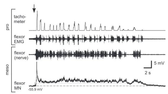
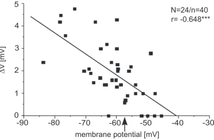
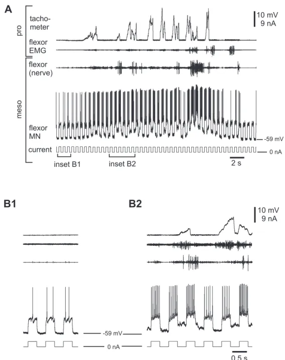
This dissertation investigates the mechanisms that underlie the tonic modulation of neu- ronal activity, which is the basis of the rhythmic activity. A single-legged preparation of the stick insect was used, that allows the analysis of neuronal activity in mesothoracic motoneurons during front leg stepping, without the influence of local sense organs. In- tracellular recordings of the membrane potential in flexor motoneurons ipsilateral and contralateral to the stepping front leg revealed a mean tonic membrane depolarization of 1.8 ±1.1 mV that could outlast the stepping sequence by several seconds. Furthermore, a phasic modulation of membrane potential occurred on top of the tonic depolarization, which was variably coupled to front leg steps. The tonic depolarization was associated with a decrease in input resistance, its amplitude depended on membrane potential and its mean reversal potential was found to be −41 mV. These properties of the tonic de- polarization indicate that it is based on a nonselective cation conductance or a mixed inward and outward current through different channels. Furthermore, the tonic depolari- zation increased the excitability of the membrane to depolarizing input and was found to have long repolarization time constants ( ¯τ=800 ms).
Pharmacological experiments were performed to identify the participating transmitter(s) and to test for a possible involvement of second messengers. Flexor motoneurons were recorded intracellularly while superfusing pharmacological agents, which were restricted to the mesothoracic ganglion. The muscarinic antagonist atropine decreased the tonic de- polarization amplitude, thus indicating a role for acetylcholine in mediating the tonic de- polarization via metabotropic receptors. Other transmitters/receptors might be involved too, as octopamine increased the tonic depolarization amplitude. This was supported by the reduction of the tonic depolarization amplitude by an octopaminergic antagonist (mi- anserin). Serotonin had an opposing effect on the tonic depolarization amplitude. It was shown that serotonin increased, but also decreased the tonic depolarization amplitude in different experiments.
Several second messenger pathways might be involved in mediating the tonic depolari- zation, one of which seems to include calcium in the flexor motoneurons. Furthermore, the increase of the tonic depolarization amplitude by 8-Br-cAMP suggests a role for cAMP in mediating the tonic depolarization. An involvement of an IP3/DAG pathway was indicated by the increase in tonic depolarization in the presence of neomycin and U-73122.
Additional experiments addressed the role of the brain (supraesophageal ganglion) in controlling neuronal activity in the mesothoracic ganglion induced by front leg stepping.
Especially the relevance of the brain for generating the tonic depolarization was tested in lesion experiments. Brain removal decreased the tonic depolarization amplitude, thus indicating a role for descending pathways in influencing the tonic depolarization. An- other preparation was used to investigate the neuronal activity in neck connectives dur- ing front leg stepping. The neuronal activity increased and was correlated to front leg stepping velocity. In brainless animals, however, the increased activity in neck connec- tives was diminished.
Fortbewegung beruht auf der rhythmischen Aktivität der Fortbewegungsorgane. Wäh- rend des Laufens treten rhythmisch alternierende Aktionspotentialsalven in Beinmo- toneuronen auf, die aus der Integration von Signalen zentraler Rhythmusgeneratoren, Sinnesorganen und koordinierenden Signalen aus benachbarten Segmenten resultieren.
Das rhythmische Muster von Aktionspotentialsalven wird durch rhythmisch erregende und hemmende Eingänge, sowie durch eine lang anhaltende (tonische) depolarisierende Modulation strukturiert.
Die vorliegende Dissertation untersucht die der tonischen Modulation neuronaler Ak- tivität unterliegenden Mechanismen, welche die Basis der rhythmischen Aktivität sind.
Es wurde ein Einbein-Präparat der Stabheuschrecke verwendet, welches die Analyse neuronaler Aktivität in mesothorakalen Motoneuronen ohne die Einflüsse der lokalen Sinnesorgane während des Vorderbein-Laufens erlaubt. Intrazelluläre Ableitungen des Membranpotentials von Flexor Motoneuronen ipsi- und kontralateral zum laufenden Vorderbein zeigten eine mittlere tonische Depolarisation der Membran von 1.8 ±1.1 mV, welche die Schrittsequenz um mehrere Sekunden überdauern konnte. Außerdem trat eine phasische Modulation des Membranpotentials auf, die variabel an Vorderbeinschritte gekoppelt war. Die tonische Depolarisation ging mit einer Abnahme des Eingangswider- stands einher, ihre Amplitude hing vom Membranpotential ab und das Umkehrpotential lag bei −41 mV. Diese Eigenschaften der tonischen Depolarisation weisen darauf hin, dass sie entweder auf einer unselektiven Kationen-Leitfähigkeit oder einem gemischten
Einwärts- und Auswärtsstrom durch verschiedene Kanäle basiert. Außerdem steigerte die tonische Depolarisation die Erregbarkeit der Membran für depolarisierende Eingänge und zeigte lange Repolarisations-Zeitkonstanten ( ¯τ= 800 ms).
Es wurden pharmakologische Experimente durchgeführt um die beteiligten Transmit- ter zu identifizieren und die mögliche Mitwirkung von second messengern zu testen.
Während einer auf das mesothorakale Ganglion beschränkten Applikation von Phar- maka wurde intrazellulär von Flexor-Motoneuronen abgeleitet. Der muskarinerge An- tagonist Atropin reduzierte die Amplitude der tonischen Depolarisation und deutet auf eine Rolle von Acetylcholin bei der Übertragung der tonischen Depolarisation über meta- botrope Rezeptoren hin. Es scheinen aber auch andere Transmitter bzw. Rezeptoren beteiligt zu sein, da Oktopamin die Amplitude der tonischen Depolarisation vergrößerte.
Die Reduktion der Amplitude der tonischen Depolarisation durch den oktopaminer- gen Antagonisten Mianserin unterstützte diesen Befund. Serotonin hatte gegensätzliche Wirkung auf die Amplitude der tonischen Depolarisation. Es wurde gezeigt, dass Sero- tonin die Amplitude der tonischen Depolarisation in verschiedenen Experimenten ver- größerte oder reduzierte.
Verschiedene second messenger Wege könnten an der Übertragung der tonischen Depo- larisation beteiligt sein, möglicherweise unter Nutzung von Calcium in den Motoneu- ronen. Eine Erhöhung der tonischen Depolarisation durch 8-Br-cAMP weist zudem auf eine Beteiligung von cAMP bei der Übertragung der tonischen Depolarisation hin. Eine Beteiligung eines IP3/DAG Weges lässt der Anstieg der tonischen Depolarisation in Ge- genwart von Neomycin und U-73122 vermuten.
Ergänzende Experimente behandelten die Rolle des Gehirns (Oberschlundganglion) bei der Kontrolle neuronaler Aktivität im mesothorakalen Ganglion, die durch Schreiten des Vorderbeins induziert wurde. Speziell die Relevanz des Gehirns für die Erzeugung der tonischen Depolarisation wurde in Läsionsexperimenten untersucht. Das Entfernen des Gehirns reduzierte die Amplitude der tonischen Depolarisation, und deutet damit auf eine Rolle deszendierender Bahnen bei der Beeinflussung der tonischen Depolarisation hin. In einer weiteren Präparation wurde die neuronale Aktivität in Halskonnektiven während Schreitens des Vorderbeins untersucht. Die neuronale Aktivität zeigte eine mit
Abstract 3
Zusammenfassung 5
1 Introduction 13
1.1 Descending control of motor activity . . . . 14
1.2 Modulatory influences . . . . 15
1.2.1 Intrinsic properties . . . . 15
1.2.2 Neuroactive substances . . . . 17
1.2.3 Second messenger pathways . . . . 20
1.3 Stick insect walking . . . . 22
1.3.1 Objectives . . . . 23
2 Materials and Methods 25 2.1 Experimental animal . . . . 25
2.2 Preparation and experimental setup . . . . 26
2.2.1 General preparation . . . . 27
2.2.2 Non-pharmacological experiments . . . . 27
2.2.3 Pharmacological experiments . . . . 27
2.2.4 Lesion experiments . . . . 29
2.2.5 Connective recordings . . . . 30
2.3 Treadmill . . . . 30
2.4 Solutions . . . . 31
2.5 Electrophysiology . . . . 32
2.5.1 Extracellular recordings and electromyograms . . . . 32
2.5.2 Intracellular recordings . . . . 35
2.5.3 Experimental setup . . . . 35
2.6 Data recording and evaluation . . . . 36
2.6.1 Analysis of intracellular recordings . . . . 37
2.6.2 Statistics . . . . 38
3 Results 41 3.1 Activity in mesothoracic flexor MNs . . . . 41
3.1.1 Tonic depolarization in contralateral flexor MNs . . . . 42
3.1.2 Rhythmic modulation in mesothoracic flexor MNs . . . . 50
3.1.3 Summary . . . . 52
3.2 Transmitters . . . . 54
3.2.1 Role of Acetylcholine . . . . 54
3.2.2 Effect of Octopamine . . . . 59
3.2.3 Effect of Serotonin . . . . 65
3.2.4 Effect of Mianserin . . . . 70
3.2.5 Summary . . . . 74
3.3 Second messengers . . . . 75
3.3.1 Role of Calcium . . . . 75
3.3.2 Role of cAMP . . . . 79
3.3.2.1 Effect of 8-Br-cAMP . . . . 80
3.3.2.2 Effect of H-89 and SQ22,536 . . . . 83
3.3.3 Role of IP3/DAG . . . . 85
3.3.3.1 Effect of Neomycin . . . . 89
3.3.3.2 Effect of U-73122 . . . . 93
3.3.4 Summary . . . . 93
3.4 Neuronal activity in lesioned animals . . . . 98
3.4.3 Summary . . . 114
4 Discussion 119 4.1 Activity in mesothoracic flexor MNs . . . 119
4.1.1 Tonic depolarization . . . 120
4.2 Pharmacological experiments . . . 123
4.2.1 Involved transmitters/receptors . . . 124
4.2.2 Second messenger pathways . . . 134
4.2.3 Direct or indirect influences? . . . 136
4.3 Rhythmic modulation . . . 138
4.4 Neuronal activity in lesioned animals . . . 142
4.4.1 Influence of lesions on the tonic depolarization . . . 142
4.4.2 Correlation of neuronal and stepping activity . . . 146
4.5 Conclusions . . . 149
Bibliography 151 Appendices 171 A Effect of Riluzole . . . 171
B Spike frequency adaptation . . . 175
Abbreviations 177
List of Figures 181
List of Tables 185
Teilpublikationen 187
Acknowledgements 189
Erklärung 191
Curriculum vitae 193
Introduction
Throughout the animal kingdom, movement through the environment - locomotion - is a necessary part of behaviors such as foraging, escape or reproduction. Different forms of locomotion have evolved, such as walking, running, flying, swimming and crawling.
Rhythmic muscle contractions lead to movements of limbs or appendages respectively, and/or the body itself. In vertebrates and invertebrates, these movements are controlled by central pattern generator networks (CPGs), that lead to coordinated, repetitive con- tractions of antagonistic muscles (Pearson 1993; Grillner 2003; Selverston 2005; Büschges 2005; Marder et al. 2005). Motor rhythms are the expression of oscillatory discharges from an ensemble of neurons wired together to generate a coherent motor output.
Rhythmic motor patterns play also a crucial role in nonlocomotory systems as respira- tion, heartbeat, chewing or saccadic eye movements (Pearson 1993; Calabrese et al. 1995;
Grillner 2006). Interaction of the centrally generated activity and sensory information from various sense organs leads to a functional motor output (e.g., reviewed in Grillner and Wallen 2002; Fouad et al. 2003; Pearson 2004; Zill et al. 2004; Büschges 2005). Sense
organs ensure a functionally, coordinated and efficient behavior by monitoring and ad- justing it to the actual environment. The degree of sensory information needed depends on the complexity of the locomotor organs and on the properties of the environment through which the animal moves (Orlovsky et al. 1999). Furthermore, central commands and neuromodulators influence the patterns produced by CPGs.
1.1 Descending control of motor activity
Motor activity can be influenced in its initiation, maintenance and adaptation by descend- ing signals from the brain. In the lamprey, the transition from the quiescent to the active state is induced by activation of the reticulospinal system (RS) via mesencephalic (MLR) and diencephalic locomotor regions (Deliagina et al. 2002; Grillner et al. 1998; Grillner and Wallen 2002). The RS is the main descending system and activates glutamatergic receptors on both excitatory and inhibitory interneurons and the output motoneurons (MNs) in the spinal cord. Furthermore, a range of behaviors that include coordinated motor activity, can be elicited by stimulation of discrete brain regions in vertebrates and invertebrates. Stimulation of the MLR in cats activates RS neurons and evokes locomo- tion (Mori et al. 1998; Jordan 1998), and wing flapping in birds can be induced by stimu- lating brainstem regions (Steeves et al. 1987).
Stimulation of single neurons was shown to evoke motor activity in invertebrates, such as the activation of swimmerets in the crayfish (Hughes and Wiersma 1960; Wiersma and Ikeda 1964). Those neurons were termed "command neurons" by Wiersma and Ikeda (1964), and Kupfermann and Weiss (1978) defined them to be both, necessary and suffi- cient for evoking a motor act. Only few examples are known up to now that meet both criteria, for example the dorsal ramp interneuron inTritoniathat elicits the swim motor program due to stimulation above a certain frequency (Frost and Katz 1996). In general, these neurons usually act parallel with other elements of the network, thus distributing the command properties. Leech swimming can be initiated and terminated/suppressed by trigger neurons located in the subesophageal ganglion (SEG) (Tr1 and Tr2, respec- tively; Kristan and Weeks 1983; Brodfuehrer et al. 1995; Taylor et al. 2003). Evidence for descending influences on motor activity in insects was obtained from several species
2000; Hedwig 2000; locust walking: Kien 1983). In stick insects, the influence of higher brain centers on walking activity was analyzed by the means of lesion experiments.
According to these experiments, transection of head-connectives suggests an inhibitory influence of the supraesophageal ganglion (brain) on spontaneous walking activity and an opposing effect of the SEG (Graham 1979a;b; Bässler 1983; Graham 1985). Up to now, nothing is known about the activity of single descending interneurons (DINs) in stick in- sects, but Altman and Kien (1979) showed that locusts’ DINs in the SEG are active during leg movements with varying response properties.
1.2 Modulatory influences
Neuronal networks have to produce flexible outputs for behaviors adapted to varying environments. There are several ways of altering the motor output, for example by sen- sory and descending influences (see above) or neuromodulatory action. Furthermore, the output of the motor network depends on the interplay of all involved neurons (synaptic interaction) and of course on intrinsic membrane properties of single neurons. The lat- ter is determined by channel function, and involves the generation of plateau potentials, post-inhibitory rebound spiking and spike frequency adaptation. All these aspects can be subject to modulation by several neuroactive substances.
1.2.1 Intrinsic properties
Neurons with plateau potentials make rapid transitions between two relatively stable membrane potentials (Marder 1991). The ability to produce a plateau potential depends on the respective status of the neuron, but usually it can be induced by brief excitation (from an electrode respectively synaptic input) and terminated by a short hyperpolariza- tion. Once a plateau potential is initiated, the neuron can fire action potentials without continuous excitation. Modulation of plateaus can influence for example specific phase relationships in a rhythmic motor pattern or influence frequencies. Several neuroactive
substances promote plateau potentials in MNs, for example serotonin (crab: Zhang and Harris-Warrick 1995, snail: Arshavsky et al. 1998, turtle: Hounsgaard and Kiehn 1989, cat: Hounsgaard et al. 1988), and influencing actions of glutamate, ACh and GABA were shown for spinal MNs (Alaburda et al. 2002). In locusts, octopamine causes plateau po- tentials in flight interneurons (Ramirez and Pearson 1991a). Plateau properties were also shown for neurons in the cockroach and moth (Hancox and Pitman 1991; Mills and Pit- man 1997; Mercer et al. 2005). Although up to now, there is no evidence for plateau po- tentials in stick insect neurons, sustained depolarizations were observed in motor- and interneurons associated with motor activity (Büschges et al. 2004; Ludwar et al. 2005b).
One mechanism by which plateau potentials are generated is a persistent inward current (PIC). PICs are depolarizing currents that are generated by voltage-sensitive channels, which stay open as long as the membrane potential is above the threshold for their ac- tivation (Heckmann et al. 2005). The threshold for a PIC is usually close to the voltage for initiating action potentials and most of the PIC is generated in dendritic regions. In vertebrate MNs, sodium or calcium ions play an important role in generating the PIC (e.g., turtle MNs: Hounsgaard et al. 1984). Evidence for PICs in invertebrates comes from studies on leech interneurons, and from MNs in the stomatogastric ganglion (STG) of the lobster (Lu et al. 1999; Elson and Selverston 1997). Furthermore, PICs are subject to neuromodulation, as shown for serotonergic modulation of spinal MNs by DINs in cats (Hyngstrom et al. 2007).
Neurons with post-inhibitory rebound (PIR) properties show increased excitability af- ter inhibition. It can be produced by several mechanisms, such as activation of hyper- polarization-activated depolarizing conductances (Ih, e.g., snail MNs: Straub and Ben- jamin 2001, mice brainstem neurons: Sekirnjak and du Lac 2002) or deinactivation of depolarization-activated inward currents. For leech swim MNs PIR is the product of a combination of low-threshold sodium and calcium currents (Angstadt et al. 2005). PIR plays an important role in locomotor rhythms, e.g., in the spinal cord (lamprey: Mat- sushima et al. 1993, xenopus: Roberts and Tunstall 1990, the crustacean STG: Hooper and DiCaprio 2004, or leech heartbeat and swimming: Calabrese et al. 1995; Angstadt and Friesen 1993a;b). Evidence for PIR in insects comes from studies on locusts,Manduca
2001). Serotonin modulates PIR responses in leech swim neurons (Angstadt et al. 2005) and dopamine enhances the PIR property in pyloric neurons of lobsters (Harris-Warrick et al. 1995). Spike frequency adaptation (SFA) is known to influence the final output of many neurons, for example by determination of burst duration in MNs of vertebrates (lamprey: el Manira et al. 1994, rat: Sawczuk et al. 1995; reviewed in Hultborn et al. 2004) and invertebrates (crab: Krans and Chapple 2005, stick insect: Schmidt et al. 2001). In- terneurons involved in motor acts can also exhibit SFA and are subject to modulatory action (e.g., interneuron C2 inTritonia: Katz and Frost 1997). SFA can result from an out- ward current that develops during the spike train and causes spike frequency to decrease.
These outward currents are often expressed as postspike afterhyperpolarization (AHP) occurring after stimulus termination, but there is also evidence for AHP-independent SFA in other neuronal types (rat: Melnick et al. 2004; mouse: Fleidervish et al. 1996).
1.2.2 Neuroactive substances
Most neuroactive substances can exert their effects via ionotropic and metabotropic re- ceptors, which are divided into two distinct superfamilies. Ionotropic receptors are li- gand-gated ion channels that are responsible for fast neurotransmission (milliseconds), whereas metabotropic receptors are linked with ion-channels and exert their effect via signal transduction cascades (seconds to minutes).
The major neurotransmitter in the nervous system of vertebrates and invertebrates is acetylcholine(ACh). It acts on distinct receptors, which are named according to their activation by the respective agonist, nicotinic and muscarinic ACh receptors (n- and mAChRs). A third receptor type shows mixed pharmacological properties (mixed nico- tinic/muscarinic receptor). nAChRs usually mediate rapid and short-lived depolariza- tions, whereas mAChR activation exerts slow and prolonged effects (Gundelfinger and Schulz 2000; Felder 1995). ACh is an excitatory transmitter in insects, and is not involved in neuromuscular transmission (Sattelle 1980). Several studies revealed ACh as a trans-
mitter between sensory- and interneurons, or sensory- and motoneurons respectively (e.g., cricket: Meyer and Reddy 1985, locust: Parker and Newland 1995,Manduca sexta:
Trimmer and Weeks 1989). There is also evidence that ACh is released by DINs, thereby effecting interneurons in the locust (Baines and Bacon 1994). In the insect nervous system, nAChRs are found in high density and far exceed the number of mAChRs (Breer 1981;
Orr et al. 1991), whereas for both, different receptor subtypes were identified (Benke and Breer 1989). A functional role for nAChRs in synaptic transmission was found in sev- eral neuron types (cockroach: David and Sattelle 1984; Grolleau et al. 1996, Manduca sexta: Waldrop and Hildebrand 1989, cricket: Cayre et al. 1999, honey bee: Goldberg et al. 1999). A postsynaptic effect for nAChRs was found for the fast coxal depressor MN in cockroaches (Butt and Pitman 2005). In insects, mAChRs exert their effect pre- or postsynaptically. Presynaptic mAChRs can act as negative feedback autoreceptors (in Breer and Sattelle 1987; Osborne 1996). Postsynaptic mAChRs mainly regulate spike threshold and excitability of moto- and interneurons (Trimmer 1995). Evidence for coup- ling of mAChRs to second messenger pathways is found in a range of different species.
Muscarinic stimulation can lead to increased excitability and membrane depolarizations (Trimmer 1994; David and Pitman 1996), which could be mediated by activation of phos- pholipase C (PLC). An inhibitory action of mAChRs was found in locust synaptosomes, where they presynaptically inhibited transmitter release by reducing the cyclic adenosine monophosphate (cAMP) level (Knipper and Breer 1988).
The function of insect mAChRs might be in the rhythm-generating networks, since ap- plication of the muscarinic agonist pilocarpine induces rhythmic activity in MNs in stick insects (Büschges et al. 1995), locusts (Ryckebusch and Laurent 1993) andManduca sexta (Johnston and Levine 1996). Rhythmic activity influenced by pilocarpine was also shown in some crustacean preparations, for example in the STG (Marder and Hooper 1985).
Trimmer and Weeks (1993) and Wenzel et al. (2002) suggest a functional role for mAChRs as being the basis for specific arousal.
In some studies, receptors were found which were equally affected by nicotinic and mus- carinic agonists, e.g., in cockroach DUM neurons (Lapied et al. 1990) or in housefly brains (Eldefrawi and O’Brien 1970).
lar chemical structure and effects, it is thought that the octopaminergic system is homolo- gous to the noradrenergic system of vertebrates. The respective receptors are G-protein coupled. Actions of octopamine are versatile, as it plays a role as neurotransmitter, neu- rohormone and neuromodulator (Orchard 1982; Roeder 1999).
In the insect central nervous system octopamine is involved in flight and walking initia- tion (locust: Sombati and Hoyle 1984) and in memory processes (honey bee: Menzel et al.
1990). In the periphery it modulates muscle contraction and functions of the oviduct and fatbody. In the locust extensor tibiae muscle preparation, octopamine presynaptically modulates the release of transmitters from MNs (Evans and O’Shea 1977), whereas octo- pamine affects muscle fibers postsynaptically. Octopamine release into the hemolymph due to stress exerts neurohormonal effects, for example in the locust, cockroach and honey bee (Davenport and Evans 1984; Hirashimai and Eto 1993; Harris and Woodring 1992) and an aggression related role was described by Stevenson et al. (2005) in crick- ets. In the thoraco-abdominal nervous system, octopamine is synthesized and released by DUM and VUM (ventral unpaired median) neurons, which for example supply leg and flight muscles in locusts (Hoyle and Barker 1975; Morton and Evans 1984). Mentel et al. (2003) showed that locust flight muscles are poised for flight activity by the release of octopamine from DUM neurons, and Ramirez and Pearson (1991a;b) described octo- pamine induced bursting and plateau potentials. The only known effect of octopamine in stick insects so far is a suppression of the pathways involved in the resistance reflex of the femur-tibiae control loop (Ramirez et al. 1993; Büschges et al. 1993). Responses in- duced by octopamine last from milliseconds to seconds and the effect of octopamine is known to be mainly mediated by activation of adenylate cyclase (AC), but in some sys- tems also other second messenger pathways play a role. Rhythmic motor behavior can be influenced by octopamine, as shown for eliciting complete flight motor output in an iso- lated metathoracic ganglion in locusts (Stevenson and Kutsch 1986) or the mesothoracic ganglion inManduca sexta(Claassen and Kammer 1986).
The neuroactive substanceserotonin(5-hydroxytryptamine, 5-HT) is known to play an
important role in modulating feeding, postural and aggressive behaviors, and generally arousal levels in vertebrates and invertebrates (Weiger 1997). The effects of serotonin, as those of octopamine, include that of a neurotransmitter, neuromodulator and neurohor- mone. Modulatory action of neuronal activity was shown in context with learning and memory inAplysia(Kandel 2001). Serotonin influences rhythmic motor behaviors, e.g., the expression of swim activity in the leech is facilitated amongst others by serotonin (Brodfuehrer et al. 1995), although also an inhibitory influence was described by Crisp and Mesce (2003) when applied only to the brain. In the swim CPG ofTritoniaa role for serotonergic receptors is suggested (Clemens and Katz 2001). With the exception of one receptor type (5-HT3, which is known to induce quick opening of an ion channel), all serotonin receptors are G-protein coupled and mediate their effects by second messen- gers (Hoyer et al. 1994; Barnes and Sharp 1999).
In insects, serotonin affects neurons in the central nervous system, the periphery and neuromuscular junction. Brain structures, such as the optic and antennal lobes, mush- room bodies and the central body, show intense serotonin immunoreactivity (see review by Osborne 1996). Within the central nervous system it antagonizes the effects of other transmitters in the control of rhythmic behaviors, e.g., suppression of the dopamine in- duced production of flight motor output inManduca sexta(Claassen and Kammer 1986).
Application of serotonin to the nerve cord ofDrosophilastimulates locomotion (Yellman et al. 1997), and Lundell and Hirsh (1998) could show that mutants with reduced num- bers of serotonin cells in the ventral nerve cord are inactive. Isolated neuronal somata of locusts respond with three different membrane currents to serotonin application, thus indicating different receptor subtypes (Bermudez et al. 1992). Furthermore, Parker (1995) showed that properties of leg MNs in the locust are modulated by serotonin, which leads to a potentiation of synaptic transmission between MNs.
1.2.3 Second messenger pathways
The activation of a second messenger system leads to a cascade of intracellular events that induce delayed effects on neurotransmission. The involved receptors usually acti- vate guanosine nucleotide-binding proteins (G-proteins), which causes the synthesis of
ence enzyme production, protein synthesis and regulate genes (Kandel et al. 1996). NO and CO are membrane permeable second messengers and influence guanylyl cyclase in the cytoplasm.
In second messenger systems, the binding of a first messenger (neurotransmitter) to the receptor induces a range of events. In most cases, the receptor interacts with the G-protein and this binds to its effector enzyme:
1) In the cAMP cascade, AC is activated (or inhibited) due to interaction with the G- protein. The function of AC is to convert adenosine triphosphate (ATP) to adenosine diphosphate (ADP). The latter is metabolized to cAMP, and activation of another en- zyme or protein kinase occurs. This, in turn, can lead to phosphorylation of ion chan- nels or transcription factors. The involvement of cAMP pathways was shown for many systems, as for the other second messengers. One example is the modulation of respira- tory MNs in rats by cAMP-dependent protein kinase A (Bocchiaro et al. 2003), another comes from insects, where mAChRs mediate excitation in the grasshopper brain by an AC/cAMP/PKA dependent pathway (Wenzel et al. 2002).
Two other second messenger pathways involve the hydrolysis of phospholipids in the inner leaflet of the plasma membrane. The hydrolysis is catalyzed by phospholipase C and A2.
2) Phospholipase C produces IP3or DAG. IP3serves as another second messenger and in- duces the release of calcium from internal stores (endoplasmatic reticulum). DAG on the other hand remains within the plasma membrane and activates protein kinase C (PKC).
Furthermore, the activation of protein kinase C also requires phospholipids. The active form of PKC is translocated to the membrane and forms an active complex with DAG, thus phosphorylation of proteins in the cell is possible. Genetic studies from vertebrate and invertebrate model systems suggest that coordinated rhythmic motor functions are most susceptible to changes in intracellular Ca2+, released from internal stores (endo- plasmatic reticulum) by IP3receptors (in Banerjee and Hasan 2005). Levi and Selverston (2006) showed that lobster gastric mill neurons are excited by metabotropic glutamate
receptors acting via phospholipase and IP3, and Qazi and Trimmer (1999) investigated the role of inositol signaling in the central nervous system of larvalManduca sexta.
3) Receptors that activate phospholipase A2 induce the release of arachidonic acid from the cell membrane, which is rapidly converted intoeicosanoidmetabolites.
The different second messengers can interact with each other, their synthetic pathways are intertwined. Thus a combination of different modulators can act together, the level of one second messenger may for example influence the response mediated by another one, which gives rise to a large range of behavioral possibilities.
1.3 Stick insect walking
The muscles of the three leg pairs of stick insects are innervated by MNs from the re- spective thoracic ganglia. The stick insect leg is divided into five segments, whereas only three proximal leg joints are crucial for walking movements. A single step consists of the so called stance and swing phase. During stance phase the animal is pushed forward by developing force with the leg towards the ground. The leg is then lifted during swing phase and moves forward to the starting position for the next step. A characteristic mo- toneuronal and muscle activity pattern can be attributed to each phase (Graham 1985;
Fischer et al. 2001; Büschges 2005). There is strong evidence that each leg joint is asso- ciated with an individual CPG (Büschges et al. 1995). Sensory information from several sources is involved in inter- and intra-leg coordination, e.g., femoral chordotonal organ (fCO) and campaniform sensilla (CS) (Akay et al. 2004; Akay and Büschges 2006). The fCO is a stretch receptor that provides information about angle and movement of the femur tibia joint, whereas CS are strain sensors that report signals about the load of the leg.
The rhythmic bursting pattern observed in leg MNs during walking is due to an alternat- ing rhythmic (phasic) excitation and inhibition (Schmidt et al. 2001; Gabriel 2005), and an additional long-lasting (tonic) depolarization (Ludwar 2003; Büschges et al. 2004; Gabriel 2005; Ludwar et al. 2005b). Phasic excitatory input seems to originate from sensory or- gans in the leg (Akay et al. 2001). It is suggested, that the phasic inhibition as well as
of head or abdomen, and was based on phasic inhibition with an underlying tonic exci- tation (Büschges et al. 2004). Furthermore, the pilocarpine induced rhythmicity in MNs is also based on a phasic inhibitory and tonic excitatory input (Büschges 1998).
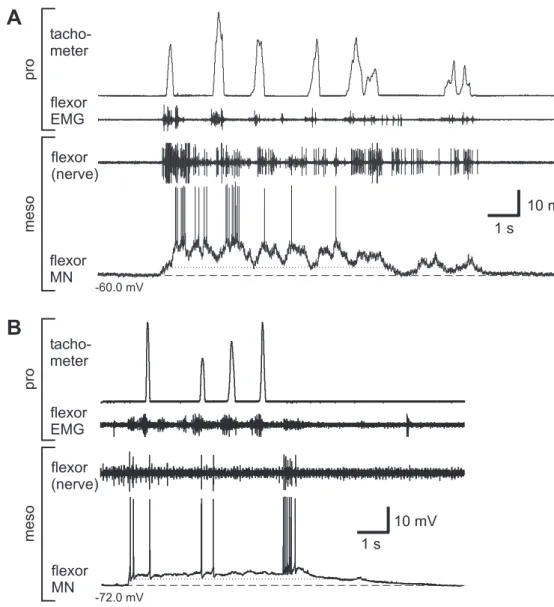
Previous studies using the single-legged preparation showed, that the activity in meso- thoracic MNs increased during stepping sequences of the ipsilateral front leg (Ludwar 2003). Furthermore, intracellular recordings revealed that this increased activity consists of a phasic and tonic modulation (depolarization). The phasic modulation was usually coupled to front leg steps and the tonic depolarization outlasted the complete stepping sequence. Recently, Borgmann (2006) could show that the activity in ipsi- and contralate- ral coxae MNs of all thoracic ganglia is increased during stepping sequences of a single leg.
1.3.1 Objectives
The focus of this thesis was on the state-dependent, long-lasting tonic depolarization observed in leg MNs during walking in stick insects. In the light of recent studies, the tonic depolarization became of particular interest, because it might be ubiquitous during locomotion.
All experiments were conducted on reduced preparations. The single-legged preparation of the stick insect allowed an analysis of activity in the mesothoracic segment induced by front leg stepping, without sensory influence from other segments. The experiments were conducted solely on flexor MNs and should shed light onto the following issues:
1. Characterization of the membrane potential modulation incontralateralmesothora- cic flexor MNs during front leg stepping and analysis of the properties of the tonic depolarization.
2. Pharmacological analysis of the tonic depolarization to answer the following ques- tions:
a) Which are the involved transmitters / receptors, that participate in the tonic depolarization?
b) Are second messengers involved in mediating the tonic depolarization?
3. Analysis of the influence of lesions (of the brain or connectives) on neuronal activity, especially on the tonic depolarization.
Materials and Methods
2.1 Experimental animal
The experiments were carried out on adult female stick insects of the speciesCarausius morosus(syn.Dixippus morosus) andCuniculina impigra(syn.Baculum extradentatum) from a colony maintained at the University of Cologne. The colonies were kept at temperatures between 20°C and 25°C and at an artificial light/dark rhythm. The forage plant for the colony was blackberry (Rubus fructiosus).
Carausius morosus andCuniculina impigrabelong to the phylum: arthropoda, class: in- secta, subclass: pterygota, superorder: orthopteroidea, order: phasmida, family: phas- matidae, subfamily: phasmatinae. Both are wingless, nocturnal animals. The native place ofCarausius morosusis southern India, adults grow up to 8 cm and the reproduction is generally parthenogenetic. The origin ofCuniculina impigrais Vietnam, females grow up to 10 cm, males up to 7 cm (but they are seldom). The lifetime is 12 to 14 months. The
A
B
Figure 2.1:Stick insects on blackberry leafs.A:Cuniculina impigra andB:Carausius morosus
body is divided into three main parts - head, thorax and abdomen, whereas the head consists of six segments and carries the brain, sense organs, antennae and mouthparts.
The thorax is divided into pro-, meso- and metathorax, and each segment carries a leg pair. The abdomen serves for reproduction and digestion and consists of 11 segments.
The central nervous system (CNS) is composed of the ventral nerve cord (including the subesophageal ganglion = SEG) and the dorsally positioned brain (supraesophageal gan- glion). The brain is a fused ganglion composed of the protocerebrum, deutocerebrum and tritocerebrum. The anterior three pairs of ganglia in the ventral chain are also fused, forming the cephalic SEG. The brain and the SEG are linked via the circumoesophageal connectives, whereas the SEG is linked to the prothoracic ganglion via the neck connec- tives.
2.2 Preparation and experimental setup
This chapter describes first the general preparation which had to be performed for all experiments. Then slight differences for the individual experiments are listed.
The experiments were carried out under dimmed light conditions at room temperature (20-25°C). A semi-intact preparation was used with one intact front leg walking, while all other legs were amputated at the level of the mid-coxa. The thorax was fixed ventrally on a platform using dental cement (PROTEMP, 3M ESPE, Seefeld, Germany). The cuticule was cut dorsally and removed from the middle of the mesothorax to the middle of the metathorax. The gut was left intact, moved aside and the cavity was filled with saline (composition according to Weidler and Diecke (1969), see chapter 2.4, table 2.1). Connec- tive tissue and fat were removed to allow access to the mesothoracic ganglion and leg nerves. Care was taken to leave as much as possible trachea intact, and cut main trachea were exposed to air. All lateral nerves but thenervus cruris (ncr, nomenclature accord- ing to Graham 1985; Marquardt 1940) ipsi- or contralateral to the remaining leg were cut near the mesothoracic ganglion to exclude sensory input in this segment. In most cases the tissue between the two connectives had to be carefully cut with fine scissors to al- low lifting of the mesothoracic ganglion on a movable waxed platform. The surrounding connective tissue was pinned down with small cactus spines (Nopalea dejecta) to stabilize the ganglion for penetration with sharp microelectrodes.
2.2.2 Non-pharmacological experiments
For recording of mesothoracic MNs the ganglion sheath had to be softened to allow pen- etration of intracellular electrodes. Therefore saline was quickly removed from the cavity and the ganglion sheath was treated with crystals of a proteolytic enzyme (PRONASE E, MERCH, Darmstadt, Germany) for 60-90 s. The enzyme was thoroughly washed out and saline was applied again.
2.2.3 Pharmacological experiments
The ganglion sheath in stick insects serves as a blood-brain barrier and displays there- fore a diffusion barrier for ions (Schofield 1979; Dörr et al. 1996) and pharmacological agents. Due to that, the first step to be able to perform pharmacological experiments in
MN intracellular MN
extracellular
pro- meso- meta- thoracic segment
treadmill flexor EMG
inflow
outflow silicon-gel
silicon-gel
A
flexor nerve extracellular
pro
meso meta
saline outflow flexor MN
intracellular
ganglionholder
silicon - gel
silicon-gel
tachometer
flexor EMG saline
inflow
B
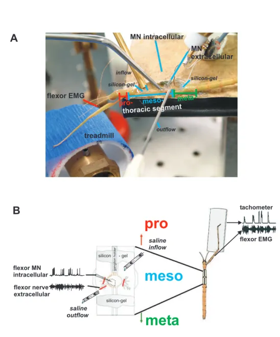
Figure 2.2:Split-bath preparation of the experimental animal.A:Picture of the split-bath preparation. B:
Schematic of a split-bath setup. A-B:Rostral and caudal of the mesothoracic (meso) ganglion silicon-gel barriers are built to allow superfusion with saline containing drugs of only the mesothoracic ganglion.
intracellular recordings. Tissue and fat along the connectives were removed toward the pro-and metathoracic ganglion. Saline was removed from the cavity and silicon-gel bar- riers (BAYSILONE-PASTE, hochviskos, Bayer AG, Leverkusen) were quickly built with a modified syringe (Fig. 2.2). Three separate compartments were formed, one rostral and caudal to the mesothoracic ganglion and one around the ganglion. This allowed a sep- arate superfusion of drugs only to the mesothoracic ganglion. Then saline was added again (chapter 2.4, table 2.1), and the superfusion system was adjusted and started. Be- fore desheathing the ganglion it was made sure that the system worked and the gan- glion was completely covered with saline because exposure of the desheathed ganglion to air would damage the nerve cells immediately. By using very fine scissors (SuperFine VANNAS SCISSORS straight, Harry Fein WORLD PRECISION INSTRUMENTS, Berlin, Germany) a first cut was made along the midline from posterior to anterior of the gan- glion, then the sheath was cut either towards the ipsi- or contralateral side. The loosened sheath was fixed with fine forceps and finally removed by a third cut. In this way at least one third of the ganglion was desheathed. The solutions were exchanged by a gravity driven perfusion system, positioned 50 cm above the platform and conducted via a tube system toward the cavity (0.75 mm inner diameter of the inflow tube). The flow rate was 2 ml/min, and it took 20 s for a new solution to enter the cavity. The volume of the cavity was 100 µL, the volume was therefore exchanged 20 times per minute. A continuous flow rate was achieved by an adequate positioning of the outflow.
2.2.4 Lesion experiments
In one set of experiments, it was necessary to have access to the supraesophageal gan- glion (brain) during recording from mesothoracic flexor MNs. Therefore the head was stabilized with plasticine while the cuticule of the head capsule was carefully cut with a sharp razor blade and removed to expose the brain. The cuts ran between the eyes along the front rim and bilateral toward the posterior end of the head. During the experiment, care was taken to add saline into the cut regularly. Referring to Graham (1979a), immobile
antennae indicated a successful preparation. The animal was then prepared according to chapter 2.2.3. As soon as the intracellular recording of a flexor MN was stable, modula- tion in its membrane potential was recorded during a few stepping sequences of the front leg. Then the brain was removed by holding it with fine forceps and cutting the connec- tives with sharp scissors. Care was taken to induce as less perturbations as possible and not to lose the intracellular recording. If the lesion was successful and the quality of the intracellular recording did not change, again the membrane potential was recorded dur- ing stepping sequences of the front leg. Successful experiments were analyzed according to chapter 2.6.1.
2.2.5 Connective recordings
To perform extracellular recordings from both, the ipsi- and contralateral neck connec- tives (connectives between the prothoracic ganglion and the SEG) the general prepara- tion was extended. The cuticule was cut dorsally and removed from the middle of the mesothorax towards the headcapsule. The gut was moved aside and the cavity was filled with saline. One front leg performed stepping sequences on a treadmill.
2.3 Treadmill
A lightweight low-friction treadmill (Gabriel et al. 2003) was used, which consisted of two styrofoam drums (∅ 40 mm; width 28 mm), each mounted on a micro DC-motor (DC1516, FAULHABER, Schönaich, Germany), that had a center distance of 50 mm (Fig- ure 2.3 A). A belt made of light crêpe paper was placed around the styrofoam drums. The tangential force that had to be applied to overcome belt friction was 4.0 ± 0.3 mN. The moment of inertia of the system, which is determined by the effective mass of the tread- mill was 1.1 g and thus approximately equal to the mass of an adult animal (Gabriel et al.
2003). One DC-motor served as a tachometer, with the other belt friction could be var- ied. The tachometer signal was digitized with a MICRO1401 A/D converter (sampling rate: 6.5 kHz) and recorded with SPIKE2 software (both CAMBRIDGE ELECTRONIC DESIGN, Cambridge, UK) on a personal computer. The treadmill was positioned 45° be-
AD/DA-Converter
Personal Computer recording
of belt velocity
current command amplifier/
filter
voltage-current converter
tachometer flexor EMG
0.5 s stance
phase
stance phase
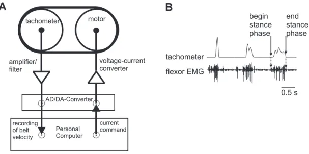
Figure 2.3:Treadmill. A:Tachometer. The signal from the tachometer was filtered and digitized prior to recording. A current could be applied to the other motor to decrease or increase belt friction. (Picture modified after Gabriel et al. 2003).B:Tachometer trace.
low the intact leg, and the height was adjusted so that the animal could easily pull the belt and performed walking like movements. The tachometer trace in combination with the flexor EMG (see chapter 2.5.1) allowed determining the stance phase of a step (Fig. 2.3 B). The beginning of stance was defined as the beginning of activity in the flexor EMG.
The end of stance was defined as the last maximum of the trace before it decreases back to zero. The falling edge is just determined by the inertia of the treadmill and does not contain any information about the status of the step. In all experiments, not single steps were used for analysis but stepping sequences. A stepping sequence was defined as a minimum of three consecutive steps that had a maximum temporal distance of 3.5 s.
2.4 Solutions
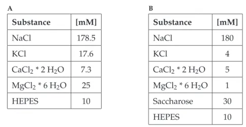
The composition of the physiological saline is listed in table 2.1. Different saline com- positions were used in non-pharmacological (chapter 2.2.3) and pharmacological experi- ments, both with a pH of 7.2 (chapter 2.2.2). A different saline composition was necessary in those experiments where the ganglion was desheathed.
A
Substance [mM]
NaCl 178.5
KCl 17.6
CaCl2* 2 H2O 7.3 MgCl2* 6 H2O 25
HEPES 10
B
Substance [mM]
NaCl 180
KCl 4
CaCl2* 2 H2O 5 MgCl2* 6 H2O 1
Saccharose 30
HEPES 10
Table 2.1:Saline composition used inA:non-pharmacological experiments according to Weidler and Diecke (1969), andB:pharmacological experiments; saline modified after Schmidt.
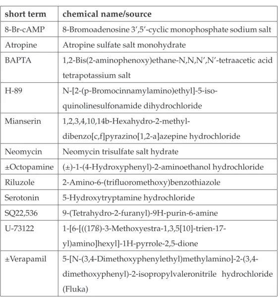
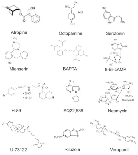
Drugs for pharmacological experiments were dissolved in saline prior to the experiment or added as aliquots, stored at −20° C and thawed prior to the experiment. H-89 and U-73122 were dissolved in DMSO, mianserin was dissolved in ethanol and verapamil in methanol, whereas the solvents had a final concentration in the experiment of less than 1 %. This solvent concentration in saline alone had no effect on membrane potential of flexor MNs. All drugs were obtained from SIGMA (Sigma-Aldrich, Schnelldorf, Ger- many) with the exception of verapamil (FLUKA, Switzerland). The chemical structures of the drugs are shown in figure 2.4.
2.5 Electrophysiology
2.5.1 Extracellular recordings and electromyograms
Extracellular recordings were made from a lateral nerve or from connectives with mo- nopolar hook electrodes (custom built, modified after Schmitz et al. (1988; 1991)) filled with silicon-gel (BAYSILONE-PASTE, hochviskos, Bayer AG, Leverkusen). Recordings were made from the mesothoracic leg nerve ncr (nervus cruris), ipsi- or contralateral to the intact front leg. The ncr contains the axons of flexor motoneurons (MNs). In another set of experiments, different connectives were recorded.
Electromyograms (EMGs) were recorded with two twirled copper wires (∅50 µm), which
short term chemical name/source
8-Br-cAMP 8-Bromoadenosine 3’,5’-cyclic monophosphate sodium salt Atropine Atropine sulfate salt monohydrate
BAPTA 1,2-Bis(2-aminophenoxy)ethane-N,N,N’,N’-tetraacetic acid tetrapotassium salt
H-89 N-[2-(p-Bromocinnamylamino)ethyl]-5-iso- quinolinesulfonamide dihydrochloride Mianserin 1,2,3,4,10,14b-Hexahydro-2-methyl-
dibenzo[c,f]pyrazino[1,2-a]azepine hydrochloride Neomycin Neomycin trisulfate salt hydrate
±Octopamine (±)-1-(4-Hydroxyphenyl)-2-aminoethanol hydrochloride Riluzole 2-Amino-6-(trifluoromethoxy)benzothiazole
Serotonin 5-Hydroxytryptamine hydrochloride SQ22,536 9-(Tetrahydro-2-furanyl)-9H-purin-6-amine U-73122 1-[6-[((17ß)-3-Methoxyestra-1,3,5[10]-trien-17-
yl)amino]hexyl]-1H-pyrrole-2,5-dione
±Verapamil 5-[N-(3,4-Dimethoxyphenylethyl)methylamino]-2-(3,4- dimethoxyphenyl)-2-isopropylvaleronitrile hydrochloride (Fluka)
Table 2.2:Drugs used in the experiments. All drugs were obtained from Sigma if not indicated differently.
8-Br-cAMP Atropine
H-89 Mianserin
Neomycin Octopamine
Riluzole
Serotonin
SQ22,536
U-73122 Verapamil
BAPTA
Figure 2.4:Chemical structures of the used drugs. The molecular weight varied between 190 and 909.
(∅0.1 mm). The copper wires were inserted and fixed with dental cement.
Both the extracellular recordings and the EMG recordings were amplified and filtered (250 Hz - 5 kHz, 50 Hz - 1 kHz).
2.5.2 Intracellular recordings
Intracellular recordings from flexor MNs were made using thin-walled glass microelec- trodes (GC100TF-10, HARVARD APPARATUS ltd, Edenbridge, Kent, UK), which were pulled on a P-97 filament puller (SUTTER INSTRUMENTS, Novato, USA). Microelec- trodes were filled with a solution of 3 M potassium acetate (KAc) and 0.1 M potassium chloride (KCl) or 1.5 M KAc and 1.5 M KCl. The electrode resistances were 15-25 MΩ. The recordings were made from the aborizations of the flexor MNs in the neuropilar region of the mesothoracic ganglion (ipsi- or contralateral to the walking front leg). An SEC-10L amplifier (NPI, Tamm, Germany) was used to amplify the signals in bridge or discon- tinuous current clamp (DCC) mode (switching frequency 12-25 kHz). The DCC mode was used during current injections. The MNs were identified by a one-to-one relation- ship of intracellularly recorded spikes with spikes in the appropriate extracellular nerve recording. Recordings were made from fast, semi-fast and slow flexor MNs, whereas no discrimination were made for data analysis.
Recordings where no stable membrane potential was reached or where the input resis- tance decreased without external stimulus were discarded. A total of 77 flexor MNs for non-pharmacological experiments and 67 flexor MNs for pharmacological experiments were recorded in 144 animals.
2.5.3 Experimental setup
The equipment (platform, electrodes etc.) was placed on an air table (MICRO-g, TMC, Peabody, MA, USA), which was surrounded by a Faraday cage. Thus vibrations and movements of the recording electrodes relative to the animal were minimized. Depend- ing on the experimental conditions, next to the platform where the animal was fixed the
following devices were positioned: three micromanipulators for the extracellular elec- trode, the ganglion holder and the outflow, a flexible magnetic stand for the inflow, and a micromanipulator for the intracellular electrode (LEICA Micromanipulators, Wetzlar, Germany). A lamp with optical fibers illuminated the setup during the preparation. A treadmill was positioned next to the platform under the front leg of the animal in an ap- proximately 40° angle to the animal as this corresponds to the mean position of the front leg in a standing animal (Cruse 1976).
2.6 Data recording and evaluation
The voltage output of the treadmill and the electrophysiological data were recorded us- ing a MICRO 1401 A/D converter and SPIKE 2 data-acquisition/analysis software (ver- sions 3.13 - 4.12, CAMBRIDGE ELECTRONIC DESIGN, Cambridge, UK) and a personal computer. The second DC-motor of the treadmill was connected to a voltage-current con- verter. A SPIKE 2 sequencer script was written to apply continuous current to the motor.
For the A/D conversion of electrophysiological data, a sampling rate of 12.5 kHz was used for extracellular, and of 6.25 kHz for intracellular data.
The extracellular recordings and the tachometer trace were preprocessed in SPIKE2 for further data evaluation. Neuronal activity of the extracellular recordings (only for con- nective recordings) and beginning and end of a stance phase in the tachometer trace were displayed as event channels.
Some data were analyzed with respect to the start of the front leg stance phase, to describe the modulation in membrane potential in mesothoracic flexor MNs. A time window of 1 s before and after the beginning of each stance phase was analyzed. The animals some- times performed spontaneous stepping sequences, but generally they had to be elicited by gently touching the abdomen with a paintbrush. The paintbrush was removed as soon as the animal started a sequence of stepping movements. Steps that were elicited due to stimulation with a paintbrush were discarded for the analysis of the phasic modulation.
In order to estimate the gross activity of a connective recording, it was rectified and smoothed (first order low pass filter, time constant τ = 0.07 s). Custom SPIKE2 script programs were written to analyze the recordings.
SOFT) and Microcal Origin (version 6.0, ORIGINLAB CORPORATION Northampton, MA, USA).
2.6.1 Analysis of intracellular recordings
The analysis in this thesis focuses mainly on the tonic membrane potential modulation in mesothoracic MNs, namely the tonic depolarization. This tonic depolarization is only induced by a behavior of the animal itself and did not occur in resting animals nor could it be induced artificial by current injection or the like. Flexor MNs exhibited the tonic depolarization either during stepping sequences of the front leg on a treadmill, or it oc- curred during searching movements (Büschges et al. 2004) of the front leg. The latter was not included in the analysis of this thesis, because without reference to steps, there was no evidence of comparing the amplitudes of the tonic depolarization in different sequences. If possible, the amplitude of the tonic depolarization was measured and com- pared during similar stepping sequences. Once a stable intracellular recording could be established, the success of an experiment depended on the behavior of the animal.
The determination of the amplitude of the tonic depolarization is shown in figure 2.5.
It was measured as the offset between the resting membrane potential before onset of a stepping sequence and the lower edge of the phasic modulation during a stepping se- quence. One could argue, that this is an underestimation of the real amplitude of the tonic depolarization. It is possible that the tonic depolarization amplitudes are higher than determined in this thesis, but I wanted to exclude a contamination by any phasic modulation. This was achieved in all probability by using this analysis (see also discus- sion, chapter 4.1.1).
Each data point in the graphics (see e.g., Fig. 3.10) represents the amplitude of the tonic depolarization in membrane potential induced by one complete stepping sequence (3 to 20 steps) of the front leg at certain times. For the pharmacological experiments, it was