The role of sensory influences in the control of motor activity of a stepping insect leg
Inaugural - Dissertation zur
Erlangung des Doktorgrades
der Mathematisch-Naturwissenschaftlichen Fakultät
der Universität zu Köln
vorgelegt von
Joscha Schmitz
aus Lippstadt
Köln
2018
Berichterstatter: Prof. Dr. Ansgar Büschges Prof. Dr. Peter Kloppenburg
Mündliche Prüfung: 16. Mai 2018
v
Table of Contents
Zusammenfassung ... ix
Abstract ... xi
1. General Introduction ... 1
1.1. Central pattern generating networks ... 2
1.2. Summary of the leg sense organs of the stick insect ... 3
1.2.1. Tactile Hairs ... 4
1.2.2.Campaniform sensilla ... 4
1.2.3. Chordotonal organs ... 6
1.2.4.Other sense organs ... 7
1.3. Femoro-tibial control network ... 8
1.4. Sensory processing during curve walking ... 9
1.5. Interleg and central influences ... 9
1.6. Experimental Approach ...10
1.7. Aim of the study ...11
2. The influence of touchdown in the middle leg of the walking stick insect for the control of the local locomotor activity ...12
2.1. Abstract ...12
2.2. Introduction ...12
2.3. Material and Methods ...15
2.3.1.Experimental animals ...15
2.3.2. Experimental setup ...15
2.3.3.Preparation ...16
2.3.4. Evaluation of muscle latencies and magnitude of muscle activity ...17
2.3.5.Statistics ...18
2.4. Results ...18
2.4.1.Ground contact influences on the latency of stance muscle activation ...19
2.4.2. Ground contact influences on the strength of stance muscle activation ...23
vi
2.4.3. Ground contact influences on FlxTi in different phasmid species ...25
2.4.4.Potential sources of TD feedback for FlxTi activation ...29
2.5. Discussion ...34
2.5.1.Dependence of stance muscle latency on TD ...34
2.5.2. Effects of lack of ground support on stance muscles ...35
2.5.3. Sensory influences on FlxTi activation ...37
2.5.4.FlxTi activation during SIH ...38
3. Task dependent changes in sensorimotor processing of movement feedback ...41
3.1. Introduction ...41
3.2. Materials and Methods ...43
3.2.1.Positioning of the experimental animal for walking ...43
3.2.2. Preparation for fCO stimulation and recording of motor activity ...43
3.2.3.Curve walking on a slippery surface ...45
3.2.4. Curve walking on a ball ...46
3.2.5.Experiments with different movement parameter stimulations ...46
3.2.6. Extracellular recordings of motor nerves to investigate intraleg influences of fCO feedback .46 3.2.7.Data acquisition ...46
3.3. Results ...47
3.4. Discussion ...58
3.4.1. Transition angle from stance to swing phase ...60
3.4.2.Influence of movement parameters is task specific ...60
3.4.3.Influence of the starting angle ...61
3.4.4. Influence of the stimulus velocity ...61
3.4.5.Processing of the stimulus amplitude ...61
3.4.6. Task dependent processing of fCO feedback ...62
3.4.7.Interjoint influences of the fCO ...62
3.4.8. Conclusion ...63
4. Mechanisms altering the task dependent processing of fCO feedback ...65
vii
4.1. Abstract ...65
4.2. Introduction ...65
4.3. Materials and Methods ...67
4.3.1.Positioning of the stick insect for stepping experiments ...67
4.3.2. Mechanical stimulation of the fCO with extracellular and intracellular recording of motor activity ...69
4.3.3.Electrical stimulation of the fCO afferents with extracellular and intracellular recording of motor activity ...69
4.3.4.Curve walking. ...70
4.3.5. Data Acquisition ...71
4.3.6.Statistical analysis ...71
4.4. Results ...71
4.5. Discussion ...89
4.5.1. The femoro-tibial joint control network ...90
4.5.2.Investigation of a monosynaptic connection from fCO to ExtTi MN ...91
4.5.3. Presynaptic inhibition...92
4.5.4.Influence of other leg sense organs ...92
4.5.5. Interleg and central influences ...93
4.5.6. Membrane input resistance ...94
4.5.7. Influence of nonspiking and spiking interneurons ...95
4.5.8.Task dependent changes in NSI ...95
4.6. Conclusion ...96
5. General Discussion ...97
5.1. Sensory influence on magnitude and timing of muscle activation ...97
5.1.1. Sensory influence on the timing of muscle activity ...98
5.1.2.Task-dependent processing of the magnitude and timing of muscle activity ... 100
5.2. Mechanisms for changing the task-dependent sensorimotor processing ... 102
5.3. Conclusion ... 103
List of Figures ... 105
viii
List of Tables ... 107
Bibliography ... 108
Abbreviation ... 127
Appendix ... 128
Danksagung ... 131
ix
Zusammenfassung
Im Diskurs um die Erforschung der Bewegungskontrolle standen lange Zeit stereotype Bewegungsabläufe im Mittelpunkt, wie zum Beispiel Vorwärtslaufen. In dieser Dissertation wird beleuchtet, wie verschiedene Ebenen der neuronalen Bewegungskontrolle zur Bewegungsveränderungen beitragen. Daraus ergaben sich drei Hauptfragen. Erstens, wie sensorische Rückkopplung die Aktivierung und Aktivität von Muskeln im Allgemeinen beeinflusst, zweitens, wie diese Rückkopplung bei aufgabenspezifischen Bewegungsveränderungen verarbeitet wird und drittens, welche Mechanismen im neuronalen Netzwerk genutzt werden, um flexible Bewegungsaktivität zu gewährleisten.
Zentrale Mustergeneratoren erzeugen eine rhythmisch alternierende Motoraktivität, welche durch sensorische Rückkopplung zeitlich und in der Aktivitätsstärke moduliert wird.
Im Mittelbein der Stabheuschrecke Carausius morosus messen zwei Hauptgruppen von Sinnesorganen entweder die Belastung oder die Bewegungs- und Positionsparameter des Beins. Die Rolle dieser Sinnesorgane wurde in dieser Arbeit untersucht.
In der ersten Studie wurde der Einfluss von campaniformen Sensillen (CS) auf die Stärke der Aktivität und den Aktivierungszeitpunkt von Stemmphasenmuskeln herausgestellt.
Hierfür wurde ein Versuchsaufbau mit einer absenkbaren Glitschplatte genutzt. Während des Experiments lief das Tier entweder mit Bodenkontakt auf dieser Glitschplatte oder trat in ein Loch, wenn die Platte während der Laufsequenz abgesenkt wurde. Durch den Bodenkontakt wurden die Beine belastet und die sensorische Rückkopplung durch CS aktiviert. Diese Rückkopplung fehlte bei dem Schritt ins Loch. Durch Ablationsexperimente konnte ich neben CS ein weiteres Sinnesorgan identifizieren, die durch ihre sensorische Information über den Bodenkontakt den Flexor tibiae aktivierten. In allen Stemmphasenmuskeln, mit Ausnahme des Depressor trochanteris, erhöhte sich die Stärke der Muskelaktivität durch den Bodenkontakt.
In der zweiten Studie wurde die Kontrolle durch die Rückkopplung des femoralen Chordotonalorgans auf die Aktivierung, insbesondere des Extensor tibiae Muskels, untersucht. Hierbei wurde ein Fokus auf die Verarbeitung dieser Rückkopplung bei Bewegungsveränderungen, insbesondere beim Kurvenlaufen, gelegt. Das fCO misst verschiedene Parameter der Kniegelenkbewegung und –position (Femur-Tibia [FT] Gelenk).
Durch die Rückkopplung vom fCO werden zum einen Widerstandsreflexe generiert, die zur Aufrechterhaltung der Positur genutzt werden. Zum anderen, wird die Reflexumkehr beobachtet, die im aktiven Tier zur Unterstützung der Stemmphase beiträgt. Während des Kurvenlaufens haben die Beine auf jeder Seite des Tieres eine unterschiedliche Kinematik.
Das Innenbein wird hauptsächlich im FT Gelenk bewegt, während das Außenbein
x
größtenteils im Hüftgelenk (Thorax-Coxa Gelenk) bewegt wird. Die Aktivierung von Extensor tibiae Motorneuronen (MN) während einer Reflexumkehr wurde bei einer bestimmten Winkeldifferenz ausgelöst. Dies geschah unabhängig von der Funktion des Beines als Innen- oder Außenbein. Darüber hinaus induzierte die fCO Stimulation häufiger AR, wenn das Bein als Innenbein fungierte. Beim Außenbein traten hingegen RR häufiger auf. Außerdem wurden mehr AR ausgelöst, wenn das fCO mit langsameren Geschwindigkeiten und größeren Startwinkeln stimuliert wurde. Der Protraktor coxae zeigte im Außenbein eine erhöhte Aktivierung durch die fCO-Stimulation, während der Levator coxae keinen Unterschied in der Reaktion zwischen den beiden Laufsituationen zeigte. Somit konnte in diesem Teil meiner Arbeit gezeigt werden, dass Rückkopplung vom fCO aufgabenspezifische Motoraktivität hervorrufen kann.
In einer letzten Studie wurden die Mechanismen untersucht, die für die Abnahme der Reaktion auf die fCO Stimulation verantwortlich sein könnten, wenn das Bein als Außenbein genutzt wird. Die direkte Verbindung von fCO Afferenzen zu ExtTi MN, sowie zu Nicht- Spikenden-Interneuronen (NSI), war abgeschwächt. Ein ähnliches Bild zeigte sich über die gesamte Länge der fCO Stimulation in ExtTi MN. Ferner erhielten die ExtTi MN eine größere tonische Depolarisation und der Membran Eingangswiderstand wurde verringert.
In meiner Dissertation konnte ich somit zeigen, dass Motoraktivität durch verschiedene Sinnesorgane beeinflusst wird. Diese Beeinflussung kann an Bewegungsveränderungen angepasst werden, wobei ein Mechanismus aufdeckt werden konnte, der zu dieser Anpassung beiträgt. Welche Einflüsse NSI oder eine präsynaptische Inhibierung auf aufgabenspezifische Motoraktivitäten haben, bleibt offen.
xi
Abstract
For a long time the focus of the discourse in motor control research was on stereotypical movements, such as forward walking. My thesis emphasizes how different levels of neuronal motor control contribute to the processing of task-dependent locomotor behavior. This resulted in three main questions. First, how sensory feedback affects the timing and magnitude of muscle activity in general. Second, how this feedback is processed in changes of task-dependent movement behavior. And third, what mechanisms are used in the neural network to evoke an adaptive capability.
Central pattern generators (CPGs) generate a rhythmic, alternating motor activity that is in turn modulated by sensory feedback in timing and in magnitude. In the middle leg of the stick insect Carausius morosus, two major groups of sense organs measure either the load or the movement and positional parameters of the leg. The role of these sense organs was examined in my work.
In the first study, the influence of campaniform sensilla (CS) on magnitude and the timing of stance phase muscles was examined. For this purpose, a trapdoor setup with a slippery surface was used. The animal either stepped on a slippery surface with ground contact or they stepped into a hole when the trapdoor was lowered (SIH). Through ground contact, the legs are loaded and thereby activate the leg CS. During SIH this sensory feedback is missing. Through ablation experiments, I was able to show, in addition to CS, an additional sense organ that activated the flexor tibiae (FlxTi) through their sensory information of the touchdown (TD). In all stance phase muscles, except for the depressor trochanteris (DepTr), the strength of muscle activity increased through the TD.
In the second study, the control on the timing of activation, especially of the extensor tibiae (ExtTi) muscle by sensory feedback of the femoral chordotonal organ (fCO) was investigated. Here, a focus was set on the processing of this feedback to movement changes, in particular curve walking. The fCO measures various parameters of the knee joint movement (femorotibial [FT] joint) and position. Feedback from the fCO generates resistance reflexes (RR), which are used to maintain the posture. Assistance reflex or reflex reversals (AR), which assist stance phase activity in the active animal, were also observed. During curve walking, the legs on each side of the animal have different kinematics. The leg inside of a curve (inside) is mainly moved around the FT joint, while the leg outside of the curve (outside) is mostly moved in the hip joint (thoracocoxal [ThC] joint). ExtTi motor neurons (MN) were activated during an AR at a certain angular difference. This was independent of the function as inside or outside leg. Further, the fCO stimulation induced ARs more often in the inside leg while RRs occurred more frequently in the outside leg. In addition, more ARs were generated through fCO stimulation at slower velocities and larger starting angles. The protractor coxae (ProCx) showed increased activation through fCO stimulation in the outside leg, while the levator coxae (LevTr) showed no difference in the reaction between the two leg
xii
functions. Thus, it could be shown in this part of my thesis that feedback from the fCO can cause task-specific motor activity.
In the third study, the mechanisms that might be responsible for the decrease in the response to fCO stimulation when the leg is used as the outside leg were investigated. The monosynaptic connection of fCO afferents to ExtTi MNs, as well as to nonspiking interneurons (NSIs), was reduced. Similar observations were made in ExtTi MNs throughout the complete fCO stimulation. Furthermore, ExtTi MNs received greater tonic depolarization and their membrane input resistance was reduced.
In my dissertation I could show that motor activity is influenced by various sense organs. These influences can be adapted to changes in movement, whereby I could reveal one mechanism leading to task-dependent motor activity. The influence of NSIs or presynaptic inhibition on changes in task dependent motor output remains unclear.
1
1. General Introduction
Every animal navigates its environment using locomotion. Depending on the habitat, this can be swimming, crawling, flying or walking. Each of these behaviors consists not only of one type of locomotion, but it can be adapted to the environmental needs. An animal can not only move straight forward, but can change the pattern in a task-dependent manner to backwards locomotion (lamprey: Islam et al., 2006; salamander: Ashley-Ross and Lauder, 1997; humans: van Deursen et al., 1998; Pang and Yang, 2002; Choi et al., 2008; crayfish:
Ayers and Davis, 1977; stick insect: Rosenbaum et al., 2010), climbing (cockroach: Watson et al., 2002 a,b; fruit fly: Pick and Strauss, 2005; stick insect: Bläsing and Cruse, 2004a, b) and turning (fish: Orger et al., 2008; human: Lamb and Yang, 2000; cockroach: Mu and Ritzmann, 2005; fruit fly: Frye and Dickinson, 2001; 2004; Bender and Dickinson, 2006; stick insect: Dürr and Ebeling, 2005; Gruhn et al., 2009 a, 2016). This behavioral plasticity, not only in walking, but also in the other forms of locomotion, emerges from an interaction of neuronal networks, sense organs and muscles (Orlovskiĭ et al., 1999). The neuronal networks consist of rhythm generating networks that activate motor neurons (MN) in a rhythmical manner, which then initiate alternating muscle activity. The timing and magnitude of the rhythmic output of this neuronal network, is modulated by sensory feedback. The sensory feedback measures position, load and movement parameters, and originates from sense organs that correspond to the locomotor organs.
In vertebrates, the command centers for locomotion are located in the higher brain centers. Signals are transmitted from the basal ganglia, via the motor cortex, the cerebellum and the brainstem to the local networks in the spinal cord, where central pattern generators (CPGs) generate a rhythmic motor output (c.f. Grillner et al., 1995, Grillner, 2003, Kiehn, 2006). For invertebrates, especially insects, it is known that the signals, initiating locomotion descend from the central complex (Strauss, 2002) to the ventral nerve cord, where local networks that contain CPGs generate the motor output (Büschges and Bässler, 1998).
Another adaptation to stereotyped locomotion is the use of different coordination patterns between the legs. In vertebrates these so called gaits, like walking, trot, gallop in horses, are correlated with a distinct movement speed (Alexander, 1989, Hoyt and Taylor, 1981), to decrease energy costs (Hreljac, 1993). Invertebrates, like crustaceans, arachnids or insects, on the other hand, show continuous transition between gaits, which are thus here better described as coordination patterns (Cruse et al., 2009, Grabowska et al., 2012, Graham, 1985, Hughes, 1952, Wendler, 1964, Wosnitza et al., 2013). Insects use three major coordination patterns. For fast walking speeds, this is a tripod, for intermediate velocities a tetrapod, and for slow velocities a metachronal coordination (Cruse et al., 2009,
2
Delcomyn, 1971, Grabowska et al., 2012, Graham, 1972, 1985, Hughes, 1952, Wendler, 1964, 1966, Wosnitza et al., 2013). During the tripod coordination, three legs are in stance phase, whereas, during the tetrapod coordination, four legs are in stance phase at the same time (Cruse et al., 2009, Graham, 1985). An increase in walking speed is not only marked by change in the coordination pattern, but also by changes in the step cycle.
One step cycle consists of two phases. The first is the stance phase, during which the leg has ground contact and generates propulsion to push the body forward. The second is the swing phase, during which the leg is moved through the air to a new position to generate the next stance phase (Büschges and Gruhn, 2008, Epstein and Graham, 1983, 1985).
In stick insect, the local control of a step cycle in a single leg is well described, partially down to the level of single contributing neurons (reviews in Bässler and Büschges, 1998, Büschges et al., 2008, Büschges and Gruhn, 2008). The single leg is moved by antagonistic muscles around three mayor leg joints. Around the thoraco-coxal (ThC) joint the leg is moved posteriorly by the retractor coxae (RetCx) and anteriorly by the protractor coxae (ProCx), respectively. The next leg segment, the fused trochantero-femur is moved around the coxa- trochanter (CTr) joint. Here, the depressor trochanteris (DepTr), lowers the leg and the levator trochanteris (LevTr) lifts it The last mayor leg joint is the femoro-tibial (FT) joint.
Through it, the flexor tibiae (FlxTi), and the extensor tibiae (ExtTi) flex, respectively extend the tibia of the leg (Büschges et al 2008). When the stick insect is walking forward, the RetCx, the DepTr and the FlxTi are used as stance phase muscles, coordinated in a specific activity pattern (Graham, 1985, 1985). During backward walking the same muscles are used, only the RetCx and ProCx are switched as respective stance and swing, as the leg is moved anteriorly during stance (Rosenbaum et al., 2010).
1.1. Central pattern generating networks
The activity of the antagonistic muscles in a leg is often controlled and timed by the activity of CPGs (Grillner and Zangger, 1979, Jordan et al., 1979, Matsushima and Grillner, 1992). These networks are located in the spinal cord of vertebrates or the ventral nerve chord of invertebrates. Examples for the importance of CPGs in locomotion has been found in vertebrates (review in Grillner, 2011, Rossignol et al., 2006), such as cats (Grillner and Zangger, 1979, Pearson and Rossignol, 1991), mouse (Kiehn, 2006), tadpole (Roberts et al., 1998) and lamprey (Grillner et al., 1981, Wallén and Williams, 1984) or invertebrates (Bässler and Büschges, 1998, Pearson, 1993), such as crayfish (Chrachri and Clarac, 1987), cockroach (Pearson and Iles, 1970, Zill, 1986), in locust (Ryckebusch and Laurent, 1993), hawk moth (Johnston and Levine, 1996) and stick insect (Büschges, 1995c). In stick insects, the neurons of the CPG have not yet been identified, although single nonspiking interneurons (NSIs) were found, which contribute to the function of the CPG (Berg et al., 2015, Büschges,
3 1995a). In other invertebrates, such as the stomatogastric nervous system of crustaceans (Marder and Bucher, 2007), the leech heartbeat (Kristan et al., 2005) or the swimmeret system of the crayfish (Mulloney and Smarandache-Wellmann, 2012), the cellular basis of the CPG is known better. The cells of the CPG express particular intrinsic properties, like endogenous bursting, spike frequency adaptation, postinhibitory rebound, plateau potentials and synaptic interaction between the cells of the CPG, which allow them to show rhythmic behavior (Marder and Calabrese, 1996, Marder and Bucher, 2001). Two types of networks are known, the pacemaker driven CPG, as it is found in the pyloric system of the stomatogastric nervous system (Marder and Eisen, 1984) and the half center oscillator, like it is found in the lamprey spinal cord, which oscillates through reciprocal inhibition between network components (Grillner et al., 1991).
In stick insects, it has been shown though pharmacological activation with the muscarinic agonist pilocarpine that each leg joint is driven by it's own CPG (Büschges, 1995c). In the stick insect, the rhythmic motor activity is the joint result of tonic descending excitatory drive, inhibitory patterning influence from the CPG and sensory inputs, and excitatory patterning influences from sensory inputs. (Büschges, 1998, Büschges et al., 2004, Ludwar et al., 2005a, Ludwar et al., 2005b).
1.2. Summary of the leg sense organs of the stick insect
As alluded to above, the activity pattern of the CPGs for each joint in a stick insect leg are strongly modulated by sensory feedback from the leg sense organs. Campaniform sensilla (CS) are known to activate and even entrain the CPG activity (FT joint: (Akay et al., 2001, Berendes et al., 2013, Zill et al., 2011, Zill et al., 2013, Zill et al., 2015); ThC joint:
(Akay et al., 2004, Zill et al., 2004); CTr joint: (Borgmann et al., 2011, Zill et al., 2011, Zill et al., 2012, Zill et al., 2015). The femoral chordotonal organ (fCO) in the stick insects femur has been shown to induce phase transition in the CTr joint, from LevTr activity to DepTr (Bucher et al., 2003, Hess and Büschges, 1999). In the active behavioral state of the stick insect, fCO activation also induces a transition of motor activity in the FT joint. During extension of the tibia, feedback of the fCO is first increasing the FlxTi activity, which is at a certain position of the tibia, switched to ExtTi activity. This reaction to extension of the tibia in the active animal is called “reflex reversal” or "active reaction" (AR) (Bässler, 1976, 1988).
However, depending on the behavioral state, in the inactive animal, the same fCO feedback, measuring an extension of the tibia, directly initiates a strong activation of the ExtTi muscle, which is thought to maintain posture and is thus called “resistance reflex” (RR) (Bässler, 1977b, 1988). Moreover, the sense organs of the leg can also provide sensory feedback to establish an accurate coordination of the leg joints.
4
1.2.1. Tactile Hairs
Hair rows
Four hair rows have been described on the coxa of the stick insect Carausius morosus (Bässler, 1965) that react to coxal movement in a posterior direction with increasing activity (Cruse et al., 1984). Bässler (1977a, b) described additional tactile hairs on the femur, which show a phasic response to a bending stimulus, but without a preferred directional response.
In some animals, the slow extensor tibia (SETi) was increasing its firing rate slightly as a result of the stimulation of these tactile hairs (Bässler, 1977b). The physiology was similar to tactile hairs found on the tarsi of the locust (Runion and Usherwood, 1968) and trochanteral hair receptors in cockroach (Spencer, 1974).Tactile hairs have also been described on the tarsus of the stick insect (c.f. Bässler, 1983).
Hair plates
Several hairplates (HPs) are located on the stick insect leg. Two HPs (ventral and dorsal coxal HP) are located on the coxa (Büschges and Schmitz, 1991, Wendler, 1964) that both react to an anteriorly directed bending of the coxa (Bässler, 1965, Bässler, 1977b, Cruse et al., 1984, Wendler, 1964). The ventral coxal HP (vcxHP) was also found to presynaptically inhibit afferents of the fCO (Stein and Schmitz, 1999). One HP is located anteriorly on dorsal surface of the trochanter (trHP), close to the CTr joint, and one HP is located on the ventral trochanter (Schmitz, 1986b). The trHP has proprioceptive function (Wendler, 1964) and reacts to lifting of the leg (Schmitz, 1986a, b). Ablation experiments demonstrated the importance of the trHP for the feedback about the ThC angle, as the activation of DepTr MNs failed after ablation (Schmitz, 1986a).
1.2.2. Campaniform sensilla
Campaniform sensilla (CS) are bipolar sensory cells with a dendritic ending in caps on the cuticle of insects (Zill et al., 2004), first described by Pringle on the cockroach palps (Pringle, 1938a) and legs (Pringle, 1938b). The CS react to resisted force which is applied to the cuticle of the animal as well as to resisted muscle contractions. Both produce strains of the cuticle, which deform the cuticular cap and thereby activate the sensory cell beneath (Barth and Blickhan, 1984, Moran and Rowley III, 1975, Zill et al., 2004). Most of the CS have a preferred direction, in which the CS are easier to activate, depending on the oval shape of the cuticular cap. The direction, the CS is easiest to activate is the axis perpendicular to the longitudinal axis of the oval cap (Spinola and Chapman, 1975) .The Cs measure load of the leg and muscle forces (Zill et al., 2012).
5 Trochanteral campaniform sensilla
The trochanteral campaniform sensilla (trCS) consist of four groups of CS (Groups 1- 4). Group 1 is located on the posterior, group 2 on the anterior trochanter, and groups 3-4 are located on the dorsal side of the trochanter (Hofmann and Bässler, 1982, Zill et al., 2012). Groups 1 and 2 were found to measure load of the leg mainly in anterior and posterior direction (Delcomyn, 1991, Hofmann and Bässler, 1982, 1986, Schmitz, 1993), whereas groups 3 and 4 are found to measure load mainly in dorsal and ventral direction (Zill et al., 2012).
Stimulation of the trCS can reset the timing of the ThC joint CPG and switch the MN activity from RetCx to ProCx MN, or vice versa (Akay et al., 2004). This, however, depends on the walking direction. The trCS could provide either excitatory or inhibitory drive to ThC MN (Schmitz and Stein, 2000). Sensory feedback from group 3 trCS was also described to be dependent on the behavioral state of the animal, in that lifting of the leg activates slow DepTr MN in the resting animal, and LevTr MNs when the animal is in the active state (Akay et al., 2007, Zill et al., 2012).
Femoral campaniform sensilla
The femoral campaniform sensilla (feCS) are located on the posterior side of the proximal femur (c.f. Bässler, 1983, Petryszak and Fudalewicz-Niemczyk, 1994, Zill et al., 2017). During stance phase, when the posterior femur is loaded in an upward direction, the feCS initiate FlxTi and terminate ExtTi activity and thereby induce a transition from ExtTi to FlxTi activity (Akay et al., 2001, Berendes et al., 2013, Zill et al., 2015). The caps and forms of the feCS can be divided into three groups, one ventral (caps' orientation with great angles to the autotomy plane of the trochantero-femoral joint, one dorsal group (caps' orientation with angles in line to the autotomy plane) and one central group with round caps (Zill et al., 2017). Through this, the feCS are capable of detecting forces at the CTr as well as at the trochantero-femoral joint, whereas the last joint is immobilized in stick insects and forms one plane from the CTr to the FT joint (Cruse and Bartling, 1995, Zill et al., 2017).
Tibial campaniform sensilla
The two main groups (6A and 6B) of tibial campaniform sensilla (tiCS) are located on the dorsal tibia, where group 6A consists of two subgroups, located more proximal to the FT joint, one anteriorly and one posteriorly, and group 6B is located more distally on the central tibia (Zill et al., 2011). The tiCS respond to muscle contractions of the tibial muscles and also to forces applied onto the plane of the FT joint, however, tiCS also react to unloading of the leg with increasing firing rate and could thereby contribute to the stance-swing transition (Zill et al., 2011, Zill et al., 2013). The different tiCS groups have different effects on tibial and trochanteral MN. Group 6B stimulation increases the activity in ExtTi and DepTr MN, while
6
the anterior group 6A stimulation opposes this effect by decreasing the activity in ExtTi and DepTr MN (Zill et al., 2011, Zill et al., 2013).
Tarsal campaniform sensilla
Tarsal campaniform sensilla (taCS) are found on tarsal segments 1 - 4 and on the Arolium. They are located dorsally at the distal part of each segment and the Arolium (Zill et al., 2014). By application of force on the taCS, the FlxTi, the DepTr and the retractor unguis (RetUng) are activated. taCS could also be activated by applying force to the RetUng tendon (Zill et al., 2014, Zill et al., 2015). The latencies until the activation of the different muscles were between 35 ms in the FlxTi to 43 ms in the RetUng (Zill et al., 2015). Functionally, by activation of the FlxTi, which occurs in parallel to an inactivation of the ExtTi, the leg is pulled towards the body and by activation of the RetUng, the claw is bent, which both lead to a higher adhesion to the ground (Zill et al., 2014, Zill et al., 2015).
1.2.3. Chordotonal organs
Femoral chordotonal organ
A chordotonal organ in the femur was first mentioned in the work of Borchard (1927) about heteromorphism in Dixippus (today Carausius) morosus. The fCO lies in the dorsal part of the proximal femur and is connected to the proximal tibia with a receptor apodeme (Bässler, 1965, Field and Matheson, 1998, Füller and Ernst, 1973). A single scolopidium within the fCO consists of two sensory neurons, one fiber cell and one or more sheet cells (Füller and Ernst, 1973). The fCO consists of a ventral scoloparium with 80 sensory neurons and a dorsal scoloparium with 420 sensory neurons (Kittmann and Schmitz, 1992). The sensory cells of the dorsal scoloparium measure manly vibration (Büschges, 1994, Field and Pflüger, 1989, Sauer and Stein, 1999, Stein and Sauer, 1999) and the cells of the ventral scoloparium measure velocity, position and acceleration of the tibia, or a combination of these three parameters (Büschges, 1994, Hofmann and Koch, 1985, Hofmann et al., 1985).
The sensory feedback of the ventral scoloparium, is used in the FT control loop (Kittmann and Schmitz, 1992). In the inactive animal, the feedback about extension of the tibiae, measured by the fCO, elicits a RR, to maintain posture (Bässler, 1972a, b, Büschges, 1989, Büschges, 1990, Kittmann, 1991), while in the active animal, a behavioral state elicited by stimulation or by walking, the same feedback can generate a AR that assists FlxTi activity (Bässler, 1974, Bässler, 1983, 1988, Cruse and Schmitz, 1983, Nothof and Bässler, 1990, Schmitz, 1985, Weiland and Koch, 1987). In addition to the influence on the FT joint, the feedback of the fCO also produces inter-joint influences. The elongation of the fCO increases the activity of the LevTr with a phasic and a tonic component, and also entrains LevTr MN activity, while the opposite was found for DepTr (Bucher et al., 2003, Hess and Büschges,
7 1997, Hess and Büschges, 1999). RetUng activity is increased by the fCO elongation, eliciting a AR (Bässler, 1988). The ProCx MN are only found to be weakly affected by fCO feedback (Bässler, 1986, 1988). The fCO was also shown to have intersegmental influences.
Stimulation of the front leg fCO could elicit an increase in FlxTi and RetCx, and simultaneously a decrease ProCx activity (Ludwar et al., 2005a).
Subgenual and distal organ
The subgenual organ is located as a hemicircle in the proximal tibia and consists of 41 to 44 sensory neurons. The distal organ is located distally to the subgenual organ in the tibia and consists of 16-17 sensory neurons. The subgenual organ is thought to be sensitive to substrate vibrations (Field and Matheson, 1998, Strauß and Lakes‐Harlan, 2013).
1.2.4. Other sense organs
Apodeme receptor
The apodeme receptor is connected to the distal part of the ExtTi muscle tendon, and reacts to both flexion and extension of the tibia. Its minimum firing frequency is reached at a FT joint angle of around 90° (Bässler, 1977a, 1983), but the reaction was only found in Cuniculina impigra.
Tension receptor
One sense organ, reacting to tension of muscle fibers of the FlxTi muscle was found by Bässler (1977a).
Récepteur dorso-antéro-latéral
The Récepteur dorso-antéro-latéral has first been described by Coillot and Boistel (1968) in the locust. In the stick insect, the Récepteur dorso-antéro-latéral was described by Bässler (1977a). It's firing frequency increases through extension of the tibia and putting pressure onto the membrane of the FT joint. It's effect on SETi was reported to be either increasing or decreasing SETi firing frequency (Bässler, 1977a).
Récepteur dorso-postéro-latéral
The Récepteur dorso-postéro-latéral has first been described by Coillot and Boistel (1968) in the locust. In the stick insect, the Récepteur dorso-postéro-latéral firing frequency is increased by a fast extension of the tibia, and by putting pressure onto the membrane of the FT joint. Stimulation of this receptor decreased the firing frequency of the SETi in most of the experiments (Bässler, 1977a).
8
Récepteur ventro-postéro-latéral
The Récepteur ventro-postéro-latéral has first been described by Coillot and Boistel (1968) in the locust. Bässler (1977a) described the Récepteur ventro-postéro-latéral to possibly react to deformation of the FT joint membrane, if the leg is completely flexed.
In summary, there are many sense organs in and on the stick insect leg, however, for the control loop of the legs motor network, the main sense organs are probably the hair plates, responsible for giving positional information, CS, reporting on load, and the fCO, giving feedback about movement and position of the tibia.
1.3. Femoro-tibial control network
The FT control network consists of five levels of parallel and antagonistic interaction.
The first level is the information of the sense organ itself. Bässler (1993) described the FT control network with fCO as afferents, but also the CS and HP sensory feedback is processed in the same fashion (review see Büschges and Gruhn, 2008, Schmitz and Stein, 2000). As described above, the fCO has sensory cells measuring position and movement of the FT joint in the ventral scoloparium (Büschges, 1994, Hofmann and Koch, 1985, Hofmann et al., 1985). The afferents receive presynaptic inhibition at their terminals from afferents of the same type (Sauer et al., 1997). Additionally, the fCO afferents also receive presynaptic inhibition from other leg sense organs, like CS and HP (Stein and Schmitz, 1999). The second level of processing is the synaptic connection from the fCO afferents onto NSI, spiking interneurons (SI) and ExtTi and FlxTi (as well as MN of the other leg joints). The NSI receive inputs from fCO afferents with the same parameter, which are acceleration, velocity and position, or a combination of these parameters. The postsynaptic cells can either receive direct excitatory or delayed inhibitory inputs (review in Bässler and Büschges, 1998, Büschges, 1990, Sauer et al., 1995, Sauer et al., 1996). The third level is the transmission of information from the NSI onto the ExtTi MN. The NSI provide parallel excitatory and inhibitory drive to the ExtTi MN (Büschges, 1990, Driesang and Büschges, 1996, Sauer et al., 1996).
The fourth level is the force produced by the ExtTi muscles. The muscle is innervated by the common inhibitor 1, inhibiting the ExtTi muscle, as well as the fast extensor tibiae (FETi) and the SETi MN, which excite the ExtTi (Bässler and Storrer, 1980). The last level is the movement of the tibia as a result of ExtTi muscle contraction, and that of its antagonist, the FlxTi muscle (Bässler and Stein, 1996). As this neuronal network is not only capable of producing the RR, which was analyzed in the present study, but also its reversal, it's FT joint control was described by Bässler (1993) as one that was controlled through distributed processing in a parliamentary manner, consisting of these five levels that act in parallel and also antagonistically (cf. Kristan, 2000, Morton and Chiel, 1994).
9
1.4. Sensory processing during curve walking
The kinematics of the stick insects middle leg on the inside of the turning animal differs strongly from the one on the outside. The leg on the inside is mostly moved around the FT joint, while the movements of other leg joints are often rather small. The outside middle leg, on the other hand is retracted and protracted strongly, while the FT joint is only moved very little (Dürr and Ebeling, 2005, Gruhn et al., 2009 a). Moreover, when the leg functions as inside leg (inside) the frequency of ARs is increased over straight walking, while ARs in leg functioning as outside leg (outside) are rare (Hellekes et al., 2011, Hellekes, 2012).
A mechanism underlying this change in motor activity is likely to be the relative weighting of excitatory and inhibitory synaptic inputs to NSI, which is known to change during the generation of the AR (Bässler and Büschges, 1990, Driesang and Büschges, 1996).
Hellekes (2012) gave some insight into the task specificity of fCO afferent feedback processing in NSI during curve walking. She could show a change in weighting of synaptic inputs to some NSI in the mesothoracic ganglion also during turning. During outside stepping the NSI E2/3 was inhibited less, whereas a strong inhibition was visible during inside- stepping, when the fCO was stimulated. For an another NSI, E5/6, the membrane potential changed from a slight hyperpolarization during inside-stepping to a slight depolarization during outside-stepping (Hellekes, 2012). NSI E8, did show a hyperpolarization for outside- stepping during fCO stimulation, which was not found for the inside-stepping leg. However, for this neuron, only a qualitative analysis was possible (Hellekes, 2012). For other NSI (E4, E9/10, I2 and I) no differences were found between the outside- and inside-stepping leg (Hellekes, 2012). Even though this was not tested during curve walking, the NSI E4 shows three different reactions during generation of the AR, where in two out of three possibilities also APs of the SETi were shown during the inactive phase (Driesang and Büschges, 1996).
1.5. Interleg and central influences
In addition to local network activity, also influences from other leg joints, neighboring legs, and other descending central influences have an impact on the motor output of the FT joint motor network.
In the stick insect, stepping of a single front leg can elicit alternating activity in the MN pools of the middle leg, and also increase general activity in MN of the hind leg. The MN in the mesothoracic segment receive a tonic depolarization, which is then phasically modulated by front leg stepping (Borgmann et al., 2007, Borgmann et al., 2009, Gabriel, 2005, Ludwar et al., 2005a, Ludwar et al., 2005b). With pharmacological experiments, Westmark et al.
(2009) could show that the tonic depolarization is evoked by acetylcholine (ACh) and enhanced by the neuromodulator octopamine (OA). OA was found to modulate motor output and arouse motor activity in locust as well as stick insects (Büschges et al., 1993, Sombati
10
and Hoyle, 1984). A source for OA in the stick insect is likely to be dorsal unpaired median (DUM) neurons, which originate in the suboesophageal ganglion. By stimulation of these neurons, two classes of neurons were found that influence the gain of the ExtTi MN activity during fCO stimulation (Stolz el al., in prep).
1.6. Experimental Approach
The stick insects used in the present study, Carausius morosus, Cuniculina impigra and Aretaon asperrimus have the advantage to be relatively large in size for an insect, and that they are easy to breed and to keep.
In the present study, different approaches have been used to investigate the influence of leg sense organs on the activity of the motor control network of phasmid species. In all experiments, a slippery surface setup (Berendes et al., 2013, Gruhn et al., 2006) has been used.
The slippery surface setup has been used to study backward walking (Graham and Epstein, 1985, Rosenbaum et al., 2010), escape responses of cockroach (Camhi and Nolen, 1981), curve walking of cockroach and stick insects (Gruhn et al., 2009 a, Hellekes et al., 2011, Tryba and Ritzmann, 2000), and changes in velocity of stick insects (Gruhn et al., 2009 b). These setups have the advantage that the animal is free to move its legs, and the legs are not coupled mechanically via the substrate, the animal is walking on. This reduces sensory load feedback and unmasks the influence of central output during walking.
The first study used a trapdoor setup, to reveal the influence of load on the timing and magnitude of motor output in the middle leg. The investigated leg is stepping on a platform, which is giving the exact time of ground contact via a current circuit. The platform then can be lowered, while the animal is in swing-phase. The next ground contact is then measured by a laser light sheet which the leg passes through (Berendes et al., 2013). Through this method, the impact of the missing ground on the motor output of this step could be investigated.
In the second and third study, a slippery surface setup was used without measuring the ground contact. The slippery surface was used to allow stationary animals to freely move their legs and be able to perform turning movements (Gruhn et al., 2009 a, Hellekes et al., 2011, Hellekes, 2012). Here, I used mechanical (Bässler, 1977b, Hellekes et al., 2011) as well as electrical stimulation (Sauer et al., 1995) of the fCO to investigate the task-dependent processing of fCO feedback. In the different approaches, neuronal and muscle activity was monitored by electromyographic, extracellular and intracellular recordings.
11
1.7. Aim of the study
Sensory feedback is known to influence the muscle activity and phase transitions in stick insects. For the FT motor network, the fCO and the CS of the leg have an mayor influence on the MN output. However, only little was known about the influence of load sensors on the timing and the magnitude of muscle activity. By using a trapdoor setup, the impact of load sensing on muscle activation and activity could was to be compared for steps with and without loading of the leg. In addition, by ablating load sensing CS selectively, experiments should reveal the source FlxTi activation and modulation in the middle leg.
Furthermore, the influence of leg sense organs seems to be task-dependent (Hellekes et al., 2011). The influence of the parameters of the tibial movement and position could influence the likelihood of occurrence of ARs and RRs, depending on the task, the leg is performing. It was an additional aim to test this and to reveal the mechanisms, underlying the task-dependent changes of fCO afferent processing.
12
2. The influence of touchdown in the middle leg of the walking stick insect for the control of the local locomotor activity
12.1. Abstract
A lot is known about how the activation of MN pools in the locomotor control system of stick insects is influenced by sensory input. However, the neuronal connectivity of the sensory feedback as well as the exact origin is unknown. In the present study, I make use of a trap door setup to expose the role of ground contact for the intensity and timing of the activation of stance phase muscles in different stick insect species. I also investigate the afferent inputs that contribute to the respective changes. When the animal steps into the trap door, only the timing of the activation of the FlxTi muscle was changed. In addition, the magnitude of the activity of all stance phase muscles (RetCx, DepTr, FlxTi and RetUng) is changed, if the leg unexpectedly lacks ground contact. Ablations of single and multiple force sensors on the leg were performed to reveal the source of sensory feedback. The Ablations show that the feCS have a major role in timing of the activation of the FlxTi, by increasing the latencies to activation during ground and air steps significantly. Our results show that the swing-to-stance transition of the FlxTi is timed by load feedback. However, the ablation experiments also show that additional sensory feedback may be involved in FlxTi muscle activation. With respect to timing, the DepTr, RetUng and RetCx are controlled by sensory feedback other than that elicited through ground contact.
2.2. Introduction
Movement of legs during stepping in vertebrates as well as invertebrates is the joint result of central rhythmic activity influenced by sensory feedback. Central rhythmic activity and the sensory feedback are also responsible for the activation of all muscles, which then cause movements of limbs and the body (for review see: Kiehn and Kjaerulff, 1998, Pearson, 2008), for example in mammals (Brown, 1911), in turtle (Robertson et al., 1985) and in insects (Büschges, 1995a, 2005). The discussion about the relative importance of central output over sensory feedback for the timing of the coordinated muscle movements has been
1 The chapter is already published: Schmitz J, Gruhn M, Büschges A. The role of leg touchdown for the control of locomotor activity in the walking stick insect. J Neurophysiol. 2015 Apr 1;113(7):2309- 20. doi: 10.1152/jn.00956.2014. Epub 2015 Feb 4. The authors contributions to the paper are as follows: JS, MG, and AB designed research; JS performed experiments, analyzed data and prepared figures; JS, MG, and AB wrote manuscript. The chapter is a modified version of the original paper.
13 discussed since the early days of neuroscience, starting with Brown and Sherrington (Brown, 1911, Prochazka et al., 2000, Sherrington and Sowton, 1911). Since then, it has been shown that proprioceptive feedback from sense organs of the leg is shaping the CPG activity by modifying the magnitude of motor output as well as the timing of phase transitions between muscle antagonists in each individual leg joint (for detailed reviews, see Büschges, 2005, Büschges and Gruhn, 2008, Duysens et al., 2000, Ekeberg et al., 2004, Orlovskiĭ et al., 1999). In addition, much is known about the phase transition of a step cycle, between the stance and swing phases, and how it is affected by load and position feedback measured by sense organs in the same and also in neighboring legs (Bässler, 1967, 1993, Conway et al., 1987, Cruse, 1985, Cruse, 1990, Duysens and Pearson, 1980, Gorassini et al., 1994, Hiebert and Pearson, 1999, Wendler, 1964, Zill et al., 2009). In vertebrates, an experiment with cats used a trap door setup to reveal the relative contribution of peripheral over central influences during transitions between step cycles. In this experiment, a cat was walking on a tread wheel for several steps until the wheel was taken away mimicking a step into a hole in the ground. This demonstrated an effect of sensory feedback on the leg ExtTi activity later than 30 ms after touchdown (TD), however, before that the leg ExtTi activity was driven centrally (Gorassini et al., 1994, Hiebert et al., 1994). In the present study, only the magnitude of the muscle activity was influenced by loading of the leg, not the timing. In invertebrates, namely the stick insect, there is knowledge about several sense organs that provide input and thus contribute to phase transitions of muscle antagonists (for review, see Büschges, 2005). It is known that the transition from stance to swing phase is affected by load and position feedback, on top of to coordinating influences from the neighboring legs (Bässler, 1967, 1977b, Cruse, 1985, Cruse, 1990, Wendler, 1964). On the other hand, during the transition from swing to stance, the timing of the FlxTi muscle was found to be linked to leg TD (Bässler et al., 1991, Berendes et al., 2013, Cruse, 1985, Gruhn et al., 2006, Rosenbaum et al., 2010). However, in the case of the FlxTi coupling to TD, it is unresolved to what extent this is affected by afferent feedback or by central drive. Therefore, Berendes et al (2013) developed a laser-assisted trap door setup, which is similar to the one used by Gorassini et al. (1994) for the cat, to study how local sensory feedback influences the activation of different leg muscles of the stick insect (Berendes et al., 2013). With this experimental setup it was shown that the timing of the activation of the FlxTi in stick insects was strongly delayed, or in some cases absent when ground contact was unexpectedly missing for one stance cycle. Berendes et al. (2013) proposed that the absence of load feedback due to the missing ground contact might be the reason of the delayed or missing FlxTi activation (Berendes et al., 2013). In vertebrates, tendon organs can provide load feedback (Prochazka et al., 1997). In insects the load sensing organs are CS (Büschges and Gruhn, 2008, Zill et al., 2004). In the cockroach as well as in the stick insect, ground contact could be signaled by
14
feCS and also trCS. These CS have been shown to play a crucial role for the stance initiation and its maintenance by influencing the MN activity (Akay et al., 2001, Akay et al., 2004, Rosenbaum et al., 2010, Zill et al., 2004, Zill et al., 2009). An increase in load on the leg has been demonstrated to initiate and increase the activity of DepTr MN (Borgmann et al., 2011, Cruse et al., 1993, Rosenbaum et al., 2010, Watson et al., 2002a, Zill et al., 2004, Zill et al., 2009), and the duration of the activity (Pearson and Bradley, 1972, Rosenbaum et al., 2010, Watson and Ritzmann, 1997, Zill et al., 1999). Also, the FlxTi muscle electromyogram (EMG) magnitude was reduced after ablation of feCS (Akay et al., 2001). However, until now, there is neither knowledge on how the activation of stance phase muscles is controlled through the TD signal in general, nor on the influence of CS as load sensors or other leg sense organs with regard to TD in particular (for review, see Büschges, 2005).
In the present study, I have used the trap door setup to systematically investigate the influence of the missing ground contact on the activity of the stick insect´s major stance phase muscles. The investigated muscles were RetCx, DepTr, FlxTi as described above, and the RetUng, all during forward stepping and the ProCx during backward stepping.
Moreover, I analyzed the influence of the leg sense organs on FlxTi muscle activation, by selectively ablating either single or multiple sense organs, and also amputated parts of the leg, to remove multiple sense organs. I compared the latencies to the onset of the FlxTi activity between normal steps on ground (SG) and steps into the hole (SIH). Finally, to determine if the findings from Carausius morosus are transferrable to other phasmid species Aretaon asperrimus and Cuniculina impigra, which are often used interchangeably, I compared the response in all three species.
15
2.3. Material and Methods 2.3.1. Experimental animals
For the experiments in the present study adult, female stick insects of three species C.
morosus, A. asperrimus and C. impigra were used. The animals were kept in a colony at the Biocenter of the University of Cologne under a 12:12-h light-dark cycle at 20–22°C and were fed with blackberry leaves (Rubus fructiosus) ad libitum.
2.3.2. Experimental setup
The trap door setup used in these experiments has been described in detail in Berendes et al. (2013). In brief, the animals were fixed on a wooden animal holder (see below) above a slippery surface. The slippery surface had a separate platform for the left middle leg (49 mm × 34 mm, stainless steel surface) which was integrated at the same height. This platform in the slippery surface is lowerable pneumatically (SLS-6-25-P-A;
FESTO mini slide, Esslingen, Germany). It could be lowered from the original height in 2-mm intervals. The touchdown of the leg was detected electrically using a lock-in amplifier (Gruhn et al., 2006) (electronics workshop, Zoological Institute, Cologne, Germany). A small current is applied to the slippery surface and during TD a small voltage could be detected at the animal at any platform level. In addition, a sheet of laser light (LG series, 1 mW, 660 nm, Lasertechs, Aschaffenburg, Germany) was used to measure virtual touchdown (vTD) after the platform was lowered (photo detector SLCD-61N4, Silonex, Montreal, Canada). After a manual initialization of the setup control, the lowering of the platform occurred after the tarsus was lifted off from the platform the next time. The lift off was also measured by the ground contact detection circuit. After the platform was lowered, the passing of the tarsus through the level of normal ground level was detected by the laser light sheet. To allow more trials, the experimenter could bring the platform pneumatically back up to its original position at any time. The virtual and tarsal ground contact detection signals were converted by an analog-to-digital converter (Micro1401, CED, Cambridge, UK), and then it was recorded with the Spike2 software (version 7, CED, Cambridge, UK). The swing phase was marked by the period of maximum amplitude (Amax), where the tarsus is lifted off. Stance phase was marked by the minimum amplitude, where the tarsus had contact with the surface. When the tarsus crossed the laser light sheet, a digital pulse was produced, marking the first virtual ground contact. Additional deflection signals were possible, which could be caused by either a misreading of the laser signal, when the leg was changing its position within the laser light sheet or by additional leg movement through the light sheet. The moving leg on the slippery platform was recorded with a high-speed camera (AOS S-PRI, AOS Technology AG, Baden
16
Daettwil, Switzerland, resolution: 400 × 1,024 pixel, frame rate: 500 fps, shutter speed: 2000 μs) to ensure for correct ground contact detection.
2.3.3. Preparation
As described in Berendes et al. (2013), the stick insect was glued dorsal side up (two component glue, ProTemp II, ESPE, Seefeld, Germany) onto a balsa stick. Autotomy of the animals hind and front legs was induced as described in Schmidt and Grund (2003). If autotomy induction was unsuccessful, the legs were removed at the level of the coxa with scissors. Walking was elicited by tactile stimulation, either by stroking the abdomen with a brush for forward, or pulling on the antennae for backward walking. Electromyographic recordings of the stance phase muscles RetCx, DepTr, FlxTi, and the RetUng at its tibial branch for forward and the ProCx in backward walking, were done using two twisted copper wires (51 μm outer Ø) (Rosenbaum et al., 2010). The EMG recording sites were chosen according to locations described before (Radnikow and Bässler, 1991, Rosenbaum et al., 2010). The wires were freshly cut-off and the tips were inserted into the muscles through small holes in the cuticle, as described above, and fixed with ProTemp II glue. The recorded EMG signal was amplified 100-fold with an insulated preamplifier MA101 (electronics workshop, Zoological Institute, Cologne, Germany), and additionally amplified 10-fold with a signal conditioner/main amplifier MA102 (electronics workshop, Zoological Institute, Cologne, Germany). The EMG signals were band-pass filtered between 100 Hz and 1,000 Hz in all experiments. In most of the experiments ExtTi muscle cross talk was visible in FlxTi EMG recordings. However, it was easily distinguishable from FlxTi activity, based on its smaller amplitude and higher frequency. Over its length, the FlxTi muscle, which is multiply innervated, shows different innervation patterns depending on the location of the muscle recording (Debrodt and Bässler, 1989, Goldammer et al., 2012). As it has been reported previously by Berendes et al. (2013), the latencies to the activation of the FlxTi muscle can vary depending on whether the recording is proximal or distal within the femur. For the experiments in the present study, the recording wires of the EMG were always placed at the end of the proximal third of the femur. To determine the first large FlxTi unit of every stance phase and to distinguish the FlxTi from the ExtTi, a threshold was placed above the ExtTi potentials.
17 Fig. 2.1: EMG recording sites and current flow for detecting TD.
(A) Schematic drawing of the stick insects right middle leg with marked EMG recording sites for the stance phase muscles and (B) scheme of current flow during TD, lift off and the vTD; the left arrows mark leg movement and right arrows mark current flow. This is a modified figure, already published in Schmitz et al., 2015.
All experiments, in which ablations or amputations were carried out, were performed 24 h after surgery. These animals had not been used for control experiments previously, because the EMG signals could deteriorate over this period of time.
Ablations of CS were performed as described by Zill et al. (2011). The caps of the CS were destroyed with an insect pin, which was subsequently inserted into the destroyed cap.
The CS, which were ablated included trCS (groups 1-4, Bässler, 1977a, Zill et al., 2012), feCS (Akay et al., 2001) and tiCS (groups 6A and B, Zill et al., 2011), together with two groups located anterior and posterior from these. After the CS ablation experiments, the tendon of the RetUng was cut. For this procedure the cuticle was opened at the tibia by cutting a window. Without damaging muscles or nerves in the tibia, the tendon was slightly lifted with an insect pin and cut with a fine pair of scissors. After cutting the RetUng tendon, the cuticle window was closed. For ablation of the tarsus, the tarsus and the distal part of the tibia were cut just above the tibia-tarsus joint. The different sense organs as well as the RetUng tendon were either destroyed in combinations or alone, as it is described below in the results section.
2.3.4. Evaluation of muscle latencies and magnitude of muscle activity
The latencies from TD to activation of the stance phase muscles were calculated by using the first unit in the EMG visible above noise level, TD as described above. For comparison of muscle activities between SG and SIH, the original EMG recordings were rectified (rect) and smoothed (sm) with a time constant of 20 ms and the integral of muscle
18
activity between the first 200 ms after TD and the vTD were compared (grey shaded area in Fig. 2.2). However, if the antagonistic muscle was activated before the time window of 200 ms ended and a second step was initiated within this time window, the steps were excluded from the analysis. The 200ms time window was chosen after the length of stance phase was compared to the time before the swing phase muscle activity started. In addition to the integral, the maximum value of the integrals was compared for the time window of 200ms.
The values were exported to Origin for statistics (version 8.5, Origin Lab, Northhampton, MA).
For the analysis of the activation and activity of the stance phase muscles, 3 different experiments were performed in which I used three different species. In total, 16 animals of C.
morosus were used. Three of them for the RetCx, ProCx and DepTr experiments, nine for the FlxTi, and four for the RetUng experiments. The FlxTi muscle activity was also analyzed in four animals of the species A. asperrimus, and five stick insects of C. impigra to compare cross-species differences. Furthermore, ablation experiments were performed to find the sensory source that influences the muscle activation and its magnitude. These experiments were carried out solely in C. morosus. Single ablations of the trCS and the tiCS were carried out in five animals, respectively, and the feCS was ablated in three different animals. In three experiments the trCS were ablated together with the feCS, and in five animals, the tiCS and the tarsus were ablated additionally. The RetUng tendon was cut in seven animals.
2.3.5. Statistics
The program Origin (version 8.5, Origin Lab, Northhampton, MA) was used for analysis and statistics. The acquired data sets were checked for normal distribution using the Shapiro-Wilk test. As none of the data sets were distributed normally, nonparametric test were used. For the dependent data sets the Wilcoxon signed-rank test, and for the independent data sets, the Mann-Whitney-U-test was used. Dependent data sets were the latencies, the integral and the amplitude compared between TD and vTD. As there were more TD before the vTD, these values were pooled for each walking sequence. Significant changes were marked in Figs. 2–5 as following: * for significance levels of P < 0.05; ** for significance levels of P < 0.01, and *** for significance levels of P < 0.001. The number of animals was labeled with N, the number of steps with n.
2.4. Results
During walking, stance phase muscles are activated in a specific sequence, which is modulated by sensory feedback (Büschges, 2005, Rosenbaum et al., 2010). In this regard, ground contact has been shown to have a major influence for the FlxTi muscle activation (Berendes et al., 2013).
19 Fig. 2.2 Example of a ProCx muscle recording during backward walking.
Traces of three consecutive SG [after TD, marked with TD] and one SIH [after vTD, marked with vTD)]. Top trace marks laser signal [asterisk marks the activation of the laser light sheet], 2nd trace from top is the electrical tarsal contact trace with the slippery surface, 3rd trace from top is the rect and sm EMG from the 4th trace, the original EMG recording. Gray shaded area under the rectified and smoothed trace marks the window of 200-ms for the integral analysis. This is a modified figure, already published in Schmitz et al., 2015.
However, it needs to be investigated if sensory input from the ground contact also affects the activity of other stance phase muscles (ProCx and RetCx, DepTr, RetUng).
Besides, it is still unclear, which sense organ signals link ground contact to the FlxTi muscle activation. For this purpose, I used the trap door setup described by Berendes et al. (2013) to investigate how ground contact influences the different stance phase muscles and to reveal sensory sources, which could affect these muscles.
First, the timing of the activation of all stance phase muscles was investigated using EMG recordings. Here, the RetCx, which was only investigated during forward walking, the ProCx, only during backward walking, and DepTr, FlxTi and RetUng were recorded. The recording sites of the EMGs are shown in Fig. 2.1. In Fig. 2.2 an example recording of a ProCx EMG is shown. Next to the original recording, the rectified and smoothed trace with the calculated integrals are depicted together with the respective tarsal contact trace for steps on the ground (SG) and one corresponding SIH (Fig. 2.2).
2.4.1. Ground contact influences on the latency of stance muscle activation
The RetCx, responsible for moving the leg from the back to the front by moving the coxa posteriorly was activated due to the TD. However there was no difference in activation latency of this muscle between SG and SIH (Fig. 2.3). RetCx activation latency measured in the three animals was on average -34.0 ms before SG (SD = 32.1; N = 3, n = 27), and with - 33.5 ms to SIH not significantly different (SD = 21.0; N = 3, n = 9) (Fig. 2.3). During backward walking, the ProCx takes over the role of the RetCx, but moves the leg forward during stance
20
phase. An example recording of the activity of the ProCx is shown in Fig. 2.2 and Fig. 2.4, with a larger scale.
Fig. 2.3: Example of a RetCx muscle recording during forward walking.
Traces of one SG [after TD, marked with TD] and one SIH [after vTD, marked with vTD)].
Top trace marks laser signal [asterisk marks the activation of the laser light sheet], 2nd trace from top is the electrical tarsal contact trace with the slippery surface, 3rd trace from top is the rect and sm EMG from the 4th trace, the original EMG recording. This is a modified figure, already published in Schmitz et al., 2015.
On average, in three animals, the latency of the activation of the ProCx was −19.4 ms (SD = 24.7; N = 3, n = 27) before TD. There was no significant change to SIH with an average latency of −34.1 ms before the SIH (SD = 34.3; N = 3, n = 9). The DepTr, a stance phase muscle, which is located in the coxa and lowers the leg is activated a long time before TD and vTD (Fig. 2.5). The average latency was -93.5 ms before the SG (SD = 35.6; N = 3, n
= 27) and −105.5 ms before SIH (SD = 39.9; N = 3, n = 9), which is not significantly different.
In previous studies, a correlation of FlxTi activation and TD has been demonstrated (Berendes et al. 2013; Gruhn et al. 2006; Rosenbaum et al. 2010). In the present study the results of Berendes et al. (2013) could be confirmed.
21 Fig. 2.4: Second example of a ProCx muscle recording during backward walking.
Traces of one SG [after TD, marked with TD] and one SIH [after vTD, marked with vTD)].
Top trace marks laser signal [asterisk marks the activation of the laser light sheet], 2nd trace from top is the electrical tarsal contact trace with the slippery surface, 3rd trace from top is the rect and sm EMG from the 4th trace, the original EMG recording. This is a modified figure, already published in Schmitz et al., 2015.
The FlxTi latency during SG was significantly shorter compared to SIH (P < 0.001). The average latency of FlxTi to SG was 10.2 ms (SD = 3.0; N = 9, n = 171) after the TD and 36.3 ms (SD = 19.1; N = 9, n = 57) after vTD (Fig. 2.6). The last leg muscle tested, the RetUng, which is distributed over three different locations in the stick insect leg (proximal femur, proximal tibia and distal tibia), is responsible for flexing and lowering the claw. The RetUng EMG was recorded at the proximal tibia. The average latency of RetUng activation in respect to TD was −130.3 ms (SD = 63.3; N = 4, n = 36) before TD, which is shortly after the lift off.
The average latency to the SIH was also around the time of the lift off with −142.1 ms (SD = 74.5; N = 4, n = 12), however, this was not significantly different from the latency of the SG (Fig. 2.7).
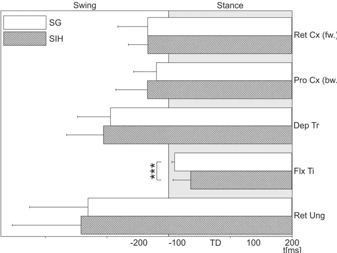
A summary of the results from all investigated stance muscles is shown in Fig. 2.8.
Only the FlxTi had a significantly changed latency between stepping on the ground and stepping into a hole, which confirms the results of Berendes et al. (2013). The other stance phase muscles were not influenced in their activation, as they are all activated before the TD.
Summarizing, these results indicate, with respect to the timing of muscle activation that only the FlxTi is affected by ground contact.