The role of the femoral chordotonal organ in motor control, interleg coordination, and leg kinematics in
Drosophila melanogaster
Inaugural-Dissertation zur
Erlangung des Doktorgrades
der Mathematisch-Naturwissenschaftlichen Fakultät der Universität zu Köln
vorgelegt von
Alexander Seldon Chockley
aus Cleveland, Ohio, Vereinigten Staaten von Amerika Köln, Deutschland
2020
Berichterstatter:
Prof. Dr. Ansgar Büschges PD Dr. Benjamin Altenhein
Tag der mündlichen Prüfung:
10. September, 2020
The years of work that went into this dissertation are dedicated to my mother,
Lynn Michelle Chockley, who always encouraged me
to go anywhere possible and do what I love
ABSTRACT ... I ZUSAMMENFASSUNG ... III LIST OF ABBREVIATIONS ... VI
1 GENERAL INTRODUCTION ... 1
1.1 LOCOMOTION AND SENSORIMOTOR NETWORKS IN INSECTS ... 1
1.2 PROPRIOCEPTION... 6
1.3 THE FEMORAL CHORDOTONAL ORGAN ... 9
1.4 FCO NEURONS IN LOCOMOTOR CONTROL ... 16
1.5 LINKING ENCODING AND FUNCTION ... 19
1.6 AIMS OF THE PRESENT WORK ... 21
2 GENERAL METHODS ... 23
2.1 CHOICE OF MODEL ORGANISM... 23
2.2 EXPERIMENTAL ANIMALS... 25
2.3 VNCPREPARATION... 27
2.4 LEG PREPARATION ... 27
2.5 FLUORESCENCE MICROSCOPY AND IMAGE PROCESSING ... 28
2.6 OPTOGENETICS EXPERIMENTS ... 28
3 CHAPTER 1: ANATOMY OF THE DROSOPHILA FCO ... 30
3.1 IDENTIFYING DRIVER LINES ... 30
3.2 RESULTS... 31
3.3 SUMMARY ... 41
4 CHAPTER 2: NEUROPEPTIDES IN LEG SENSORY ORGANS ... 43
4.1 SENSORY STRUCTURES AND NEUROPEPTIDES ... 44
4.2 METHODS ... 45
4.3 RESULTS... 46
4.4 DISCUSSION ... 49
5 CHAPTER 3: FCO ACTIVATION ... 52
5.1 INTRODUCTION ... 52
5.2 METHODS ... 54
5.3 RESULTS... 56
5.4 DISCUSSION ... 59
6 CHAPTER 4: THE ROLE OF THE FCO IN WALKING ... 61
6.1 INTRODUCTION ... 61
6.2 METHODS ... 62
6.3 RESULTS... 71
6.4 DISCUSSION ... 83
6.5 CONCLUSIONS ... 88
7 CHAPTER 5: FCO ENCODING OF PROPRIOCEPTIVE STIMULI ... 89
7.1 INTRODUCTION ... 89
7.2 METHODS ... 90
7.3 RESULTS... 93
7.4 DISCUSSION ... 94
8 SUMMARY AND CONCLUSIONS... 97
8.1 ANATOMY AND CONNECTIVITY OF THE FCO ... 97
8.2 ROLE OF THE FCO IN LEG MOVEMENTS AND MOTOR CONTROL ... 98
8.3 OUTLOOK AND FUTURE INVESTGATIONS ... 98
LIST OF FIGURES ... 101
LIST OF TABLES ... 102
REFERENCES ... 103
ACKNOWLEDGEMENTS... 115
ERKLÄRUNG ... 116
CURRICULUM VITAE ... 117
Abstract | i
Abstract
Legged locomotion in terrestrial animals is often essential for mating and survival, and locomotor behavior must be robust and adaptable in order to be successful. The behavioral plasticity demonstrated by animals’ ability to locomote across diverse types of terrains and to change their locomotion in a task-dependent manner highlights the flexible and modular nature of locomotor networks. The six legs of insects are under the multi-level control of local networks for each limb and limb joint in addition to over-arching central control of the local networks. These networks, consisting of pattern-generating groups of interneurons, motor neurons, and muscles, receive modifying and reinforcing feedback from sensory structures that encode motor output.
Proprioceptors in the limbs monitoring their position and movement provide information to these networks that is essential for the adaptability and robustness of locomotor behavior.
In insects, proprioceptors are highly diverse, and the exact role of each type in motor control has yet to be determined. Chordotonal organs, analogous to vertebrate muscle spindles, are proprioceptive stretch receptors that span joints and encode specific parameters of relative movement between body segments. In insects, when leg chordotonal organs are disabled or manipulated, interleg coordination and walking are affected, but the simple behavior of straight walking on a flat surface can still be performed. The femoral chordotonal organ (fCO) is the largest leg proprioceptor and monitors the position and movements of the tibia relative to the femur. It has long been studied for its importance in locomotor and postural control. In Drosophila melanogaster, an ideal model organism due its genetic tractability, investigations into the composition, connectivity, and function of the fCO are still in their infancy. The
Abstract | ii fCO in Drosophila contains anatomical subgroups, and the neurons within a subgroup demonstrate similar responses to movements about the femur-tibia joint. Collectively, the experiments laid out in this dissertation provide a multi-faceted analysis of the anatomy, connectivity, and functional importance of subgroups of fCO neurons in D.
melanogaster.
The dissertation is divided into four chapters, representing different aspects of this complex and intriguing system. First, I present a detailed analysis of the composition of the fCO and its connectivity within the peripheral and central nervous systems. I demonstrate that the fCO is made up of anatomically distinct groups of neurons, each with their own unique features in the legs and ventral nerve cord. Second, I investigated the neuropeptide profile of the fCO and demonstrate that some fCO neurons express a susbtance that is known to act as a neuromodulator. Third, I demonstrate the sufficiency of subsets of fCO neurons to elicit reflex responses, highlighting the role of the Drosophila fCO in postural control. Lastly, I take this a step further and look into the functional necessity of these neuronal subsets for intra- and interleg coordination during walking. The importance of the fCO in motor control in D. melanogaster has been considered rather minor, though research into the topic is very limited. In the work laid out herein, I highlight the complexity of the Drosophila fCO and its role in the determination of locomotor behavior.
Zusammenfassung | iii
Zusammenfassung
In den meisten terrestrischen Tieren ist zwei- oder vierbeinige Fortbewegung essentielle Grundlage für Verhaltensweisen wie Paarung und Überleben. Um dessen Erfolg zu garantieren, muss das Fortbewegungsverhalten robust und anpassungsfähig sein. Die Fähigkeit eines Tieres, seine Fortbewegung spezifisch gemäß den Ansprüchen diverser Untergründe zu verändern, demonstriert die Plastizität dieses Verhaltens und unterstreicht den flexiblen und modularen Aufbau der Fortbewegungsnetzwerke.
Bei Insekten, sowie bei vielen anderen Wirbellosen und Wirbeltieren, wird jedes Gelenk und jedes Bein, zusätzlich zur übergreifenden zentralen Kontrolle, von mehrstufigen lokalen Netzwerken kontrolliert. Letztere bestehen aus Rhythmus- generierenden Gruppen von Interneuronen, Motoneuronen und Muskeln, deren Aktivität von sensorischen Strukturen modifiziert und verstärkt wird. Propriozeptoren in den Gliedmaßen versorgen diese Netzwerke mit Informationen zu Bewegung und Position, welche essentiell für die Anpassungsfähigkeit und Robustheit des Verhaltens sind.
Propriozeptoren der Insekten formen eine diverse Klasse und die exakte Funktion jedes einzelnen Typs für die Kontrolle der Motorik ist weiterhin offen.
Chordotonalorgane, funktional analog zu den Muskelspindeln der Wirbeltiere, sind propriozeptive Streckrezeptoren an den Gelenken, die spezifische Parameter der relativen Bewegungen zwischen Segmenten signalisieren. Wenn die Chordotonalorgane im Insektenbein funktional ausgeschaltet oder manipuliert werden, beeinflusst dies das Laufen und die Koordination zwischen den verschiedenen Beinen; das simple geradeaus Laufen auf einer flachen Ebene ist jedoch weiterhin möglich. Das femorale Chordotonalorgan (fCO) ist der größte Propriozeptor im Bein
Zusammenfassung | iv und misst Position und Bewegungen der Tibia relativ zum Femur. Zahlreiche Studien belegen seine Relevanz für Fortbewegung und zur Kontrolle der Körperhaltung.
Untersuchungen zur Zusammensetzung, Konnektivität und Funktion des fCO sind bei Drosophila melanogaster, einem aufgrund seiner genetischen Formbarkeit idealen Modellorganismus, noch in den Kinderschuhen. Das fCO bei Drosophila ist in anatomische Untergruppen unterteilt und die Neuronen innerhalb einer produzieren ähnliche Signale in Reaktion auf Bewegungen des Femur-Tibia Gelenks.
Zusammengenommen liefern die Experimente dieser Dissertation eine vielseitige Analyse der Anatomie, Konnektivität und funktionalen Relevanz der Neuronen in Untergruppen des fCO in D. melanogaster.
Die vorliegende Dissertation ist in vier Kapitel unterteilt, welche sich mit unterschiedlichen Aspekten dieses komplexen und faszinierenden Systems beschäftigen. Im ersten Kapitel stelle ich eine detaillierte Analyse der Zusammensetzung des fCO und seiner Konnektivität im peripheren und zentralen Nervensystem dar. Ich zeige, dass das fCO aus anatomisch abgetrennten Gruppen von Neuronen besteht, jede mit individuellen Merkmalen im Bein und im ventralen Nervensystem. Der zweite Abschnitt beschäftigt sich mit dem Neuropeptid-Profil des fCO und belegt, dass einzelne Neurone des fCOs ein Neuropeptid exprimieren, welches ein bekannter Neuromodulator ist. Im dritten Teil demonstriere ich, dass Teilmengen der fCO Neurone ausreichend sind, um Reflexantworten auszulösen. Letzteres unterstreicht die Rolle des Drosophila fCOs für die Kontrolle der Körperhaltung. Im letzten Abschnitt erweitere ich dies auf die funktionale Notwendigkeit der neuronalen Untergruppen für die Kontrolle innerhalb eines und zwischen allen Beinen während des Laufens.
In der motorischen Kontrolle von D. melanogaster wurde das fCO bisher als von geringer Relevanz eingeschätzt, trotz oder wegen der begrenzten Anzahl der Studien
Zusammenfassung | v zu diesem Thema. In dieser Arbeit verdeutliche ich die Komplexität des Drosophila fCOs und seine Bedeutung für die Kontrolle des Laufens.
List of Abbreviations | vi
List of Abbreviations
AEP Anterior extreme position
BDSC Bloomington Drosophila Resource Center
BL Body length
ChAT Choline acetyltransferase CNS Central nervous system
CO Chordotonal organ
CPG Central pattern generator
CS Campaniform sensilla
DAPI 4′,6-diamidino-2-phenylindole
DLC DeepLabCut
DMSO Dimethyl sulfoxide DNA Deoxyribonucleic acid
DN Descending neuron
EMG Electromyogram
fCO Femoral chordotonal organ
Flp Flippase
FRET Fluorescence resonance energy transfer FRT Flippase recognition target
GECI Genetically encoded calcium indicator GFP Green fluorescent protein
GtACR1 Guillardia theta anion channelrhodopsin
IN Interneuron
IR Infrared
List of Abbreviations | vii
MN Motor neuron
NOMPC No mechanoreceptor potential C PBS Phosphate-buffered saline PBT PBS with Triton X-100 PEP Posterior extreme position
PFA Paraformaldehyde
RFP Red fluorescent protein RMSE Root mean squared error
RNA Ribonucleic acid
ROI Region of interest
RT Room temperature
TA Tyramine
tCO Tibial chordotonal organ
TNT Tetanus toxin
TRP Transient receptor potential (channel) UAS Upstream activating sequence
VNC Ventral nerve cord
Introduction | 1
1 General Introduction
1.1 Locomotion and Sensorimotor Networks in Insects
Locomotion, be it flight, swimming, or walking, is necessary for the survival of animals.
Legged locomotion in terrestrial animals is a ubiquitous, flexible, and highly optimized behavior (Alexander, 1989). With the ability to move, animals can access mates and resources, escape predators or find prey, and seek out habitats and environments that fit their current needs. Highlighting the importance of locomotion, most animals exhibit some degree of motility in their life cycle, and even sponges have been shown to move across the substrate (Bond and Harris, 1988).
In order to be effective, locomotion must be both resistant and resilient to environmental factors, such as unexpected perturbations, as well as internal factors, such as mismatch between the “intended” and actual output of the locomotor system (af Klint et al., 2010; Blaesing and Cruse, 2004; Hellekes et al., 2012). This behavioral plasticity is reflected in the ability of animals to change the employed type of locomotion in a task-dependent manner (Ashley-Ross and Lauder, 1997; Islam et al., 2006; Orger et al., 2008; Pick and Strauss, 2005). Particularly in terrestrial legged locomotion, walking movements must almost continuously be adapted to the irregular and often unpredictable nature of the substrate. Legged animals encounter many types of terrain and walking situations, so the networks and appendages supporting locomotion must be quite flexible to be successfully used under different conditions.
In addition to the dynamic appendages and physical characteristics that contribute to this flexibility, the underlying neuromuscular networks contain multiple levels of control loops and exhibit experience-dependent plasticity (Bidaye et al., 2018; Hooper and Bueschges, 2017).
Introduction | 2 The neural networks underlying limb movements contain many interneurons (INs), motor neurons (MNs), and sensory neurons. These networks are largely under the influence of central descending neurons (DNs), and additionally provide ascending innervation to these brain networks. In insects, movements of the six legs are under the multi-level control of local networks for each limb and limb joint in addition to over-arching central control of the local networks. These networks controlling locomotion consist of pattern-generating networks that activate MNs, which, in turn, activate the muscles of the appendages. These central pattern generators (CPGs) consist mainly of INs located in the thoracic ganglia, analogous to the spinal cord of vertebrates (Bassler and Buschges, 1998; Bidaye et al., 2018; Brown, 1911; Kiehn, 2006). Additional INs coordinate the activity of groups of muscles between segments of individual legs or between different legs. CPG activity is responsible for the rhythmic activation of MNs, and these MNs regulate the contractions of skeletal muscles that are attached to the skeleton either directly or via connective tissue, resulting in propulsion of the animal. This loop circuit is completed by various sensory structures in the limbs that monitor their position and movement, providing modifying or reinforcing feedback signals directly onto CPGs, coordinating INs, or MNs (Figure 1.1; Bidaye et al., 2018; Burrows, 1996; Buschges et al., 2011; Tuthill and Azim, 2018; Tuthill and Wilson, 2016).
Introduction | 3 Figure 1.1 Schematic of neuromuscular networks underlying movement creation and modification. Neurons (circles) making up central pattern generators (CPGs) create patterns of neural activity, which in turn activate or inhibit motor neurons (MNs). MNs activate muscles, producing movement. Sensory organs (multi-colored circles) read out the position and movement of segments, and project onto MNs and CPG neurons, modifying or reinforcing their activity. In this manner, movements are fine- tuned, and posture is adjusted.
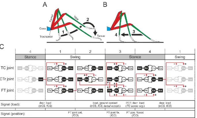
The neuromuscular networks underlying stepping in insect limbs contain all of these parts; this has been shown in larger insects such as stick insects and locusts but has yet to be empirically determined in Drosophila. A step, consisting of a swing phase (Figure 1.2A) and a stance phase (Figure 1.2B), requires the coordination of many CPGs, MNs, and muscles (Figure 1.2C). Muscles in antagonistic pairs, such as depressors and levators or extensors and flexors, alternate their activity to lift legs up, push them away from the body, move them down, and pull the body in the direction of movement. These phases of stepping and the transitions between them require precise temporal sequential activation of the MNs controlling the muscles that produce movement around each leg joint (Figure 1.2C). Signals from the locomotor network components
Introduction | 4 are used to coordinate activity between limb segments and whole limbs, and additional control is introduced by load and position sensors that signal the state of the limb or transitions between the phases of stepping (Bidaye et al., 2018; Mantziaris et al., 2017)
Figure 1.2 Schematic of stepping in an insect leg. A stepping cycle consists of a swing phase (A), in which the tarsus is lifted (1) and moved over the substrate (2); and a stance phase (B), in which the tarsus is placed on the substrate (3) and the body is moved relative to the tarsus (4). The muscles (squares) and CPGs (circles) required for these movements are shown in C, with antagonistic pairs in white and black. Movements around each joint are controlled by individual CPGs, which drive motor neuron and muscle activity. Sensory signals transduced by CS and the fCO are indicated in the lower rows. The fCO, phasically active during extension and flexion of the femur-tibia joint and tonically active at positions throughout the movement range, signals transitions between the beginning and end of each stepping phase (1-2 and 3-4) as well as during the transitions between the phases, signaled by a reduction in tibial extension (2-3). Figure used with permission from Bidaye et al. (2018)
This feedback from sensory organs monitoring the position and state of limbs, as well as from exteroceptive organs monitoring the surrounding environment, is essential for the adaptability and robustness of locomotor behavior. Exteroception can, in principle,
Introduction | 5 provide feedback about and modify motor output, especially during goal-directed movement. This is exemplified by optic flow, the pattern of apparent motion of the visual scene. As animals move, the movement of their surroundings relative to their body is represented as optic flow across photoreceptors; visual neurons have even been shown to encode information about locomotion behavior independently of vision, suggesting ascending signals from locomotor networks to the visual system (Fujiwara et al., 2017). When an animal is moving forward and the optic flow does not match forward motion, descending neurons from central brain networks provide modulatory signals to local locomotor networks; these locomotor networks can modify their output accordingly to match the optic flow (Poggio and Reichardt, 1976; Reichardt and Poggio, 1976). A prime example of this is the optokinetic reflex response in Drosophila, where animals will orient toward the direction of optic flow stimuli (Götz, 1968). Further, when animals are following olfactory plumes when foraging, the olfactory stimulus can provide feedback about self-motion and modulate motor programs (Jung et al., 2015).
Regarding precise positions and movements of the effectors and limbs producing locomotion, however, these systems cannot be effectively employed. Constant visual monitoring of limb position and movement, for example, is highly inefficient, and makes successful locomotion slow and difficult (McNeill et al., 2010). Humans with sensory neuropathy affecting proprioception can learn to perform coordinated movements, but still show large deviations in executed movement trajectories (Ghez et al., 1990; Ghez and Sainburg, 1995; Miall et al., 2018). This highlights the importance of limb proprioceptors in the production of robust, adaptive movements.
Introduction | 6
1.2 Proprioception
Proprioception provides information on the positions and movements of body parts that is crucial for the neural control loops regulating appendages, enabling precise and appropriate adjustments of posture and movements (Bassler, 1993; Bässler et al., 1982). Originally thought to be based on precise monitoring of motor output commands, postural control, fine motor control, and limb coordination are all highly dependent on specific mechanosensitive proprioceptors (Graham and Bassler, 1981;
Pearson, 1995; Steeves and Pearson, 1982). Proprioceptors exist throughout the animal kingdom, serving as sensors of movement, load, and body position (Tuthill and Azim, 2018; Tuthill and Wilson, 2016).
Vertebrates have three main classes of limb proprioceptors, each with distinct function.
Muscle spindles are stretch receptors located within skeletal muscle fibers and encode changes in length of the muscle (Ellaway et al., 2015). Golgi tendon organs act as load sensors by encoding relative tension at tendons (Mileusnic and Loeb, 2006). Lastly, joint receptors, or joint afferents, encode extreme positions of joint rotation (Burgess and Clark, 1969). In recent decades, evidence has additionally been put forth that cutaneous stretch receptors also play a significant role in proprioception (Collins and Prochazka, 1996; Collins et al., 2005; Edin and Abbs, 1991). It seems that there are evolutionary advantages of having proprioceptors that encode information about these aspects of body position and movement, as invertebrates have many analogous proprioceptors in their limbs and appendages.
In insects, proprioceptors are highly diverse, and the exact role of each type in motor control has yet to be determined. Insect legs contain five main classes of proprioceptors: campaniform sensilla (CS), hair plates, bristles, multipolar receptors, and chordotonal organs (COs). CS, analogous to Golgi tendon organs, are the major
Introduction | 7 load sensors in insects. Compression of the cuticle is transduced by CS neurons when self-generated movements are resisted or body load is changed (Delcomyn et al., 1996).
Bristles, or trichoid sensilla, are mainly exteroceptive mechanosensors that function as tactile surface receptors on the cuticle. Some bristles contain blunt, hollow shafts that are connected to gustatory neurons; these bristles belong to a different class called basiconic sensilla (Hannah-Alava, 1958; Tuthill and Wilson, 2016). Hair plates, structurally similar to mechanosensory bristles, are small regions of many densely packed mechanosensitive “hairs” that activate their underlying neurons when they are deflected. Using a transduction mechanism similar to bristles, hair plates get their main function from their location on the body—on the cuticular surface near the creases of joints—and are deflected when joint angles reach their extremes (Tuthill and Wilson, 2016). Multipolar receptors are a rather under-researched class of mechanosensors. They are non-ciliated neurons with multiple dendritic branches and are found throughout the body embedded in different tissues and structures. Their main function as proprioceptors is through their encoding of parameters related to stretching of tissue, though their exact role and details of their transduction and signaling mechanisms are not well understood. COs, functionally analogous to vertebrate muscle spindles (and mildly to cutaneous stretch receptors), are proprioceptive stretch receptors that span joints and encode positions of and relative movements between body segments. CO neurons encode specific parameters of these relative movements and positions, and as such are rather diversely employed proprioceptors. Among the most well-researched COs are the Johnston’s organ of the antenna, the largest CO in Drosophila and main detector for sound, gravity, and wind;
and the femoral chordotonal organ, a major player in the control of locomotion (Field and Matheson, 1998). Upon first glance, these many proprioceptor types appear to
Introduction | 8 have their own function; however, the leg proprioceptive system of insect contains a surprising amount of redundancy of function.
Theoretically, some of these proprioceptors could effectively replace others while maintaining a functional proprioceptive system. Bristles, for example, could effectively be replaced by CS; bristle neurons are activated by their deflection, via exogenous particles coming into contact with the legs or the legs touching against something. CS are highly sensitive to cuticular deformations and could functionally perform the same task, assuming the stimulus deforms the cuticle. Hair plates are responsible for measuring extreme joint positions, but CO neurons encoding joint angles do the same.
CS can read out muscle activity, and CO neurons provide a proxy of this in encoding actual movement about joints. Lastly, in elegant experiments artificially increasing body load in Drosophila melanogaster, Mendes et al. (2014) demonstrated that flies can adapt their posture and locomotion to two-fold body load increases. They further demonstrate the necessity of the fCO in these postural adaptations. Despite these redundancies in the proprioceptors of the legs, these many different organs exist. This suggests that they, as individual organs, provide specific information to the system that may be lost without them. If these signals were somehow pooled together by a more generalized proprioceptive organ, specific information might be lost; while a hair plate can signal an extreme position in a joint, it cannot signal anything between the extreme positions—that is the role of the fCO.
In insects, when leg proprioceptors are disabled, interleg coordination and walking are affected, but the simple behavior of straight walking on a flat surface can still be performed (Cruse et al., 1984; Mendes et al., 2013; Mendes et al., 2014; Usherwood et al., 1968). Disabling leg COs alone, however, produces effects similar to disabling multiple leg sensory structures (Field and Matheson, 1998; Mendes et al., 2013;
Mendes et al., 2014). Only two COs can be found on the legs, the small tibial CO (tCO)
Introduction | 9 and the much larger fCO; many of the detrimental effects from disabling leg proprioceptors likely come from disabling the fCO.
1.3 The Femoral Chordotonal Organ
The femoral chordotonal organ (fCO; Bässler, 1965) has long been studied for its involvement in locomotor control. It consists of a group of cells with mechanosensitive dendrites in the femur and encodes various movement parameters (position, velocity, and acceleration) about the femur-tibia joint (Figure 1.3A; Field and Matheson, 1998).
fCO dendrites attach to either a receptor apodeme attached to the tibia or to surrounding muscles and the distal femoral epicuticle. When the tibia rotates about the femur-tibia joint and strain on the receptor apodeme changes, mechanosensitive ion channels (Akitake et al., 2015; Cheng et al., 2010) in the dendritic tips open, leading to changes in membrane potential in the neurons (Field and Matheson, 1998). Due to its functional connectivity with various components of locomotor networks, signals from the fCO play a major role in inter- and intraleg coordination during walking.
1.3.1 Signal Transduction and Transmission
The fCO, as all COs, consists of one or more closely packed groups (scoloparia) of sensory neurons embedded in a matrix of supporting cells. In Drosophila, around 152 primary sensory neurons are grouped into three scoloparia attached to femoral muscles and epicuticle via connective apodemes (Figure 1.3B; Mamiya et al., 2018;
Shanbhag et al., 1992). Each scoloparium consists of paired sensory neurons organized into scolopales. At the proximal end of the scolopales lie the neurons’ cell bodies, and they project their dendrites distally into a scolopale cell, which attaches to the apodeme via an attachment cell (Field and Matheson, 1998). fCO dendrites contain a cilium at
Introduction | 10 their distal end within the scolopale cell, and it is at the level of this cilium where signal transduction occurs.
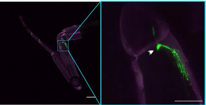
Figure 1.3. Schematic and detailed view of the femoral chordotonal organ in D. melanogaster. (A) Schematic showing segments of the leg and the placement of the femoral chordotonal organ (fCO) and tibial chordotonal organ (tCO), as labeled by iav- GAL4. (B) Confocal laser scanning microscope view of iav-GAL4; UAS-RedStinger (left) showing the locations of fCO cell nuclei, and iav-LexA; LexAop-myrGFP (right) showing
Introduction | 11 the membranes of fCO neurons; three lobes of the fCO can be seen—one elongated, thin lobe (asterisk), and two more round, clustered lobes (brackets); color in (B) represents relative z-stack depth in maximum intensity z-projections.
Exactly how mechanical signals are transduced by COs remains debated, but both theories include mechanosensitive ion channels located in the cilium of the dendrite.
Several such channels have been implicated in the proper functioning of COs, mostly in the Johnston’s organ, and they belong to the transient receptor potential (TRP) superfamily of ion channels. The two channels in question are NOMPC (no mechanoreceptor potential C; Gong et al., 2013; Göpfert et al., 2006) and a heterodimeric TRPV channel that is formed by the proteins Inactive and Nanchung (iav/nan; Warren and Matheson, 2018). Both of these are mechanically activated, cation permeable ion channels localized to CO dendritic cilia. It has been proposed, but not empirically determined, that when the cilia are stretched or relaxed, mechanical gates on the ion channels open or close and thus regulate the influx of cations into the dendrite. In this way, the mechanical stimulus is coupled to the sensory neuron, transduced into a receptor potential, and the receptor potential is coded into an action potential (Field and Matheson, 1998; French, 1992).
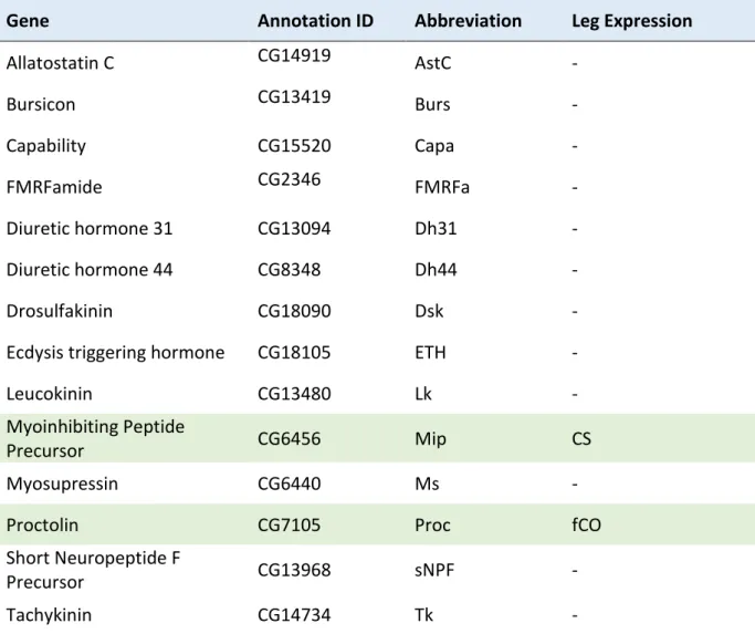
The fCO contains primarily excitatory neurotransmitters, and evidence hints toward the presence of biogenic amines and neuropeptides. In the locust Schistocerca gregaria (Lutz and Tyrer, 1988), some afferent neurons from the fCO are immunoreactive to choline acetyltransferase (ChAT) antiserum. In Drosophila, Johnston’s organ neurons are cholinergic (Sivan-Loukianova and Eberl, 2005), and a driver line under control of ChAT labels the fCO (Mamiya et al., 2018). Further, a subset of fCO neurons in D. melanogaster can be labeled using genetic driver lines based on tdc2 (tyrosine decarboxylase) expression (Pauls et al., 2018). Tyrosine decarboxylase is necessary for the synthesis of Tyramine (TA) from tyrosine.
Introduction | 12 Interestingly, TA can function as a neurotransmitter and a neuromodulator depending on the receptor to which it binds, and it is known to regulate a variety of physiological functions in invertebrates (Zhang and Blumenthal, 2017). Another biogenic amine, serotonin, has also been found in the fCOs of S. gregaria and D. melanogaster and has been shown to affect locomotor behavior (Howard et al., 2019; Lutz and Tyrer, 1988).
Lastly, the presence of neuropeptides, a family of molecules that act as neurohormones and neuromodulators, has been suggested due to the finding of dense-core vesicles in an electron microscopy study of the fCO of D. melanogaster (Shanbhag et al., 1992).
The finding of dense-core vesicles in fCO axon by Shanbhag et al. (1992) was coupled with the discovery of a peripheral processing center of sorts between fCO neurons. A transmission electron microscopy analysis of the fCO in D. melanogaster led to the discovery that all axons from the fCO form a peripheral glomerulus before entering the leg nerve. This glomerulus contains reciprocal and serial synapses, and rarely contained monosynapses between fCO axons. It was in this glomerulus that dense-core vesicles were identified in addition to the clear vesicles, in which classical neurotransmitters are found. The complex neurochemical and neuroanatomical composition of the fCO, specifically in Drosophila, deserves the attention of researchers, especially when looking at the transmission of signals from the fCO into the central nervous system. It remains to be investigated exactly what occurs in this glomerulus, but peripheral transformations of signals before they reach axon terminals could throw a wrench into our young understanding of the Drosopholid fCO.
Introduction | 13
1.3.2 Encoding of Proprioceptive Signals
Extracellular single-unit recordings in Cuniculina impigra were the first to show that fCO neurons encode various parameters of joint movement and position. Cells were found that responded to positions, velocities, and accelerations, with some cells sensitive even to positive or negative velocities or accelerations (Bueschges, 1994;
Hofmann and Koch, 1985; Hofmann et al., 1985). Position sensitive neurons respond tonically with increased firing rates as the joint reaches their preferred angle, and velocity sensors respond phasically to their preferred velocity (Bueschges, 1994;
Hofmann et al., 1985). Acceleration receptors tend to respond with single or a few spikes (Hofmann and Koch, 1985). Intriguingly, many cells respond to combinations of these parameters while still maintaining directional selectivity (Bueschges, 1994;
Hofmann et al., 1985). In a study in S. gregaria, white-noise stimuli were applied to the fCO and responses were analyzed with a Wiener kernel analysis to investigate non- linear dynamics in neural response characteristics. In their analysis, (Kondoh et al., 1995) concluded that fCO neurons can change stimulus encoding properties depending on the patterns of stimuli applied. This is also reflected in the presence of response hysteresis in fCO neurons, a phenomenon in which neurons fire at a certain rate for a given joint position when approached from one direction and fire at a different rate when the position is approached from the other direction (Field and Matheson, 1998;
Mamiya et al., 2018).
Little work has been done on the encoding properties of fCO neurons in Drosophila, but a recent seminal study used calcium imaging to demonstrate similarities to other insects studied. In larger insects, such as the stick insect and locust, the fCO is normally stimulated by opening the leg, clamping the receptor apodeme of the fCO around a hook, and moving the hook back and forth with a piezoelectric actuator to simulate the
Introduction | 14 natural stretching and relaxation of the fCO during tibial movement. Because of its small size, this is nearly impossible in D. melanogaster; however, one advantage of this animal is the ability to express genetically encoded calcium sensors that change their fluorescence intensity in response to the internal calcium concentration of a neuron.
In this manner, Mamiya et al. (2018) were able to record activity of fCO neurons at their axon terminals in the VNC while moving the tibia with a magnet. They showed that the fCO also contains tonically and phasically active neurons responding to position and velocity, as well as some cells that are directionally sensitive velocity sensors. Unfortunately, they were unable to address the presence of acceleration- sensitive neurons due to the kinetics of the calcium sensor.
Intriguingly, they imaged activity of a majority of fCO neurons (using a driver line based on inactive) and deduced all patterns of activity using a correlation-based K- means clustering analysis. This analysis provided an unbiased grouping of response patterns based on pixel intensity changes throughout fCO axon terminals in a single hemiganglion. From this, the authors found that pixels that were grouped in their responses corresponded to different anatomical projection patterns of fCO afferents.
These projection patterns, termed club, claw, and hook (Phillis et al., 1996), were shown to make up the entire pattern of inactive expression in the VNC (Mamiya et al., 2018). Similar to the stick insect and locust, the D. melanogaster fCO contains tonic position sensors and phasic velocity sensors, in addition to phasically-responding vibration-sensitive neurons, as can be found in the dorsal scoloparium of the stick insect fCO (Kittmann and Schmitz, 1992). From the analysis described above, no phasic-tonic responses were noted, though this could be an artefact of the clustering analysis. Interestingly, fCO neuron projection patterns clearly corresponded to response types. Claw neurons encode the angle of the femur-tibia joint with tonic responses. Club neurons act as phasically responding, directionally insensitive
Introduction | 15 movement sensors. Hook neurons also respond to movement, but exhibit directional sensitivity; the hook neurons tested by (Mamiya et al., 2018) were mostly flexion sensitive, though a more detailed response analysis using other subpopulations of hook neurons is still needed to parse out their preferred stimuli.
Matheson (1992) demonstrated in a locust that neurons in the fCO that have similar response characteristics demonstrate morphological similarities. The morphological distinction between response types in the locust, however, is not as clear as that seen in D. melanogaster (Mamiya et al., 2018; Shanbhag et al., 1992). In addition to this morphological grouping, Drosophila is thus far unique regarding the presence of a peripheral hub of fCO synapses in the leg; its role in fCO signal processing, however, has not been investigated (Shanbhag et al., 1992). Whether the different neuron types are represented differently in the glomerulus remains an open question.
1.3.3 fCO Connectivity and Signal Processing
In the stick insects Carausius morosus and C. impigra and the locust species Locusta migratoria and S. gregaria, the animals in which pioneering work on the fCO was done, fCO afferents project ipsilaterally within their segmental ganglion (Braunig et al., 1981; Hustert, 1978; Pfluger et al., 1988). In D. melanogaster, the majority of fCO neurons project locally to their respective hemiganglion, some have interhemiganglionic projections, and some (~3%) ascend directly to the brain (Tsubouchi et al., 2017). In the local projection areas in the VNC, fCO neurons have been shown to affect MNs, spiking and non-spiking local INs, and intersegmental INs (Bassler, 1993; Burrows, 1985b, 1994). In S. gregaria, it has been shown that fCO neurons connect with tibial extensor and flexor MNs, sometimes monosynaptically, fitting with their role of the major mediators of femur-tibial joint movements (Burrows, 1987; Field and Matheson, 1998). Connections, both direct and indirect,
Introduction | 16 with local INs are where transformations of fCO signals and effects on other local locomotor and proprioceptor networks occur, with these largely being responsible for the fCO’s role in intraleg coordination. Individual fCO afferents have also been shown to synapse with both MNs and INs; additionally, afferents from multiple fCO neurons sometimes converge on single INs (Burrows, 1985a; Field and Matheson, 1998). The role of the fCO in interleg coordination likely results from connections with intersegmental INs, and the fCO has been shown to have excitatory effects on such cells (Laurent and Burrows, 1988). On top of these local connections between the fCO and MNs or INs, another layer of processing occurs between pools of INs.
There are many parallel pathways of information processing that exist between INs that receive direct input from fCO neurons, and it is thought that behavioral flexibility is largely dependent on this level of signal processing (Field and Matheson, 1998). By way of polysynaptic connections via inhibitory INs, the fCO can exert inhibitory effects on MNs (Burrows and Siegler, 1982). It has further been shown that a single non- spiking IN can simultaneously excite one MN while inhibiting another, adding more degrees of freedom to the potential effects of fCO signals on locomotor networks (Burrows, 1989). Taken together, these examples illuminate only a portion of the myriad parallel processing pathways from fCO neurons via local and intersegmental INs onto other ascending and descending INs and MNs controlling leg muscles. In this regard, understanding the entirety of the locomotor networks receiving and transforming signals from the fCO is quite a daunting task.
1.4 fCO neurons in locomotor control
In D. melanogaster, owing to recent advances in neurogenetic techniques, research addressing the role of the fCO in locomotor behavior is just getting underway. In the larger insects studied, however, experiments using reduced, tethered preparations
Introduction | 17 have shed light on the functional connectivity between the fCO and other locomotor network components. Moreover, investigations of the fCO’s involvement in interleg coordination and reflexes has set the stage for studies on the roles of individual fCO neuron types in motor control.
Drosophila leg proprioceptors have been shown to affect coordination parameters during walking. Using broad manipulations with tetanus toxin light chain (TNT), an inhibitor of synaptic transmission, and mechanoreceptor mutants, it has been shown that removal of leg sensory signals and, specifically, fCO signals leads to modified kinematic parameters during walking (Mendes et al., 2013). Synaptic inhibition using a pan-sensory driver line (5-40-GAL4) with expression restricted to the femur and proximal tibia (dac-Flp, UAS-FRT-stop-FRT-TNT; see section 3.2) caused flies to walk with longer and less linear steps, as well as increased swing duration. In flies expressing the Nanchung mutation nan36a and, therefore, with non-functional COs, step length and swing duration were also increased, and stances were less linear compared to wild- type flies, though not to the extent caused by the pan-sensory manipulations.
Represented by gait patterns, which were defined by the number of tarsi concurrently on the ground at any given time during walking, flies exhibited largely normal interleg coordination. The authors deduced that, because “gait parameters and interleg swing phase coupling were largely unimpaired”, interleg coordination in the fly is “not dominated by sensory feedback” (Mendes et al., 2013). While these conclusions are arguable based on the clear walking impairments shown, the main takeaway from this study is that removal of leg sensory activity and leg CO mechanotransduction does result in altered leg movement and coordination parameters, if not to a surprisingly large extent.
Intriguingly, the changes seen in CO-deficient flies largely matched those seen in flies lacking functional CS, hair plates, bristles, and COs. The functional role of CS, hair
Introduction | 18 plates, and COs in motor networks has been previously demonstrated in multiple insects (see Tuthill and Wilson, 2016); these results thus beg the question of the usefulness of these sensors in the walking behavior of the fly. This could partially be explained by the methodology used—the authors restricted TNT expression to the limbs using a transcription factor that is active in leg imaginal discs, dachshund (dac;
Giorgianni and Mann, 2011; Rauskolb, 2001). Expression of dac starts in the larval stage of development, leaving open the possibility of plasticity in the functionally redundant sensory networks of the leg (Mardon et al., 1994). Thus, such experiments would ideally be repeated using transient inhibition of these sensory neurons.
However, the results laid out in Mendes et al. (2013) suggest that these leg sensory organs—COs excluded—play only a minor role in locomotor coordination in D.
melanogaster.
As sensory signals representing leg joint angles and movements are thought to mediate coupling between local motor control circuits and interleg coordination (Brunn and Dean, 1994) and the fCO is clearly necessary for normal walking behavior (Mendes et al., 2013), the question remains of the importance of the distinct types of fCO proprioceptive neurons in this behavior. As only broad manipulations of fCO activity are possible in larger insects, Drosophila provides a novel way to investigate sub- population level effects of fCO neurons in freely behaving animals (Mamiya et al., 2018).
The fCO of Drosophila contains neurons that respond to position and velocity, as well as some velocity-sensitive cells that are directionally selective. Mathematically speaking, velocity and acceleration are the first and second derivatives of position, and neural networks theoretically have the ability to perform this calculation (Areas et al., 2001). However, this would likely require a few processing steps within central networks and, consequently, extra time. Drosophila walk at a range of speeds between
Introduction | 19 roughly 4 and 15 body lengths (BL) per second, or roughly 8-30 mm/s, leading to a step cycle duration of 60-300 ms (Wosnitza et al., 2013). In the shortest of these, signal transfer through a few chemical synapses would simply take too long (up to about 5 ms per synapse). Coupled with the presence of neurons that encode individual first- and second-order derivatives of positional information, this suggests that specific information on speed, velocity, and acceleration of leg movements are useful for the regulation of locomotion.
1.5 Linking Encoding and Function
The encoding of tibial position, velocity, and acceleration by fCO neurons suggests distinct roles of these cells in the processing and regulation of limb movements. First, tonically active position sensors are likely less important for walking as they are for postural control. Simply put, signaling the current angle of a joint during stepping movements seems to be not very informative unless changes in angle can be inferred from these signals. In D. melanogaster, claw neurons fire with increasing frequency as the femur-tibia joint reaches its extreme positions and are less active (or not active) in the middle of the joint’s range of motion (Mamiya et al., 2018). Response hysteresis seen in these neurons can provide information to locomotor networks about the direction of movements based solely on positional information, though this would pose a computational issue for downstream circuits without combined input from directionally selective hook neurons. As such, claw neurons alone are likely not important for signaling the current phase of the step cycle to other limbs and to central networks. Interestingly, communication of extreme joint angles is already done by hair plates, so the necessity of such signals from fCO neurons during walking is still unclear.
Introduction | 20 Phasically responding speed sensors (no directional sensitivity), club neurons in Drosophila, likely simply signal that the femur tibia joint is changing its angle. This could be used to distinguish between the phases of the stepping cycle; however, without directional information, these signals can only communicate that a change in position is occurring. It is likely that signals from these neurons are used to signal to neighboring legs, for example, when it is appropriate to initiate or end a swing or stance phase. Directionally sensitive speed sensors, on the other hand, could signal when a leg or leg joint is approaching the end of a step phase or when the leg is in stance or swing phase. Encoding of this by hook neurons in Drosophila is likely to be important for interleg coordination and for determining the switch from stance to swing (and swing to stance). Notably, these two phases involve different movements in Drosophila legs because the three leg types (front, middle, and hind) are not parallel to each other with respect to their relative angles to the thorax. The swing phase in front legs, for example, involves an extension movement as the femur-tibia joint changes from fully flexed to fully extended; in hind legs, the swing phase involves movement from an extended to a flexed position. Therefore, signals from neurons encoding the same velocities in these different limbs would need to be differentially processed in central networks.
Acceleration sensors have yet to be demonstrated in the fCO of D. melanogaster, but they are likely to play a role in signal lift-off and touch-down events of the tarsus in addition to vibration-related stimuli from the substrate. These neurons could be useful for stability during walking, but it is likely that they are not essential for walking behavior. It has been shown, however, that vibration-sensitive neurons of the fCO (club neurons) exhibit a topographical frequency tuning along their VNC axon terminals, so information about vibrational stimuli in the femur tibia joint must be useful to the underlying neural networks (Mamiya et al., 2018).
Introduction | 21 It is currently unclear if these signals from fCO neurons are preprocessed before entering VNC. Potential transformation of signals in the glomerulus, for example, indicate that the calcium activity induced by tibial movements as seen by (Mamiya et al., 2018) could represent signals that have already been shaped by synaptic transmission or neuromodulation. Further, it remains to be determined if all fCO neurons are important for walking or if some are specifically important for other behaviors. The present dissertation addresses these questions.
1.6 Aims of the Present Work
In the present dissertation, I aimed to answer some of the open questions regarding the fCO of D. melanogaster and its importance for locomotor behavior. The dissertation is divided into four chapters, representing different aspects of this complex and intriguing system. In the first chapter, I aimed to investigate the composition of the fCO and its connectivity within the peripheral and central nervous systems. For this, I identified various transgenic driver lines with expression within subsets of fCO neurons. Such driver lines provide the ability to break up the fCO into parts and allow for investigations into the functional and anatomical characteristics of the organ as a whole. Using anatomical techniques befitting transgenic model organisms, I demonstrate that the fCO is made up of anatomically distinct groups of neurons, each with their own unique features in the legs and VNC. Moreover, I investigated the peripheral and central connectivity of these neurons. In the second chapter of this dissertation, I investigated the neuropeptide profile of the fCO and demonstrate that, in addition to its central and peripheral connectivity, the fCO contains a neuropeptide that acts as a neuromodulator. In the third chapter of this dissertation I demonstrate the sufficiency of subsets of fCO neurons for the production of reflex responses. For this, I activated specific neurons in the fCO and measured leg
Introduction | 22 movements and muscle activity to demonstrate that stimulation of parts of the fCO is sufficient for producing reflex movements and muscle activity. In the fourth chapter, I take this a step further and look into the functional necessity of these neuronal subsets.
For this, I inhibited these neurons in fully intact, behaving flies during natural walking behavior and investigated their effects on leg movements and coordination. I demonstrate that some, but not all, fCO neurons are important for normal walking behavior. In the final chapter of this dissertation, I describe my attempts to perform live imaging of neural activity in the periphery. While these experiments were cut short due to methodological difficulties, I have set the stage for future investigations into the activity of fCO neurons at the level of their cell bodies. Based on the peripheral connectivity of the fCO, it is essential that we understand encoding of proprioceptive stimuli in the periphery. Collectively, the experiments laid out in this dissertation provide a multi-faceted analysis of a proprioceptor that has previously received much attention in other insects. Moreover, they highlight the importance of the fCO in locomotor behavior of D. melanogaster.
General Methods | 23
2 General Methods
The following section contains a detailed description of the methods that were employed in the experiments described in this dissertation. In cases where methods were used only for experiments described in an individual chapter, the applicable methods are described in the chapter itself.
2.1 Choice of Model Organism
Despite their small size, Drosophila make excellent model organisms for locomotion research thanks to the extensive genetic tools available, low cost, ease of access, and very large foundation of knowledge of their development and physiology. Moreover, they are highly active animals, and they locomote often even when confined or restrained (e.g., Ali et al., 2011; Jones and Grotewiel, 2011; Woods et al., 2014;
Wosnitza et al., 2013). D. melanogaster, thanks to decades of work in genetics, provides us with an almost fully tractable model nervous system. There are countless, ever-developing genetic tools available in D. melanogaster that make manipulations of the animal’s anatomy and physiology rather straight-forward. The primary genetic tool used in the present work is the GAL4/UAS system (review: Duffy 2002). GAL4 is a transcription factor found in yeast that binds DNA and activates transcription via recruitment of the RNA polymerase transcriptional complex. The GAL4 sequence can be inserted into the Drosophila genome behind known promoters, for example, to restrict expression to a certain cell type or population. The GAL4 protein then binds DNA in those cells in which it is expressed and activates the transcription of downstream sequences. The DNA binding domain of GAL4 binds specifically to a so- called upstream activating sequence (UAS), which can be inserted into the genome artificially. Downstream of the UAS sequence, we can place a reporter gene, effector,
General Methods | 24 or other genes of interest. Once the DNA binding domain is bound to the UAS sequence, the activation domain of GAL4 then recruits RNA polymerase. Further, due to the two domains being necessary to activate UAS transcription, it is possible to express each part of GAL4 behind different promoters to find overlap and to create very sparse labeling (the split-GAL4 system; Luan et al. 2006). Additionally, another yeast transcription factor, GAL80, can be used to suppress GAL4, adding another level of control to the system (Lee and Luo, 1999). This can be further modulated via selective expression or using a temperature-sensitive variant of GAL80, GAL80ts, which is inert below temperatures of around 30 °C (McGuire et al., 2004). The binary GAL4/UAS system can also be used concurrently with the LexA/LexAop system, a similar binary transcription system, without any cross-effects (Yagi, Mayer, and Basler 2010). A multitude of reporter genes and genes of interest can be driven by these systems, including interfering RNAs, channelrhodopsins, fluorescent calcium or voltage indicators, fluorophores, and various proteins of interest. These can all be used for both morphological and functional studies of multiple systems in Drosophila.
Importantly, these systems allow researchers to alter the activity of neurons in a completely non-invasive manner—this is the main basis for using this organism in the present work.
General Methods | 25
2.2 Experimental Animals
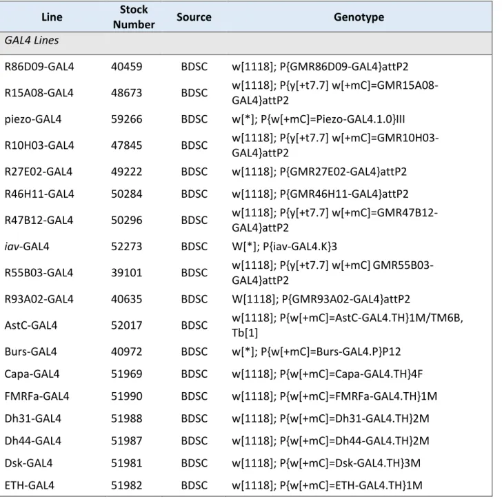
The genotypes and sources of all transgenic fly lines used in the present work are listed in Table 1. For all experiments described herein, I used female flies aged 3-8 d post- eclosion. Animals were reared on a standard yeast-based medium (Backhaus et al., 1984) at 25 °C on a 12-h/12-h light/dark cycle.
Table 1. Transgenic animals
Line Stock
Number Source Genotype
GAL4 Lines
R86D09-GAL4 40459 BDSC w[1118]; P{GMR86D09-GAL4}attP2 R15A08-GAL4 48673 BDSC w[1118]; P{y[+t7.7] w[+mC]=GMR15A08-
GAL4}attP2
piezo-GAL4 59266 BDSC w[*]; P{w[+mC]=Piezo-GAL4.1.0}III R10H03-GAL4 47845 BDSC w[1118]; P{y[+t7.7] w[+mC]=GMR10H03-
GAL4}attP2
R27E02-GAL4 49222 BDSC w[1118]; P{GMR27E02-GAL4}attP2 R46H11-GAL4 50284 BDSC w[1118]; P{GMR46H11-GAL4}attP2 R47B12-GAL4 50296 BDSC w[1118]; P{y[+t7.7] w[+mC]=GMR47B12-
GAL4}attP2
iav-GAL4 52273 BDSC W[*]; P{iav-GAL4.K}3
R55B03-GAL4 39101 BDSC w[1118]; P{y[+t7.7] w[+mC]GMR55B03- GAL4}attP2
R93A02-GAL4 40635 BDSC W[1118]; P{GMR93A02-GAL4}attP2
AstC-GAL4 52017 BDSC w[1118]; P{w[+mC]=AstC-GAL4.TH}1M/TM6B, Tb[1]
Burs-GAL4 40972 BDSC w[*]; P{w[+mC]=Burs-GAL4.P}P12 Capa-GAL4 51969 BDSC w[1118]; P{w[+mC]=Capa-GAL4.TH}4F FMRFa-GAL4 51990 BDSC w[1118]; P{w[+mC]=FMRFa-GAL4.TH}1M Dh31-GAL4 51988 BDSC w[1118]; P{w[+mC]=Dh31-GAL4.TH}2M Dh44-GAL4 51987 BDSC w[1118]; P{w[+mC]=Dh44-GAL4.TH}2M Dsk-GAL4 51981 BDSC w[1118]; P{w[+mC]=Dsk-GAL4.TH}3M ETH-GAL4 51982 BDSC w[1118]; P{w[+mC]=ETH-GAL4.TH}1M
General Methods | 26 Lk-GAL4 51993 BDSC w[1118]; P{w[+mC]=Lk-GAL4.TH}2M
MIP-GAL4 51984 BDSC w[1118]; P{w[+mC]=Mip-GAL4.TH}2M Ms-GAL4 51986 BDSC w[1118]; P{w[+mC]=Ms-GAL4.TH}6Ma Proc-GAL4 51972 BDSC w[1118]; P{w[+mC]=Proc-GAL4.TH}6M sNPF-GAL4 51991 BDSC P{w[+mC]=sNPF-GAL4.TH}2, w[1118]
UAS Lines
UAS-mCD8::GFP 32189 BDSC y1 w* P{10XUAS-IVS-mCD8::GFP}su(Hw)attP8 UAS-GtACR1 -
A.
Claridge- Chang
w1118;;P{20x-UAS-GtACR1-EYFP}attp2, Sb[1]
UAS-FRT-stop-FRT-
mCD8::GFP 30032 BDSC
y[1] w[1118]; Pin[1]/CyO;
P{w[+mC]=UAS(FRT.stop)mCD8-GFP.H}14, P{w[+mC]=UAS(FRT.stop)mCD8-GFP.H}21B UAS-syt.eGFP 6926 BDSC w*; P{ UAS-syt.eGFP}3
UAS-RedStinger 8545 BDSC
P{w+mc=UAS-RedStinger}3, w1118
UAS-trans-Tango 77124 BDSC
y[1] w[*] P{y[+t7.7] w[+mC]=UAS-myrGFP.QUAS- mtdTomato-3xHA}su(Hw)attP8; P{y[+t7.7]
w[+mC]=trans-Tango}attP40 LexA/LexAop Lines
iav-LexA 52246 BDSC
y[1] w[*]; wg[Sp-1]/CyO, P{Wee-P.ph0}Bacc[Wee- P20]; PBac{y[+mDint2] w[+mC]=iav-
lexA::p65}VK00013 LexAop-myrGFP
(III) 32209 BDSC
y[1] w[*]; wg[Sp-1]/CyO, P{Wee-P.ph0}Bacc[Wee- P20]; PBac{y[+mDint2] w[+mC]=iav-
lexA::p65}VK00013 Other
Dac[RE]-Flp - R. Mann Genotype not provided Berlin-K 8522 BDSC Wild type
BDSC, Bloomington Drosophila Resource Center, Bloomington, IN, USA
General Methods | 27
2.3 VNC Preparation
For the dissection, staining and visualization of VNCs, flies were anesthetized on ice and briefly (< 1 min) soaked in 70% EtOH to de-wax the cuticle. VNCs were dissected out in 0.1 M PBS and fixed in 4% paraformaldehyde (PFA) for 30 min on ice. Following three 15-min washing steps in 0.5% Triton X-100 in 0.1 M PBS (0.5 % PBT), VNCs were blocked in 10% normal goat serum (blocking solution; ThermoFisher Scientific, Waltham, MA) in PBT for 20 min at RT and incubated for 48 h at 4° C in primary antibodies diluted in blocking solution (mouse anti-nc82, 1:500; rabbit anti-GFP, 1:500). After three washes in PBT, they were incubated for 48 h at 4° C in secondary antibodies (goat anti-mouse AlexaFluor 633, 1:500; goat anti-rabbit AlexaFluor 488, 1:500) followed by three washes in PBT, before being mounted in Vectashield (Vector Laboratories, Burlingame, CA) and coverslipped.
2.4 Leg Preparation
Whole flies were skewered on insect pins, briefly soaked in 70% EtOH to de-wax the cuticle, fixed in 4% PFA for 45 min on ice, and washed in 0.1 M PBS (3 x 15-min). After washing, the tibia, tarsus, and distal femur were removed with microscissors. Legs were then mounted in Vectashield (Vector Laboratories) and coverslipped. For immunohistochemistry experiments in legs, the following was done after fixation in PFA: flies were then incubated in a blocking solution of 10% normal donkey serum (Jackson ImmunoResearch Laboratories, West Grove, PA) and 5% PBT for 2 h at RT with nutation then incubated with primary antibody (mouse anti-Choline acetyltransferase [ChAT], 1:50) in 0.5% PBT and 1% donkey serum for 72 h at 4° C.
After 4 washes in 0.5% PBT of 1.5-2 h each at RT, flies were incubated in the secondary antibody (anti-mouse AlexaFluor 488, 1:50) 0.5% PBT and 1% normal donkey serum for 96 h at 4° C. Nuclei were then stained with DAPI (5 mg/ml solution in DMSO)
General Methods | 28 diluted in 0.1 M PBS (1:10000) overnight at 4° C, after which samples were washed 3 x 1 h in PBS, dissected off of the thorax, mounted in Vectashield, and coverslipped.
2.5 Fluorescence Microscopy and Image Processing
Confocal stacks (SP8; Leica, Wetzlar, Germany) were taken of samples with a 63x oil immersion objective (Legs) or a 20x glycerol immersion objective (VNCs). Maximum intensity projections were created using Fiji (http://fiji.sc; Schindelin et al., 2012). All figures containing microscopy images were compiled using CorelDraw (X6; Corel Corporation, Ontario, Canada).
Some confocal stacks of VNCs were registered to a standard VNC to ensure comparability between them. This followed a process similar to that described by Boerner and Duch (2010) and Boerner and Godenschwege (2010).
2.6 Optogenetics Experiments
Light-activatable ion channels (channelrhodopsins) provide researchers with a tool for transiently activating and inactivating cells using pulses of light (Riemensperger et al., 2016). Here, I take advantage of two such channels: Chrimson and GtACR1. Chrimson, a red-shifted, cation-permeable channelrhodopsin, is useful for fast activation of neurons (Klapoetke, 2014). With an activation wavelength in the red spectrum, it has an activation spectral peak at 590 nm; in this dissertation, I activated it with 658-nm light. In contrast, GtACR1 (Guillardia theta anion channelrhodopsin) provides a sensitive and fast way to inhibit neurons in behaving flies (Mohammad et al., 2017).
GtACR1 is sensitive to wavelengths in the green spectrum and was activated using 525- nm light in the experiments described herein.
For experiments employing light-activatable channelrhodopsins, flies (0-2 d post- eclosion) were transferred to vials containing food and 0.14 mM all-trans retinal for
General Methods | 29 three days before experiments. These vials were wrapped in aluminum foil to block out light and kept at 25 °C for at least two days before experiments were performed.
Chapter 1 | 30
3 Chapter 1: Anatomy of the Drosophila fCO
3.1 Identifying Driver Lines
The fCO is a multi-faceted proprioceptive organ and, as such, contains many diverse cell types, as described above. Attempting to decipher and manipulate the function of each of the cells in the fCO is a daunting task, and being able to only manipulate the entire fCO (or most of it) can only inform us about the general role of the fCO in locomotor networks. To be able to test the different types of neurons within the fCO and their specific morphologies and functions, it is necessary to put together a driver- line library of lines with distinct expression patterns. This has been done for other similar systems, in which creating a modular system of driver lines has proven essential in discoveries of circuit construction and functions. Catalogues of GAL4 driver lines have been created for larval motor neurons (Pérez-Moreno and O’Kane, 2019), the mushroom body (Aso et al., 2009), and the Johnston’s organ (Kamikouchi et al., 2006), among others.
The FlyLight project at the Howard Hughes Medical Institute’s Janelia Research Campus (HHMI Janelia, Ashburn, VA, USA) has generated more than 10,000 transgenic D. melanogaster lines. These are enhancer trap lines in which GAL4 is expressed under the control of different transcriptional enhancers that are often expressed only in small groups of neurons (Jenett et al., 2012; Pfeiffer et al., 2008).
These GAL4 lines, as described in section 2.1, are essential tools for neuroscience researchers studying D. melanogaster.
To find GAL4 lines with expression in the fCO, I manually screened thousands of images from the FlyLight database of expression patterns in the VNC of various GAL4 lines. Based on the expression pattern of iav-GAL4, I manually searched through
Chapter 1 | 31 images and selected GAL4 lines with iav-GAL4’s characteristic patterns (Mamiya et al., 2018). In addition to this manual search, I also found several lines in the literature that were not previously identified as labeling the fCO, such as several lines with labeling in the Johnston’s organ. After identifying candidate lines, I crossed them with a UAS-mCD8::GFP line and checked for expression in the legs (see sections 2.4 and 2.5).
3.2 Results
3.2.1 Leg Labeling
In total, I found about 20 GAL4 lines with variable expression patterns in the legs. Of these, nine had labeling restricted to the fCO (and the tCO in one case; Figure 3.1).
These lines range in the amount of fCO labeling from very few cells (Figure 3.1D,E,G,J) to many cells (Figure 3.1C,I). Because there are many GAL4 lines that label fCO neurons, it can also be said that the fCO exhibits a high degree of genetic diversity.