Walking in Insects
Inaugural – Dissertation
zur
Erlangung des Doktorgrades
der Mathematisch-Naturwissenschaftlichen Fakultät der Universität zu Köln
vorgelegt von
Anne Wosnitza
aus Neuss
Köln
Mai 2013
Tag der mündlichen Prüfung: 5.7.2013
Contents
1 Zusammenfassung 7
2 Abstract 9
3 Introduction 11
4 Published Studies 17
4.1 Activity Patterns and Timing of Muscle Activity in the Forward Walking
and Backward Walking Stick Insect Carausius morosus 17 4.2 Control of Stepping Velocity in the Stick Insect Carausius morosus 31 4.3 Inter-leg Coordination in the Control of Walking Speed in Drosophila 43 4.4 Segment-specific and state-dependent targeting accuracy of the stick insect 55
5 Discussion 69
5.1 Similarities between Carausius and Drosophila 70 5.2 State dependent differences in coordination 72 5.3 Segment specific differences in coordination 74
5.4 Conclusion 75
6 Bibliography 77
7 Teilpublikationen 95
Danksagung 97
Beteiligung an den publizierten Studien 98
Erklärung 99
Um eine erfolgreiche Fortbewegung gewährleisten zu können, müssen Bewegungen kontinuierlich an die Bedingungen der Umgebung angepasst werden. Eine sinnvolle räumliche und zeitliche Koordination von verschiedenen Körperteilen ist hierfür notwendig. Bisher ist nicht bekannt, wie neuronale Strukturen diese sinnvollen Anpassungen verwirklichen. Der genaue Beitrag von Nervensystem, Muskulatur und mechani- schen Randbedingungen ist unklar. Durch die Verwendung von Präparationen, mit denen spezielle Formen adaptiven Verhaltens unter Bedingungen untersucht werden können, die gezielt externe Einflüsse wie z. B.
die mechanische Kopplung der Beine oder Unterschiede in der Körpermasse ausschließen, können Rück- schlüsse auf die Organisation der jeweils zugrunde liegenden neuronalen Strukturen gezogen werden.
In der vorliegenden Arbeit werden vier Publikationen vorgestellt, die jeweils Hinweise auf Mechanismen der zeitlichen oder räumlichen Koordination der Beinbewegungen bei der Stabheuschrecke Carausius morosus oder der Fruchtfliege Drosophila melanogaster unter verschiedenen Versuchsbedingungen geben. Zunächst wurden zustandsabhängige, lokale koordinierende Mechanismen untersucht. Anhand von elektromyogra- phischen Messungen wurden die drei wichtigsten antagonistischen Beinmuskelpaare in der vorwärts und rückwärts laufenden Stabheuschrecke untersucht. Es wird deutlich, dass sich beim Wechsel der Laufrichtung nur die Aktivität des proximalsten Beingelenks ändert. Dies ist ein Beleg für die modulare Organisation der neuronalen Netze, die für die Bewegung der einzelnen Beine zuständig sind.
Der zweite Abschnitt beschäftigt sich mit Mechanismen, die die Fortbewegungsgeschwindigkeit der einzelnen
Beine und die Koordination der Geschwindigkeit zwischen den verschiedenen Beinen bei der Stabheuschre-
cke beeinflussen. Elektrophysiologische und Verhaltensexperimente mit dem intakten Tier oder reduzierten
eines schreitenden Beins und der neuronalen Aktivität im benachbarten Ganglion, sowie Korrelationen zwi- schen den Geschwindigkeiten verschiedener Beine während Läufen mit kontinuierlicher Geschwindigkeit oder mit deutlichen Beschleunigungen. Es konnte gezeigt werden, dass die Schreitgeschwindigkeit eines Bei- nes weder in der Aktivität der Motoneurone anderer Beine noch in deren Schreitgeschwindigkeit widerge- spiegelt wird. Nur bei einer Zunahme der Fortbewegungsgeschwindigkeit konnte eine Korrelation zwischen den Schreitgeschwindigkeiten verschiedener Beine gefunden werden.
Im Anschluss zeigt die Untersuchung der Veränderungen in der zeitlichen Koordination der Beine während verschiedenen Fortbewegungsgeschwindigkeiten, dass das Lokomotionssystem von Drosophila einen breiten Bereich an Geschwindigkeiten abdecken kann und dabei sehr ähnlichen Regeln folgt, wie das Lokomotions- system der Stabheuschrecke. Die Laufgeschwindigkeit wird durch Veränderungen in der Stemmphasendauer variiert, während Schwingphasendauer und Schrittweite nahezu unverändert bleiben. Änderungen in der Koordination der Beine sind graduell und systematisch mit der Fortbewegungsgeschwindigkeit und können gravierenden biomechanischen Änderungen, wie etwa der Amputation eines Beines, angepasst werden.
Im letzten Abschnitt war es das Ziel, die Rolle der neuronalen Mechanismen bei der Orientierung und räumlichen Koordination der Aufsetzpositionen der Beine bei der Stabheuschrecke zu verstehen. Die Po- sitionierung der Mittel- und Hinterbeine wurde in Bezug auf die Position ihres entsprechenden anterioren Nachbarbeins bei zwei verschiedenen Aktivitätszuständen untersucht. Es wurden segment- und zustandsab- hängige Unterschiede in der Zielgenauigkeit von Mittel- und Hinterbeinen gezeigt. Dies weist auf Unter- schiede in den zugrunde liegenden neuronalen Strukturen der verschiedenen Segmente, sowie die Bedeutung der Bewegung im Ziel-Bein für die Verarbeitung der Positionsinformation hin.
Zusammenfassend können aus den Arbeiten gemeinsame Gesetzmäßigkeiten für die Beinkoordination wie
z. B. Ähnlichkeiten zwischen verschiedenen Organismen und segment- oder zustandsabhängige Modifikati-
onen im Fortbewegungssystem abgeleitet werden. Diese können als Beleg für die starke Anpassungsfähigkeit
und die modulare Struktur der zugrunde liegenden neuronalen Strukturen angesehen werden.
Locomotion depends on constant adaptation to different requirements of the environment. An appropriate temporal and spatial coordination of multiple body parts is necessary to achieve stable and adapted behavior.
To date, it is unclear how the underlying neuronal structures can achieve these meaningful adaptations. The specific roles of the nervous system, muscles and mechanical constrains are not known. By using preparations in which special forms of adaptations are considered under experimental conditions that selectively exclude external influences, like mechanical interactions through the ground or differences in body mass, one can draw conclusions about the organization of the respective underlying neuronal structures.
In the present thesis, four different publications are introduced, focusing on mechanisms of temporal or spatial coordination of leg movements in the stick insect Carausius morosus and the fruit fly Drosophila me- lanogaster in different experimental paradigms. First of all, state dependent local coordinating mechanisms were analyzed. Electromyographic measurements of the three major antagonistic leg muscle pairs of the forward and backward walking stick insect were evaluated. It became evident that only the motor activity of the most proximal leg joint is changed when walking direction is changed from forward to backward. This demonstrates that the neuronal networks driving movement in each individual leg seem to be organized in a modular fashion.
In the second part, mechanisms that influence movement speed of the individual leg and coordination of
speed between the different legs of the stick insect come into focus. Electrophysiological and behavioral ex-
periments with the intact and reduced stick insect were used to examine relationships between the velocity
of a stepping front leg and neuronal activity in the mesothoracic segment, as well as correlations between the
shown that stepping velocity of single legs were not reflected in motoneuron activity or stepping velocity of another leg. Only when an increase in walking speed was induced, clear correlations in the stepping velocities of the individual legs were found.
Subsequently, the analysis of changes in temporal leg coordination during different walking speeds in the fruit fly revealed that the locomotor system of Drosophila can cover a broad range of walking speeds and seems to follow very similar rules as the locomotor system of the stick insect. Walking speed is controlled by modifying stance duration, whereas swing duration and step amplitude remain largely constant. Changes in inter-leg coordination are gradual and systematical with regard to walking speed and can be adapted to major biomechanical changes, like the amputation of one leg.
In the final part, the aim was to understand the role of neuronal mechanisms for the orientation and spatial coordination of foot placement in the stick insect. Placement of middle and hind legs with respect to the position of their respective rostrally neighboring leg were analyzed under two different conditions. Segment and state dependent differences in the aiming accuracy of the middle and hind legs could be shown. This indicates differences in the underlying neuronal structures in the different segments and the importance of movement in the target leg for the processing of the position information.
Taken together, common principles in inter-leg coordination were found, comprising similarities between
different organisms and segment specific or state dependent modifications in the walking system. These com-
mon principlesc can be interpreted as evidence for a highly adaptive and modular design of the underlying
neuronal structures.
If animals want to navigate through any kind of environment, they need to constantly adapt their motor output to produce appropriate temporal and spatial coordination of their movements. During evolution different species have developed different ways of locomotion, but regardless whether swimming, crawling, flying, or walking, all ways of locomotion have to meet the same prerequisites. Locomotion always emerges from a complex interplay of the activities of nervous system, muscles, and sense organs with the environment (Orlovsky et al. 1999). During locomotion, antagonistic muscles of specialized body parts have to be activa- ted in recurrent patterns of coordinated, rhythmical contractions. These contractions move the body, multi- jointed limbs or other appendages. For locomotion a complex rhythmic motor pattern has to be generated, coordinated intersegmentally and adapted to the environment the animal locomotes in.
Terrestrial vertebrates and invertebrates have developed different numbers of multi-jointed limbs, ranging from
two in humans to up to 750 in myriapods. The cyclic pattern of a walking leg consists of two phases: stance
(power stroke) and swing (return stroke). During stance phase the leg is on the ground to produce propulsion
of the animal while during swing phase the leg is moved to the starting position of the next stance. The mo-
vements of the legs have to be not only coordinated intra- and intersegmentally, but also adapted to different
walking terrains, body postures and behavioral situations to allow speed changes, reversed walking direction,
and goal-directed locomotion. Especially, when navigating through an uneven terrain or when slow explorative
walking has to be changed into a fast escape run, the temporal and spatial adaptations in the movement of the
limbs are drastic. For example when an animal has to escape a predator or cross terrain without cover, it has
to distinctly increase its movement speed. Legged animals can achieve a change in walking speed by changing
cycle period or stride length. Changes in cycle period are found in some vertebrates, insects, or crustaceans,
(cat: Halbertsma 1983; dog: Maes et al. 2008; stick insect: Wendler 1964; locust: Burns 1973; lobster: Clarac
& Chasserat 1983; Chasserat & Clarac 1983; reviewed in Orlovsky et al. 1999). However, a decrease in swing duration has also been reported as a means to decrease cycle period in alligators (Reilly & Elias 1998), mice (Herbin et al. 2004, 2008), horses (Robilliard et al. 2007), and elephants (Hutchinson et al. 2006).
A well established system for slow walking behavior is the stick insect. The simply organized and easily ac- cessible nervous system of the stick insects shows only a comparatively narrow behavioral repertoire. In their natural habitat, stick insects walk and climb on the bushes they feed on. They have six multisegmented legs that have to be coordinated properly to achieve a stable locomotor pattern. The insect leg consists of five main segments: the coxa, the trochanter, the femur, the tibia, and the segmented tarsus. In the stick insect Carausius morosus the trochanter is fused with the femur and hence, in this organism, leg movements are mainly con- trolled by muscles of the thorax-coxa (ThC) joint, the coxa-trochanter (CTr) joint and the femur-tibia (FTi) joint. The muscles of the ThC joint move the leg forwards through activity of the protractor coxae muscle and backwards through that of the retractor coxae muscle. The levator and depressor trochanteris muscles lift and lower the leg through the CTr joint and the flexion and extension of the FTi joint is mediated by the flexor and extensor tibiae muscles (Graham & Epstein, 1985). These antagonistic muscle pairs are active in alternation du- ring the generation of a step but very little is known about the timing of leg muscle activity during walking of the intact animal (Epstein & Graham 1983; Graham & Epstein 1985). However, to understand how sensory input induces the transitions between the different phases of a step, it is necessary to know the exact timing of muscle activities (Büschges & Gruhn 2008). With detailed knowledge of the muscle activity during straight walking it is possible to interpret the alterations in muscle activity that occur during alterations and adapta- tions in walking, e.g. changes in walking direction (Cruse et al. 2009; Gruhn et al. 2009a; Mu & Ritzmann 2005; Ridgel et al. 2007; Akay et al. 2007) or changes in walking speed (Gruhn et al. 2009b).
The leg of an insect is equipped with different sense organs like femoral and trochanteral campaniform sensil- la, which provide information about load or forces (Tatar 1976; Bässler 1977; Hofmann & Bässler 1982; Akay et al. 2004), hair plates and hair rows, which measure the relative position of leg segments (Wendler 1964;
Tatar 1976; Bässler 1977), and the femoral chordotonal organ, which measures angle and movement of the
FTi joint (Borchardt 1927; Bässler 1965, 1967; Füller & Ernst 1973). Sensory feedback from these sense organs
contributes both to coordination of motor activity of the single stepping leg (Büschges et al. 2008) as well as
to intersegmental coordination between legs (Dürr et al. 2004). Behavioral studies have led to the proposition
of a set of coordination rules, which suggest that signals from these sense organs contribute to the coordina-
tion between legs (Cruse 1990; Dürr et al. 2004). Furthermore, studies with reduced mechanical interaction
between the legs have demonstrated the importance of intersegmental neural pathways (Graham & Cruse
1981; Cruse & Epstein 1982; Gruhn et al. 2006, Gruhn et al. 2009a). Further evidence confirmed the impor-
tance of central inter-segmental neural pathways for the coordination of local networks controlling walking
movements in the cockroach Periplaneta americana (Pearson & Iles 1973), the locust Schistocerca americana
(Ryckebusch & Laurent 1993) and Manduca sexta (Johnston & Levine, 2002). However, different studies have
also demonstrated the role of local sensory feedback in establishing inter-leg coordination, e.g. in the hawk
moth (Johnston & Levine 1996; 2002) and the stick insect C. morosus (Borgmann et al. 2009; Büschges et al.
legs play important roles, their specific contribution for the generation of leg coordination is not clear.
Even though sensorimotor control of walking in general is fairly well understood in the stick insect (Büsch- ges et al. 2007; for review see Büschges & Gruhn 2008), very little is known about the neural mechanisms underlying fast specific adaptations of the walking pattern that are necessary, for example, to change the walking speed. Bender and coworkers (2010) identified brain structures in the central complex of cock- roaches, which are involved in the control of locomotor speed. Foth and Bässler (1985a,b) showed that the cycle period of all six legs adjusts to whole number ratios in a situation in which five legs are stepping on a passive treadmill, while a single hind leg is stepping on a separate treadmill with a given speed. However, it is unclear if this is due to a control of stepping velocity commonly shared between the legs, as it might as well be a consequence of coordinating influences between the legs. Gabriel and Büschges (2007) could show in the single middle leg preparation of the stick insect, that stance phase motor neuron activity is responsible for stepping velocity. They also discovered that mechanisms for altering the velocity become effective only during an already ongoing stance phase. However, exactly how the motor neurons and their activity patterns are affected in the course of changes in walking speed is still largely unresolved. One mechanism for velocity adjustments without neural origin, which should not be neglected are muscle characteristics, especially the force–velocity relation (Blümel et al. 2007; Guschlbauer et al. 2007; Hooper et al. 2007, 2009). Forces gene- rated by the stepping front legs could be transferred to the posterior legs by altering the forces acting on them and their muscles as a result of mechanical coupling through the ground. This might in turn change the mu- scle contraction velocity, as predicted by the force-velocity curve of the respective muscles. To exclude these mechanical properties, experimenters have used preparations with mechanically uncoupled legs. This can be achieved by using single leg preparations (Bässler 1993; Fischer et al. 2001), isolated nerve cords (Bässler &
Wegener 1983; Büschges et al. 1995) or a slippery surface setup (Graham & Cruse 1981; Cruse & Epstein 1982; Gruhn et al. 2006). The slippery surface setup reliably removes effects of ground contact-mediated mechanics and hence facilitates the study of the neuronal control of leg movements.
Changes in walking speed usually also entail changes in the coordination between several or all legs. De- pending on the movement speed, quadrupeds like cats, dogs or horses, for instance, often use specific gaits (Alexander 1989). Leg coordination is changed from slow to fast speeds using walking and pace gaits at slow speeds, trotting gaits at intermediate speeds and gallop at high speeds to select the energetically optimal gait at a given speed (Hoyt and Taylor 1981). The temporal coordination of the front and hindlegs changes from anti-phase in walking to nearly in-phase during gallop (Orlovsky et al. 1999). It has been found that the mechanism underlying speed changes varies with the gait (dogs: Maes et al. 2008; cats: Halbertsma 1983;
Yakovenko et al. 2005; reviewed in Orlovsky et al. 1999; mice: Herbin et al. 2004, 2006; and elephants:
Hutchinson et al. 2006). During walking and trot, speed is increased by a decrease in cycle period, whereas during gallop, speed is increased by an increasing stride length.
At first glance, in hexapods, i.e. insects, the situation appears to be comparable as they also show different
preferred patterns of intersegmental coordination during different walking speeds. During very slow walking
a coordination pattern called wave gait is generated. This gait is characterized by a metachronal wave from
When the walking speed increases, the coordination of the legs is changed in a way that the number of legs that are simultaneously on the ground is reduced, i.e. the number of legs that perform a swing phase are increased at the same time. At medium speeds, primarily the so called tetrapod coordination occurs. This coordination is characterized by the fact that four legs perform a stance phase while a diagonal, contralateral pair of legs is simultaneously in swing (Burns 1973; Graham 1972; Hughes 1952; Spirito & Mushrush 1979;
Wendler, 1964, 1966). The tripod coordination, with three legs in stance while the three remaining legs are in swing, prevails at high speeds (Bender et al. 2011; Delcomyn 1971; Graham 1985).
However, while quadrupeds show a distinct, discontinuous, and speed dependent switch between two patterns of inter-leg coordination, this is not the case in invertebrates. Invertebrates appear to display a speed-dependent continuum of inter-leg coordination and the specific patterns together with intermediate forms of coordination are part of this continuum. By simply modifying stance duration insects can seamlessly transition between tetrapod and tripod coordination without changing the locomotion speed (Cruse 1990; Graham 1985; Wend- ler 1966). Many insect species like stick insects (C. morosus), cockroaches (P. americana), ants (Cataglyphis, Formica, Lasius and Myrmica), and fruit flies (Drosophila melanogaster) are known to use tripod coordination during fast locomotion, while at lower speeds, leg coordination becomes much more variable, even approa- ching tetrapod coordination (Wendler 1964; Graham 1972; Bender et al. 2011; Strauss & Heisenberg 1990;
Zollikofer 1994). Although it is the current notion that invertebrates show a speed-dependent continuum of interleg coordination, this idea is yet unproven because it has never been shown to be present in a single species.
One aspect why the neural mechanisms underlying inter-leg coordination could not be analyzed in more detail is the fact that insect species at given developmental stages (Graham, 1985) often show a rather narrow range of preferred walking speeds. For example, under natural conditions cockroaches mostly use tripod coordination (Bender et al., 2011) although they can use the full range of inter-leg coordination from meta- chronal wave gait to tripod coordination (Hughes, 1952). Adult stick insects almost exclusively use tetrapod coordination during level walking, while at high speeds they also use tripod coordination (Graham, 1972, Grabowska et al. 2012). Although tripod coordination is less frequent in adult stick insects, the much smaller larval stages tend to use tripod coordination much more frequently (Graham, 1972). As a consequence, only small ranges of walking speeds could be investigated reliably in the species studied so far. However, as the inter-leg coordination is often used as indicator of how the neural mechanisms generating walking behavior may be structured (Zollikofer, 1994) it is crucial to capture a large range of walking speeds in a single species.
To capture such a large range of walking speeds in a single species one could either use species that show a
broad range of walking speeds, or use genetically different strains of the same species. Both is possible in the
fruit fly Drosophila melanogaster which already shows a broad range of walking speeds in the wild type and
additionally numerous transgenic organisms are available, which show altered walking behavior. Previous
studies on Drosophila have already analyzed inter-leg coordination (Strauss & Heisenberg, 1990; 1993) and
global parameters of locomotor activities (Martin, 2004; Martin et al., 1999) in the two wild-type strains
Canton-S and Berlin. In addition, one neuromodulator that is implicated to have an effect on the higher-level
control of locomotor activity of insects is octopamine (Brembs et al., 2007; Gal & Libersat, 2008; 2010).
range of observable walking speeds to lower values. The two mutant Drosophila strains, white
1118and w
1118, Tbh
nM18meet these conditions. w
1118flies have reduced levels of octopamine (Sitaraman et al., 2008), while w
1118, Tbh
nM18lacks this neuromodulator altogether (Monastirioti et al., 1996). As an extensive amount of transgenic flies have a w
1118background, characterization of this strain is also necessary as control for future studies with other transgenic strains of Drosophila.
However, not only the temporal coordination of the legs and the muscles within is necessary to locomote, also spatial coordination has to be achieved. Especially when moving through an unpredictable environment this is crucial to reliably find foothold. In several species, it is known that targeting of leg movements is primarily mediated by visual information (e.g. human: Mohagheghi et al. 2004, Patla & Vickers 2003; cat: McVea &
Pearson 2007, McVea et al. 2009, Wilkinson & Sherk 2005, fruit fly: Pick & Strauss 2005, Triphan et al 2010;
locust: Niven et al. 2010). However, how do animals find appropriate foothold when visual information is not available? In the same study as mentioned above, Niven et al. (2010) also observed that placement of the middle leg in locusts was not visually guided. Information on where to place the middle legs has therefore to be acqui- red differently. Being a nocturnal animal, the stick insect Carausius morosus primarily relies on mechanosensory information from the antennae to guide its front legs towards an appropriate foothold and does not use vision for this purpose (Bläsing & Cruse 2004, Dürr 2001, Schütz & Dürr 2011). How it guides its hind legs towards an appropriate foothold has also been the focus of several investigations (e.g. Cruse 1979, Cruse et al. 1984, Dean 1984 & 1989, Dean & Wendler 1983). From work on the stick insect it is known that proprioceptive in- puts of several sensory structures in the leg influence the protraction endpoint of all legs (Wendler 1964; Bässler 1977; Dean and Wendler 1983). This implies that the nervous system has information about the position of all legs and integrates it at all times during walking to target the tarsi. This information can be provided by seve- ral kinds of sense organs and different animals use different sources. Cats, for example, use information from muscle receptors and cutaneous receptors in the skin from different joints to be integrated and reliably represent the position of the limb relative to the body in the dorsal root ganglia (Stein et al. 2004). The stick insect uses information from hair rows and hair fields on the coxa to measure the position of the leg parallel to the body axis (Cruse et al. 1984, Dean & Wendler 1983) and the femoral chordotonal organ to measure the position perpendicular to the body axis (Cruse et al. 1984). However, it is still unclear how information from sense or- gans of different legs is integrated to achieve appropriate spatial coordination. Three types of interneurons are known that each signal the angle of one single leg joint and hence together are able to encode the tarsus position (Brunn & Dean 1994). And, at least for the middle leg, this information is transmitted caudally via the ipsila- teral connective (Dean 1989). The touchdown position of the hind leg depends on the position of the standing middle leg (Cruse 1979), but it is not known how stick insects guide their middle legs towards an appropriate foothold, e.g. if they use position information from the front legs. In addition, many studies have shown that the behavioral state of the animal is important for the effectiveness of sensory input on the motoneurons (e.g.
Hellekes et al. 2012; for review, see, e.g., Büschges & El Manira 1998, Clarac et al. 2000, Duysens et al. 2000,
Pearson 1993) but it is not known to what extend movement of the anterior leg influences the targeting accuracy
of the middle or hind leg. Until now the question has also been neglected as to what extend targeting behavior
might be a result of limb joint constraints or mechanical coupling via the ground or if it is an effect that actually
arises only from properties of the neuronal system.
leg movements in the stick insect Carausius morosus and the fruit fly Drosophila melanogaster during different experimental paradigms. Starting with local coordinating mechanisms of antagonistic muscle pairs within the individual leg, I will continue with mechanisms that influence movement speed of the individual leg and coordination of speed between the different legs. Then, I will analyze how changes in walking speed are implemented in the fruit fly and compare those with the stick insect. Finally, I will focus on spatial leg coordination of the stepping stick insect. The four parts of the thesis each consist of one publication.
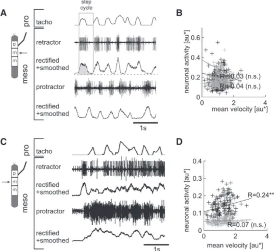
The first publication (Rosenbaum et al. 2010) compares the activity of the three major muscle pairs of the stick insect middle leg between straight forward and backward walking. It shows that the timing of protrac- tor and retractor is inverted while timing of the other muscle pairs remains largely unchanged. In this study, the slippery surface setup is used together with electromyographic measurements to investigate the activity of protractor and retractor coxae, levator and depressor trochanteris and flexor and extensor tibiae of the stick insect C. morosus. The measurements were evaluated with respect to touchdown and liftoff.
The second publication (Gruhn et al. 2009) gives evidence for a neural control mechanism to change step- ping speed. In this study, electrophysiological and behavioral experiments with the intact and reduced stick insect (Carausius morosus) where used. Extra- and intracellular recordings in single-leg stick insect prepara- tions where used to examine relationships between the velocity of a stepping front leg and the motoneuronal activity in the ipsi- or contralateral mesothoracic protractor and retractor, as well as flexor and extensor MNs.
I performed experiments with intact stick insects tethered above a slippery surface to effectively remove mechanical coupling through the ground and to elucidate correlations between the stepping velocities of different legs during walks with constant a velocity or with distinct accelerations.
The third publication (Wosnitza et al. 2013) shows that Drosophila changes the coordination of its legs gradually and systematically with walking speed and can adapt its coordination to major biomechanical changes in its walking apparatus. In this study, I used four different Drosophila strains in order to capture as large a range of walking speeds as possible in a single species. The two wild-type strains Canton-S and Berlin represented the typical behavior in the wild. In addition, two mutant Drosophila strains, white
1118and w
1118, Tbh
nM18where used to extend the range of observable walking speeds to lower values. Furthermore, in some individuals of the wild-type strain Canton-S, I removed one of the hindlegs to analyze if Drosophila is capable of adapting to major biomechanical changes in its walking apparatus.
The fourth and final publication of my thesis (Wosnitza et al, in prep.) gives evidence for differences in the
targeting accuracy of the middle and hind leg of Carausius morosus in first steps of a sequence and during
continuous walks. I investigated the placement of middle and hind legs in the stick insect C. morosus in a
slippery surface setup to understand how important neuronal mechanisms are for the orientation and spatial
coordination of foot placement without visual guidance. I measured the targeting accuracy of the middle
leg towards the front leg and the targeting accuracy of the hind leg towards the middle leg and compared
their performance with each other under two behavioral conditions. First, targeting was investigated in the
resting animal when the anterior leg was standing on one of seven defined positions. Second, to identify to
which extend the state of the animal influences the targeting accuracy, I also looked for dependencies of the
touchdown position on the position of the rostrally adjacent leg during continuous walks.
4.1 Activity Patterns and Timing of Muscle Activity in the Forward Walking and Backward Walking Stick Insect Carausius morosus
Philipp Rosenbaum, Anne Wosnitza, Ansgar Büschges, Matthias Gruhn Published in Journal of Neurophysiology (104(3):1681-1695, 2010)
Für diese Publikation habe ich die Versuchsserien und das Konzept der Arbeit zusammen mit den
Koautoren entwickelt. Ich habe die Versuche gemeinsam mit Philipp Rosenbaum durchgeführt (Vor-
wärtslaufen – Rosenbaum; Rückwärtslaufen – Wosnitza), was auch für die Datenauswertung gilt. Die
Ergebnisse wurden gemeinsam diskutiert. Ich habe alle Abbildungen fertig gestellt sowie gemeinsam
mit Philipp Rosenbaum die ersten Versionen der Methoden, Ergebnis- und Diskussionsteile der Arbeit
entworfen.
Activity Patterns and Timing of Muscle Activity in the Forward Walking and Backward Walking Stick Insect Carausius morosus
Philipp Rosenbaum,* Anne Wosnitza,* Ansgar Büschges, and Matthias Gruhn Department of Animal Physiology, Zoological Institute, University of Cologne, Cologne, Germany Submitted 19 April 2010; accepted in final form 23 July 2010
Rosenbaum P, Wosnitza A, Büschges A, Gruhn M. Activity patterns and timing of muscle activity in the forward walking and backward walking stick insect Carausius morosus. J Neurophysiol 104: 1681–1695, 2010. First published July 28, 2010; doi:10.1152/jn.00362.2010. Understanding how animals control locomotion in different behaviors requires under- standing both the kinematics of leg movements and the neural activity underlying these movements. Stick insect leg kinematics differ in forward and backward walking. Describing leg muscle activity in these behaviors is a first step toward understanding the neuronal basis for these differences. We report here the phasing of EMG activities and latencies of first spikes relative to precise electrical measurements of middle leg tarsus touchdown and liftoff of three pairs (protractor/
retractor coxae, levator/depressor trochanteris, extensor/flexor tib- iae) of stick insect middle leg antagonistic muscles that play central roles in generating leg movements during forward and backward straight walking. Forward walking stance phase muscle (depressor, flexor, and retractor) activities were tightly coupled to touchdown, beginning on average 93 ms prior to and 9 and 35 ms after touchdown, respectively. Forward walking swing phase muscle (levator, extensor, and protractor) activities were less tightly coupled to liftoff, beginning on average 100, 67, and 37 ms before liftoff, respectively. In back- ward walking the protractor/retractor muscles reversed their phasing compared with forward walking, with the retractor being active during swing and the protractor during stance. Comparison of intact animal and reduced two- and one-middle-leg preparations during forward straight walking showed only small alterations in overall EMG activ- ity but changes in first spike latencies in most muscles. Changing body height, most likely due to changes in leg joint loading, altered the intensity, but not the timing, of depressor muscle activity.
I N T R O D U C T I O N
Freely behaving animals often display much more complex locomotor outputs than those observed in reduced preparations because of the needs to respond to environmental contingen- cies and to produce goal-directed movements. Despite consid- erable work devoted to understanding how this behavioral plasticity arises (humans: Lamb and Yang 2000; van Deursen et al. 1998; salamander: Ashley-Ross and Lauder 1997; fish:
Orger et al. 2008; lamprey: Islam et al. 2006; fruit fly: Frye and Dickinson 2004a,b; cockroach: Watson et al. 2002a,b; stick insect: Dürr and Ebeling 2005; Gruhn et al. 2009a,b), we are still only beginning to understand the underlying mechanisms on the neuronal level (Akay et al. 2007; Pick and Strauss 2005;
Ridgel and Ritzmann 2005; Ridgel et al. 2007; Schaefer and Ritzmann 2001).
For the stick insect Carausius morosus substantial knowl- edge exists about leg kinematics during adaptive locomotor behaviors such as different walking directions, turning, and gap climbing (Blaesing and Cruse 2004; Cruse 1976a; Cruse et al.
2009; Dürr and Ebeling 2005; Gruhn et al. 2009b; Jander 1985). Substantial information also exists about the central neural mechanisms that generate the locomotor output (for review, see Bässler and Büschges 1998; Büschges 2005;
Büschges and Gruhn 2008), although very little is known about the timing of leg muscle activity during walking (Epstein and Graham 1983; Graham and Epstein 1985). This information is important because 1) the exact timing of muscle activities during swing-to-stance transitions is needed to assess how sensory input induces them (Büschges and Gruhn 2008) and 2) detailed knowledge of straight walking muscle activity is required to correctly interpret the alterations in muscle activity that occur during locomotor output changes such as turns (Cruse et al. 2009; Gruhn et al. 2009a; Mu and Ritzmann 2005; Ridgel et al. 2007), changes in walking speed (Gruhn et al. 2009b), and switches between tunneling and climbing (Harley et al. 2009) and forward and backward walking (Akay et al. 2007).
In the present study we set out to bridge this gap in knowledge by recording stick insect leg muscle activity during forward and backward walking. Studying neuronal or muscular activity in behaving animals requires recording techniques that do not unduly interfere with animal movement. Two tech- niques that have been successfully applied to relatively large animals are to use implantable electrodes and then to transmit the data along a long tether (Böhm et al. 1997; Clarac et al.
1987; Duch and Pflüger 1995; Gruhn and Rathmayer 2002) or using telemetric devices (Fischer et al. 1996; Hama et al. 2007;
Kutsch et al. 1993; Tsuchida et al. 2004; Wang et al. 2008).
These methods are difficult to apply to small animals such as stick insects because smaller animals become easily entangled in the long, often heavy tethers and the weight of telemetric devices is often very large. One solution to this problem is to conduct experiments with tethered animals on a slippery sur- face, where the animal is free to walk but the body nonetheless stays stationary. This has been used to study escape responses (Camhi and Levy 1988; Camhi and Nolen 1981), turning (Gruhn et al. 2009a; Tryba and Ritzmann 2000a,b), backward walking (Graham and Epstein 1985) , and changes in velocity (Gruhn et al. 2009b). It also allows easy combination of intra- and extracellular physiology with kinematics analyses, partic- ularly when coupled with our electronic measurement of tarsus ground contact (Gruhn et al. 2006). Furthermore, this approach allows one to study the neuronal control of a leg movement without the effects of ground contact mediated mechanics
* These authors contributed equally to this work.
Address for reprint requests and other correspondence: M. Gruhn, Univer- sitaet zu Koeln, Biozentrum, Institut für Zoologie/Abt.Tierphysiologie, Zim- mer 1.514, Zülpicher Straße 47 b, 50674 Koeln, Germany (E-mail: mgruhn
@uni.koeln.de).
on September 15, 2010 jn.physiology.orgDownloaded from
because the respective interleg interactions through the surface are not present and the moving legs therefore exert no force on the body.
We have used the slippery surface setup to investigate the activity of the three major muscle pairs of the stick insect middle leg with respect to touchdown and liftoff during for- ward and backward straight walking as a first form of adaptive behavior.
M E T H O D S
Animals
All experiments were performed on adult female stick insects (Carausius morosus). Animals were reared in the animal facility of the institute in a 12-h/12-h light/dark cycle at 20 –22°C and were fed with blackberry leaves (Rubus fructiosus) without restriction.
Experimental setup
In all experiments, animals walked on a 13.513.5 cm polished nickel-coated brass plate divided into two halves. To allow unimpeded walking under tethered conditions and remove mechanical coupling between the legs, the plate was covered with a lubricant composed of 95% glycerin, 5% saturated NaCl, and a small amount of electrode cream (Marquette Hellige, Freiburg, Germany). This created a slip- pery surface and also allowed recording of tarsal contact by electric current flow during ground contact (Gruhn et al. 2006). The animal was glued ventral side down on an 803 mm (lengthwidth) balsa rod using dental cement (ProTempII, ESPE, Seefeld, Germany) so the legs and head protruded from the rod and all joints were unrestrained.
Animal height above the substrate was adjustable, but was typically 10 mm. Experiments were performed in a darkened Faraday cage at room temperature.
Walking was elicited by projecting a progressive striped pattern (pattern wave length 21°) onto two 13.5-cm diameter round glass screens (Scharstein 1989) placed at right angles to each other and at a 45° angle to the walking surface, about 6 –7 cm away from the eyes of the animal. Reflections on the polished brass plate further increased the field of view. Alternatively, a single white stripe on dark back- ground (toward which the animals orient with straight walking se- quences) was placed in front of the animal. If the animal did not begin locomotion spontaneously, walking was elicited by light brush strokes to the abdomen. Backward walking was elicited by gentle pulls on the antenna (Graham and Epstein 1985).
Electrophysiology
Muscle activity (electromyogram [EMG]) was recorded using two twisted, coated copper wires (OD: 57 or 49m) placed in each muscle about 1 mm apart and held in place with dental cement (ProTempII, ESPE) or tissue adhesive (Vetbond; 3M, St. Paul, MN). Figure 1A shows the approximate sites for the EMG wire placement in the cuticle of the leg and thorax. All recordings were differentially amplified. The EMG signal was preamplified 100-fold (electronics workshop, Zoological Institute, Cologne, Germany), band-pass fil- tered (100 to 2,000 Hz), when necessary further amplified 10- to 1,000-fold, and imported into Spike2 (version 5.05, CED, Cambridge, UK) through an AD converter (Micro 1401k II; CED). A reference electrode was placed in the abdomen of the stick insect.
In most experiments, two antagonistic joint muscles were recorded simultaneously. Protractor coxae and retractor coxae EMGs were recorded in the thorax, depressor trochanteris and levator trochan- terisin the coxa, and extensor tibiae and flexor tibiae in the femur. In two experiments three muscles, in three experiments four muscles,
and in one all six muscles were recorded from simultaneously. These experiments gave the same results as the others.
Recording tarsal contact
To determine the exact moment of the switch between stance and swing we used middle leg tarsal contact as a switch to open and close an electric circuit (Gruhn et al. 2006). Briefly, we used a 2- to 4-mV amplitude square-wave signal generated with a pulse generator (Model MS501, electronics workshop; Zoological Institute) and ap- plied to one half of the slippery surface and a lock-in amplifier (electronics workshop; Zoological Institute) as a reference signal. We tied a copper wire (OD: 49m) with its insulation removed at the tip around the tibia of the leg being monitored and connected it to the lock-in amplifier with an alligator clip. The resistance between the cuticle and copper wire was reduced with a drop of electrode cream (Marquette Hellige) placed at the area of contact, allowing a 2- to 4-nA current to pass through tarsus and tibia. During stance, current flowed from the plate through tarsus and tibia into the copper wire, but during swing, when the leg was in the air, the circuit was discon- nected. Amplifier output was fed into a CED AD converter and digitized using Spike2.
Due to the low-pass filter properties of the lock-in amplifier and the gradual liftoff/touchdown of the tarsus, the signal was not exactly square. We therefore used thresholds set close to the transition point to define the timing of tarsal contact and manually checked each event. Touchdowns could be determined at a resolution of1 ms.
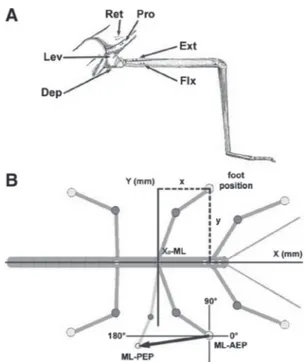
FIG. 1. A: drawing of the stick insect middle leg and the adjacent meso- thorax with the approximate placement sites for the electromyographic (EMG) electrodes for recordings of the main leg muscles. Pro, protractor coxae; Ret, retractor coxae; Ext, extensor tibiae; Flx, flexor tibiae; Lev, levator trochan- teris; Dep, depressor trochanteris. B: schematic drawing of the stick insect with the tracked reference points for the analysis of leg kinematics marked as gray dots. x-values are always points along the length of the animal, whereas y-values mark points perpendicular to the animal. The x0-value was set at the level of the middle leg coxa to give a clear reference point. As an example for the determination of the step length vector and its direction, the right middle leg is drawn at 2 arbitrary positions, one anterior extreme position (ML–AEP) and one posterior extreme position (ML–PEP). The vectors for all steps connecting the 2 positions, normalized to the origin in the AEP, gave direction in degrees and step length in millimeters. The 0 –180° axis was always parallel to the body axis and crossed the AEP, 90° always points toward the animal perpendicularly.
on September 15, 2010 jn.physiology.orgDownloaded from
Liftoff transitions were less steep and more delayed because of delayed tearing of the lubricant from the tarsus due to a capillary action and occasional upward movements of the leg during stance without complete liftoff. To have comparable liftoff times in all experiments we therefore always defined liftoff as the time point with the steepest ascending slope.
Optical recording and digital analysis of leg movements Optical recordings of forward and backward walking were per- formed and analyzed as in Gruhn et al. (2009a). In brief, we recorded walking sequences with a high-speed video camera (Marlin F-033C;
Allied Visions Technologies, Stadtroda, Germany) that was externally triggered at 100 fps. Insect head, thorax, and legs were marked with fluorescent pigments (Dr. Kremer Farbmühle, Aichstetten, Germany) mixed with dental cement. During the recording of walking se- quences, the animal was illuminated with blue light-emitting diode arrays (12 V AC/DC; Conrad Electronic, Berlin). The video files were analyzed using motion-tracking software (WINanalyze 1.9; Mikro- mak Service, Berlin). AEP describes the anterior extreme position of the leg at touchdown, whereas PEP is the posterior extreme position at liftoff. In forward stepping AEP in stance is anterior to PEP, whereas in backward stepping AEP in stance is posterior to PEP. AEP and PEP values are always given in millimeters in the form xx.x; yy.y (SDx; SDy); x-values are given with respect to the length of the animal, a virtual 0 line being drawn across the animal at the level of the coxa. Positive and negative x-values indicate points anterior and posterior to the coxa, respectively; y-values are given with respect to the axis perpendicular to the length of the animal. Larger y-values denote more distal points, smaller values more central points. Figure 1B shows a schematic drawing of the stick insect with the tracked reference points for the analysis of leg kinematics marked as gray dots. As an example for the step length vector determination and its direction, the right middle leg is drawn at two fictive positions, one anterior (ML–AEP) and one posterior (ML–PEP). The vectors for all steps connecting the two posi- tions, normalized to the origin in the AEP, gave direction in degrees and step length in millimeters. The 0–180° axis was always parallel to the body axis and crossed the AEP; 90° always points inside perpendicularly.
The simultaneous recordings of the EMG trace and the camera trigger and tarsal contact signals allowed frame-by-frame correlation of filmed movement and EMG and tarsal contact traces. In calculating middle leg movement vectors all steps were transposed to reflect walking as a right leg regardless of which leg was being recorded from.
Data analysis and figure preparation
Leg positions were measured with their x and y coordinates. Care was taken to choose animals of the same size and leg lengths. The number of animals used for a given condition (N) and the number of steps evaluated (n) are given in the figures. The sample size for the kinematics analysis of straight forward walks was N5 (n125), for backward walks N3 (n83).
Cycle period was calculated from touchdown to touchdown, as determined from the tarsal contact trace. For comparisons of EMG activity of the six different muscles between intact forward and backward walking and between intact and reduced forward stepping preparations, EMG traces were rectified and smoothed (50 ms) and each single data point of each step was exported in Excel (Microsoft, Redmond, WA) to allow averaging. In each step the minimum muscle activity was set to zero and the maximum to one. In several cases, weak cross talk from the antagonist muscle was re- moved mathematically using the EMG trace from the antagonist: the activity of the EMG in the antagonist was triggered to the same point in time as that of the EMG in the agonist (i.e., liftoff or touchdown of the tarsal contact trace, common for both EMGs) and exported in the same way as before. Then its minimum activity was set to 0, but its maximum to an arbitrary value of 0.5, due to the smaller size of the
antagonist signal in the agonist EMG. The normalized activity of the antagonistic muscle was then subtracted from the corresponding value of the muscle under investigation (see Supplemental Fig. S1).1
First spike latencies with respect to liftoff or touchdown were calculated relative to the tarsal contact signal (see preceding text). The absolute latency was then normalized with respect to the correspond- ing step cycle and averaged for the plot in Fig. 11C. Average swing/stance phase duration was calculated from each evaluated step from liftoff to touchdown for swing and from touchdown to liftoff for stance.
All angles were analyzed using the Watson–Williams test, the circular analogue of the two-sample t-test (Matlab, circular statistics toolbox; Berens 2009). Circular variance of vector angles was tested using the variance test in the same toolbox (Matlab, circular statistics toolbox; Berens 2009). For all other statistical analyses, a nonpara- metric Wilcoxon U test (Matlab, Statistics toolbox; The MathWorks, Natick, MA) was used, except for the comparison of integrals of depressor activity, where a standard Student’s t-test was used. Statis- tical significance was assumed at values of P0.01. Figures were prepared in Origin 6.1 (OriginLab, Northampton, MA) and Photoshop 6.0 (Adobe Systems, San Jose, CA).
R E S U L T S
Understanding how animals adapt their motor behavior to changing environmental conditions requires measuring limb kinematics and muscle activity in different behaviors. We have shown elsewhere that stick insect leg kinematics differ in straight and curve walking and examined the effect of reducing leg number on these changes (Gruhn et al. 2009a). Here we compare middle leg kinematics during forward and backward walking in intact animals and then examine muscle activity in these two behaviors in the intact and reduced preparations.
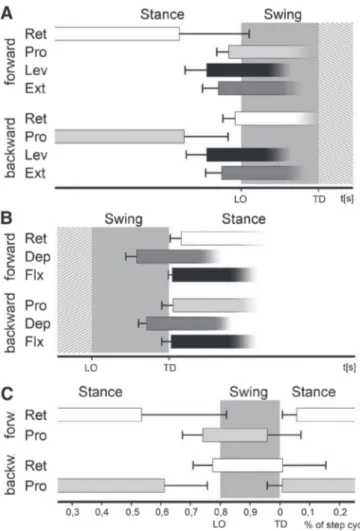
Kinematics of straight forward versus backward walking in the middle leg
Figure 2A shows a schematic drawing of the stick insect with marked anterior and posterior extreme positions (AEP and PEP plus SD) of the right middle leg in forward and backward straight walking. The data for forward walking (gray) were taken from Gruhn et al. (2009a). AEP is defined as tarsus position at touchdown and PEP as tarsus position at liftoff, always with respect to the direction in which the animal moves.
During forward walking (FW), the leg is moved anteriorly during swing and posteriorly during stance. This order of leg movements is reversed during backward walking (BW). In backward walking each step’s AEP is therefore more caudal along the long axis of the animal than the PEP. Forward steps were significantly longer (mean step length FW: 16.2 5.4 mm; BW 9.9 4.7 mm, P 0.0001) and their movement direction was on average more parallel to the body length axis than were backward steps (Fig. 1A). To compare movement vector angles, we mirror-imaged the forward step movement angles in Fig. 2B along the horizontal axis. The resulting mean angles of forward (8.817.3°, gray) and backward (36.1 20.3°, black) steps are shown in Fig. 2D and differed signifi- cantly (P0.0001) from each other. The variability between the movement vector angles of single steps is similar in both directions and spans angles over a range of 83° during FW and 88.5° during BW between the respective extremes. Mean touchdown position along the transverse axis was significantly
1The online version of this article contains supplemental data.
on September 15, 2010 jn.physiology.orgDownloaded from
closer to the midline in FW versus BW (y-positions: AEPFW 16.9 3.3 mm; AEPBW 19.6 2.9 mm, P 0.0001), but mean liftoff position was not significantly different (y-posi- tions: PEPFW 14.4 2.9 mm; PEPBW 13.8 2.6 mm, P 0.32). However, because during backward walking the move- ment is more inward directed in each step, the PEP is generally reached after a shorter step length (Fig. 2, B–D). Taken together, these data show that in intact animals middle leg backward stepping is not simply reversed forward walking, but is instead altered to having shorter and more inward directed steps, albeit with a similar degree of variability as seen for forward stepping.
Stance duration alone determines cycle period
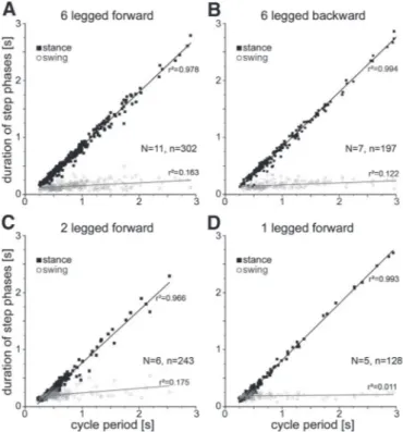
In stick insects walking on nonslippery surfaces, in which the different legs are coupled mechanically through the ground on which the animal walks, step cycle period depends on stance duration (Graham 1972, 1985; Wendler 1964). We tested whether this relationship is also present in slippery surface forward and backward walking and, to test for interleg interactions, in animals with reduced leg numbers (only the
two middle legs or only one single middle leg). In all these cases, the cycle period varied linearly with stance duration but did not depend on swing duration, which was essentially constant at all cycle periods (Fig. 3).
Phasing of leg muscle activity
EMG recordings of various leg muscles during walking have been made (Fischer et al. 2001; Graham and Epstein 1985), but with few exceptions, only the activities of single muscle pairs were recorded (Bässler 1993; Cruse and Pflüger 1981; Epstein and Graham 1983). In addition, the timing reference for the beginning or end of muscle activity relative to step cycle, if present at all, was not precise. To remedy this lack we made comprehensive paired EMG recordings of all three major muscle pairs controlling leg movements: the protractor/retrac- tor coxae, levator/depressor trochanteris, and the extensor/
flexor tibiaemuscles at a time during both forward and back- ward walking.
Figure 4 shows the activity of the muscles of the most proximal leg joint, the thorax– coxa joint, the protractor and retractor coxaemuscles, which serve to protract and retract the leg, respectively. The traces in Fig. 4A show raw EMG activity, those in Fig. 4B rectified and smoothed (50 ms) activity, and those in Fig. 4C mean rectified activity from one stepping sequence (gray trace) and from five animals (black trace). In forward walking protractor activity began before the liftoff of the leg, reached its main activity during swing, and then decreased toward the end of swing. In backward walking the protractor was barely active during swing but began at the transition between swing and stance and reached peak activity
FIG. 3. Cycle period depends on stance, not swing, duration. Gray circles represent swing phase, filled black boxes stance. A: straight forward walking, 6-legged animal. B: backward walking, 6-legged animal. C: straight forward walking, 2-legged (only middle legs) animal. D: one-legged (middle leg) animal. Nanimal number, nstep number.
FIG. 2. Kinematics of a forward and backward walking stick insect middle leg on a slippery surface. A: schematic drawing of a stick insect with the mean anterior extreme position (AEP) and posterior extreme position (PEP) values (and SD error bars) of the right middle leg for forward (gray) and backward (black) walking. Note that for the backward walking animal the AEP is posterior to the PEP; the gray line marks the X0-value for the middle leg. B and C: step-to-step variability in angle and length of stance movement of forward (B) and backward (C) steps normalized to touchdown position (AEP); the average stepping vector is drawn in black in both cases. D: average stepping vectors for forward (gray) and backward (black) walking from B and C; the average vector for forward walking from B was mirrored across the horizontal plane for easier comparison. Nanimal number, nstep number.
on September 15, 2010 jn.physiology.orgDownloaded from
about 100 ms into stance. This activity pattern was the same for the retractor muscle except that it showed stance activity during forward walking and swing activity during backward walking.
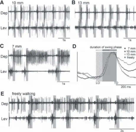
Figure 5 shows the activity of the muscles of the next most distal leg joint, the coxa–trochanter joint, the depressor tro-
chanteris and levator trochanteris muscles, which serve to depress and lift the leg, respectively. The traces in Fig. 5A again show raw EMG activity, those in Fig. 5B rectified and smoothed ( 50 ms) activity, and those in Fig. 5C mean rectified muscle activity from one stepping sequence (gray trace) and from five animals (black trace). Depressor activity
FIG. 4. Right middle leg protractor and retractor EMG recordings during forward (left column) and backward (right column) walking on a slippery surface. Gray boxes mark swing phase. A: raw EMG recordings. B: rectified and smoothed traces of EMGs in A. C: mean rectified and smoothed traces of recordings (gray) from one and from 5 animals (black). Gray boxes mark the average swing duration; shaded area shows swing duration SD. Double asterisks mark where cross talk from the retractor was removed mathematically from the pro- tractor traces. Nanimal number, nstep number.
FIG. 5. Right middle leg levator and depressor activity during forward (left column) and backward (right column) walking on a slippery surface. Gray boxes mark swing phase. A: raw EMG recordings. B: rectified and smoothed traces of EMGs in A.
C: mean rectified and smoothed traces of recordings from one (gray) and from 5 animals (black). Gray boxes mark the average swing duration; shaded area shows swing duration SD. N animal number, nstep number.
on September 15, 2010 jn.physiology.orgDownloaded from
began very shortly after swing beginning, was active through- out swing, and declined shortly after stance beginning. Pro- vided the animal was maintained at a constant height about the substrate (see following text), depressor activity was the same in forward and backward walking. Moderate levator muscle activity was present at the beginning and middle of stance, with a substantial peak of activity occurring just before the stance to swing transition. Levator activity decreased and reached a minimum shortly after swing beginning. As with the depressor, levator activity was the same in forward and backward walk- ing.
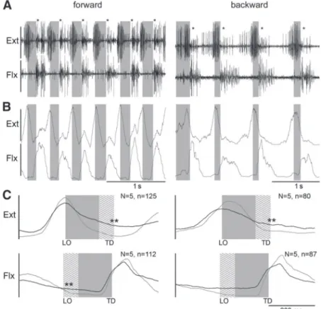
The last muscles analyzed (Fig. 6) were the extensor tibiae and flexor tibiae muscles, which move the femur–tibia joint and extend and flex the tibia, respectively. The traces in Fig. 6A again show raw EMG activity, those in Fig. 6B rectified and smoothed ( 50 ms) activity, and those in Fig. 6C mean rectified muscle activity from one stepping sequence (gray trace) and from five animals (black trace). Peak extensor activity occurred around liftoff in forward and in backward walking, whereas flexor activity peaked during the first half of stance in forward and backward walking.
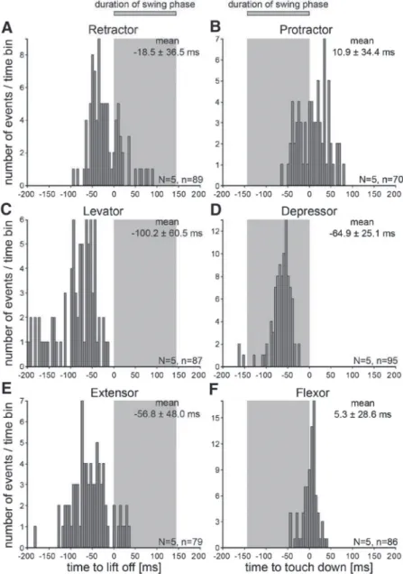
Latency of muscle timing during forward and backward walking
Reliably comparing muscle activity in different walking directions and across preparations requires determining the exact timing of muscle activity within the step cycle. Swing to stance and stance to swing transitions are two such points.
Figures 7 and 8 show first spike latencies relative to these points for all six muscles in forward and backward walking, respectively, from five animals each. The gray areas mark mean swing duration averaged across all steps and animals.
The protractor, levator, and extensor muscles move the middle leg forward, up, and extend the femur–tibia joint.
During forward walking these movements occur during swing.
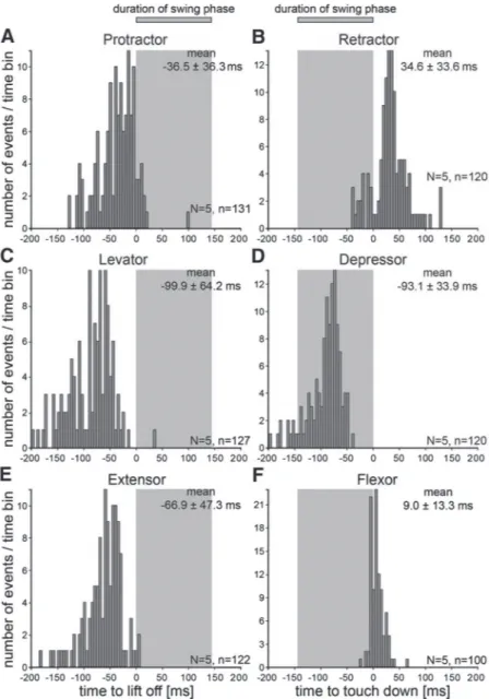
We therefore measured the first spikes in these muscles rela- tive to liftoff (Fig. 7, A, C, and E). Activity occurred earliest in the levator (mean first muscle potential 99.9 64.2 ms before liftoff), followed by the extensor (66.9 47.3 ms) and then the protractor (36.5 36.3 ms). The retractor, depressor, and flexor muscles move the leg backward, down, and flex the femur–tibia joint. During forward walking these movements occur during stance. We therefore measured the first spikes in these muscles relative to touchdown (Fig. 7, B, D, and F). Activity occurred earliest in the depressor with the mean first muscle potential 93.1 33.9 ms before touchdown, 22% into the swing phase. The first flexor activity occurred next with mean first muscle potential 9.013.3 ms after touchdown. Single first spikes occurred just before touch- down, confirming previous findings for the timing of this muscle (Gruhn et al. 2006). First retractor activity was more variable, with mean first muscle potential 34.633.6 ms after touchdown and first activity occurring50 ms before touch- down. The joint activation sequence in swing is thus the same as that for stance, i.e., first coxa–trochanter, then femur–tibia, and finally thorax– coxa. The high SD values result from the high variability in the stepping of the stick insect on the slippery surface. Walking sequences with many consecutive straight forward steps do not occur often and every step has a slightly different direction and stance duration.
As was shown earlier in the kinematics and EMG data, in backward walking protractor and retractor timing is the reverse of that in forward walking. To continue to compare the timing of functional swing and stance muscles in the two walking
FIG. 6. Right middle leg extensor and flexor activity during forward (left column) and backward (right column) walking on a slippery surface. Gray boxes mark swing phase. A: raw EMG recordings; asterisks mark cross talk from the flexor in the extensor trace. B: rectified and smoothed traces of EMGs in A.
C: mean rectified and smoothed traces of recordings from one (gray) and from 5 animals (black). Gray boxes mark the average swing duration; shaded area shows swing duration SD.
Double asterisks mark where cross talk from the antagonist muscle was removed mathematically. Nanimal number, n step number.
on September 15, 2010 jn.physiology.orgDownloaded from
directions, in backward walking sequences we therefore refer- enced retractor activity to liftoff and protractor activity to touchdown, but continued to reference the activity of the other muscles as before (Fig. 8, A–F). Sequence of levator and extensor activation as well as the latencies for the first muscle potential (100.260.5 ms, Fig. 8C; 56.848.0 ms, Fig. 8E, respectively) were the same as in forward walking (PLev 0.98; PExt 0.31). During backward walking the retractor activated 18.536.5 ms before liftoff (Fig. 8A), dramatically different from this muscle’s activation in forward walking (Fig.
7B), but barely not significantly different from the timing of the functionally analogous protractor during forward walking (P 0.012) (Fig. 7A).
Except for the difference mentioned earlier that the protrac- tor is a stance phase muscle in backward walks, the timing and activation sequence of the functional stance phase muscles were also similar in forward and backward walking. The depressor again activated first (Fig. 8D), although only halfway through swing at 64.9 25.1 ms before touchdown, signifi- cantly later than that in forward walking (P 0.0001). The protractor and flexor activated next at almost the same time:
10.934.4 and 5.328.6 ms after touchdown (Fig. 8, B and F). Flexor timing did not differ significantly from that in forward walking (P0.12). Despite their large SDs, protrac- tor timing in backward walking (10.934.4 ms) and retractor timing in forward walking (34.633.6 ms) did differ signif- icantly (P0.0001).
In summary, these data show that 1) only the muscles controlling the thorax– coxa joint showed large changes when walking direction changed, and 2) with respect to liftoff and touchdown, the timing of functionally analogous muscles in swing and stance is almost the same in both directions.
Muscle activity in reduced preparations
Many studies on stick insect walking have been conducted in preparations with reduced leg number (e.g., Akay et al. 2001, 2004; Fischer et al. 2001; Gabriel and Büschges 2007; Gabriel et al. 2003; von Uckermann and Büschges 2009). Because these preparations lack interleg sensory interactions, it is im- portant to test whether data from such experiments are appli- cable to intact animals. Leg kinematics in straight forward
FIG. 7. Histograms of the latency distribution of the first muscle potentials in the EMG traces of the 6 analyzed leg muscles during forward walking. Timing values of the first spikes in protractor, levator, and extensor were measured with respect to the time of liftoff. Retractor, depressor, and flexor activity spikes were measured with respect to leg touchdown.
Gray boxes mark the average swing phase length. Average latency of the first spike is given with SD. Nanimal number, nstep number.
on September 15, 2010 jn.physiology.orgDownloaded from