Mechanisms for Intersegmental Leg Coordination in
Walking Stick Insects
INAUGURAL-DISSERTATION zur
Erlangung des Doktorgrades
der Mathematisch-Naturwissenschaftlichen Fakult¨at der Universit¨at zu K¨oln
vorgelegt von Bj¨orn Ludwar aus Augsburg
K¨oln 2003
Berichterstatter: Prof. Dr. Ansgar B¨uschges Prof. Dr. Peter Kloppenburg
Tag der m¨undlichen Pr¨ufung: 5. Juni 2003
Contents
Zusammenfassung 4
Abstract 6
1 Introduction 7
1.1 Intersegmental Coordination . . . . 7
1.2 Possible mechanisms . . . . 7
1.3 Evidence for mechanisms used by invertebrates . . . . 8
1.3.1 Coordination of leech swimming movements . . . . 8
1.3.2 Coordination of locomotion in crustaceans . . . . 9
1.3.3 Coordination of walking in insects . . . 11
1.4 Studies of vertebrate systems . . . 14
1.4.1 Intersegmental coordination of swimming in lamprey . . . 15
1.4.2 Intersegmental coordination of leg movements of cats . . . 15
1.5 Question and experiments . . . 17
2 Materials and Methods 18 2.1 Animals and experimental procedures . . . 18
2.2 Experimental conditions . . . 18
2.3 Extracellular recordings . . . 19
2.4 Intracellular recordings . . . 19
2.5 Electromyograms (EMG) . . . 19
2.6 Experiments using a treadmill . . . 20
2.7 Pharmacological stimulation in ’split bath’ experiments . . . 21
2.7.1 Meso- and metathoracic ganglia . . . 21
2.7.2 Pro- and mesothoracic ganglia . . . 21
2.8 Stimulation of the femoral chordotonal organ . . . 23
2.9 Stimulation of the campaniform sensilla . . . 23
2.10 Data acquisition and processing . . . 25
3 Results 27 3.1 Semi-intact walking preparation: extracellular recordings . . . 27
3.1.1 Protractor coxae motoneurons . . . 28
3.1.2 Depressor trochanteris motoneurons . . . 30
3.1.3 Extensor tibiae motoneurons . . . 33
3.1.4 Antagonistic motoneuron pools . . . 35
CONTENTS 3
3.2 Semi-intact walking preparation: intracellular recordings . . . 37
3.2.1 Protractor coxae motoneurons . . . 37
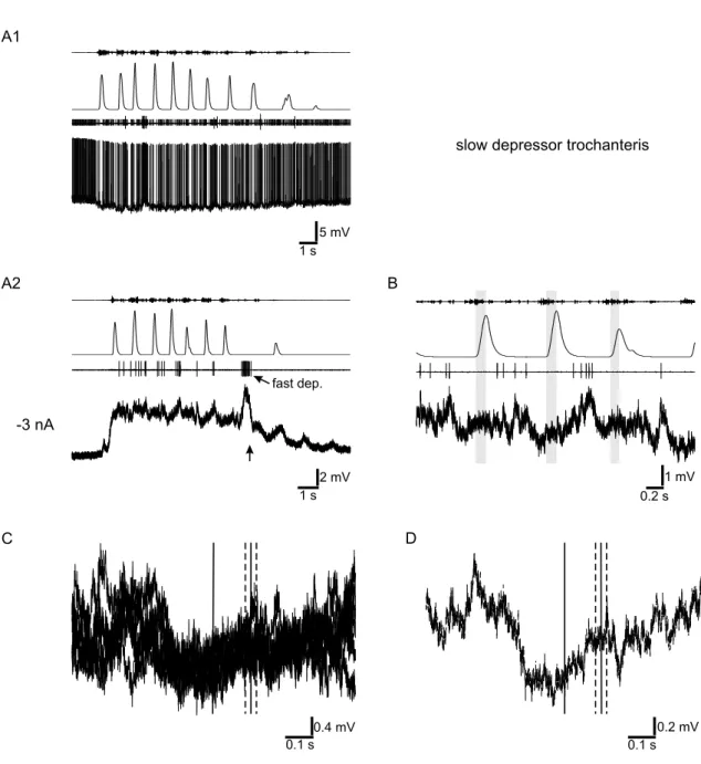
3.2.2 Depressor trochanteris motoneurons . . . 42
3.2.3 Levator trochanteris motoneurons . . . 46
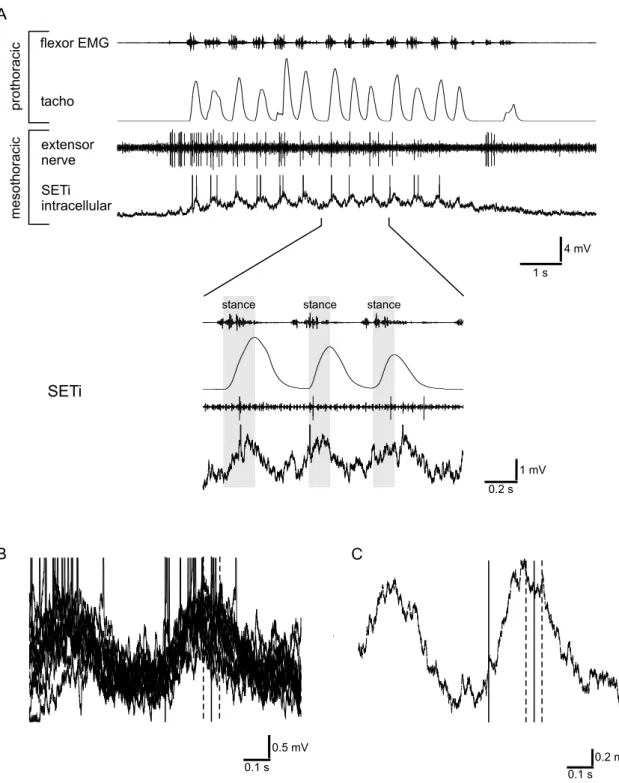
3.2.4 Extensor tibiae motoneurons . . . 47
3.2.5 Flexor tibiae motoneurons . . . 54
3.3 Semi-intact walking preparation: recordings of non-spiking interneurons 56 3.4 Mutual influences of segmental central pattern generators . . . 66
3.4.1 Interaction between meso- and metathoracic ganglia . . . 66
3.4.2 Interaction between pro- and mesothoracic ganglia . . . 71
3.5 Intersegmental influences of the chordotonal organ . . . 73
3.6 Local and intersegmental influences of campaniform sensilla . . . 75
3.6.1 Local effect of campaniform sensilla signals in the hind leg . . 75
3.6.2 Intersegmental effect of campaniform sensilla signals . . . 76
4 Discussion 83 4.1 Correlation of mesothoracic motoneuron activity and front leg walking 83 4.2 Origins of coordinating signals . . . 85
4.2.1 Intersegmental effects of sensory organs . . . 85
4.2.2 Coupling of segmental central pattern generators . . . 86
4.2.3 Peripheral vs. central coordinating mechanisms . . . 87
4.3 Inputs on motoneurons . . . 88
4.4 The role of the pre-motor network . . . 89
4.5 Conclusions . . . 93
Bibliography 94
Danksagung 103
Erkl¨ arung 104
Teilpublikationen 105
Lebenslauf 106
Zusammenfassung
Zur effizienten Fortbewegung m¨ussen Bewegungen einzelner Beine w¨ahrend dem Laufen koordiniert werden, was in einem Schrittmuster oder einer Gangart resul- tiert. Diese Dissertation untersucht die neuronalen Mechanismen, die der Bildung einer solchen Gangart unterliegen, besonders die neuronalen Grundlagen der Kop- pelung von ipsilateralen Beinbewegungen.
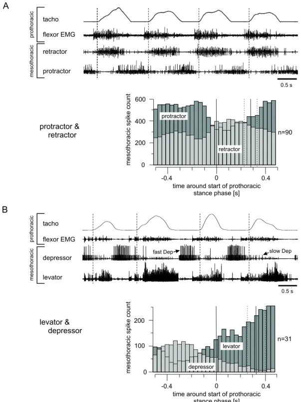
In einer semi-intakten Laufpr¨aparation der Stabhauschrecke Carausius morosus wurde die Korrelation von Aktivit¨at ipsilateraler, mesothorakaler Motoneurone und Laufbewegungen eines Vorderbeines beschrieben. Extrazellul¨are Ableitungen zeigten eine ausgepr¨agte Ankoppelung mesothorakaler protractor coxae und extensor tibiae Motoneuronen. Depressor trochanteris Motoneurone zeigten eine variablere Ankop- pelung. Mesothorakale retractor coxae und levator trochanteris Motoneurone waren antizyklisch zur ihren jeweiligen Antagonisten aktiv.
Intrazellul¨are Ableitungen zeigten zwei unterschiedliche Arten der Modulation des Membranpotentials mesothorakaler Motoneurone: Eine tonische Modulation, die w¨ahrend der Laufaktivit¨at des Vorderbeins anhielt, und eine rhythmische Modula- tion, die mit einzelnen Schritten des Vorderbeins korreliert war. Es wurden Hinweise auf tonisch erregende und hemmende, wie auch auf rhythmisch erregende und hem- mende Eing¨ange auf verschiedenen Motoneurone gefunden.
Intrazellul¨are Ableitungen von mesothorakalen nicht-spikenden Interneuronen, des den Motoneuronen vorgeschalteten Netzwerks, zeigten, daß auch diese Neuronen in- tersegmental koordinierende Signale erhalten. Tonische wie auch rhythmische Mod- ulationen des Membranpotentials, die mit der Laufaktivit¨at des Vorderbeins korre- liert waren, wurden gefunden. Die nicht-spikenden Interneuronen wurden zum Teil morphologisch identifiziert und es ist bekannt, daß sie lokale sensorische Informa- tion verarbeiten. Sie stellen daher eine Grundlage zur Integration von Information lokaler Sinnesorgane und intersegmentaler Signale dar.
Weiterhin wurden Versuche durchgef¨uhrt um die Herkunft intersegmentaler Signale aufzukl¨aren. In Experimenten mit einer isolierten Ganglienkette, die pharmakolo- gisch mittels Pilocarpine aktiviert wurde, wurde die Interaktionen von zentralen, Rhythmus generierenden Netzwerken untersucht. Sensorische Eing¨ange waren in dieser Pr¨aparation ausgeschlossen. Es wurden keine Hinweise auf eine starke Kop- pelung zentraler Mustergeneratoren zwischen mesothorakalen und metathorakalen, sowie prothorakalen und mesothorakalen Segmenten gefunden.
Zwei weitere Versuchsreihen besch¨aftigten sich mit der Rolle von sensorischen Sig-
nalen bei der intersegmentalen Koordination. Signale vom mesothorakalen femoralen
Chordotonalorgan, welches die Position und Bewegung im Femur-Tibia Gelenk mißt,
zeigten im ’aktiven’ Tier keinen deutlichen Einfluß auf die Aktivit¨at metathorakaler
Motorneurone. Signale der Campaniformen Sensilla, welche die Belastung eines
CONTENTS 5
Beines messen, zeigten nur einen schwachen intersegmentalen Einfluß auf die Ak-
tivit¨at mesothorakaler Motoneurone. Letzteres wurde im ruhenden Tier und mit
umgekehrtem Effekt im ’aktiven’ Tier, wie auch w¨ahrend durch Pilocarpine in-
duzierter rhythmischer Aktivit¨at, gezeigt.
Abstract
For efficient locomotion, the movements of single legs need to be coordinated during walking, which results in a stepping pattern or gait. This dissertation explores the neural mechanisms underlying the formation of a gait, in particular the neural basis of coupling of ipsilateral leg movements.
In a semi-intact walking preparation of the stick insect Carausius morosus, correla- tions between ipsilateral mesothoracic motoneuron activity and walking movements of a front leg were described. Extracellular recordings showed a dedicated coupling of activity for mesothoracic protractor coxae and extensor tibiae motoneurons. De- pressor trochanteris motoneurons showed a more flexible coupling. Mesothoracic retractor coxae and levator trochanteris motoneurons were active in anti-phase with their respective antagonists.
Intracellular recordings revealed two different modulations of membrane potentials of mesothoracic motoneurons: a tonic modulation, lasting during the stepping ac- tivity of the front leg, and a rhythmic modulation, correlated with individual steps of the front leg. Evidence for tonic excitatory and inhibitory, as well as for rhythmic excitatory and inhibitory inputs were found for different motoneurons.
Intracellular recordings of mesothoracic non-spiking interneurons of the pre-motor network revealed that these interneurons receive intersegmental coordinating signals.
A tonic as well as a rhythmic modulation of their membrane potential, correlated with the walking activity of the ipsilateral front leg, were found. The non-spiking interneurons were in part morphologically identified and are known to process local sensory information. Hence, they could provide the basis for integration of local sensory and intersegmental signals.
Additionally experiments were performed to investigate the origin of intersegmental signals. In experiments with an isolated chain of ganglia which was pharmacologi- cally activated with pilocarpine, interaction between the central rhythm generating networks were studied. Sensory input was excluded in this preparation. No evi- dence was found for strong coupling of central pattern generators in mesothoracic and metathoracic segments, nor in prothoracic and mesothoracic segments.
Two more sets of experiments focused on the role of sensory signals for intersegmen-
tal coordination. Signals from the mesothoracic femoral chordotonal organ, measur-
ing position and movement of the femur-tibia joint, showed no clear influence on the
activity of metathoracic motoneurons in the ’active’ animal. Sensory signals from
the metathoracic campaniform sensilla, measuring load on the leg, showed only a
weak intersegmental influence on mesothoracic motoneuron activity, but a clear in-
fluence on local protractor and retractor motoneuron activity. The latter was found
in the resting animal and with reversed effects in the ’active’ animal, as well as
during rhythmic activity evoked by application of pilocarpine.
Chapter 1 Introduction
1.1 Intersegmental Coordination
Locomotion, whether in the form of swimming, crawling, flying, or walking, requires two simultaneous actions. First, the part of the body responsible for locomotion must be moved in an appropriate way to produce propulsion. A wing must rotate around its longitudinal axis while it is moving upward and downward to advance the animal. A leg must perform stance and swing phases, which means it must be on the ground while the body is pushed forward (stance) and lifted off while it does not support the body and is moved forward (swing). Second, multiple body parts responsible for locomotion must act in a coordinated way. Left and right wings need to move upwards and downwards in a synchronized way to assure stable flight. Left and right legs need to alternate in their cycles to guarantee a biped, slowly walking individual that at least one leg is on the ground all the time, so the individual does not fall. If an animal has more than two legs, additional coordination is required: the multiple pairs of legs, e.g. front, middle, and hind legs of an insect have to be coordinated. This coordination is termed intersegmental coordination as one segment of an animal usually contains the circuitry to control one pair of legs.
In a multi legged organism this coordination between segments leads to so called stepping patterns. Examples for a horse would be trot or gallop, for an insect tripod or tetrapod gait. In animals with other forms of locomotion, for example snakes or lampreys, this coordination creates a wave of body undulation that propels the animal. The neural basis of intersegmental coordination of walking is the topic of this thesis.
1.2 Possible mechanisms
How is intersegmental coordination achieved? Of several proposed mechanisms, some have been demonstrated to exist in various animals.
A central command unit, located in the central nervous system, could activate
local circuitry in the segments. The local circuitry could then execute the motor
program to perform a step. The higher level central command unit ’supervises’ the
segments, thus ensuring coordination (reviewed by Pearson, 1993).
1. INTRODUCTION 8
Such a central command unit is not necessarily required, as the segmental circuitries could exchange information between each other. A segment commonly contains central oscillators that generate rhythmic activity underlying locomotion. These oscillators are referred to as central pattern generators (Delcomyn, 1980; Pearson, 2000; Marder, 2001; MacKay-Lyons, 2002). A mechanism ensuring intersegmental coordination is the coupling of central pattern generators, that is maintaining a stable phase relationship between their oscillators. This can be achieved by specific connections and suitable intrinsic frequencies (Marder and Calabrese, 1996; Friesen and Cang, 2001).
A disadvantage of these central coupling mechanisms becomes clear for locomotion on irregular terrain. For an insect climbing in shrub, it might not be possible to place a leg at the predetermined location. Walking on rock, a leg might not find the support needed. Feedback from sense organs is required to adjust the movements to the given environment. In most systems a variety of such sense organs, measuring position, movement, and load, are found. Their information is processed locally in the segments and intersegmentally in neighboring segments. Sense organs are often components of reflex loops or they interact with central pattern generators.
The sensory information can also be shared with other segments and used for intersegmental coordination. This can be done directly through projections of sensory cells in adjacent segments (Br¨aunig et al., 1981; Pfl¨uger et al., 1981) or via intersegmental interneurons (Laurent, 1986; B¨uschges, 1989; Brunn and Dean, 1994;
Matheson, 2002).
Coordinating information does not necessarily have to be transmitted electrically (via neurons) between the segments. A mechanical coupling exists and if for example one leg of a multi legged organism is lifted off the ground, the load on the other legs increases. This is sensed by local sensory organs such as campaniform sensilla in an insect leg or muscle spindles in a vertebrate muscle and influences the local control (Cruse et al., 1998; Friesen and Cang, 2001). A similar coupling exists if the active movement of one limb passively moves another limb. This passive movement may trigger reflexes and thereby cause active movements (’reflex chain theory’, reviewed by Grillner (1981)). In both cases coordination of movements of both segments results.
1.3 Evidence for mechanisms used by invertebrates
In the last few decades several invertebrate systems have been used to study the question of how intersegmental coordination is achieved and which of the mecha- nisms mentioned above are realized.
1.3.1 Coordination of leech swimming movements
A well studied system is the leech. Besides mechanisms for intersegmental coordina-
tion of heartbeat and crawling behavior, swimming especially has been extensively
studied. Leeches swim by alternating contractions of the dorsal and ventral muscles
of their body segments. The timing of these contractions is controlled by a chain of
1. INTRODUCTION 9
18-21 segmental ganglia containing the swim network. Each ganglion contains local interneurons that drive motoneurons to produce the dorsal-ventral alteration. It also contains intersegmental interneurons that project to approximately five ganglia in either direction of the chain. These intersegmental connections are asymmetric:
correlated with its activity phase, a neuron either projects caudally or rostrally; sole excitatory neurons only project caudally (Brodfuehrer et al., 1995).
If the nerve cord is isolated and lateral nerve roots are briefly stimulated electrically, it responds with a fictive motor pattern similar to the one produced by a swimming animal. This suggests that coupling can be achieved in the absence of sensory in- puts, solely by central mechanisms (Kristan and Calabrese, 1976). The timing and phase relations of the motor pattern are thought to result from the asymmetry of in- tersegmental connections mentioned, and from a rostrocaudally-decreasing intrinsic cycle period of the segmental central pattern generators (Friesen and Pearce, 1993).
However, the motor pattern of the isolated nerve cord shows a longer period and a smaller phase lag when compared to an intact preparation. This suggests that sensory information, e.g. from the ventral stretch receptors, which are known to influence the phase lag, is required for appropriate coordination (Cang and Friesen, 2000; Friesen and Cang, 2001).
By severing the nerve cord Yu et al. (1999) showed that proper coordination can be achieved by local sensory feedback alone. They suggest that six to eight pairs of segmental stretch receptors provide the information used for this coordination. As these stretch receptors do not project to other segments (Blackshaw and Thomp- son, 1988; Huang et al., 1998), information has to be transmitted mechanically and perceived locally by the sense organ. Thus coordination results from mechanical coupling.
In summary, intersegmental coordination in the leech seems to be achieved by both, central coupling of segmental pattern generators and mechanical coupling via sensory organs - the ventral stretch receptors.
1.3.2 Coordination of locomotion in crustaceans
Another group of animals intensively studied with regard to the coordination of locomotion movements is crustacea, especially crayfish and lobsters.
The crayfish swimmeret system
The swimmerets of crayfish are paired, jointed limbs on four or five abdominal segments, used for swimming. They rhythmically perform stereotyped power- and return-strokes and thereby produce forward thrust. Swimmerets of neighboring seg- ments move with the same period and constant phase relation (reviewed by Cattaert et al., 2001).
Ikeda and Wiersma (1964) showed that a deafferented and isolated chain of abdom-
inal ganglia spontaneously produces an activity pattern which parallels the motor
pattern of a swimming animal. Thus no sensory input or ’single master center’ is re-
quired. It was proposed that the coordination between the ganglia could derive from
1. INTRODUCTION 10
a excitability-gradient - that is the neurons driving the more posterior swimmerets are more excitable and reach thresholds before their more anterior counterparts (Wiersma and Ikeda, 1964). But Mulloney (1997) showed that the excitability is identical for all segments and thus proved this hypothesis incorrect. Instead, the coordination between the segments is achieved by the actions of three types of in- tersegmental interneurons (Stein, 1971; Namba and Mulloney, 1999).
The observation that the normal motor pattern keeps existing even after shorten- ing the chain of ganglia to only two ganglia, led to the proposal that only nearest neighbors are coupled (Paul and Mulloney, 1986). This idea was incorporated in mathematical models which produced the characteristic phase differences and rela- tive durations of the swimmeret system (Skinner et al., 1997; Mulloney et al., 1998).
Although the coupling of only nearest neighbors seems to be sufficient to explain the intersegmental coordination in the swimmeret system, Tschuluun et al. (2001) showed that interneurons project beyond the neighboring ganglion and maintain coordination even if the synaptic transmission is blocked in the nearest neighboring ganglion.
In the crayfish swimmeret system intersegmental coordination is achieved by central coupling of segmental pattern generators. The coordinating interneurons link not only nearest neighbors, but also more distant ganglia. Sensory feedback might be unnecessary for basic coordination.
Walking of lobsters and crayfish
Both, lobster and crayfish, were used for studying terrestrial locomotion. For walk- ing, decapods perform periodical stepping movements with all or only a subset of their ten walking legs. Each single leg alternates between a stance and a swing phase. On a smooth, flat surface, the left and right legs of a pair move in anti- phase, while the phase lag of adjacent ipsilateral legs is approximately one-quarter of a cycle (Clarac and Chasserat, 1983; Chasserat and Clarac, 1980). This coordi- nation produces the specific gait. Experiments in which left and right legs walked on separate treadmill belts, driven with different speeds, revealed that left and right legs were able to step at different frequencies. Thus separate leg controllers must exist (Clarac and Chasserat, 1986).
Chrachri and Clarac (1987) showed that bath application of cholinergic agonists, such as pilocarpine or oxotremorine, evoked rhythmic activity of motoneuron pools in an isolated thoracic ganglion preparation of crayfish. This rhythmic activity resembled the motor pattern seen in an intact, walking animal. Even a single iso- lated ganglion produced a rhythmic activity pattern after application of cholinergic agonists. Therefore, segmental central pattern generators are responsible for the generation of basic motor output patterns, and interactions between them lead to an intersegmental coordination of motor activity (Chrachri and Clarac, 1990).
Although coordinated patterns of activity are found in the isolated chain of ganglia, sensory signals strongly affect the inter-leg coordination (reviewed by Clarac, 1985;
Cattaert et al., 2001). An ’in phase’ coordination with the next anterior intact leg is
observed when legs are not able to generate propulsive forces. This is the case when
1. INTRODUCTION 11
legs are held up off the ground (’waving behavior’), or when legs are autotomized and only the stumps move. The observations suggest that substrate contact and/or power stroke forces are necessary for the production of an alternating pattern in the intact animal. Studies by Cruse et al. (1983) showed that the position of one leg influences the coordination of the remaining legs. While a lobster was walking on a driven treadmill, they isolated one leg from the walking legs by placing it on a platform. Moving the platform, and thereby changing the position of the leg, they could alter the stepping pattern of the walking legs. In other experiments Cruse and M¨uller (1986) analyzed the effects of disturbing the stance phase of individual legs of a walking crayfish. Both studies led to the description of two coordinating influences: a forward directed influence, facilitating a swing phase of the anterior leg while the posterior leg is in stance phase, and a backward directed influence, facilitating a stance phase in the posterior leg at the transition from stance to swing phase of the anterior leg (Cruse and M¨uller, 1986).
In summary, it appears that a central coupling mechanism, as well as information from sensory organs, are involved in the coordination of leg movements of decapods.
Both seems to be important for the production of the gait seen in the intact, walking animal.
1.3.3 Coordination of walking in insects
Many studies of motor systems and locomotion have been performed with insects - namely stick insects, locusts, and cockroaches. Their six legs are coordinated into a tripod gait, a tetrapod gait, or an intermediate form - depending on species, age and speed of locomotion of the animal tested (Graham, 1985). Most experimental designs to unravel mechanisms responsible for this intersegmental coordination rely on describing changes of stepping patterns caused by alteration or disruption of sen- sory feedback.
Experiments in which one leg was amputated, led to slight changes of the spatial and temporal pattern of movements of the remaining legs. Apparently no learn- ing period is required to establish the new coordination and it was shown that the changes are not due to a change of walking speed or alteration of the load distribu- tion. Interestingly, normal coordination was reestablished if a prothesis was glued to the stump of the amputated leg. If a middle leg was not amputated, but just im- mobilized, coordination between the legs was completely destroyed and movements became disorganized. These findings suggest that available sensory information, es- pecially from proximal sensory organs, is used for intersegmental coordination, but that the system generating the stepping pattern does not rely upon a complete set of sensory information to produce a stable pattern of movements (stick insect: von Buddenbrock (1921); Wendler (1964); B¨assler (1972); Graham (1977, 1985), cock- roach: Hughes (1957); Pearson (1972); Delcomyn (1991a,b), locust: Macmillan and Kien (1983)).
To investigate if mechanical coupling is used to coordinate walking movements in
stick insects, Graham and Cruse (1981) analyzed the stepping pattern of a tethered
stick insect walking on a mercury surface. The surface tension provided force feed-
1. INTRODUCTION 12
back for every individual leg, while isolating it from the forces applied by other legs - ruling out mechanical coupling of the legs. Epstein and Graham (1983) conducted similar experiments using a slippery oiled glass surface instead of the mercury sur- face to exclude mechanical coupling. Under these conditions the animal walked with fully coordinated leg movements. The only difference, compared to the stepping pat- tern of an animal walking on solid substrate, was a shorter stance phase, which was accredited to the lower friction between tarsus and substrate. Mechanical coupling appears not to be a relevant mechanisms for coordination of walking movements of stick insects.
Additional evidence comes from experiments in which one of the paired thoracic con- nectives was cut. After the operation, severe changes in step coordination occurred.
A coordination of leg movements as it is seen in the intact animal, apparently re- quires exchange of information between the thoracic ganglia (mantis: Roeder (1937), stick insect: Dean (1989), cockroach: Greene and Spirito (1979).
A comprehensive set of ’rules’ for the coordination of leg movements in a walking arthropods was proposed by Cruse (1990). He investigated coupling between ipsilat- eral legs by briefly interrupting the stance phase of an animal walking on a slippery surface. Observing how the legs return to normal coordination following this distur- bance, he deduced three ’rules’ (Cruse and Schwarze, 1988; Cruse and M¨uller, 1986):
1. A forward directed influence inhibits the start of the swing phase as long as the posterior leg is in swing phase.
2. A forward directed influence excites the start of the swing phase when the poste- rior leg starts the stance phase.
3. A backward directed influence excites the start of a swing phase, the influence being stronger the further the anterior leg has moved backwards.
Three more proposed ’rules’ are based on older observations:
4. During walking, the tarsus of the posterior leg is placed directly behind the middle leg tarsus. (Cruse, 1979; Dean and Wendler, 1983)
5. An increase in the motor output of a leg immediately leads to an increase in the motor output of neighboring legs during their stance phase. (B¨assler, 1979; Cruse, 1985)
6. If at the end of a stance phase a tarsus is placed onto the tarsus of the anterior leg, the posterior leg is lifted again and placed slightly to the rear (treading-on-tarsus reflex). (Graham, 1979a,b)
Although little is known about a neuronal basis for these ’rules’, it was shown with hardware and software simulations that they are sufficient to produce a pattern of leg movement similar to the one seen in walking stick insects (Cruse et al., 1995, 1996, 1998).
With their experiments B¨assler et al. (1985) demonstrated the existence of a path-
way from the prothorax to the metathorax which, together with the suboesophageal
ganglion, induces the hind legs to walk in a forward direction. They found that
in decapitated animals mounted above a slippery surface, the front legs appear to
1. INTRODUCTION 13
walk forward (retraction during stance phase), while the hind legs appear to walk backwards (protraction during stance phase). In contrast, decerebrated insects still possessing the suboesophageal ganglion did produce a normal stepping pattern. De- capitated animals on solid substrate showed no walking-like behavior, which is ex- plained by the forward pull of the front legs being cancelled by the backward pull of the hind legs. B¨assler et al. (1985) suggested that signals from prothoracic sense organs could reach the metathoracic ganglion directly or via the suboesophageal ganglion. In the first case the suboesophageal ganglion could gate the signals of a direct pathway. These signals could determine the walking direction of the metatho- racic legs. Details about the neuronal basis of this pathway are not known.
Two other coordinating channels were described by Foth and B¨assler (1985a,b).
They used an experimental setup in which a stick insect walked on a ’double tread- wheel’ with five legs, while the remaining hind leg walked on a motor driven belt.
Analyzing the coordination of movements of the legs, they described one posterior and one anterior directed influence between the ipsilateral middle and hind leg that inhibit the transition from stance to swing phase of a leg.
The experiments mentioned so far were behavioral studies, most of them describ- ing the effects of manipulation of sensory input. To study the interaction between central rhythm generators, preparations with an isolated thoracic nerve cord were used (locust: Ryckenbusch and Laurent (1994), stick insect: B¨uschges et al. (1995a), hawkmoth: Johnston and Levine (1996a, 2002)). Sensory feedback was completely removed in these preparations. Rhythmic activity of leg motoneurons was evoked by superfusion with the muscarinic agonist pilocarpine, and monitored with extra- cellular electrodes. In locusts activity of levator motoneurons, on the same side of different segments, appeared to be uncorrelated in most experiments (Rycken- busch and Laurent, 1994). In the experiments where a correlation was seen, levator activity in one segment immediately preceded or followed levator activity in an ad- jacent segment. Analyzing the correlation of protractor motoneuron activity on the same side of different segments in the stick insect, B¨uschges et al. (1995a) found no stereotyped or fixed coupling, although in some preparations they observed long periods of in-phase activity. In juvenile as well as in adult hawkmoth, Johnston and Levine (1996a, 2002) found patterns of activity that resembled the patterns seen in intact animals. When activated with pilocarpine, leg motoneurons of the isolated nerve cord of adult Manduca sexta produced activity that parallels the coordination of a tripod gait. Although the patterns differed in some aspects, the experiments demonstrate that in Manduca sexta coupling of central pattern generators exists, and is sufficient to produce a basic tripod-like activity pattern.
A neural basis for the transmission of sensory information between segments was described by Br¨aunig et al. (1981) and Pfl¨uger et al. (1981) for locusts. With mor- phological studies they showed that exteroceptive tactile hairs on the thorax form intersegmental projections. The same is true for receptor cells in chordotonal organs of ventral thoracic parts and proximal leg joints.
More evidence for neurons that could mediate the neural information used for inter- segmental coordination comes from experiments performed by Laurent (1986, 1987).
He recorded intracellularly from interneurons in the mesothoracic ganglion of locusts
1. INTRODUCTION 14
which, as he could show by dye filling, project into the metathoracic segment. Con- versely, for interneurons in the metathoracic ganglion, he could show projections into the mesothoracic ganglion. These intersegmental interneurons receive presum- ably direct inputs from specific local mechanosensors, like exteroceptive hairs or the femoral chordotonal organ, a proprioceptor in the femur of the leg (Laurent and Burrows, 1988). Further studies, using simultaneous recordings with two intracellu- lar electrodes, showed that they make direct synaptic connections with motoneurons and non-spiking interneurons in the adjacent segment. The latter integrate local and intersegmental information and could modify the effectiveness of the intersegmental pathway (Laurent and Burrows, 1989a,b). The described population of intersegmen- tal interneurons thus provides a neural basis for the transfer of information used to coordinate leg movements.
In the stick insect, B¨uschges (1989) found a different population of spiking interneu- rons that show intersegmental processes and respond to stimulation of the femoral chordotonal organ. He could show that these interneurons respond to very specific aspects, like position, velocity, and acceleration of the tibia in respect to the femur of a leg. This population also provides a pathway for coordination of leg movements.
Another effort to clarify the neuronal basis of intersegmental coordinating pathways was done by Brunn and Dean (1994). They intracellularly recorded from interneu- rons in the metathoracic ganglion of stick insects, while systematically moving the ipsilateral mesothoracic leg. Several interneurons were found which code for the po- sition and movement of joints of the mesothoracic leg. Dye injection revealed that all but one of the recorded interneurons span several ganglia. These intersegmental interneurons could be part of the mechanism for targeting behavior which assures that the tarsus of the hind leg of a walking stick insect is placed directly behind the tarsus of the middle leg.
In summary, coordination between insect leg pairs has been well studied on the behavioral level. Experiments suggest that a mechanical coupling mechanism is of minor importance. Coupling of segmental oscillators seems to exist in the hawkmoth Manduca sexta, and is able to generate a basic stepping pattern. Studies of other insect systems did not reveal an important role of central mechanisms for intersegmental coordination. Sensory input appears to be a major source of signals for intersegmental coordination, and studies have shown that neural pathways for the exchange between the segments exist.
1.4 Studies of vertebrate systems
Vertebrates face the same problem of coordination of body segments or limbs as
invertebrates; and they presumably use the same mechanisms to solve it. As verte-
brate nervous systems are often more complex and their behavior less stereotyped,
it is more difficult to study these mechanisms. Nevertheless experiments, especially
with lampreys and cats, have advanced the understanding of vertebrate locomotion.
1. INTRODUCTION 15
1.4.1 Intersegmental coordination of swimming in lamprey
The lamprey, a lower vertebrate, swims by alternately contracting muscles on left and right side of its body segments. An intersegmental phase lag of about one per- cent of the cycle period, from anterior to posterior, creates the undulation needed to produce forward thrust (Grillner et al., 1995). How this phase lag is achieved and controlled was the topic of several studies.
Neuronal connectivity was described with E and CC-type interneurons (reviewed by Grillner and Wall´en, 2002). The first ideas for a possible mechanism came from a mathematical model, simulating phase-coupled oscillators (Cohen et al., 1982;
Kopell and Ermentrout, 1988). In this model, an oscillator in each segment, con- trolling the alternating contractions, was coupled in both directions to its nearest neighbor oscillators. The neighbor oscillators had identical properties, e.g. the same intrinsic frequency, but the coupling was asymmetric in that the ascending coupling dominated in setting the phase lags. The model produced an output similar to the motor output observed in pharmacologically activated in vitro spinal cord prepa- rations. But experiments in which rostral and caudal segments were differentially activated showed that the assumption of a dominating ascending coupling was not correct for the lamprey (Sigvardt and Williams, 1996 ). Furthermore long range coupling, which was not implemented in the model, was found to contribute to co- ordination (McClellan and Hagevik, 1998; Miller and Sigvardt, 2000). Another the- ory was proposed with the ’trailing oscillator hypothesis’ (Matsushima and Grillner, 1990). It was based on the assumption of symmetric coupling and different intrinsic frequencies of the segments’ oscillators. The phase lag was explained by ascending intrinsic frequencies in the caudal direction.
One aspect not included in these theories was the role of sensory feedback. Earlier experiments showed that stimulation of stretch receptive cells, located at the lat- eral margin of the spinal cord (edge cells), had an influence on the rhythmic activity (reviewed by Wall´en and Williams, 1986). Thus it seems likely that sensory informa- tion assists intersegmental coordination in the intact animal. However, a simulation study using a neuro-mechanical model that considers the visco-elastic properties of the muscles as well as the viscous properties of water, suggested that sensory feed- back has no apparent influence on the pattern of movement. Yet, sensory feedback was found to be of critical importance, as it enabled the model lamprey to compen- sate for perturbations (reviewed by Grillner and Wall´en, 2002).
Mainly simulation studies have shown that central coupling could be the ma- jor mechanism used by lampreys to coordinate their segments’ movements. The models reflect many physiological aspects found in the animal and produce similar coordination patterns.
1.4.2 Intersegmental coordination of leg movements of cats
In tetrapod locomotion a variety of different coordination patterns between fore and
hind limbs can be observed. They can be divided into alternating gaits and non
alternating gaits. In the former case, left and right legs show a clear phase differ-
1. INTRODUCTION 16
ence and the left leg touches the ground while the right is lifted. Examples include walk, pace and trot. In non alternating gaits, left and right leg touch the ground approximately simultaneously. Examples include gallop, half bound and leaping. In general, animals can switch between different gaits, and different gaits are often used for different speeds of locomotion. This, and the need to keep in balance, require a flexible or selective control of coordination and can not be explained by a ’hard wired’ network (reviewed by Grillner, 1981).
Lesion experiments in which the dorsal column of the spinal cord, containing as- cending axons that carry somatic sensory information, was cut in the lower tho- racic region, caused changes in the pattern of fore-hind limb coordination. This suggests that propriospinal pathways are used to transmit coordinating signals (En- glish, 1980).
An effect of sensory input was shown for a leg pair. In experiments with decere- brated and awake cats walking on a treadmill, perturbations (mechanical taps) were applied to the paw. This stimulus changed the coordination of leg muscles in the walking leg, as well as in the contralateral leg (Matsukawa et al., 1982). Indirect ev- idence for influence of sensory information on homolateral coordination comes from experiments in which such information was removed by deafferentation. Wetzel et al. (1976) used cats, in which they removed the dorsal root ganglia carrying the sensory information from the left hind leg. Analyzing the stepping pattern of these animals on a motor driven treadmill, they described the observed inter-limb coordi- nation as blurred, although distinct patterns were still seen. Orsal et al. (1990) used a thalamic cat preparation, in which all higher centers of the brain were removed.
They recorded extracellularly from stumps of cut motor nerves of all four limbs during periods of spontaneous activity - described as fictive locomotion. Any phasic sensory input could be ruled out. They found distinct patterns of activity such as strict alternation between left and right front leg nerves. A small number of pat- terns involved nerves of all four limbs and corresponded to walking or trotting gaits of the intact cat. The experiments demonstrate that different forms of inter-limb coordination, including coupling between front- and hind legs, exist during fictive locomotion and do not depend on sensory feedback.
Though coordinated leg movements can be observed in high spinal cats, in which the spinal cord is transected in the cervical region (Miller and Van der Meche, 1976), supraspinal structures may have a crucial role in fore-hind limb coordination. An example includes neurons of the medullary reticular formation. Their activity is correlated with activity of motoneurons controlling muscles of different limbs. Stim- ulation of the medullary reticular formation may evoke simultaneous responses of such motoneurons (Rossignol et al., 1993; Armstrong, 1986).
In summary, the experiments show that sensory information has a strong influ-
ence on coordination of the leg pairs, as a lack of such strongly affects the stepping
pattern. Yet, some coupling must be achieved by central mechanisms to explain the
coordination seen in fictive locomotion. Supraspinal structures could play a stronger
role for inter-limb coordination in cats than higher brain structures in lower verte-
brates or invertebrates.
1. INTRODUCTION 17
1.5 Question and experiments
The intention of this thesis is to further investigate the neural basis of mechanisms used by animals to coordinate walking movements of different segments. It de- scribes influences of walking movements on the activity of ipsilateral motoneurons in the adjacent segment in a semi-intact walking preparation of the stick insect (3.1).
Evidence for how the motoneurons are modulated is presented (3.2). Non-spiking
interneurons of the pre-motor network are studied as possible mediators of inter-
segmental signals (3.3). To assess the origin of the coordinating signals described,
coupling of central pattern generators in adjacent segments is examined with a phar-
macologically activated preparation (3.4), and the role of signals from two sensory
organs for intersegmental coordination is investigated on the neuronal level (3.5 and
3.6).
Chapter 2
Materials and Methods
2.1 Animals and experimental procedures
All experiments were performed on adult, female Indian stick insects (Carausius morosus Brunner 1908, syn.n. Dixippus morosus) from the breeding colony at the University Cologne. For most experiments the animals were fixed dorsal side up on a foam platform using dental cement (Protemp II, 3M ESPE, Seefeld) and placed on a vibration isolating table (MICRO-g, TMC, Peabody, MA, USA) in a Faraday cage (custom made). The thorax of the animal was opened by cutting along the midline of the tergum. Both sides were folded apart and fixed with insect pins. The gut was lifted out of the animal and placed parallel to the body. Connective tissue was removed to expose the nerve cord. Care was taken to do as little damage as possible, especially with regard to tracheae. The body cavity was filled with saline (pH 7.2) composed according to Weidler and Diecke (1969), for some experiments (partly sections 3.2 and 3.4.2) 10 mM HEPES instead of TRIS buffer was used. Leg nerves were identified according to Marquardt (1940) and Graham (1985). All experiments were carried out under daylight conditions and at temperatures between 18
◦C and 24
◦C.
2.2 Experimental conditions
Some experiments (sections 3.4, 3.5, and 3.6) were carried out with animals in dif-
ferent behavioral states: resting, in an ’active’ state after the animal was tactilely
activated by touching the abdomen with a paint brush, or in a state of pharmaco-
logically evoked rhythmic activity. The latter was achieved by application of the
muscarinic agonist pilocarpine hydrochloride (Sigma-Aldrich Chemie GmbH, Stein-
heim). Drops of stock solution with a concentration of 10
−3M were delivered into
the bath, to reach a final concentration in the range of 10
−4M to 5 ∗ 10
−3M . This
concentration evokes in stick insects rhythmic activity of antagonistic motoneuron
pools (B¨uschges et al., 1995a).
2. MATERIALS AND METHODS 19
2.3 Extracellular recordings
Activity of motoneurons was monitored by recording from nerves containing their axons. Nerves used for this purpose were nl2 (protractor), nl5 (retractor), C1 (le- vator), C2 (depressor), nl3 or F2 (extensor), and nCr (flexor). A monopolar hook electrode (custom built, modified after Schmitz et al. (1988, 1991)) was brought close to the nerve and electrically isolated from the surrounding medium with vase- line (’Baysilone-Paste hochviskos’, Bayer AG, Leverkusen). The nerve was either cut or crushed distal to the electrode. The signals were amplified and filtered (high- pass: approx. 400 Hz, lowpass approx. 1 KHz) with a custom built pre- and filter-amplifier.
2.4 Intracellular recordings
For intracellular recordings from the neuropilar processes of motoneurons, the gan- glion was fixed on a small wax coated platform with cut minutien pins or fine cactus spines. The ganglion sheath was treated with Pronase E (Merck, Darmstadt) for 45 to 90 seconds to allow electrode penetration. Microelectrodes were pulled from glass capillaries (GB100TF-8P, Science Products GmbH, Hofheim) and filled with 0,05 M KCl and 3 M KAc, which resulted in an electrode resistance of 15 to 25 MΩ.
Recordings were made in bridge and discontinuous current clamp (DCC) mode with a SEC-10L, SEC-5L, or SEC-1L/H intracellular amplifier (npi electronic GmbH, Tamm). Motoneurons were identified by a one to one correlation with spikes in an extracellular recording of nerves nl2 (protractor), nl5 (retractor), C1 (levator), C2 (depressor), nl3 (extensor), or nCr (flexor). Non-spiking interneurons were identified by their effect on extensor motoneuron activity during hyperpolarization or depo- larization. Furthermore, dextran tetramethylrhodamine (3000 MN, lysine fixable, Molecular Probes Inc., Eugene, OR, USA) was injected at the end of the experi- ment and the interneurons were viewed with an UV-microscope, after fixation with 4% paraformaldehyde in 0.1M phosphate buffer, dehydration with an alcohol series, and clearing in methylsalycilate. A catalog with camera lucida drawings of known interneurons was available for identification (catalog compiled by A. B¨uschges, in part published in B¨uschges (1990)).
2.5 Electromyograms (EMG)
To record activity of the flexor muscle, two small holes with a distance of approxi-
mately 2 mm were drilled in the femur right above the flexor muscle. Two 65 µm
copper wires, insulated except for the tip, were inserted and fixed with dental ce-
ment. The signals were amplified and filtered (highpass: approx. 400 Hz, lowpass
approx. 1 KHz) with a custom built pre- and filter-amplifier.
2. MATERIALS AND METHODS 20
2.6 Experiments using a treadmill
Some experiments (sections 3.1, 3.2, and 3.3) required the animal to perform walking like movements with a single front leg on a treadmill (Fig. 2.1). Here, a custom made treadmill with two lightweight drums (Ø 40 mm x 28 mm, shaft distance 51 mm) covered with crepe paper was used. Inside each drum a motor was installed:
one to measure speed, the other one to slightly drive the treadmill to reduce friction.
The signal from the tachometer was amplified and filtered (lowpass approx. 20 Hz).
For a more detailed description see Gabriel (2002). The left front leg, middle and hind legs were cut. The animal was then fixed on the edge of a foam platform, in a way that the remaining right front leg could move mostly unrestricted - in contrast to the single leg preparation described eg. by Fischer et al. (2001). The height of the treadmill was adjusted, so that with horizontal femur, the angle of femur - tibia and tibia - tarsus were both 90
◦. Wires for a EMG recording were fixed on the front leg and the mesothoracic ganglion was exposed as described in section 2.1. All leg nerves in the mesothoracic segment were either cut or crushed to exclude local sensory input.
mesothorax:
extra and intracellular recordings of protractor, retractor, extensor and depressor motoneurons
prothorax:
treadmill tachometer and flexor EMG
Figure 2.1: The animal walked on the treadmill with the mostly unrestricted right
front leg. All other legs were cut. Inside the two drums of the treadmill motors were
installed: one was used as tachometer the other one slightly drove the treadmill to
reduce friction.
2. MATERIALS AND METHODS 21
2.7 Pharmacological stimulation in ’split bath’ ex- periments
To investigate central coupling mechanisms (section 3.4), a ’split bath’ preparation was developed that allowed pharmacological stimulation of a single ganglion and monitoring the activity of motoneurons in an adjacent ganglion, while excluding all sensory input.
2.7.1 Meso- and metathoracic ganglia
After fixing the animal on a foam platform, decapitating it and opening the thorax, the connectives between pro- and mesothoracic ganglia and between metathoracic and second abdominal ganglion were cut (the first abdominal ganglion is fused with the metathoracic ganglion in Carausius morosus and in future I will refer to it sim- ply as metathoracic ganglion ). All nerves of the meso- and metathoracic ganglia were cut, leaving an isolated chain of two ganglia. Next, a stripe of cuticula with a width of approximately 2 mm was removed between meso- and metathoracic gan- glion. All tissue in this region was removed, but care was taken not to damage the connective, so that the front and rear part of the animal was now only connected by the connective. Around this connective a barrier from purified vaseline (Engelhard Arzneimittel GmbH & Co. KG, Niederdorfelden)was modelled, to obtain two sep- arate baths - one for the mesothoracic and one for the metathoracic ganglion (Fig.
2.2A). In most experiments the dye Janus Green B (Eastman Chemical Company, Rochester, NY, USA) was delivered into either one of the baths to verify that the fluids were not connected.
Pharmacological stimulation was achieved by application of pilocarpine as described in section 2.2. The success of the stimulation was controlled with two extracellular recordings from the stumps of nerve C1 and C2 - containing the axons of leva- tor and depressor motoneurons. Preparations were only used when a stable rhythm with clear alternation between levator and depressor motoneuron activity was estab- lished. The activity of motoneurons in the ganglion not stimulated was monitored by extracellular recordings of the stumps of nerve nl2 (protractor), nl3 (extensor) or C1 (levator).
2.7.2 Pro- and mesothoracic ganglia
Because of the more complex internal morphology of the prothoracic segment, and therefore worse accessibility of the nerves, a different preparation had to be used.
The body of the animal was cut posterior to the head and between the meso- and
metathoracic segment. The gut was removed and the body cavity flushed with
saline to remove unintentionally spilled gut content. After removal of the tergum,
the remaining part was pinned out on a foam platform. The two ganglia were
exposed by removing the overlaying tissue and some trachae. Cuticula and tissue
were carefully cut posterior to the prothoracic ganglion and anterior to mesothoracic
ganglion, without damaging the connective. This resulted in two pieces of cuticula,
each containing some muscles, trachae, and a ganglion connected via the connective
2. MATERIALS AND METHODS 22
vaseline barrier
prothorax:
extracellular recordings of pro- and retractor motoneurons
mesothorax:
extracellular recordings of pro- and retractor motoneurons
mesothorax metathorax
vaseline barrier Pilocarpine
extracellular recordings of levator and depressor motoneurons
extracellular recordings of protractor, levator, and extensor motoneurons
A
B
saline saline
+ Pilocarpine
Figure 2.2: To study central coupling mechanisms a ’split bath’ preparation was
used. (A) The meso- and metathoracic ganglia were isolated in the animal and
separated with a vaseline barrier. After the mesothoracic ganglion was made rhyth-
mically active by application of pilocarpine, activity of motoneurons was recorded
extracellular from the nerve stumps. (B) To study coupling of pro- and mesothoracic
ganglia, they were taken out of the animal together with some tissue and cuticular
structures. They were placed in a special dish and constantly superfused. Rhyth-
mic activity was induced in the prothoracic ganglion by superfusion of pilocarpine
dissolved in saline.
2. MATERIALS AND METHODS 23
to the other ganglion. This preparation was now transferred to a special dish (Fig.
2.2B).
The dish consisted of two compartments separated by a wall. A gap located in the wall was filled with vaseline so that no exchange of fluids between the two compartments was possible. Separate superfusion devices allowed a constant flow of fresh saline through each compartment.
Each piece of cuticula was pinned out in one compartment, and the connective put in the vaseline filled gap. After removal of some more tissue, all nerves except for nl2 and nl5 were cut at both ganglia, so that no sensory input was possible.
Extracellular electrodes were placed at the latter nerves, which were then crushed distal to the electrodes. This allowed monitoring of the activity of protractor and retractor motoneurons in both the pro- and mesothoracic ganglion.
Rhythmic activity of motoneuron pools was evoked by superfusion of pilocarpine at a concentration of approximately 5 ∗ 10
−4M (see also section 2.2) on the prothoracic ganglion. Preparations were only used when a stable rhythm, with clear alternation between protractor and retractor motoneuron activity, established.
2.8 Stimulation of the femoral chordotonal organ
In a set of experiments (section 3.5) the femoral chordotonal organ, a sensory organ measuring position, velocity and acceleration of the femur-tibia joint, of the right middle leg was stimulated. For that, the receptor apodeme was exposed by cutting a small window into the distal part of the femur, fixed in a clamp, and cut distally (Fig.
2.3A). The clamp was moved with a stimulation device as described by Hofmann and Koch (1985a); Hofmann et al. (1985b). The zero position corresponded to an angle between femur and tibia of approximately 120
◦. A movement of 300 µm simulated a flexion of approximately 60
◦, that is, to a femur-tibia angle of 60
◦(Weiland et al., 1986). Typical velocities used were between 140
◦/s and 300
◦/s. An extracellular recording of nerve F2 - carrying the axons of the extensor motoneurons - was used as a control. It showed a resistance reflex response during stimulation of the resting animal as described by e.g. by B¨assler and Storrer (1980).
Front legs were fixed with insect pins, the left middle leg and hind legs were cut.
The meso- and metathoracic ganglia were exposed as described in section 2.1 and all leg nerves, except for the right mesothoracic nCr carrying the afferents of the chordotonal organ, were cut or crushed to restrict sensory input. The experiments were carried out in resting animals as well as in ’active’ animals (see section 2.2).
2.9 Stimulation of the campaniform sensilla
In other experiments (section 3.6), campaniform sensilla, strain sensors located in
three groups (anterior, posterior and dorsal) on the trochanter and one group on
the femur of the right hind leg, were stimulated. The leg was cut at the proximal
tibia and positioned perpendicular to the body axis. The coxa was embedded in
dental cement to impede pro- and retraction of the leg. Care was taken not to cover
the campaniform sensilla. A piezoelectrical element (Physik Instrumente GmbH &
2. MATERIALS AND METHODS 24
rostrad caudad
piezo300mm
A
B
metathorax:
extracellular recordings of protractor, extensor, and levator motoneurons
mesothorax:
stimulation of chordotonal organ and extracellular recording (F2) of extensor motoneurons
metathoracic segment:
stimulation of campaniform sensilla, extracellular recordings of pro- and retractor motoneurons
mesothoracic segment:
extracellular recordings of protractor, extensor, and levator motoneurons
Figure 2.3: To study intersegmental influences of sensory organs, (A) the femoral
chordotonal organ of the right middle leg and (B) campaniform sensilla of the right
hind leg were stimulated, while the activity of motoneurons in the adjacent segment
was monitored with extracellular electrodes.
2. MATERIALS AND METHODS 25
Co. KG, Karlsruhe) was attached to the distal end of the femur and, by horizontal deflection of the femur by approximately 200 µm, strain was applied to the cuticula and thus the campaniform sensilla stimulated (Fig.2.3B, see also Schmitz (1993).
Front, middle, and left hind legs were cut. The meso- and metathoracic ganglia were exposed as described in section 2.1. All leg nerves, except the right metathoracic nCr which carries the afferents of the campaniform sensilla, were either cut or crushed to restrict sensory input. Extracellular recordings of nerves nl2 and nl5 - carrying axons of protractor and retractor motoneurons respectively - were used to monitor a local reflex response similar to the one described for the mesothoracic segment by Schmitz (1993). The experiments were carried out with resting animals, ’active’
animals, and under pharmacologically evoked rhythmic conditions (see section 2.2).
2.10 Data acquisition and processing
Electrophysiological and stimulus data from the semi-intact walking preparation (sections 3.1, 3.2, and 3.3) were viewed online on an oscilloscope (Tektronix 5103n series, Beaverton, OR, USA), and A/D converted and stored on a computer (Mi- cro 1401 hardware and Spike II software version 3.13 - 4.12, Cambridge Electronic Design Limited, Cambridge, England). The same data acquisition was used for ex- periments investigating central interaction between pro- and mesothoracic ganglia (section 3.4.2). In experiments that studied the central interaction between meso- and metathoracic ganglia (section 3.4.1), data were additionally viewed online on a digital scope (DL 708E, Yokogawa Electric Corporation, Tokyo, Japan), recorded on video tape (StorePlus VL, Racal Elektronik System GmbH, Bergisch-Gladbach) and A/D converted online or offline. Data on the intersegmental influences of the chordotonal organ (section 3.5) were recorded on 1/2” magnetic tape (Store 7DS, Racal) and A/D converted offline (1401 plus hardware and Spike II software, Cam- bridge Electronic Design Limited, Cambridge, England). During the experiments studying the influence of campaniform sensilla (section 3.6), data were recorded on digital audio tape (DAT 1800, Bio-Logic - Science Instruments SA, Claix, France) and A/D converted online (again Micro 1401).
For the A/D conversion of electrophysiological data, a sampling rate close to 12.5 KHz was used for extracellular data and close to 6.25 KHz for intracellular data.
At these sample rates no loss of information compared to the signal displayed on the oscilloscope could be detected. The evaluation was done with custom written scripts within the Spike 2 software and through printouts (DL2300 or OR1400 ther- mal printer, Yokogawa Electric Corporation, Tokyo, Japan or with Spike II on a laser printer).
Data plotted in the form of a histogram was processed with the Spike 2 software, and plotted with Grapher version 4 (Golden Software Inc., Golden, CO, USA). Changes described in ’per cent’ relate to the ratio between the most and the least filled class.
Prior to averaging data from intracellular recordings of spiking neurons (section 3.2),
action potentials were removed from the recording. For this, the typical durations of
depolarization (1-2 ms) and repolarization (1-3 ms) during an action potential in the
recording were determined. A Spike 2 script was used to substitute the data of each
2. MATERIALS AND METHODS 26
action potential with a linear interpolation during the determined time windows.
In sections 3.2 and 3.3, a tonic depolarization of membrane potentials during walking activity is described. To access its amplitude, a simultaneously occurring modula- tion of the membrane potential was eliminated by averaging the amplitudes during the period of walking activity. This average value was then compared to the resting potential of the neuron prior to the start of the walking activity. Time constants were used to describe the decay of the tonic depolarization. The time constant de- scribes the time until the depolarization decays to approximately 37 % of its initial amplitude. For the calculation, data were exported to Excel 2002 (Microsoft Cor- poration, Redmond, WA, USA) and an exponential function was fitted. The time constant equals |exponent of the f itted f unction|
−1.
In text and figures, a capital N denotes the number of animals used for an experi-
ment, and a lowercase n denotes the number of trials with one animal.
Chapter 3 Results
3.1 Semi-intact walking preparation: extracellu- lar recordings
Investigating the neural basis of ipsilateral coupling of walking, a correlation between front leg movements and activity of mesothoracic motoneurons was described in a semi-intact preparation. In this preparation the right front leg performed stepping movements on a treadmill. The other legs were cut. At ipsilateral nerve stumps of the deafferented mesothoracic ganglion, motoneuron activity was recorded extracel- lularly.
Usually a tactile stimulus, given by briefly touching the abdomen with a paintbrush, was necessary to elicit walking movements. Most animals responded with a sequence of eight to fifteen steps. Exceptional animals spontaneously started with walking movements, or performed sequences of 50 and more steps. More often it was neces- sary to give a continuous tactile stimulus during the walking sequence: after a first brushing, the paintbrush rested steadily on the abdomen. No difference could be found between data from such walking sequences and spontaneous sequences.
Some animals performed extensive search-movements when tactilely activated. This could often be reduced by slightly limiting the possible protraction of the leg. It was achieved by placing a pin close to the anterior side of the coxae. This mechanical barrier did not affect walking movements on the treadmill, but allowed the animal to more quickly locate the treadmill, once it had started with search-movements.
No difference was found in data from such experiments.
Activity of mesothoracic motoneurons was analyzed with respect to the start of the stance phase of the front leg. A rising tachometer trace, which indicates acceleration of the treadmill, was regarded as stance phase for this analysis, as here the leg clearly has ground contact. A maximum of the tachometer trace was regarded as end of the stance phase. No statement can be made about the swing phase of the leg, as a falling tachometer trace only reflects the physical properties of the treadmill - the leg is lifted off.
During walking sequences of the right front leg, activity of several mesothoracic
motoneurons was recorded extracellularly. Local sensory input was excluded in the
3. RESULTS 28
mesothoracic segment by cutting the leg nerves distal to the site of recording. Upon tactile activation the activity of all motoneurons increased. As EMG recordings of the front leg’s flexor muscle showed, flexor activity started shortly before the treadmill accelerated (for example Fig. 3.1A; mean advance: 0.10 s; SD ± 0.05 s) and in some experiments continued after the treadmill reached its maximum velocity.
That is, the front leg’s flexor muscles started earlier than and sometime outlasted the defined stance phase.
3.1.1 Protractor coxae motoneurons
In nine of ten animals, activity of mesothoracic protractor motoneurons was clearly modulated during stepping activity (Fig. 3.1A). To analyze this modulation with respect to the front leg’s activity, individual spikes were plotted as dots for a time window from 0.5 seconds before and after the start of a front leg’s stance phase.
The spikes of each step were plotted in one row and several rows were aligned with the start of stance phase as reference (time 0; Fig. 3.1B). This plot shows the distribution of mesothoracic protractor spikes with respect to the front leg’s stance phase; the average end of stance phase is marked by a vertical line at 0.29 seconds (SD ± 0.06 s). 117 steps of an animal are plotted. A decrease of protractor activity was seen for all steps during the stance phase of the front leg, although in some steps (e.g. step 10 to 15) this is more pronounced than in other steps (e.g. step 45 to 50). To quantify the modulation, these data were summed in time bins of 0.033 seconds to obtain a histogram (Fig. 3.1C). It shows a distinct decrease of activity by 30 %, approximately 0.1 seconds before the start of stance phase of the front leg. This decrease persists until approximately 0.3 seconds after the start of stance phase, which coincides with the average end of the stance phase (0.29 s, SD
± 0.06 s, marked again by a vertical line). The experiment plotted in Fig. 3.1 is representative for the modulation of mesothoracic protractor motoneurons seen in eight of the ten animals tested. They all showed a decrease of mesothoracic protractor activity in the range of 17 % to 88 % (average: 55 %) at time of the stance phase of the front leg. One out of ten animals showed a different modulation, to the effect that mesothoracic protractor activity clearly increased around the start of the front leg’s stance phase. Another animal out of the ten showed no distinct modulation of protractor activity at all.
In addition to the modulation described, the histogram shows the general increase
of activity after tactile activation, when compared to the spontaneous activity of
protractor motoneurons during periods in which the front leg rested. For the animal
analyzed for Fig. 3.1, this spontaneous activity was 2.1 spikes per second, calculated
from a total of 899 seconds recorded between step sequences. This spike rate plotted
in the histogram (0.033 s bin width) would result in a bar with a height of 0.07.
3. RESULTS 29
B
1 s flexor EMG
tacho mesothoracic protractor
prothoracic
A
C
-0.4 -0.2 0 0.2 0.4
0 20 40 60 80 100 120
stepnumber
-0.4 -0.2 0 0.2 0.4
0 200 400 600
mesothoracicprotractor spikecount
time around start of prothoracic stance phase [s]
time around start of prothoracic stance phase [s]
distribution of single spikes
average spontaneous spikes: 0.07