DISSERTATION ZUR ERLANGUNG DES DOKTORGRADES DER NATURWISSENSCHAFTEN (DR. RER. NAT.) DER FAKULTÄT CHEMIE UND PHARMAZIE DER UNIVERSITÄT REGENSBURG
vorgelegt von
Stefan Wilhelm aus Nabburg (Landkreis Schwandorf)
im Juni 2014
Doctoral Thesis
Stefan Wilhelm
Die Arbeit wurde angeleitet von Prof. Dr. Otto S. Wolfbeis.
Promotionsgesuch eingereicht am: 26. Juni 2014 Kolloquiumstermin: 18. Juli 2014
Prüfungsausschuss
Vorsitzende: Prof. Dr. Antje J. Baeumner Erstgutachter: Prof. Dr. Otto S. Wolfbeis Zweitgutachter: Prof. Dr. Reinhard Rachel Drittprüfer: Prof. Dr. Bernhard Dick
First of all, I want to thank Prof. Otto S. Wolfbeis for providing me with this interesting topic, for the opportunity to work independently and valuable discussions.
Furthermore, I thank Dr. Thomas Hirsch for his great help, good advices and scientific discussions, and for his excellent support and encouragement during this thesis.
I also thank my colleagues Dr. Wendy Patterson, Verena Muhr, Nadja Leibl, Rosmarie Walter, Sandy Himmelstoß, Dr. Alexander Riechers, Michael Lemberger, Alexander Zöpfl, Josef Heiland, Christoph Fenzl, Markus Buchner, and Joachim Rewitzer for their encouraging support and profound advice.
I want to thank all members of the Institute of Analytical Chemistry, Chemo- and Biosensors for the great atmosphere in both scientific and private manner.
Finally, I want to thank my family and my parents for providing never-ending support.
Contents
1. INTRODUCTION ... 1
1.1. Nanoparticles and Colloids ... 1
1.2. Nanomaterials for (bio)-analytical Applications ... 3
1.2.1. Gold Nanoparticles... 3
1.2.2. Magnetic Nanoparticles ... 4
1.2.3. Liposomes ... 6
1.3. Luminescent Nanomaterials ... 7
1.3.1. Specifications of Ideal Luminescent Labels ... 7
1.3.2. Extrinsic Luminescent Nanomaterials ... 8
1.3.3. Intrinsic Luminescent Nanomaterials ... 9
1.4. Upconverting Luminescent Nanoparticles ... 13
1.4.1. Characteristics and Composition ... 13
1.4.2. Photophysical Properties ... 15
1.4.3. Synthesis Strategies ... 19
1.4.4. Surface Modifications ... 20
1.4.5. Toxicity ... 21
2. MOTIVATION AND AIM OF THE WORK ... 23
3. MULTICOLOR UPCONVERSION NANOPARTICLES FOR PROTEIN CONJUGATION ... 24
3.1. Abstract ... 24
3.2. Introduction ... 25
3.3. Materials and Methods ... 28
3.3.1. Chemicals ... 28
3.3.2. Instrumentation ... 28
3.3.3. Synthesis of Hydrophobic β-NaYF4 Nanoparticles doped with Yb3+/Er3+ or Yb3+/Tm3+ ions... 29
3.3.4. Silica Coating of Hydrophobic UCLNPs ... 30
3.3.5. Functionalization of Silica-coated UCLNPs ... 30
3.3.6. Conjugation of Succinimidyl-functionalized UCLNPs to Streptavidin-Modified Magnetic Beads ... 30
3.3.7. Conjugation of Succinimidyl-functionalized UCLNPs to Bovine Serum Albumin ... 31
3.4. Results and Discussion ... 32
3.4.1. Synthesis and Characterization ... 32
3.4.2. Surface Engineering ... 35
3.4.3. Protein Conjugation and SPR Measurements ... 37
3.5. Conclusion ... 40
FOR MONITORING ENZYMATIC REACTIONS ... 41
4.1. Abstract ... 41
4.2. Introduction ... 42
4.3. Materials and Methods ... 44
4.3.1. Chemicals ... 44
4.3.2. Instrumentation ... 44
4.3.3. Synthesis of Nanoparticles based on α-NaYF4 ... 46
4.3.4. Synthesis of UCLNPs based on β-NaYF4 doped with Yb3+/Tm3+ ions ... 47
4.3.5. Synthesis of Core-Shell UCLNPs based on β-NaYF4(Yb3+/Tm3+)@NaYF4 ... 47
4.3.6. Surface Modification using an Amphiphilic Polymer Coating Strategy ... 48
4.3.7. Quantification of Ethanol ... 48
4.3.8. Quantification of β-D(+)-Glucose ... 49
4.4. Results and Discussion ... 49
4.4.1. Preparation and Characterization of Core-Shell UCLNPs ... 49
4.4.2. Surface Modification ... 54
4.4.3. (Bio)-analytical Applications... 54
4.5. Conclusion ... 57
5. IMPROVED SYNTHESIS OF HYDROPHILIC UPCONVERTING LUMINESCENT NANOPARTICLES, AND A STUDY ON THEIR LUMINESCENCE PROPERTIES ... 59
5.1. Abstract ... 59
5.2. Introduction ... 60
5.3. Materials and Methods ... 62
5.3.1. Chemicals ... 62
5.3.2. Instrumentation ... 62
5.3.3. Large Scale Synthesis of Oleate-coated β-NaYF4(Yb3+/Er3+) UCLNPs ... 63
5.3.4. Surface Modifications ... 64
5.3.4.1. Modification with DSPE-mPEG2000 ... 64
5.3.4.2. Modification with Silica ... 64
5.3.4.3. Modification with Amphiphilic Polymer PMA ... 65
5.3.4.4. Modification with Amphiphilic Polymer Py-PMA ... 67
5.3.4.5. Modification with Amphiphilic Polymer PEG-PMA ... 67
5.3.4.6. Modification with BF4- ... 68
5.3.4.7. Modification with Citrate ... 68
5.3.4.8. Modification with PEG-PA ... 68
5.3.4.9. Modification with PAA ... 69
5.3.4.10. Modification using a Layer-by-Layer (LbL) Coating Strategy ... 69
5.3.5. Sample Preparation for ICP-OES Measurements ... 70
5.4. Results and Discussion ... 70
5.4.1. Large Scale Synthesis of Oleate-coated β-NaYF4(Yb3+/Er3+) UCLNPs ... 70
5.4.2. Characterization of UCLNPs based on NaYF4(Yb3+/Er3+) ... 73
5.4.4. Surface Modifications ... 79
5.4.4.1. Surface Modifications via Additional Layer Strategies ... 80
5.4.4.1. Surface Modifications via Ligand Exchange Strategies ... 81
5.4.5. Luminescence Properties ... 83
5.5. Conclusion ... 86
6. PERSPECTIVES OF UPCONVERTING LUMINESCENT NANOPARTICLES ... 88
6.1. Absorption of 980 nm Excitation Light by Water ... 88
6.2. Excitation Power Density-dependent Quantum Yield ... 90
6.3. Future Directions and Perspectives ... 91
7. SUMMARY ... 94
8. ZUSAMMENFASSUNG ... 96
9. CURRICULUM VITAE ... 98
10. PUBLICATIONS ... 99
11. PRESENTATIONS ... 101
12. REFERENCES ... 102
EIDESSTATTLICHE ERKLÄRUNG ... 122
1. Introduction
1.1. Nanoparticles and Colloids
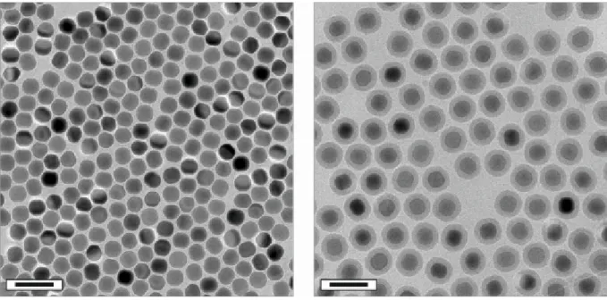
articles (organic or inorganic) with dimensions in the 1-100 nm range are referred to as nanoparticles (NPs; Greek νᾶνος: dwarf). They comprise an intermediate form of matter between individual atoms (or small molecules) and the bulk phase [1]. Exemplarily images of lanthanide-doped NaYF4 NPs acquired by transmission electron microscopy (TEM) are shown in Figure 1. TEM is a commonly utilized powerful imaging technique which can be used to directly visualize NPs. Moreover, TEM allows for the characterization of material on the nanoscale in terms of their size, shape, crystallinity, elemental composition, etc. [2].
NPs can be considered as an assembly of only a few atoms, since atomic radii are about 1 Å [3]. Using a simplified model, one can calculate the number of gold atoms (atomic radius of 144 pm) per one single gold NP (AuNP) with a diameter of 2 nm to be
~ 334 (assuming spherical AuNPs) [4,5]. Accordingly, the number of gold atoms located at the surface of the AuNP is ~ 192 (~ 57 %). This is in stark contrast to the bulk phase where the majority of atoms are located in the interior. Hence, the surface area-to-volume ratio of a spherical particle increases with 6·d-1 (d: diameter) with decreasing particle diameter.
Figure 1 | Transmission electron microscopy images of lanthanide-doped NaYF4 NPs deposited on a carbon-coated copper grid. The average particle diameter is ~ 9 nm (left) and
~ 22 nm (middle), respectively. The image on the right shows rod-like NPs with dimensions of
~ 28x54 nm. Scale bars indicate 60 nm.
P
Such a tremendous increase in the surface area-to-volume ratio can strongly alter the physical and chemical characteristics of NPs in comparison to their respective bulk phase [6]. For example, semiconductor NPs based on CdSe (referred to as quantum dots) show size dependent emissions in the visible range due to quantum confinement effects, which makes them highly attractive candidates for (bio)-imaging and sensing applications [7]. Another example are AuNPs (2-3 nm in diameter) dispersed on a titania support which have been found to show high catalytic activity for the oxidation of CO to CO2 at ambient conditions [8,9]. Today, the majority of industrial catalysts consist of metallic NPs dispersed on high surface area supports [10].
Interestingly, the application of AuNPs to make ruby glass appeared around the 5th or 4th century B.C. in Egypt and China [11]. The most famous example is the Lycurgus Cup, which is ruby red in transmitted light and green in reflected light, due to the presence of AuNPs [12]. In 1857, Faraday reported on the formation of deep-red solutions of (colloidal) gold particles by reduction of an aqueous solution of chloroaurate [13]. The term colloid was coined by Graham in 1861 [14], which is defined as one substance (e.g. NPs) evenly dispersed throughout a solution. This type of mixture can be further specified as a colloidal dispersion.
A qualitative explanation to Faraday’s observation is that AuNPs absorb visible light. In more detail, AuNPs display a broad absorption band (surface plasmon absorption) with a maximum at ~ 520 nm (for a NP diameter of ~ 15 nm), which is due to the collective oscillations of free electrons (plasmons) caused by the oscillating electric field of the irradiation light [15,16]. In 1908, Mie rationalized the nature of the surface plasmon absorption band by solving Maxwell’s equations for the absorption and scattering of electromagnetic radiation by spherical metal particles [17].
The examples of CdSe quantum dots and AuNPs demonstrate how the material properties can change on the nanoscale due to quantum mechanical effects. The impact of nanomaterials and nanotechnology – a highly interdisciplinary science which includes aspects of material science, chemistry, physics, biology, and medicine – on (bio)-analytical applications will be discussed in the next chapters.
1.2. Nanomaterials for (bio)-analytical Applications
1.2.1. Gold Nanoparticles
One of the earliest reports on colloidal AuNPs used as labelling markers (immunogold staining) for the detection of Salmonella antigens in electron microscopy dates back to 1971 [18]. Here, Faulk and Taylor successfully applied AuNPs by taking advantage of their unique properties such as: (1) High electron density and therefore clear visibility in heavy metal ion- contrasted biological structures in transmission electron microscopy (TEM); (2) Preparation of NPs with a very narrow size distribution is possible; (3) Multiplexed labelling by use of AuNPs exhibiting significant differences in diameter [19]. Today, AuNPs are extensively used in the biomedical and (bio)-analytical fields due to their unique optical and electronic properties [20]. Their applications are summarized in numerous of excellent review articles which range from sensing [21], diagnosis [22], and photothermal therapeutics [23], to catalysis [24,25], surface plasmon resonance spectroscopy (SPR) [26], and surface enhanced Raman spectroscopy (SERS) [27,28].
In more detail, AuNPs can be used for absorption-based colorimetric sensing, since the aggregation of AuNPs of appropriate sizes (diameter > 3.5 nm) induces interparticle surface plasmon coupling, resulting in a visible color change [29]. The presence of an analyte may lead to aggregation of AuNPs functionalized with corresponding recognition elements on their surface. Such a colorimetric sensing scheme has been used for the detection of ssDNA targets with detection limits in the picomolar range [30]. Here, AuNPs were functionalized with respective complementary oligonucleotide strands leading to aggregation of AuNPs due to complementary DNA base pairing with target ssDNA.
In a different fluorescence-based approach for sensing of ssDNA, a hairpin loop structure (molecular beacon) is used. This structure is formed by a self-complementary nucleic acid probe and conjugated to an organic fluorophore on one end and a AuNP on the other end (see Scheme 1). As an example, the AuNP (diameter 1.4 nm) acts as a fluorescence quencher for the organic fluorophore Rhodamine 6G due to non-radiative energy transfer from the dye to the metal nanoparticle. The hairpin structure changes to a rod-like conformation after hybridization to a ssDNA target. Accordingly, the distance between the
dye (Rhodamine 6G) and the AuNP gets larger. This results in a significant increase in fluorescence since the quenching efficiency is > 99.9 % [31].
Scheme 1 | A fluorescence-based assay for sensing of target ssDNA using a gold-quenched nucleic acid probe (molecular beacon). The hairpin structure of the molecular beacon brings the fluorophore and the AuNP in close proximity. Accordingly, upon excitation of the fluorophore, its emission is quenched by the AuNP. Through sequence-specific hybridization to a ssDNA target, the hairpin structure changes to a rod-like conformation which increases the distance between the fluorophore and the quencher (AuNP). Consequently, the fluorescence of the organic dye is restored.
1.2.2. Magnetic Nanoparticles
Magnetic nanoparticles (MNPs) based on magnetite (Fe3O4) or maghemite (γ-Fe2O3) constitute another important class of functional NPs for (bio)-analytical applications [32].
They are referred to as superparamagnetic iron oxide nanoparticles (SPIONs) since particles with diameters smaller than ~ 20 nm (single magnetic domain limit) exhibit superparamagnetism at room temperature (i.e. their magnetization can randomly flip direction under the influence of temperature, leading to a net magnetic moment of zero in absence of an external magnetic field) [33]. SPIONs can be magnetically manipulated using an external
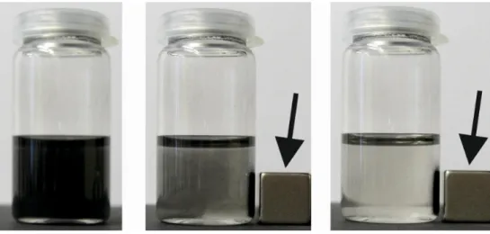
magnetic force (see Figure 2) and used for magnetic separation of target species from a complex mixture (e.g. separation of proteins from a cell lysate) [34], or remote-controlled delivery of drugs and therapeutics [35,36]. Other applications employ MNPs for hyperthermia [37], (bio)-sensing [38], or as contrast agents for magnetic resonance imaging (MRI) [39].
SPIONs for MRI diagnosis are widely used for clinical purposes (e.g. Feridex®
or Resovist®) [40]. Besides SPIONs, paramagnetic gadolinium chelates are also widely used as MRI contrast agents. However, these complexes must be administered in high dosage (0.1 mmol·kg-1 body weight for Gadovist®) because of their relatively low sensitivity [41].
Furthermore, free gadolinium ions leached from complexes can have toxic side effects like nephrogenic systemic fibrosis [42]. Finally, most gadolinium chelates are designed to have a very short circulation time, which precludes high-resolution and/or targeted MRI. In contrast, SPIONs exhibit high relaxivity and are known to be biologically well tolerated and benign.
The toxicity, metabolism, and pharmacokinetics of intravenously injected SPIONs have been well studied [43]. SPIONs can be tailored in terms of size and surface functionalization which is beneficial for targeted imaging and prolonged circulation times. Moreover, they can be used as nano-platforms for multimodal imaging (e.g. MRI, positron emission tomography (PET), fluorescence) by conjugation to radioactive tracers or fluorescent dyes [44].
Figure 2 | Magnetic nanoparticles can be collected by using an external permanent magnet (arrow). Colloidal stable, oleic acid – coated MNPs dispersed in cyclohexane are shown on the left. Snapshots taken 5 s (middle) and 10 s (right) after applying an external magnetic field, respectively.
1.2.3. Liposomes
A third example of nanomaterials suitable for medical and (bio)-analytical applications are liposomes (vesicles). In their simplest form, liposomes are composed of a phospholipid bilayer surrounding an aqueous core [45]. The size of liposomes ranges from typically 25-50 nm for small unilamellar vesicles to 100 nm – 1 µm (or even several microns) for large unilamellar vesicles. Both hydrophilic and hydrophobic compounds can be encapsulated into the inner cavity (aqueous core) or incorporated into the bilayer membrane of liposomes, respectively. Accordingly, liposomes are utilized as versatile carriers for drugs or therapeutics, which typically serves to improve the pharmacokinetics and biodistribution of a drug [46]. Currently there are ~ 11 liposomal drug formulations (e.g. chemotherapeutics) available which are approved for clinical use, and many more are in clinical or preclinical development [47].
Furthermore, liposomes provide an excellent means for signal amplification in biosensors [48]. Signal markers such as dyes, enzymes, salts, chelates, DNA, or electrochemical and chemiluminescent species can be encapsulated within liposomes.
Labelling of vesicles with biorecognition elements including bilayer incorporated gangliosides, cholesterol modified DNA oligonucleotides, and peptides, enzymes, and antibodies covalently attached to the hydrophilic headgroup of a lipid can be easily achieved.
The controlled release of liposomal cargo using phase transition, ultrasound, or lysis strategies after a one-to-one biological binding event may lead to signal amplification. Consequently, the limit of detection using liposome-based biosensor formats or assays (e.g. lateral flow assay, flow injection analysis, high-throughput microtiter plate, or microfluidic devices) is usually quite low (viz. parts-per-billion, ppb) [49].
In summary, the development of colloidal nanomaterials (e.g. AuNPs, MNPs, SPIONs, and liposomes) for applications in medicine and (bio)-analysis has shown great potential. Colloidal stability of nanomaterials in appropriate media (e.g. cell culture buffer systems, or body fluids) is an essential prerequisite for their medical and (bio)-analytical application (in vitro and in vivo). Therefore, sophisticated engineering of surface properties is
indispensable in order to avoid aggregation of nanomaterials under physiological conditions.
Beyond this, recent efforts in nanotechnology offer the possibility to combine useful chemical or physical properties of different nanomaterials within one single entity, thus allowing for synthesis of bi- or even multifunctional (hybrid) NPs (e.g. Fe3O4-Au, dumbbell-like NPs) [50,51]. Accordingly, colloidal nanomaterials are promising platforms for new and powerful theranostic agents (therapy and diagnostics) and may improve the performance and sensitivity of (bio)-analytical assays.
1.3. Luminescent Nanomaterials
1.3.1. Specifications of Ideal Luminescent Labels
Luminescence-based techniques are excellent methods to investigate fundamental processes in life sciences. They represent extremely important and powerful (bio)-analytical tools in medicine, biology, and chemistry due to their fast, sensitive (down to the single-molecule level), reliable, and reproducible detection procedures. There is a large variety of molecular chromophores (e.g. organic dyes, metal-ligand complexes, lanthanide chelates, or fluorescent proteins) from which one can choose for (bio)-imaging and sensing applications [52]. As an example, these chromophores can be employed as extrinsic luminescent labels, when the target of interest is non-luminescent or its intrinsic luminescence is not adequate for solving the analytical question of interest.
An ideal luminescent label for biological applications should fulfill the following requirements: (a) High molar absorption coefficient at a convenient excitation wavelength (without simultaneous excitation of the biological matrix); (b) Detection of luminescence with conventional instrumentation; (c) High luminescence quantum yield (number of emitted photons occurring per number of absorbed photons); (d) High brightness (product of the molar absorption coefficient at the excitation wavelength and the luminescence quantum yield); (e) Large Stokes shift between excitation and emission wavelength; (f) Solubility and stability in relevant hydrophilic media (e.g. buffers, cell culture media); (g) High photostability; (h) Functional groups for site-specific labeling; (i) Low
toxicity; (j) Reported data about its photophysics (luminescence lifetime, luminescence decay behavior, appearance of luminescence blinking); (k) Availability in reproducible quality; And (l) suitability for multiplexing (small and symmetric emission bands are favorable) [52].
During the last decades, luminescent labeling using nanoparticle-based chromophores as alternatives to conventional molecular dyes gained increasingly more attention [53,54]. The most ambitious and fascinating application of luminescent nanomaterials is probably related to medicine, molecular biology, and (bio)-analytics [55].
Here, these nanomaterials are promising tags for luminescent labeling and optical (bio)- imaging in order to enable novel techniques of non-invasive observation of complex vital functions (e.g. in vivo whole-body diagnosis, or in vitro examination of individual organs or cells) [56]. Generally, luminescent nanomaterials can be assigned to two classes, (a) extrinsic (dye-doped) luminescent nanomaterials, and (b) nanomaterials exhibiting intrinsic luminescence [57].
1.3.2. Extrinsic Luminescent Nanomaterials
The first group comprises NPs doped with (organic or inorganic) chromophores as active luminescent species [58]. Examples are NPs made out of silica or organic polymers such as polystyrene doped with organic fluorophores [59,60]. The material itself does not show any intrinsic luminescence, but rather acts as a kind of a host matrix for molecular chromophores.
Therefore, this group can be specified as extrinsic luminescent nanomaterials. The advantages of dye-doped NPs over single molecular dyes are many. Polymer- and silica-based NPs with tunable diameters from 10 to 100 nm can include tens, hundreds or even thousands of molecular luminophores, which leads to a significant gain in luminescence intensity and brightness [61]. This is an advantageous feature for (bio)-imaging and sensing applications since the signal-to-noise ratio can be greatly improved [62]. However, the concentration of dye molecules embedded into NPs must be strictly controlled in order to avoid self-quenching processes [63].
The incorporation of molecular chromophores inside a silica or polymer matrix protects them from the surrounding environment and increases their photostability. Hence, this concept is a universal and highly modular approach since physicochemical properties of
NPs (size, shape, surface chemistry, etc.) can be varied and optimized with regard to their particular application [64]. Moreover, even hydrophobic luminophores can be easily entrapped using reverse microemulsion techniques. This is a fast, simple, and elegant way for the phase transfer of hydrophobic chromophores into hydrophilic media [65,66]. Additionally, other molecules like drugs, magnetic contrast chelates, or chemotherapeutics can also be incorporated into polymer and silica NPs, which makes them promising contenders for use in smart drug delivery and therapy systems, yielding multifunctional NPs [67,68]. Furthermore, the surface of such NPs can be modified in order to introduce biorecognition ligands (e.g.
antibodies, proteins, or DNA) enabling target-oriented imaging, sensing, and active delivery of drug molecules [69,70].
1.3.3. Intrinsic Luminescent Nanomaterials
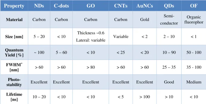
The second group covers nanomaterials displaying intrinsic luminescence. In contrast to the first group, here, the nanomaterial itself is capable of generating luminescence due to quantum mechanical or confinement effects without the need for any additional luminophore. A further classification of the second group can be made as follows: (a) semiconductor NPs; (b) metal nanoclusters; (c) carbon-based nanomaterials; and (d) metal-doped NPs. The physical and optical properties of luminescent nanomaterials including nanodiamonds (NDs) [71], carbon nanodots (C-dots) [72], graphene oxide (GO) [73], carbon nanotubes (CNTs) [74], quantum dots (QDs), and gold nanoclusters (AuNCs) [75] are compared to conventionally used organic fluorophores (OFs) including fluorescein, Cy3, Cy5, Texas Red, or Nile Red (see Table 1).
NDs are carbon NPs with a truncated octahedral architecture and are typically about 5 to 20 nm in diameter [76]. They are made from milling microdiamonds (top-down approach), chemical vapor deposition, shockwave, or detonation processes [77]. In addition, NDs exhibit superior chemical stability and excellent resistance to photobleaching along with low toxicity [78]. However, their luminescence is not easily tunable [79]. Moreover, aggregation of colloidal NDs is often a serious problem and the production of homogeneous samples with a narrow size distribution is challenging.
Table 1 | Comparison of physical and optical properties of luminescent nanomaterials and organic fluorophores.
Property NDs C-dots GO CNTs AuNCs QDs OF
Material Carbon Carbon Carbon Carbon Gold Semi- conductor
Organic fluorophor
Size [nm] 5 – 20 < 10 Thickness ~0.6
Lateral: variable Variable < 2 2 – 10 < 1
Quantum
Yield [%] ~ 100 5 – 60 < 10 < 25 < 20 10 – 90 50 - 100
FWHM*
[nm] > 60 > 60 > 80 > 60 > 60 25 – 35 35 - 100
Photo-
stability Excellent Excellent Excellent Excellent Excellent Good Medium
Lifetime
[ns] 10 – 20 < 10 < 10 < 5 > 100 > 10 < 10
*FWHM: full width at half maximum. NDs: nanodiamonds; C-dots: carbon nanodots; GO: graphene oxide;
CNTs: carbon nanotubes; AuNCs: gold nanoclusters; QDs: quantum dots; OFs: organic fluorophores. Adopted from Ref. [55].
An additional category of carbon-based nanomaterial are C-dots. They exhibit a quasi-spherical particle shape with diameters < 10 nm. C-dots display non-blinking, size and excitation wavelength dependent photoluminescence behavior, and are highly photostable.
However, the mechanisms of photoluminescence and the photophysical properties of C-dots and most other carbon-based nanomaterials are poorly understood [80]. GO offers intrinsic aqueous solubility due to the presence of functional groups (e.g. carboxyl, or hydroxyl groups) [81]. One drawback of this material is that its broad emission cannot be easily tuned [82]. CNTs do not show any photobleaching, however, the intensity of photoluminescence is relatively weak [83]. Noble metal nanoclusters (e.g. AuNCs) are smaller than 2 nm, exhibit no apparent plasmonic properties, and have excitation and emission bands similar to those of molecular dyes [84]. The luminescence properties of AuNCs are size-dependent and sensitive to their environment (i.e. pH, ionic strength, or temperature). Large Stokes shifts and long luminescence lifetimes have been observed. However, the controlled synthesis of high quality AuNCs (< 2 nm in diameter) is still very difficult [85].
Another important class of colloidal NPs are semiconductor nanocrystals with dimensions between 2 nm and 10 nm. They are referred to as quantum dots (QDs) and display size-dependent optical properties (quantum size effect), which arise from interactions between electrons, holes, and their local environment [86,87]. QDs absorb photons when the excitation energy exceeds the band gap. Electrons are promoted from the valence band to the conduction band during this process. The emission of light is due to the recombination of electron-hole pairs (excitons), which is referred to as excitonic fluorescence. For example, bulk CdSe has a band gap energy of 1.76 eV and a Bohr exciton diameter of 9.6 nm, whereas the band gap energy of 2-7 nm CdSe QDs decreases from 2.8 to 1.9 eV [88]. As a result, the wavelength of the corresponding emission can be tuned continuously from 450 to 650 nm, depending on the nanocrystal diameter. The diameter of QDs and therefore the emission wavelength can be tuned by controlling the temperature and duration of crystal growth during the synthesis. QDs are highly attractive candidates for in vitro and in vivo optical imaging [89,90,91], cell tracking [92], gene and drug delivery [93], and diagnostic applications [94,95] due to their unique optical properties, which makes them promising alternatives to conventionally used organic fluorophores.
In more detail, the absorption bands of QDs are rather broad and there is a continuous increase of absorption from their first exciton peak towards shorter wavelengths [96,97], which is in stark contrast to organic fluorophores. This broad absorption allows for free selection of the excitation wavelength, which is beneficial in order to separate emission from excitation light. Moreover, a single light source is sufficient for the excitation of QD emissions. The emission can be continuously tuned from ultraviolet (UV) to near-infrared (NIR), depending on the elemental composition and nanocrystal diameter [98]. Bawendi and coworkers reported a molar extinction coefficient for CdS QDs of ~ 105-106 M-1·cm-1, depending on the particle diameter and the excitation wavelength [99]. Hence, QDs exhibit molar extinction coefficients as high as organic fluorophores or even one order of magnitude higher [100,101]. The full width at half maximum (FWHM) of the symmetric emission peak of QDs with a Gaussian peak profile is ~ 30 nm at room temperature [102]. This makes them ideal candidates for spectral multiplexing.
The width of emission peaks of QDs is mainly determined by the size distribution of the nanocrystals. Luminescence quantum yields (QYs) of QDs in the visible range (400-700 nm) are comparable to those of fluorescent dyes. As an example, the QY of
CdSe QDs is ~ 0.8 [103,104], which is quite high. However, QYs of 0.97 or even higher can be found for organic dyes such as fluorescein under alkaline conditions [105]. In the NIR region (> 700 nm), QDs exhibit certain advantages over organic fluorophores, such as a typically higher quantum yield and superior resistance to photobleaching [106]. The excited state decay rate of QDs is typically > 10 ns and thus slightly slower than that of organic dyes (~ 1-10 ns) [107]. This enables the use of time-gated detection to separate the QDs’
luminescence from short-lived luminescence interference from scattered excitation light or cellular autofluorescence, which enhances sensitivity [108]. Another favorable feature of QDs is their large two-photon absorption cross section (103-104 GM), which is orders of magnitude larger than those of organic chromophores [109,110]. However, an inherent disadvantage of QDs is their complicated size-dependent, surface-dependent, wavelength-dependent, bi- (or even multi-) exponential decay behavior, which renders time-resolved luminescence measurements very difficult [111].
Finally, there are two additional drawbacks of QDs. First, the surface defects in the crystal structure can serve as temporary “traps” for electrons or holes. This prevents their radiative recombination and leads to a fluorescence intermittency (so-called blinking), which is apparent from single luminescent nanocrystals [112]. Second, the cytotoxicity of QDs is a serious threat, since semiconductor nanocrystals are mostly composed of toxic heavy metal ions (e.g. Cd2+, Zn2+, Pb2+) [113,114]. However, toxicity may also be problematic using organic dyes.
In conclusion, there is a large variety of different luminescent nanomaterials suitable for (bio)-analytical applications. The drawback of limited photostability of organic fluorophores can be overcome to a certain extent by using dye-doped silica or polymer NPs.
QDs, C-dots, and metal nanoclusters exhibit promising potential for (bio)-imaging and sensing due to their unique optical properties and extraordinary photostability. Therefore, they can be considered as alternatives to conventional organic fluorophores. However, their emissions are related to quantum confinement effects. Hence, a precise adjustment and control of their physicochemical properties (size, shape, surface chemistry, etc.) are important prerequisites in order to obtain NPs with defined characteristics and efficient luminescence.
1.4. Upconverting Luminescent Nanoparticles
1.4.1. Characteristics and Composition
All luminescent nanomaterials discussed so far (including molecular luminophores) require excitation by UV or visible light and show Stokes-shifted emissions (i.e. the emitted light has a longer wavelength than the excitation light). Therefore, they can be designated as downconverting luminescent (nanomaterials) nanoparticles (DCLNPs). However, in recent years, a new class of nanoscale luminophores, which are referred to as upconverting luminescent nanoparticles (UCLNPs), has gained much scientific interest. Here, the emitted light has a shorter wavelength than the excitation light, which is in stark contrast to DCLNPs.
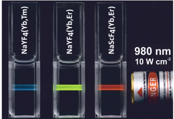
An image of UCLNPs emitting predominantly blue, green, and red emission upon 980 nm continuous wave laser excitation is shown in Figure 3. This phenomenon is called photon upconversion and was first described by Auzel, Ovsyankin and Feofilov in the 1960s [115].
There are several excellent review articles which summarize and discuss the different mechanisms of upconversion in detail [116,117,118]. Briefly the processes of photon upconversion can be roughly divided into three main classes: (1) energy transfer upconversion (ETU, see Scheme 2); (2) excited-state absorption (ESA); and (3) photon avalanche (PA). In contrast to simultaneous two-photon absorption [119,120] or second-harmonic generation [121,122], all of these three processes are based on the sequential absorption of two or more photons by existing, metastable, long-lived electronic energy states of metal ions. However, ETU is by far the most efficient UC process [123]. As a consequence of these sequential absorption steps, highly excited electronic energy states are populated from which upconversion luminescence occurs. Therefore, photon upconversion is a non-linear optical phenomenon [124].
Figure 3 | UCLNPs dispersed in cyclohexane emit visible light upon 980 nm continuous wave laser excitation (laser power density 10 W·cm-2). Predominant blue (NaYF4 doped with Yb3+/Tm3 +), green (NaYF4 doped with Yb3 +/Er3+), and red (NaScF4 doped with Yb3 +/Er3 +) luminescence can be observed by the bare eye.
UCLNPs are composed of an inorganic (crystalline) host material doped with metal ions, which act as active luminescent centers (activators). A large number of different dopants embedded into suitable host materials has been reported to show photon upconversion, for example solids doped with transition-metal ions (3d, 4d, 5d) like Ti2+, Ni2+, Mo3+, Re4+, or Os4+ [116,118]. However, the highest upconversion efficiencies at room temperature are observed for lanthanide-doped (Ln3+) solids [125]. Most commonly, upconversion (nano)-phosphors contain trivalent 4f ions such as Er3+, Tm3+, or Ho3+ as activators. The f-f transitions of lanthanide ions are strongly forbidden by the parity selection rule resulting in long lifetimes of the excited states (in the range of µs to ms) [126]. As a consequence, lanthanide ions typically show low molar absorption coefficients on the order of 1 M-1·cm-1 [127]. The energy of Ln3+ electronic levels is well defined due to the shielding of the 4f orbitals by filled 5s2p6 sub-shells (i.e. there is no significant variation of the energy levels caused by the chemical environment in which Ln3+ ions are inserted). In principle, the absorption can be greatly improved by increasing the dopant concentration of lanthanide ions per single (nano)-crystal. However, radiation-less deactivation and cross-relaxation processes
can occur at high doping concentrations [128]. Thus, strongly absorbing sensitizer ions, which should also ensure efficient non-radiative energy transfer to activator ions, are additionally doped into the crystalline host matrix in order to further increase absorption. Yb3+ ions having a molar absorption coefficient of ~ 10 M-1·cm-1 are the most commonly used sensitizers for Er3+, Tm3+, or Ho3+ doped upconverting (nano)-phosphors [129].
The efficiency of upconversion luminescence is strongly influenced by the crystalline host material and its crystal structure. The ion-to-ion distance of dopants located within the host lattice and their spatial arrangement are of great importance [130]. Therefore, a suitable host material provides a matrix to bring these dopants into optimal position with respect to one another [131]. The most efficient host material for Yb3+/Er3+ and Yb3+/Tm3+
doped upconverting (nano)-phosphors is hexagonal phase (β) NaYF4 [132]. This fluoride- based host matrix is superior to oxygen-based hosts because of its relative low phonon energy of ~ 350 cm-1, which is beneficial for long lifetimes of excited electronic states [133].
Moreover, Y3+ can be easily substituted by lanthanide ions since both exhibit similar ionic radii. Thus, the formation of crystal defects and lattice stress is prevented. The crystal structure of a host material is another important aspect for efficient upconversion luminescence. Here, NaYF4 constitutes an excellent example, since it exists in two polymorphs at ambient pressure: (a) cubic (α-phase) – a metastable high-temperature phase;
and (b) hexagonal (β-phase) – a thermodynamically stable low-temperature phase [134]. It is reported that the efficiency of upconversion luminescence is approximately one order of magnitude higher for bulk β-NaYF4 in comparison to α-NaYF4 [135,136].
1.4.2. Photophysical Properties
The “flagship” upconversion (nano)-phosphor material is undoubtedly β-NaYF4 (acting as a host matrix material) doped with either Yb3+/Er3+ or Yb3+/Tm3+ ion couples. The doping concentrations are usually ~ 20-25 mol% of Yb3+, ~ 2 mol% of Er3+, and ~ 0.3 mol% of Tm3+
ions [137]. Here, Yb3+ ions act as sensitizers which absorb excitation light at 980 nm. In contrast to other lanthanides, Yb3+ ions have a relatively simple energy level structure [138].
They undergo a transition from their 2F7/2 to 2F5/2 electronic state upon NIR excitation.
Subsequently, energy is sequentially transferred from excited sensitizer ions (Yb3+) to
adjacent activator ions (Er3+) via a non-radiative, resonant energy transfer upconversion process. The energy of 2F5/2 states of Yb3+ and 4I11/2 of Er3+ is very similar, which allows for an efficient energy transfer from excited state Yb3+ to neighboring Er3+ to occur.
Subsequently, an additional energy transfer from another excited state Yb3+ to the Er3+ can take place, resulting in further excitation to its 4F7/2 excited state. As a result of these ETU processes, Er3+ ions are promoted from their 4I11/2 ground states to 4F7/2 excited states.
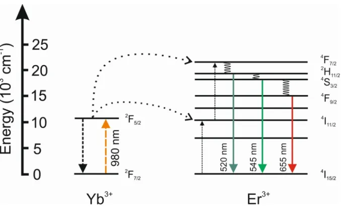
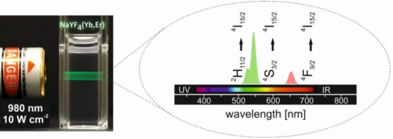
Multicolor upconversion luminescence of Er3+ activator ions can be observed in the visible range (see Scheme 2) upon relaxation to their 4I15/2 ground state. The primary emission peaks are located in the green (2H11/2/4S3/2 → 4I15/2) and red (4F9/2 → 4I15/2) region of the electromagnetic spectrum, depending on the particular relaxation pathway. Due to their unique electronic configuration, lanthanide ions display emissions with extraordinarily narrow luminescence bandwidths [139]. Here, the FWHM of the emission peaks are typically
< 20 nm (see Figure 4).
The relatively long lifetime of lanthanide ions excited states on the millisecond time scale is beneficial for these ETU mechanisms. Hence, an inexpensive continuous wave (CW) diode-laser operating at 980 nm with a moderate excitation power density of
~ 10 W·cm-2 is sufficient in order to induce upconversion luminescence based on ETU (see Figure 4). This is orders of magnitude lower than for simultaneous two-photon absorption processes. Here, expensive ultrashort pulsed lasers operating at power densities of
~ 105-109 W·cm-2 are required [140] for the excitation of dye molecules. Simultaneous two- photon absorption involves a “virtual” intermediate state of dye molecules exhibiting extremely short lifetime which is in stark contrast to the long excited state lifetime of lanthanide ions in ETU processes.
The energy levels and ETU mechanisms of Yb3+/Tm3+-doped upconversion (nano)-phosphors are shown in Scheme 3. Here, Tm3+ ions can be excited into their 1D2
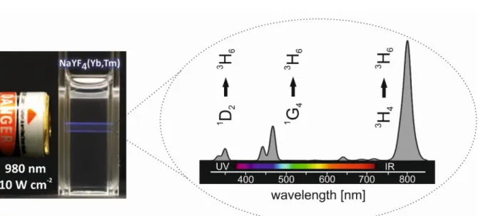
electronic state by a four times Yb3+-sensitized sequential energy transfer. As a result, emissions of Tm3+ activator ions in the UV, visible, and NIR spectral range can be detected upon relaxation to their electronic ground state (3H6). Figure 5 displays the typical predominant blue luminescence of β-NaYF4 UCLNPs doped with Yb3+/Tm3+ ions upon 980 nm CW laser excitation (laser power density 10 W·cm-2). Additionally, a characteristic luminescence spectrum of Yb3+/Tm3+ doped upconversion (nano)-phosphors is shown in
Figure 5, exhibiting distinct emission peaks at 360 nm (1D2 → 3H6), 475 nm (1G4 → 3H6), 648 nm (1G4 → 3F4), and 800 nm (3H4 → 3H6).
Scheme 2 | Energy level diagram and energy transfer upconversion (ETU) mechanisms for a Yb3+/Er3 +-doped (sensitizer/activator) system. Excitation light (980 nm) is absorbed by Yb3 + sensitizer ions and sequentially transferred to Er3+ activator ions leading to multicolor upconversion luminescence in the visible range. Arrows indicate radiative, non-radiative energy transfer, and multiphonon relaxation processes.
Excitation of UCLNPs (employed as luminescent labels, biomarkers, or sensing probes) by NIR light rather than UV radiation provides several advantages such as: (a) Photo damage of biological specimens is significantly reduced [141]; (b) The penetration depth into biological tissue is higher since excitation takes place in the so-called biological optical window (from ~ 650 to ~ 1000 nm), where the absorption coefficient of tissue is minimal [142,143]; and (c) Very weak autofluorescence background from biological tissue resulting in improved detection sensitivity due to higher signal-to-noise ratio [144]. In addition, UCLNPs do not show any blinking characteristics under continuous laser excitation and are extremely resistant to photobleaching as well as photochemical degradation even under intense excitation power densities [145]. This makes them highly attractive candidates as labels and markers for (bio)-imaging and sensing applications.
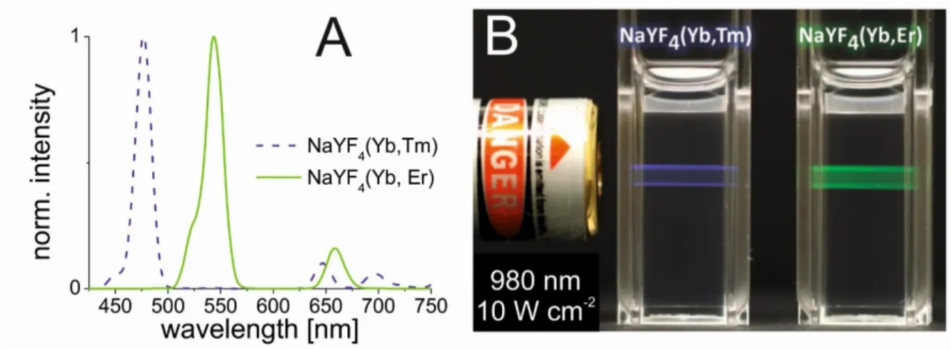
Figure 4 | Left: Colloidal dispersion of oleate-coated β-NaYF4(Yb3 +/Er3 +) UCLNPs in cyclohexane displaying predominantly green luminescence upon 980 nm CW laser excitation (10 W·cm-2). Right: Corresponding upconversion luminescence spectrum exhibiting two distinct emission peaks in the green and red spectral region. Related electronic transitions are indicated.
Scheme 3 | Energy level diagram and energy transfer upconversion (ETU) mechanisms for a Yb3+/Tm3 +-doped (sensitizer/activator) system. Excitation light (980 nm) is absorbed by Yb3 + sensitizer ions and sequentially transferred to Tm3 + activator ions leading to multicolor upconversion luminescence spanning from the UV to NIR. Arrows indicate radiative, non- radiative energy transfer, and multiphonon relaxation processes.
Figure 5 | Left: Colloidal dispersion of oleate-coated β-NaYF4(Yb3 +/Tm3 +) UCLNPs in cyclohexane displaying predominantly blue luminescence upon 980 nm CW laser excitation (10 W·cm-2). Right: Corresponding upconversion luminescence spectrum exhibiting distinct emission peaks in the UV, visible, and NIR. Related electronic transitions are indicated.
1.4.3. Synthesis Strategies
In general, there are three main components of upconversion (nano)-phosphors one should carefully consider in order to obtain efficient upconversion luminescence: (a) The inorganic host material and its crystal structure. The type and the concentration of (b) sensitizer ions, and (c) activator ions. Synthesis methods for the fabrication of UCLNPs have been developed during the last decade in order to meet all of these criteria. These methods include co- precipitation [146,147], hydro(solvo)thermal methods [148], thermal decomposition [149,150], and ionic liquids-based synthetic strategies [151,152]. The most widely used methods for the synthesis of UCLNPs are hydro(solvo)thermal based strategies and thermal decomposition procedures.
The hydrothermal/solvothermal method is a typical solution-based bottom-up approach. For the synthesis of lanthanide-doped NaYF4 UCLNPs rare-earth and fluoride precursors (e.g. rare-earth chlorides, nitrates, or oxides; HF, NH4F, or NaF), solvents and certain surfactants (e.g. ehtylenediamine tetraacetic acid; cetyltrimethylammonium bromide;
or oleic acid) are mixed. Educts are heated in a sealed autoclave above the critical point of the solvent, which increases the solubility and reactivity of the reactants. The optimization of the
synthesis parameters of this method is generally very time-consuming since the reaction times are long (up to several days). Therefore, it is difficult to synthesize high quality UCLNPs in terms of phase crystallinity and purity, particle size distribution, and particle shape [153].
Another disadvantage is that specialized reaction vessels (autoclave) are required, which makes it impossible to observe and control the nanocrystal growth during the synthesis.
An alternative synthesis strategy is the thermal decomposition of metal trifluoroacetates in solvent mixtures of oleic acid (OA) and 1-octadecene at temperatures of
~ 320 °C to corresponding metal fluorides. During the synthesis, nucleation of metal fluorides takes place, followed by the growth of nuclei into nanocrystals. These crystals are covered by oleic acid molecules which act as surfactants preventing their agglomeration. In 2006, Chow et al. reported on the synthesis of hexagonal NaYF4 UCLNPs doped with Yb3+ and Er3+ ions using a thermal decomposition strategy [154]. This method allows for the production of high quality UCLNPs based on lanthanide-doped β-NaYF4 with very narrow size distribution.
However, expensive and toxic metal precursors are used and toxic byproducts such as trifluoroacetic anhydride, trifluoroacetyl fluoride, carbonyl difluoride, tetrafluoroethylene, or hydrogen fluoride are produced [132].
However, one general drawback of all synthesis strategies is their batch-to-batch irreproducibility. This means that each batch of UCLNPs has its own particle size, size distribution, doping concentration, arrangement of dopant ions within the crystalline host lattice, and number of surface ligands, which in summary results in slightly different optical properties [155,156]. Therefore, a scale up strategy in order to produce identical UCLNPs of high quality on a large batch is highly desirable, especially since most protocols deal with the synthesis of only a small amount of UCLNPs per batch (~ 1 mmol of lanthanide precursors resulting in ~ 100 mg of UCNLPs).
1.4.4. Surface Modifications
Since most of the commonly used UCLNPs are synthesized using oil-phase based strategies with oleic acid or oleylamine molecules acting as surfactants, these NPs have neither intrinsic water dispersibility nor functional groups for further conjugation to biomolecules. Hence, post-synthesis methods for surface engineering are required which render UCLNPs
dispersible in aqueous media and colloidally stable under physiological conditions. Moreover, surface coatings should provide functional anchors for further bioconjugation to proteins, antibodies, DNA, etc. and make NPs biocompatible. Frequently used methods including silica coating (see Figure 6), ligand exchange, ligand oxidation, Layer-by-Layer coating, and coating by amphiphilic molecules and polymers have been recently summarized in several review articles [157,158,159,160]. TEM images of UCLNPs before and after silica coating are shown in Figure 6.
Figure 6 | TEM images of lanthanide-doped NaYF4 UCLNPs before (left) and after (right) silica coating. The uniform silica shell (~ 5 nm in thickness) can be clearly distinguished from the NaYF4 core, since it exhibits a different electron optical contrast. Scale bars indicate 60 nm.
1.4.5. Toxicity
The investigation of cytotoxic effects of UCLNPs is an important task for biomedical applications. In 2008, Shan et al. reported a study of in vitro cytotoxicity of silica-coated UCLNPs. Here, hydrophilic UCLNPs were incubated with human osteosarcoma cells. The results showed that silica-coated UCLNPs (concentration 1 mg·mL-1) functionalized with amine and carboxyl groups have low cytotoxicity in comparison to the control group. Further studies using different human and animal cell lines confirmed no severe adverse effects that
can be directly related to UCLNPs [161,162,163,164,165]. The long-term in vivo bio- distribution of UCLNPs was investigated by Xiong et al. in 2010. Results of toxicity studies indicated that mice intravenously injected with 15 mg·kg-1 of polyacrylic acid-coated UCLNPs survived for 115 days without any evident (observational, histological, hematological and biochemical) toxic effects. UCLNPs were found to mainly accumulate in the liver and spleen [163]. These examples demonstrate the low toxicity of UCLNPs.
This chapter described some of the basic aspects of UCLNPs with emphasis on their composition, photophysical properties, synthesis, surface modifications, and toxicity. In recent years, the number of proof-of-concept reports using UCLNPs as markers for (bio)- imaging or as donors for Förster resonance energy transfer (FRET) based sensing greatly increased [166,167,168,169]. All of these papers employ UCLNPs because of their unique optical characteristics (e.g. NIR excitation; multicolor anti-Stokes emissions; long luminescence lifetimes in the µs or ms regime). However, only a few reports investigate the photophysical properties of UCLNPs such as quantum yield, emission lifetimes, ratio of luminescence peaks, etc., which is of fundamental importance in order to fully exploit their potential for sensing and imaging applications. It is well known that photophysical properties of UCLNPs are strongly dependent on the excitation power density. Unfortunately, reports dealing with the photophysical characterization of hydrophilic UCLNPs dispersed in aqueous media are still missing.
2. Motivation and Aim of the Work
UCLNPs offer unique optical properties. They are capable of emitting anti-Stokes-shifted luminescence upon NIR excitation. This is of great advantage in comparison to commonly used luminescent labels and probes which are excited by UV or visible light. The utilization of NIR rather than UV or visible radiation maximizes the penetration depth of the excitation light into biological tissue and simultaneously minimizes photodamage of biological specimens. Moreover, the detection sensitivity is greatly improved since NIR excitation does not induce background autofluorescence resulting in excellent signal-to-noise ratio.
Within this work a bottom-up synthesis protocol should be established which allowed for the size-controlled preparation of bright UCLNPs. It is well known from in vivo experiments that NPs with hydrodynamic diameters smaller than ~ 10 nm are rapidly cleared by the kidneys or taken up by the liver. In contrast, NPs exhibiting diameters larger than
~ 150 nm are filtered out by the spleen [170]. Therefore, the aim was to synthesize small (< 50 nm) UCLNPs with narrow size distribution.
The control of surface chemistry is important in order to offer colloidal stability of NPs in biological media. Proper surface engineering which allows for further functionalization of UCLNPs is an indispensable prerequisite for providing them to (bio)- analytical applications. Thus, the aim of this work was to tailor UCLNPs for protein binding and labelling. In a second (bio)-analytical application the potential of UCLNPs for luminescence-controlled monitoring of enzymatic reactions upon NIR excitation should be evaluated. Up to now there is only little known about the influence of the surface coating on the photophysical properties of UCLNPs. Hence, one task of this work was to systematically investigate different surface modifications in terms of their impact on the upconversion luminescence.
3. Multicolor Upconversion
Nanoparticles for Protein Conjugation
3.1. Abstract
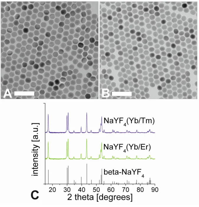
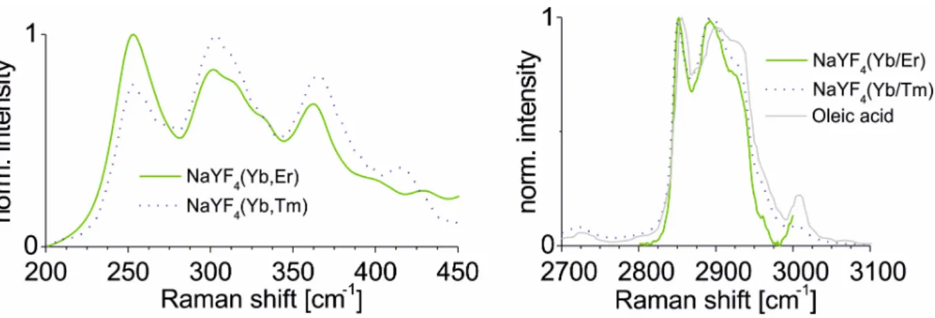
The preparation of monodisperse, lanthanide-doped hexagonal-phase NaYF4 upconverting luminescent nanoparticles for protein conjugation is described. Their core was coated with a silica shell which then was modified with a poly(ethylene glycol) spacer and N- hydroxysuccinimide ester groups. The nanoparticles were characterized by transmission electron microscopy, Raman spectroscopy, X-ray powder diffraction, and dynamic light scattering. The N-hydroxysuccinimide ester functionalization renders them highly reactive towards amine nucleophiles (e.g. proteins). Such particles can be conjugated to proteins. The protein-reactive UCLNPs and their conjugates to streptavidin and bovine serum albumin display multicolor emissions upon 980 nm continuous wave laser excitation. Surface plasmon resonance studies were carried out to prove bioconjugation and to compare the affinity of the particles for proteins immobilized on a thin gold film.
This chapter has been published.
Stefan Wilhelm, Thomas Hirsch, Wendy M. Patterson, Elisabeth Scheucher, Torsten Mayr, and Otto S. Wolfbeis. Theranostics 2013, 3, 239-248
Author contributions
SW synthesized and characterized the nanoparticles; performed conjugation experiments;
wrote the manuscript. SW and TH performed SPR measurements. WMP performed Raman measurements. ES and TM were involved in discussing the results. OSW supervised the project and is corresponding author.
3.2. Introduction
The implementation of nanotechnology to healthcare holds great promise in areas such as imaging [90,95,168,171,172,], faster diagnosis [173], targeting [174], drug delivery [175], and tissue regeneration [176], as well as the development of medical products [177,178,179]. The chemical synthesis of nanoparticles (NPs) has been studied in detail during the last decade [180,181,182,183]. Substantial efforts have been made to control the dimensions, shape, composition, particle size distribution, etc., of NPs, thereby creating new materials with size dependent electrical, optical, magnetic, catalytic, and chemical properties, which cannot be achieved by their bulk counterparts. Important classes of NPs are a) magnetic NPs, b) gold NPs, c) quantum dots, d) silica NPs, etc. [184,185,186,187,188,189,190]. In recent years, upconverting luminescent nanoparticles (UCLNPs) joined this classification. Photon upconversion has been researched ever since the 1960s. It is a process where two or more photons are sequentially absorbed, resulting in the emission of light at a shorter wavelength than the excitation light. For instance, infrared or near-infrared (NIR) light can be converted to shorter-wavelength radiation, usually in the visible range of the electromagnetic spectrum (anti-Stokes type emission) [191].
The mechanisms behind photon upconversion were first investigated in lanthanide-doped bulk materials by Auzel, Ovsyankin, and Feofilov [116]. In a sensitizer- activator system, the excitation energy is absorbed by a sensitizer ion (e.g. Yb3+) and transferred to an activator ion (e.g. Er3+ or Tm3+) via a non-radiative, resonant energy transfer process. Metastable, long-lived energy states are required, in which case energy transfer upconversion (ETU) is possible, where the combined energies of pump photons are stored, which can lead to the emission of a higher energy photon [192].
Anti-Stokes emissions from UCLNPs offer several advantages over conventional Stokes-shifted emissions from a) semiconductor quantum dots, b) organic- and protein-based fluorophores, and c) the multiphoton process employing fluorescent dyes.
UCLNPs are very attractive phosphors in terms of bioimaging due to their non-blinking emission and remarkable photostability [145,193,194]. In biological samples or tissue, there is minimal excitation of autofluorophores, since UCLNPs are usually excited by NIR (980 nm) continuous wave (CW) laser light. This scheme enables luminescence to be imaged with a
high signal to noise ratio, minimizes possible photodamage in biological systems, and allows deeper tissue penetration [169,195]. Upconversion microparticles have been used before in immunoassays [196], and enzyme activity assays [197], but their size (1 – 10 µm) and large size distribution makes their use less attractive. With respect to the relative size of a protein and upconversion microparticles, one may not speak of a label in its classical sense.
High-quality UCLNPs (with respect to crystal phase, monodispersity, geometry, etc.) are usually synthesized in high-boiling organic solvents (e.g. 1-octadecene) using ligand molecules with long alkyl chains (e.g. oleic acid), which renders them inherently functionalized with hydrophobic alkyl groups and only dispersible in non-polar organic solvents such as toluene, hexane, and the like [198,199]. In order to make them amenable to (bio)-analytical applications, surface modification is required, to make the UCLNPs water dispersible, offering a platform for further conjugation of functional chemical groups and/or (bio)-molecules. Silica is known for its biocompatibility [200], and silica coating of UCLNPs therefore offers an attractive way of functionalization [201,202]. This has already been applied to a multitude of nanoparticle systems, including gold and silver NPs [203], magnetic NPs [204], and quantum dots [205]. In addition, silica coating is a flexible coating technique that is applicable to both hydrophilic and hydrophobic NPs [206,207,208]. The coating process of hydrophilic NPs relies on the Stöber method, while a reverse-microemulsion method is typically used for coating hydrophobic NPs [128].
We describe the synthesis of protein-reactive, multicolor UCLNPs. First, monodisperse, lanthanide-doped hexagonal-phase NaYF4 nanoparticles were prepared, which were coated with oleic acid, as can be seen in Scheme 4. In the second step, the particles were silica coated using a reverse-microemulsion method, and subsequently functionalized with an amino-reactive silanization reagent. This reagent consists of a triethoxysilane conjugated to a poly(ethylene glycol) spacer (PEG) and a carboxyl group activated with an N- hydroxysuccinimide (NHS) ester. The NHS ester of the silica-coated UCLNPs renders them highly reactive towards proteins. The protein-reactive UCLNPs exhibit multicolor luminescence emission after 980 nm laser excitation. Finally, surface plasmon resonance (SPR) studies were carried out to study the binding affinity of NHS-activated nanoparticles to proteins that previously were immobilized on a gold film.
Scheme 4 | Surface engineering (including phase transfer, functionalization, and protein labelling) of initially hydrophobic UCLNPs towards protein-reactive, multicolor upconverting labels.