Nanoparticles for Assays, Sensing and Theranostic Applications
DISSERTATION ZUR ERLANGUNG DES DOKTORGRADES DER NATURWISSENSCHAFTEN (DR. RER. NAT.) DER FAKULTÄT CHEMIE
UND PHARMAZIE
DER UNIVERSITÄT REGENSBURG DEUTSCHLAND
vorgelegt von
Markus Buchner
aus Ergoldsbach
Im Jahr 2018
Regensburg.
Die Arbeit wurde angeleitet von Prof. Dr. Antje J. Bäumner und Dr. Thomas Hirsch.
Promotionsgesuch eingereicht am: 08.02.2018
Kolloquiumstermin: 23.03.2018
Prüfungsausschuss:
Vorsitzender: Prof. Dr. Hubert Motschmann
Erstgutachter: Prof. Dr. Antje J. Bäumner
Zweitgutachter: PD Dr. Miriam Breunig
Drittprüfer: Prof. Dr. Joachim Wegener
First, I want to thank Prof. Dr. Antje Bäumner and Dr. Thomas Hirsch for the opportunity to graduate at the Institute of Analytical Chemistry, Chemo- and Biosensors, for their excellent support and for countless scientific discussions.
PD Dr. Miriam Breunig and Prof. Dr. Joachim Wegener I want to thank for their acceptance as second and third reviewer. Best thanks to Prof. Dr. Hubert Motschmann for his acceptance as the chair of the exam.
I also want to thank Dr. Axel Dürkop for his often given and excellent advice throughout the last years.
Best thanks to Dr. María J. Marín for her great assistance during my “Short Scientific Mission” in Norwich. I also want to thank Dr. Paula García and Prof. Dr. David A.
Russell for their help.
I want to thank Prof. Otto Wolfbeis for his excellent support.
Thanks to Dr. Verena Muhr for the countless scientific discussions.
Dr. Umphan Ngoensawat and Milena Schenck, I want to thank for their excellent support during our nanofiber project.
Our partners at the Bundesanstalt für Materialforschung und –prüfung in Berlin and at the University of Straßbourg under the supervision of Dr. Ute Resch-Genger and Prof. Yves Mély thanks for the successful scientific cooperations.
Furthermore, I want to thank Dr. Christoph Fenzl and Sandy Himmelstoß for performing the TEM measurements and Joachim Rewitzer for his support during ICP-OES measurements.
I want to thank my former and current lab mates Dr. Alexander Zöpfl, Dr. Michael Lemberger, Lisa Wiesholler, Carola Figalist, Rosmarie Walter and Eva-Maria Kirchner for their good scientific support, but also for everything, which was not related to work.
I want to thank the whole working group for the good working atmosphere.
In the end I want to thank my family and my girlfriend for their never-ending support.
1 Introduction to Lanthanide-Doped Nanoparticles ... 1
References ... 9
2 Motivations and Objectives ... 15
3 Europium-Doped GdVO4 Nanocrystals as a Luminescent Probe for Hydrogen Peroxide and for Enzymatic Sensing of Glucose... 17
3.1 Abstract ... 17
3.2 Introduction ... 19
3.3 Experimental ... 20
3.4 Results and Discussion ... 23
3.4.1 Characterization of the GdVO
4:Eu Nanocrystals ... 23
3.4.2 Eu
3+-doped GdVO
4Nanocrystals as a Fluorescent Probe for Determination of Hydrogen Peroxide ... 27
3.5 Conclusion ... 32
References ... 33
4 Functionalization Aspects of Water Dispersible Upconversion Nanoparticles ... 37
4.1 Abstract ... 37
4.2 Introduction ... 38
4.3 Synthesis of UCNPs... 39
4.4 Surface Modifications of Hydrophobic UCNPs ... 41
4.4.1 Amphiphilic Coatings ... 42
4.4.2 Encapsulation with Silica ... 43
4.4.3 Ligand Exchange ... 44
4.5 Protein Conjugation ... 45
4.6 Conjugation to Nucleic Acids ... 48
4.7 Conjugation to Dyes ... 55
4.8 Conclusion ... 62
References ... 63
5 Photosensitiser Functionalized Luminescent Upconverting Nanoparticles for Efficient Photodynamic Therapy of Breast Cancer Cells ... 71
5.1 Abstract ... 71
5.2 Introduction ... 73
5.3 Experimental ... 76
5.4 Results ... 84
5.4.2 Synthesis and characterisation of the RB–lysine functionalised NaYF
4:Yb,Er,Gd@NaYF
4core-shell upconverting nanoparticles ... 87
5.4.3 Optical properties of the RB–lysine functionalised NaYF
4:Yb,Er,Gd@NaYF
4core-shell upconverting nanoparticles ... 90
5.4.4 Singlet oxygen production by the RB-lysine functionalised NaYF
4:Yb,Er,Gd@NaYF
4core- shell UCNPs ... 92
5.4.5 Cellular uptake of the RB-lysine functionalised NaYF
4:Yb,Er,Gd@NaYF
4core-shell UCNPs ... 93
5.4.6 Suitability of the RB-lysine functionalised NaYF
4:Yb,Er,Gd@NaYF
4UCNPs for photodynamic therapy of breast cancer cells ... 95
5.5 Conclusions ... 100
Acknowledgements ... 101
References ... 101
6 Embedded Nanolamps in Electrospun Nanofibers Enabling Online Monitoring and Ratiometric Measurements ... 105
6.1 Abstract ... 105
6.2 Introduction ... 107
6.3 Experimental Section ... 109
6.4 Results ... 114
6.4.1 Spinning of PVP Nanofibers ... 114
6.4.2 Synthesis and Surface Modification of UCNPs ... 115
6.4.3 Embedding of UCNPs inside the Nanofibers ... 118
6.4.4 Transfer to a Microfluidic System ... 120
6.4.5 Stability of PVP Nanofibers within the Microfluidic ... 121
6.4.6 Luminescence Properties ... 122
6.4.7 Online Monitoring of UCNPs in Microfluidic Channels ... 124
6.5 Conclusion ... 126
Acknowledgements ... 127
References ... 127
7 Conclusion and Perspectives ... 133
References ... 138
8 Summary ... 141
9 Zusammenfassung ... 145
Curriculum Vitae ... 148
Publications ... 150
Presentations ... 151
1
1 INTRODUCTION TO LANTHANIDE-DOPED NANOPARTICLES
Nanomaterials, especially nanoparticles, have become very popular in our modern society. Most people are not necessarily aware which products of their daily life contain nanomaterials. For example, silver nanoparticles are widely used for their antimicrobial properties in refrigerators, clothes, but also in baby bottles.
1Microscale TiO
2particles are known for their ability to strongly scatter light due to their high refractive index and are therefore utilized as white pigment in painting, food (E number: 171) and in synthetic materials.
2By downsizing to the nanoscale range their ability to efficiently scatter light gets lost, however new application possibilities in sunscreens and textile fibers are offered.
3The size-dependent properties of nanoparticles play an important role in toxicity.
Usually, smaller particles are suspected to be taken up faster by cells and therefore may accumulate in higher concentrations in the tissue as cells may not be able to expel them again. While many studies deal with this effect, the sheer breadth of nanoparticle chemistry, nanoparticle size, the number of biological and environmental systems affected is so large that much information is missing especially regarding long-term toxicity induced by nanoparticles.
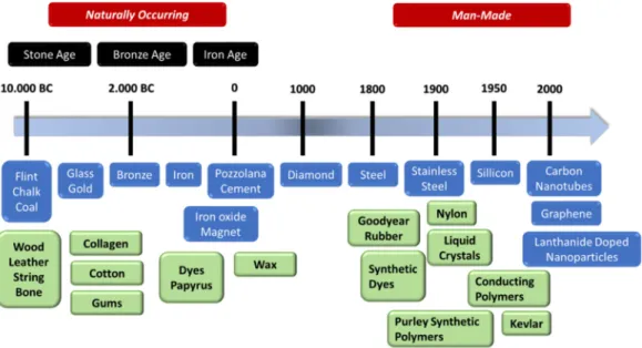
4,5,6Colloidal gold (gold nanoparticles), for example, have been used for centuries for glass coloring, however only with the development of transmission electron microscopy discovery and characterization of nanoparticles was possible in the late 20
thcentury (Figure 1.1).
Figure 1.1║ Selection of important materials discovered by humans are presented in a timeline, separating between naturally occurring and “man-made” materials.
2
Nanoparticles with size-dependent material properties attract the attention of researcher more and more. The increased surface-to-volume ratio can accelerate chemical reactions by faster mass transfer rates. Nanoparticles have also been investigated for drug delivery due their high drug loading capacity and in bioanalytical chemistry they are promising for sensor development. Here, especially their potential to lower limits of detection through inherent signal enhancement in many transduction principles is of special interest.
A specific classification of nanomaterials is based on their chemical and physical properties, e.g. between magnetic, catalytically active or luminescent nanoparticles.
Among the luminescent nanomaterials a wide variety regarding composition and optical properties exists such as semiconductor-type quantum dots,
7carbon dots
8, metal nanoclusters,
9metal-doped nanoparticles
10and organic-inorganic hybrid materials.
11The luminescence in carbon dots, quantum dots or metal nanoclusters is related to the quantum-confinement effects, where the position of the emission bands depends on the size and shape of the particles. As a consequence, it is difficult to synthesize nanoparticles with a desired diameter, without changing the optical properties. In contrast, the emission bands of luminescent organic-inorganic hybrid nanomaterials and metal doped nanoparticles are not influenced by the size; however, intensity and ratios of the bands might show size-dependent variations.
10Among the metal doped nanoparticles, the lanthanide doped nanoparticles have extraordinary optical properties. Lanthanide ions have the electron configuration [Xe] 4f
n(n = 0-14).
12The energy levels are well defined since they are shielded by the filled 5s
2and 5p
6shells. As a consequence, the inner-shell 4f – 4f transitions are sharp and specific for each lanthanide ion, ranging from the UV to the near infrared. However, the 4f - 4f transitions are formally forbidden due to Laporte´s parity rule, thereby the excited f-states have very long luminescent lifetimes, favorable for time-resolved detection systems in biological samples, minimizing background fluorescence.
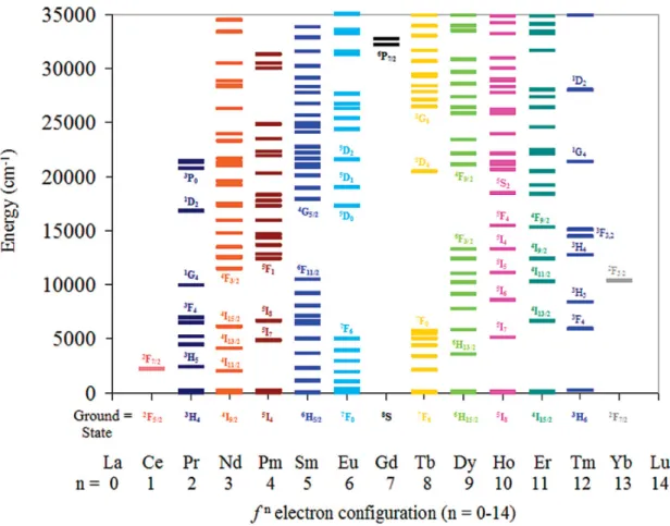
Considering the different energy levels of the lanthanide ions shown in Figure 1.2 defined energy ladders with short energy gaps can be found for most of the lanthanide ions, indicating the probability of non-radiative deactivation processes.
13Eu
3+, Gd
3+and Tb
3+are an exception among the lanthanide ions. The large energy gaps of these ions enable
luminescence measurements with large Stokes shifted emissions. Gd
3+is only able to
emit UV light and is less interesting for bioanalytical applications, since UV-light is strongly
absorbed by biological samples and can lead to phototoxicity.
3
Figure 1.2║ Energy diagram for the trivalent lanthanide ions with fn electron configuration (n = 0-14). The electronic states of the lanthanide ions are presented as horizontal lines in cm-1. Reprinted with permission from Ref. 14. Copyright 2009 American Chemical Society.
Organic dyes usually have broad absorption and emission bands with Stokes shifts less than 50 nm due to their vibrations levels attached to the excited states. With their line like emissions and their large Stokes shifted emissions lanthanide ions are thus more attractive than organic dyes for measurements in biological samples, since background luminescence is significantly reduced.
One major drawback of the lanthanides is their relatively low molar absorption coefficient
(below 1 M
-1·cm
-1). In comparison, good organic dyes have coefficients in the range of
25,000 to 250,000 M
-1·cm
-1.
15Since direct excitation of the Ln
3+ions leads to very low
luminescence signals, high energy excitation or light harvesting molecules have been
used to enhance the luminescence of the lanthanide ions. These so-called antenna
ligands are usually coordinated to lanthanide ions, absorbing and transferring the
excitation light to Ln
3+in the center. Due to the coordination of the ligands water
molecules are repressed, which is highly beneficial as the long lifetime of the lanthanide
ions can be easily quenched via OH groups in close proximity. The light harvesting
molecules typically are organic dyes, which are themselves prone to photobleaching,
4
however, as Ln
3+ions are good quenchers of the triplet states, photobleaching of the organic dyes is reduced in these complexes.
13Another strategy to overcome the low absorption coefficients of the lanthanide ions and water quenching is the entrapment of lanthanide ions in host materials. Furthermore, entrapment of Ln
3+ions inside solid host materials causes a distortion of the orbital symmetry, which can increase the probability of the f-f transitions.
16Inorganic nanoparticles like Y
2O
3,
17Gd
2O
3,
18NaYF
4,
19CeO
220or CaMoO
421nanoparticles have been synthesized as a host lattice for europium or terbium ions. A suitable secondary effect is, when the new host lattice contributes to the lanthanide emissions by charge transfer processes. This can lead to dramatically enhanced luminescent properties, for example in GdVO
4:Eu nanoparticles. Other ways to enhance the luminescence properties are antenna ligands, which can also be utilized in nanoparticles, by binding the light harvesting molecules directly to the surface of the nanoparticles.
22,23Upconversion Nanoparticles
Upconversion nanoparticles are a special group of lanthanide doped nanoparticles, able
to convert near infrared light to UV and visible light by multiphoton processes. The non-
linear process was first discovered in bulk materials doped with Yb
3+and Tm
3+ions in the
1960s.
24Generally, several mechanisms are described in literature, which are leading to
upconversion: photon avalanche (PA), excited state absorption (ESA) and energy transfer
upconversion (ETU).
25PA requires a high pump intensity to induce the cycle process and
is rarely observed.
26The ESA and the ETU mechanisms include both a sequential
absorption of two or more photons to populate higher energy levels. However, in the ESA
process the sensitizer and the emitting ion are identical. Both models require laser
irradiation due to the low molar absorption coefficient of the lanthanide ions. In the ETU
process the energy is transferred stepwise from sensitizer ions to emitting ions
(activators). Most upconversion materials are designed for utilizing the ETU process to
generate anti Stokes shifted emissions, since this is the most efficient mechanism and can
be achieved with low laser power irradiation.
5
Figure 1.3║ ETU mechanism of an Yb3+/Er3+ system for the generation of upconversion luminescence is presented. The dotted black arrows represent photon transmission. The colored arrows signify a distinct absorption or emission of a photon; the curly black lines indicate non-radiative relaxations.
In Figure 1.3 the ETU mechanism for the upconversion process of an Yb
3+/Er
3+system is shown.
27First a photon gets absorbed by a so-called sensitizer ion, which then transfers the energy to emitting ions. As sensitizer ions Yb
3+has been established as the most suitable ones, since they absorb near infrared light at a wavelength of 980 nm with a relatively high absorption coefficient (10 M
-1cm
-1) compared to other lanthanide ions.
28Another advantage is that 980 nm lasers are comparatively cheap and commercially available.
29The excited Yb
3+in the
5F
5/2state is able to transfer the energy to excited state levels of adjacent lanthanides ions, e.g. to Er
3+to populate the
4I
11/2energy level.
Subsequent absorption and transfer of another photon to the excited Er
3+leads then to the population of the
4F
7/2energy level. Two main emissions of Er
3+after non-radiative relaxation from the
4F
7/2state are the green emissions from
2H
11/2and
4S
3/2to the ground state, resulting in emissions at 525 nm and 540 nm, respectively. The third main emission leads to red luminescence at 655 nm, following the transition of
4F
9/2to the ground state.
Other popular upconversion systems are the combinations Yb
3+/Ho
3+and Yb
3+/Tm
3+, where even UV-light can be generated (
1D
23
H
6, 360 nm) by a four-photon process.
30In general, a host lattice must meet several requirements for high upconversion efficiency.
First of all, the host lattice has to be easily doped with the sensitizer and activator ions.
The dopants should be distributed homogenously throughout the whole nanocrystal to
6
achieve optimum and controllable doping ratios. This demands similar ionic radii and charge of the Ln
3+with a cation of the host lattice, also to reduce crystal defects.
31Another requirement is low phonon energy of the nanocrystals to minimize non-radiative deactivation processes and to enhance the radiative emissions. Fluoride host lattices are known to have lowest phonon energies (< 350 cm
-1).
27Considering bioanalytical applications the host material has also to be thermodynamically stable, non-toxic and chemically inert. For example, leaching of ions can significantly influence viability of cells or lead to undesired side reactions, like complexation reactions.
Furthermore, the crystal structure of the host material affects the upconversion efficiency.
The distance between sensitizer and emitting ions as well as symmetries around the Ln
3+get changed by variations of the crystal structure. Up to now the most efficient and favored host lattice is hexagonal β-NaYF
4with phonon energies (below 350 cm
-1) showing ten times higher brighter upconversion luminescence compared to cubic α -NaYF
4.
32The ionic radius of Y
3+is similar to the radii of the lanthanides ions facilitating the doping with Ln
3+, without excessive deformations of the host lattice. Other popular host lattices are Ln
2O
3, LnF
3and YVO
4, which have higher phonon energies and therefore have lower upconversion efficiency.
33In contrary to nanoparticles connected to the quantum-confinement effect the position of the emission bands of the upconverting nanoparticles are fixed and are independent of the host lattice. Only the intensity ratios of the anti- Stokes shifted emissions depend on the applied laser power (multi-photon process), the doping ratio of the lanthanide ions, the size of the nanocrystals, and the microenvironment of the lanthanide ions. The influence of this parameters demands accurate characterization of the upconverting nanoparticles or in general for all lanthanide doped nanoparticles. The size of nanocrystals can be evaluated by transmission electron microscopy (TEM) and dynamic light scattering (DLS).
While by evaluating TEM-images the exact diameter of the nanoparticles together with the particle size distribution can be determined, DLS measurements give valuable information about the dispersed nanoparticles, e.g. agglomeration of the nanoparticles. X-ray diffraction (XRD) measurements are important for the determination of the crystal phase, which can have distinct influence on the optical properties of the nanoparticles. Regarding doping ratio inductively coupled plasma optical emission spectrometry (ICP-OES) or mass spectrometry (ICP-MS) are useful techniques for the determination of the lanthanide ion content in the nanocrystals.
Thermogravimetric analysis (TGA), fourier-transform infrared spectroscopy (FTIR) and
mass spectroscopy are methods to determine the surface architecture of the
nanoparticles. After synthesis nanoparticles are stabilized by several organic ligands,
7 preventing agglomeration of the nanoparticles. The properties of the ligands determine the stability of the nanoparticles in different solvents. For bioanalytical applications surface modifications are necessary to enable colloidal stability of the nanocrystals in aqueous solutions or to functionalize the nanoparticles. The three main techniques for surface modification are ligand exchange, coating with amphiphilic polymers and growing of a silica shell.
34Detailed presentation of the different surface modification techniques is given in Chapter 4.
Enhancement Strategies for UCNPs
For bioanalytical applications reduced luminescence brightness of the UCNPs in aqueous dispersions can be observed. Two different effects contribute to this reduction: A) Continuous attenuation of the excitation light by a local absorption band of water at 980 nm accompanied by heating effects of the samples. B) Quenching of the upconversion luminescence caused due to O-H vibrations go along with changes in the peak ratios of the UCNPs.
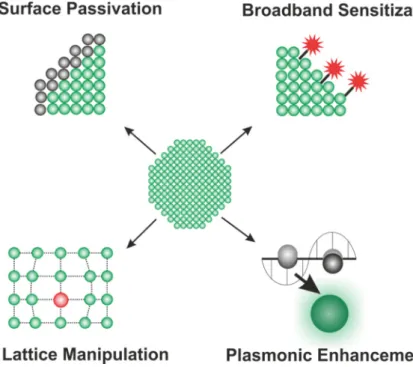
35Several strategies have been developed to enhance the luminescence properties of the UCNPs in aqueous dispersions (Figure 1.4): A) surface passivation by synthesis of core- shell nanoparticles, B) Host lattice manipulation, C) Improving the excitation efficiency or D) Plasmonic enhancement.
36Figure 1.4║ Enhancement strategies for lanthanide doped nanoparticles are presented: Surface passivation, broadband sensitization, host lattice manipulation and plasmonic enhancement.
8
Surface passivation by growing a shell around the core-particles is a well-established method to reduce crystals defects and surface quenching effects, therefore increase the emission of the UCNPs. Zhang et al. (2012) showed that by a homogenous shell growth of NaGdF
4on NaYF
4,Yb,Er core particles surface defects on the nanocrystal can be passivated, consequently an enhancement in emission intensity and lifetime can be observed.
37Fischer et al. (2016) demonstrated that the maximum upconversion quantum yield was obtained for a shell thickness of around 4 nm for NaYF
4:Yb,Er@NaLuF
4nanoparticles.
38For FRET based applications the thickness of the shell has to be adapted for high luminescent quantum yield of the particles and at the same time high energy transfer efficiency.
39The emission intensity of upconverting nanoparticles is influenced by the crystal phase, the local crystal field of Ln
3+ions in the host matrix and the Ln
3+-Ln
3+distance. Therefore, host lattice manipulation is an effective way to enhance the luminescence properties of upconverting nanoparticles. Co-doping with small alkali metal ions like Li
+alters crystal field symmetries, leading to enhanced optical properties.
40Hu et al. (2017) combined the Li
+co-doping with the growth of a passivating shell around NaLuF
4:Yb,Tm nanoparticles to enhance the upconversion luminescence synergistically by a factor of 210.
41Transition metal ions like Sc
3+, Mn
2+and Fe
3+have been also inserted to tune the upconversion emission or to selectively enhance single emission bands inside the nanocrystals.
42,43,44Improving the excitation efficiency is another strategy to enhance the luminescence properties of UCNPs in aqueous dispersions.
45This can either be achieved by enhancing the absorption at 980 nm by light harvesting NIR dyes or by shifting the excitation wavelength to shorter wavelength by co-doping of the nanocrystals with Nd
3+ions. The light harvesting NIR dyes or also called antenna ligands have to be bound directly on the surface of the nanocrystals enabling an energy transfer to the sensitizer ions, first shown by Zou et al. (2012).
46Sensitization with NIR dyes with their broad absorptions bands provides more flexibility regarding the excitation wavelength considering the heating effects of water by 980 nm irradiation. Prasad et al. (2016) achieved UCNPs with a quantum yield of 9.2% by combining the surface passivation by formation of a shell, co- doped with Nd
3+, with the light harvesting abilities of NIR dyes.
47However, NIR dyes are prone to photobleaching, especially considering higher excitation powers for the excitation, lowering the application in long-term experiments.
By co-doping of the nanoparticles with Nd
3+the excitation wavelength can be shifted to
808 nm, minimizing water absorption of the excitation light, reducing the overheating
effect.
48The energy, absorbed by Nd
3+ions, is first transferred to Yb
3+and then to the
activator ions (Er
3+, Tm
3+). The energy levels of Nd
3+are numerous compared with the
energy levels of Yb
3+limiting the doping concentrations of Nd
3+up to 1 mol%. Higher
9 concentration would lead to cross relaxations between the activator ions and Nd
3+ions, minimizing the efficiency of the upconversion process. Sophisticated core-shell architectures to separate the sensitizer and activator ions enable doping concentration of Nd
3+up to 20 mol% for effective upconversion.
49,50Yb
3+acts as a bridging sensitizer, transferring the energy from the active shell to the core of the nanocrystals.
Plasmonic enhancement is another strategy to improve the absorption and emission process of upconverting nanoparticles. Noble metal nanoparticles, mostly gold or silver nanoparticles, can influence neighboring activator ions either by concentrating the incident field or modifying the local density of the energy levels leading either to enhancing the absorption efficiency or to faster radiative decay rates. Several different UC-plasmonic structures have been developed to enhance the upconverting efficiency.
51,52,53References
1 J. I. Choi, S. J. Chae, J. M. Kim, J. C. Choi, S. J. Park, H. J. Choi, H. Bae and H. J.
Park, Potential silver nanoparticles migration from commercially available polymeric baby products into food simulants, Food Addit. Contam. Part A Chem.
Anal. Control Expo. Risk. Assess ., 2018, 1–10.
2 D. P. Macwan, P. N. Dave and S. Chaturvedi, A review on nano-TiO
2sol–gel type syntheses and its applications, J. Mater. Sci. , 2011, 46, 3669–3686.
3 M. Ge, C. Cao, J. Huang, S. Li, Z. Chen, K.-Q. Zhang, S. S. Al-Deyab and Y. Lai, A review of one-dimensional TiO
2nanostructured materials for environmental and energy applications, J. Mater. Chem. A , 2016, 4, 6772–6801.
4 L. Armand, A. Tarantini, D. Beal, M. Biola-Clier, L. Bobyk, S. Sorieul, K. Pernet- Gallay, C. Marie-Desvergne, I. Lynch, N. Herlin-Boime and M. Carriere, Long-term exposure of A549 cells to titanium dioxide nanoparticles induces DNA damage and sensitizes cells towards genotoxic agents, Nanotoxicology , 2016, 10(7), 913.
5 B. Annangi, L. Rubio, M. Alaraby, J. Bach, R. Marcos and A. Hernández, Acute and long-term in vitro effects of zinc oxide nanoparticles, Arch. Toxicol. , 2016, 90(9), 2201.
6 X. Liang, H. Wang, Y. Zhu, R. Zhang, V. C. Cogger, X. Liu, Z. P. Xu, J. E. Grice and M. S. Roberts, Short- and long-term tracking of anionic ultrasmall nanoparticles in kidney, ACS Nano , 2016, 10(1), 387.
7 B. S. Mashford, M. Stevenson, Z. Popovic, C. Hamilton, Z. Zhou, C. Breen, J.
Steckel, V. Bulovic, M. Bawendi, S. Coe-Sullivan and P. T. Kazlas, High-efficiency
10
quantum-dot light-emitting devices with enhanced charge injection, Nature Photon , 2013, 7, 407–412.
8 P. Zuo, X. Lu, Z. Sun, Y. Guo and H. He, A review on syntheses, properties, characterization and bioanalytical applications of fluorescent carbon dots, Microchim. Acta , 2016, 183, 519.
9 L.-Y. Chen, C.-W. Wang, Z. Yuan and H.-T. Chang, Fluorescent gold nanoclusters, Anal. Chem. , 2015, 87, 216–229.
10 C. Feldmann, Luminescent nanomaterials, Nanoscale , 2011, 3, 1947–1948.
11 L. Armelao, S. Quici, F. Barigelletti, G. Accorsi, G. Bottaro, M. Cavazzini and E.
Tondello, Design of luminescent lanthanide complexes, Coord. Chem. Rev. , 2010, 254, 487–505.
12 J.-C. G. Bünzli and C. Piguet, Taking advantage of luminescent lanthanide ions, Chem. Soc. Rev. , 2005, 34, 1048–1077.
13 J.-C. G. Bünzli, Lanthanide luminescence for biomedical analyses and imaging, Chemi. Rev. , 2010, 110, 2729–2755.
14 E. G. Moore, A. P. S. Samuel and K. N. Raymond, From antenna to assay, Acc.
Chem. Res. , 2009, 42, 542–552.
15 U. Resch-Genger, M. Grabolle, S. Cavaliere-Jaricot, R. Nitschke and T. Nann, Quantum dots versus organic dyes as fluorescent labels, Nat. Methods , 2008, 5(9), 763.
16 S. Han, R. Deng, X. Xie and X. Liu, Enhancing luminescence in lanthanide-doped upconversion nanoparticles. Angew. Chem. Int. Ed. , 2014, 53, 11702–11715.
17 M. G. Ivanov, I. V. Krutikova, U. Kynast, M. Lezhnina and I. S. Puzyrev, Laser- synthesized Y
2O
3:Eu
3+nanophosphors and their stabilization in water suspensions, Opt. Mater. , 2017, 74, 67–75.
18 T. K. Anh, P. T. M. Chau, N. T. Q. Hai, V. T. T. Ha, H. van Tuyen, S. Bounyavong, N. T. Thanh and Q. Le Minh, Facile fabrication and properties of Gd
2O
3:Eu
3+, Y
2O
3:Eu
3+nanophosphors and Gd
2O
3:Eu
3+/silica, Y
2O
3:Eu
3+/silica nanocomposites, J. Electron. Mater. , 2018, 47, 585–593.
19 C. Li, Z. Quan, J. Yang, P. Yang and J. Lin, Highly uniform and monodisperse beta-NaYF
4: Ln
3+(Ln = Eu, Tb, Yb/Er, and Yb/Tm) hexagonal microprism crystals:
Hydrothermal synthesis and luminescent properties, Inorg. Chem. , 2007, 46,
6329–6337.
11 20 L. Li, H. K. Yang, B. K. Moon, Z. Fu, C. Guo, J. H. Jeong, S. S. Yi, K. Jang and H.
S. Lee, Photoluminescence Properties of CeO
2:Eu
3+nanoparticles synthesized by a sol-gel method, J. Phys. Chem. C , 2009, 113, 610–617.
21 R. Wang, Red Fluorescence Enhancement via Using the Charge Compensation and Co-doping WO
3in CaMoO
4:Eu
3+Phosphor for Ultraviolet Converted LEDs, JIOP , 2017, 27, 1028–1036.
22 S. W. Li, H. J. Ren and S. G. Ju, Sensitized luminescence of LaF
3:Eu
3+nanoparticles through pyromellitic acid, J. Nanosci. Nanotech ., 2014, 14, 3677–
3682.
23 L. J. Charbonnière, J.-L. Rehspringer, R. Ziessel and Y. Zimmermann, Highly luminescent water-soluble lanthanide nanoparticles through surface coating sensitization, New J. Chem. , 2008, 32, 1055.
24 F. Auzel, Upconversion and anti-Stokes processes with f and d ions in solids, Chem. Rev. , 2004, 104, 139–173.
25 F. Zhang, Photon upconversion nanomaterials, Springer Berlin Heidelberg , 2015.
26 J. Zhou, Q. Liu, W. Feng, Y. Sun and F. Li, Upconversion luminescent materials:
advances and applications, Chem. Rev. , 2015, 115, 395–465.
27 M. Haase and H. Schäfer, Upconverting nanoparticles, Angew. Chem. Inter. Ed ., 2011, 50, 5808–5829.
28 J.-C. G. Bünzli, On the design of highly luminescent lanthanide complexes, Coord.
Chem. Rev. , 2015, 293-294, 19–47.
29 E. Y. Chan, J. C. Adams, J. M. Saint Clair, K. A. Morrison and M. Sosa, R. M. Rao, S. A. Dianat and M. D. Zoltowski, Application of COTS high-speed 980-nm pump laser diode and driver for free-space laser communication terminal, SPIE , 1999, pp. 79–86.
30 L. M. Jin, X. Chen, C. K. Siu, F. Wang and S. F. Yu, Enhancing multiphoton upconversion from NaYF
4:Yb/Tm@NaYF
4core–shell nanoparticles via the use of laser cavity, ACS Nano , 2017, 11(1), 843.
31 R. Naccache, Q. Yu and J. A. Capobianco, The Fluoride Host: Nucleation, Growth, and upconversion of lanthanide-doped nanoparticles, Adv. Opt. Mat. , 2015, 3, 482–509.
32 J.-H. Zeng, J. Su, Z.-H. Li, R.-X. Yan and Y.-D. Li, Synthesis and Upconversion
Luminescence of Hexagonal-Phase NaYF
4:Yb, Er
3+Phosphors of Controlled Size
and Morphology, Adv. Mater. , 2005, 17, 2119–2123.
12
33 F. Wang and X. Liu, Recent advances in the chemistry of lanthanide-doped upconversion nanocrystals, Chem. Soc. Rev. , 2009, 38, 976–989.
34 V. Muhr, S. Wilhelm, T. Hirsch and O. S. Wolfbeis, Upconversion nanoparticles:
From hydrophobic to hydrophilic surfaces, Acc. Chem. Res. , 2014, 47, 3481–3493.
35 C. Würth, M. Kaiser, S. Wilhelm, B. Grauel, T. Hirsch and U. Resch-Genger, Excitation power dependent population pathways and absolute quantum yields of upconversion nanoparticles in different solvents, Nanoscale , 2017, 9, 4283–4294.
36 U. Resch-Genger and H. H. Gorris, Perspectives and challenges of photon- upconversion nanoparticles - Part I: routes to brighter particles and quantitative spectroscopic studies. Anal. Bioanal. Chem. , 2017, 409, 5855–5874.
37 F. Zhang, R. Che, X. Li, C. Yao, J. Yang, D. Shen, P. Hu, W. Li and D. Zhao, Direct imaging the upconversion nanocrystal core/shell structure at the subnanometer level, Nano letters , 2012, 12, 2852–2858.
38 S. Fischer, N. D. Bronstein, J. K. Swabeck, E. M. Chan and A. P. Alivisatos, Precise tuning of surface quenching for luminescence enhancement in core-shell lanthanide-doped nanocrystals, Nano letters , 2016, 16, 7241–7247.
39 V. Muhr, C. Würth, M. Kraft, M. Buchner, A. J. Baeumner, U. Resch-Genger and T.
Hirsch, particle-size-dependent förster resonance energy transfer from upconversion nanoparticles to organic dyes, Anal. Chem. , 2017, 89(9), 4868.
40 C. Zhao, X. Kong, X. Liu, L. Tu, F. Wu, Y. Zhang, K. Liu, Q. Zeng and H. Zhang, Li
+ion doping: an approach for improving the crystallinity and upconversion emissions of NaYF
4:Yb
3+, Tm
3+nanoparticles, Nanoscale , 2013, 5(17), 8084.
41 M. Hu, D. Ma, Y. Cheng, C. Liu, Z. Zhang, Y. Cai, S. Wu and R. Wang, Synergistically enhanced upconversion luminescence in Li
+-doped core–shell- structured ultrasmall nanoprobes for dual-mode deep tissue fluorescence/CT imaging, J. Mater. Chem. B , 2017, 5(14), 2662.
42 H. Li, Q. Huang, Y. Wang, K. Chen, J. Xie, Y. Pan, H. Su, X. Xie, L. Huang and W.
Huang, Sc
3+-induced morphology, phase structure, and upconversion luminescence evolution of YF
3:Yb/Er nanocrystals, J. Mater. Chem. C , 2017, 5(26), 6450.
43 Z. Huang, H. Gao and Y. Mao, Understanding the effect of Mn
2+on Yb
3+/Er
3+upconversion and obtaining a maximum upconversion fluorescence enhancement in inert-core/active-shell/inert-shell structures, RSC Adv ., 2016, 6(86), 83321.
44 Y. Hu, X. Liang, Y. Wang, E. Liu, X. Hu and J. Fan, Enhancement of the red
upconversion luminescence in NaYF
4:Yb
3+, Er
3+nanoparticles by the transition
metal ions doping, Ceram. Int. , 2015, 41(10), 14545.
13 45 M. K. G. Jayakumar, N. M. Idris, K. Huang and Y. Zhang, A paradigm shift in the excitation wavelength of upconversion nanoparticles, Nanoscale , 2014, 6, 8441–
8443.
46 W. Zou, C. Visser, J. A. Maduro, M. S. Pshenichnikov and J. C. Hummelen, Broadband dye-sensitized upconversion of near-infrared light, Nature Photon. , 2012, 6, 560–564.
47 G. Chen, W. Shao, R. R. Valiev, T. Y. Ohulchanskyy, G. S. He, H. Ågren and P. N.
Prasad, Efficient Broadband Upconversion of Near-Infrared Light in Dye-Sensitized Core/Shell Nanocrystals, Adv. Opt. Mat. , 2016, 4(11), 1760.
48 Y.-F. Wang, G.-Y. Liu, L.-D. Sun, J.-W. Xiao, J.-C. Zhou and C.-H. Yan, Nd
3+- Sensitized Upconversion Nanophosphors: Efficient In Vivo Bioimaging Probes with Minimized Heating Effect, ACS Nano , 2013, 7(8), 7200.
49 X. Huang and J. Lin, Active-core/active-shell nanostructured design: an effective strategy to enhance Nd
3+/Yb
3+cascade sensitized upconversion luminescence in lanthanide-doped nanoparticles, J. Mater. Chem. C , 2015, 3, 7652–7657.
50 S. F. Himmelstoß, L. M. Wiesholler, M. Buchner, V. Muhr, S. Märkl, A. J.
Baeumner, and T. Hirsch. 980 nm and 808 nm excitable upconversion nanoparticles for the detection of enzyme-related reactions. Proc. SPIE 2017, 100770L.
51 K. Green, J. Wirth and S. F. Lim, Optical investigation of gold shell enhanced 25 nm diameter upconverted fluorescence emission, Nanotechnology , 2016, 27, 135201.
52 A. L. Feng, M. L. You, L. Tian, S. Singamaneni, M. Liu, Z. Duan, T. J. Lu, F. Xu and M. Lin, Distance-dependent plasmon-enhanced fluorescence of upconversion nanoparticles using polyelectrolyte multilayers as tunable spacers, Sci. Rep. , 2015, 5, 7779.
53 Q. Zhan, X. Zhang, Y. Zhao, J. Liu and S. He, Tens of thousands-fold
upconversion luminescence enhancement induced by a single gold nanorod, Laser
Photonics Rev. , 2015, 9, 479–487.
14
15
2 MOTIVATIONS AND OBJECTIVES
Lanthanide doped nanoparticles/nanomaterials have several outstanding optical properties, which have been identified. The aim of this work was to implement their unique properties in (bio)analytical applications. Assay, theranostic or sensing applications demands specific optical and structural properties of the applied particles. However, one overall basic requirement is the full characterization of the lanthanide nanoparticles regarding size distribution, crystallinity, doping ratios and optical properties.
In general, for optical assays or luminescent probes high quantum yields, fast signal responses, selectivity and reproducibility are favored properties. GdVO
4nanoparticles doped with europium are known for their high quantum yields due to the efficient charge transfer process from the vanadate groups and their large Stokes shifted emission.
Hydrogen peroxide is an efficient quencher for several europium complexes. Since dynamic quenching of luminescence has rate constants in the time range of diffusion constant, fast signal responses are obtained without changes of the probe. The objective was to investigate the suitability of the europium nanoparticles for the detection of H
2O
2and to monitor enzyme coupled reactions producing H
2O
2.
For theranostic applications of lanthanide doped nanoparticles additionally requirements
like biocompatibility, cellular uptake, high tissue penetration of the inserted light source,
low background luminescence caused by the tissue and colloidal stability of the
nanoparticles in aqueous solutions are necessary. Here, upconverting nanoparticles have
the benefit to generate light directly inside in biological tissue with minimized background
luminescence due to their near infrared excitation. Accordingly, their desired application
includes imaging, drug delivery or photodynamic therapy which requires the surface
functionalization of the nanoparticles. Either the nanoparticles have to be transferred first
from the organic phase into the aqueous phase, or the already dispersed particles must
be functionalized. Key parameters after the surface modification are the colloidal stability
in aqueous solutions of different ionic strengths and the prevention of cross-linking of the
nanoparticles. Based on the later application the thickness and the morphology of the new
surface layer have to be adapted. For example, for delivery systems thick mesoporous
structures are favorable for high drug loading capacities, however for FRET based energy
transfer systems the distance between the emitting ions of the nanocrystals and the
acceptor molecule should be as small as possible.
16
Upconverting nanoparticles are used in photodynamic therapy as carriers for the photosensitizer and to shift the excitation wavelength of the photosensitizer. The aim is to indirectly excite the singlet oxygen producing dyes with near infrared light by energy transfer from the upconverting nanoparticles for applications in deeper tissues. Therefore, several aspects have to be considered. First of all, the size of the particles plays a significant role in the cellular uptake and therefore also in the efficiency of the photodynamic therapy. Since the brightness increases with the size of the nanoparticles, but cellular uptake favors small particles, the aim is to synthesize small and bright nanoparticles. Secondly a suitable photosensitizer has to be chosen, which can produce reactive oxygen species and has an overlap of its main absorption with an emission of the UCNPs. In the next step the surface of the nanoparticles has to be modified to bind the photosensitizer, provide colloidal stability and cell permeability. The challenge is to bind as many as possible photosensitizers close to the surface with respect to the colloidal stability, since the efficiency of the energy transfer decrease in the order of six with the distance of the photosensitizer to the activator ions. In a last step the theranostic approach has to be proven by cell toxicity tests of cells incubated with the functionalized nanoparticles.
For sensing applications fast and stable signal responses are afforded from a device.
Regarding luminescent sensor application photobleaching and light scattering are major drawback of commercial light emitting materials like organic dyes. Lanthanide doped nanoparticles are resistant to photobleaching due their inorganic design, however light scattering on rough or spongy like surfaces limits their application. On the other side dense nanofiber networks for example would generate extremely high surface-to-volume ratios, which are advantageous for sensors for lower of detection limits. Upconverting nanoparticles with their large anti- Stokes shifted emissions (over 300 nm) offer the opportunities for luminescent based sensing devices with dense nanofibermats. The aim was to generate visible light directly inside a sample by entrapping UCNPs inside nanofibers. Finally, for building a sensing device, nanofibermats have to be incoperated into a microfluidic system enabling luminescent online monitoring. The entrapment of the nanoparticles should negotiate time consuming surface modifications or centrifugation steps to achieve colloidal stability in solutions with different ionic strength or pH values.
The well separated emission bands of upconverting nanoparticles are investigated regarding possible ratiometric measurements.
17
3 EUROPIUM-DOPED GDVO 4 NANOCRYSTALS AS A LUMINESCENT PROBE FOR HYDROGEN PEROXIDE AND FOR ENZYMATIC SENSING OF GLUCOSE
3.1 Abstract
The authors describe the preparation of Eu
3+-doped GdVO
4nanocrystals (NCs) by precipitation of the Gd
3+(Eu
3+)-citrate complex which was then converted to the respective vanadate by dialysis. The fractions of Eu
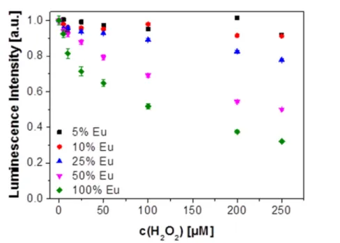
3+ranged from 5 to 100 mol%. The NCs were characterized by XRD, TEM, ICP-OES and dynamic light scattering which revealed that they possess superior colloidal stability in aqueous solutions in that no precipitation can be observed even after several months. The NCs display red and largely red-shifted fluorescence (peaking at 618 nm) on photoexcitation at around 300 nm. Fluorescence is strongly quenched by hydrogen peroxide. It is also shown that the fraction of doping with Eu
3+strongly affects quenchability. Most efficient quenching by H
2O
2is observed if the NCs are doped with 50% of Eu
3+.The findings were exploited to develop a fluorometric assay for H
2O
2that works in the 5 to 250 µM concentration range, with a limit of detection as low as 1.6 µM (at a signal-to-noise ratio of 3). The probe was further employed to design a highly sensitive enzymatic assay for glucose via measurement of the quantity of H
2O
2formed as a result of the catalytic action of glucose oxidase.
Figure 3.1║ GdVO4 nanocrystals doped with Eu(III) ions display strong red luminescence which is efficiently quenched by hydrogen peroxide. The NCs therefore are a viable probe for detection of H2O2 which often is formed by enzymatic action such as by glucose oxidase.
18
This chapter has been published.
Verena Muhr, Markus Buchner, Thomas Hirsch, Dragana J. Jovanović, Slobodan D. Dolic, Miroslav D. Dramićanin, Otto S. Wolfbeis
Author contributions
MB and VM wrote the manuscript, designed the figures and performed all optical, ICP-
OES and DLS measurements for the sensing application. DJ and SD synthesized the
nanocrystals and characterized them by XRD, quantum yield and TEM. The work was
designed and supervised by TH, MD and OW (corresponding author).
19
3.2 Introduction
Vanadates of rare earths (such as GdVO
4, YVO
4and LuVO
4) are known to display strong luminescence.
1Among these, GdVO
4and its co-doped congeners possess distinctly higher absorption cross sections than e.g. YVO
4and therefore are brighter (brightness being defined as the product of molar absorbance and quantum yield; ε·ɸ). GdVO
4can be easily doped with other luminescent lanthanide ions because of the equal valence and similar ionic radii. Such “doped” vanadates can be efficiently excited because of the strong absorption of the vanadate groups and the efficient energy transfer that occurs from GdVO
4to the lanthanide dopand. Doped GdVO
4materials have been used as phosphors (for example after doping with Eu
3+, Dy
3+, Sm
3+), as upconverters with a range of emission colors (after doping with Er
3+/Yb
3+, Ho
3+/Yb
3+, or Tm
3+/Yb
3+), and a lasing material (doped with Nd
3+).
2,3In addition, GdVO
4nanoparticles (NPs) can act as T1-positive contrast agents for magnetic resonance imaging, because Gd
3+ions possess unpaired electrons that efficiently alter the relaxation time of the surrounding water protons.
4,5Hence, such materials may be used for multimodal imaging. It is also noted that GdVO
4nanoparticles doped with Eu
3+or Er
3+/Yb
3+display a strongly temperature-dependent fluorescence.
6It is generally observed that the fluorescence of lanthanide probes is strongly temperature- dependent. Hydrogen peroxide (H
2O
2, HP) is a product of the enzymatic action of all enzymes out of the class of oxidases, and also is a “natural” contaminant in rainwater. It is being produced industrially in large quantity because it is a useful additive to various commodity products including toothpastes and household chemicals, a precursor and a decomposition product of the explosive triacetone triperoxide, a rocket propellant, and a widely used industrial chemical for purposes such as pulp- and paper-bleaching, in waste water treatment via the Fenton reaction, in odor reduction by virtue of its power to oxidize thiols, in the sterilization of surfaces and medical tools, as a safe antimicrobial (with better activity against Gram-positive bacteria), as a hair bleach (along with ammonia), and in chemiluminescence based cold light sticks. Hence, there is a substantial interest in methods for quantifying HP under various circumstances. Electrochemical
7,8and optical
9,10
detection schemes, but also chromatographic methods (for discontinuous assays), are
by far most often used to determine H
2O
2. Sensors for H
2O
2may be enzymatic (mainly
using a peroxidase as the enzyme)
11,12or non-enzymatic (via direct electroreduction of
H
2O
2).
13,14All have their respective merits. In electrochemical sensing, the trend is towards
methods working at low potential, this rendering them less prone to interferences by other
electroactive species.
15Demchenko has summarized the various kinds of metal
nanoparticles capable of fluorometric sensing and imaging of chemical species including
H
2O
2.
16A useful review on metal and metal ion-based nanomaterials for use in optical
20
probing of H
2O
2has been presented by Burmistrova et al..
17Various kinds of fluorescent carbonaceous nanomaterials also may be used,
18but small molecular chromogenic and fluorogenic molecular probes are in most common.
19In addition to enzymatic optical assays, enzyme mimics such as AgVO
3nanobelts may be employed,
20which catalyze the oxidation of tetramethylbenzidine by H
2O
2to irreversibly form a blue dye. However, direct sensing schemes are preferred, and several ones are known that are exploiting the capability of H
2O
2to dynamically or statically quench fluorescence. Examples for fluorescent probes include the Eu
3+-complex of the antibiotic tetracycline
21and certain Eu
3+-based core/shell nanoparticles which are particularly sensitive.
22In both single shot detection and in continuous sensing of H
2O
2,
23,24the trend is towards methods working at long wavelengths, as this makes methods less prone to interferences by backgrounds absorbance and fluorescence of samples, which is particularly strong in case of biomatter.
In this contribution, we describe the preparation of aqueous solutions of Eu
3+-doped GdVO
4nanoparticles, their structural and morphological properties, and long-term stability of the solution. Then we describe in detail fluorescence of NC’s and fluorescence quenching by H
2O
2. We also show that such nanoparticles are viable probes for H
2O
2and that they also may be employed as transducers in enzymatic reactions accompanied by the formation (and possibly also the consumption) of H
2O
2.
3.3 Experimental
Chemical and reagents
Ammonium vanadate (NH
4VO
3; min. 99.0%, Alfa Aesar), trisodium citrate dehydrate (Na
3C
6H
5O
7; 99%, Sigma Aldrich), gadolinium(III) nitrate hexahydrate [Gd(NO
3)
3*6H
2O;
99.9%, Alfa Aesar] and europium(III) nitrate hexahydrate [Eu(NO
3)
3*6H
2O; 99.9%, Alfa Aesar] were used without further purification. Rhodamine B (Sigma-Aldrich) was used as a standard for the determination of quantum yields.
Synthesis of colloidal Eu
3+-doped GdVO
4nanoparticles
The colloidal Eu
3+-doped GdVO
4NCs were synthesized by analogy to the method presented in our previous papers.
25,26In brief, 30 mL 0.05 M solution of trisodium citrate was added drop by drop to the mixture of 0.05 M solution of Gd(NO
3)
3and Eu(NO
3)
3(40 mL) in stoichiometric ratio (solutions were mixed in concentration of 5; 10; 25; 50 and
100 mol% Eu
3+with respect to Gd
3+ions) at room temperature. A white precipitate
consisting of the Gd
3+(Eu
3+)-Cit3−complex is formed. After vigorous stirring for 30 min, the
white precipitate is completely dissolved by the addition, drop by drop, 30 mL of a 0.05 M
21 solution of NH
4VO
3(dissolved in 0.15 M NaOH solution). A transparent solution is obtained that has a pH value of about 8. It is subsequently heated and stirred at 60 °C for 60 min. Finally, the colloidal solution is cooled down to room temperature. Slow growth of particles was accomplished by dialysis against distilled water for 24 h to remove the excess ions. Dialysis was terminated once the pH value had reached 7.0 so that such solutions can be used along with buffers of physiological pH values. Powder samples for structural characterization were obtained by evaporation of aqueous colloidal solutions.
No signs of precipitation of particles has been evidenced over the period of several months, which confirmed the superior colloidal stability of Eu
3+-doped GdVO
4NCs in aqueous solution.
Instrumentation
Powder X-ray diffraction (XRD) measurements were performed on a Rigaku SmartLab diffractometer using Cu-K
α1,2radiation ( λ = 0.15405 nm). Diffraction data were recorded with a step size of 0.02° and a counting time of 0.7 min
−1over the 2θ-range of 10° to 100°.
Transmission electron microscopy (TEM) studies were made on a Tecnai G20 (FEI) operated at an accelerating voltage of 200 kV. Luminescence measurements were performed with an Aminco-Bowman Series 2 fluorescence spectrometer (band pass 4 nm), and Fluorolog-3 Model FL3-221 spectrofluorometer system (Horiba JobinYvon), was used for the determination of quantum yields. Absorption spectra were measured with a Shimadzu UV-2600 spectrophotometer (Shimadzu Corporation, Tokyo, Japan) equipped with an integrated sphere (ISR-2600 Plus (for UV-2600)) in the 220–700 nm range with 1 nm step. Dynamic light scattering (DLS) and zeta-potential measurements were conducted on a Malvern Zetasizer nano ZS (www.malvern.com). The determination of the Eu
3+content in the particles was performed on an inductively coupled plasma optical emission spectrometer (ICP-OES) from Spectro (www.spectro.com).
Determination of quantum yields (QYs)
For the determination of quantum yields room-temperature absorption (from 250 to 700 nm) and emission spectra (330 nm excitation, emission from 500 to 750 nm) of aqueous of NCs and Rhodamine B (reference, 10
−6mol dm
−3) were measured. QYs were calculated from the following expression:
27= × × ×
where R refers to the reference (QY
R= 0.31),
28I is the integrated emission intensity, A is
the optical density, and n is the refractive index of the solvent (in both cases water,
22
n = 1.33). In order to minimize re-absorption effects, concentrations of NCs and Rhodamine B in solution were chosen to maintain optical density between 0.01 and 0.1 at 330 nm.
Quenching of fluorescence by H
2O
2The effect of the fraction of Eu
3+in the NCs was studied by dispersing the various NCs in TRIS buffer (50 mM, pH 7.4) at a total Eu
3+concentration of 0.4 mM each. A solution of H
2O
2(c = 0.1 M) was added to each cuvette to adjust the total concentrations of H
2O
2to between 5 and 500 M. The total volume of each sample was 2 mL. The emission of the particles was recorded at 618 nm. The excitation wavelength was adjusted according to the excitation maximum of the different particles (Figure 3.4).
Selectivity studies
These were performed in the following way: EuVO
4nanoparticles (containing 0.4 mM of Eu
3+) and quencher (100 M) were added to TRIS buffer (50 mM, pH 7.4) to yield a total volume of 2 mL. After mixing and incubating for 5 min, the emission intensity at 618 nm was recorded under photoexcitation at 295 nm. The data given are averages of three measurements.
Fluorometric determination of hydrogen peroxide
Time traces of the luminescence of GdVO
4(50%Eu) nanoparticles under 298/618 nm excitation/emission were acquired by dispersing the particles in 2 mL TRIS buffer of pH 7.4 and adding, after every 5 min 50 nmol of H
2O
2(equivalent to 1 µL of a H
2O
2standard solution) to the colloidal dispersion of the particles. The drop in luminescence intensity was recorded over the course of four addition steps.
Enzymatic determination of glucose
Time trace measurements of the luminescence under the same experimental conditions
as above were performed were performed by adding glucose oxidase (149.8 kU g
−1; in a
concentration of 2.3 mg mL
−1in TRIS buffer) to the nanoparticles dispersed in 2 mL of
TRIS buffer of pH 7.4. Every five minutes 50 nmol glucose (equivalent to 1 µL glucose
solution) was added to a dispersion of the particles containing a total Eu
3+concentration of
0.4 mM. The drop of the luminescence intensity was recorded over the course of four
addition steps.
23 Determination of glucose in serum
Glucose in fetal bovine serum was determined by the standard addition method. The sample was diluted by a factor of 1/80 by addition of 25 µL of serum to an aqueous solution containing 2.0 mg mL
−1of glucose oxidase (=149.8 kU g
-1) and 0.4 mM Eu
3+in the form of GdVO
4(50%Eu) particles. This sample was spiked with glucose to give final concentrations between 20 µM and 120 µM. The samples were incubated at room temperature for 5 min and fluorescence was measured at 618 nm under 298 nm excitation. The glucose concentrations of samples were also determined using a commercial glucose meter (Contour Next; Bayer, https://www.contournext.com).
3.4 Results and Discussion
3.4.1 Characterization of the GdVO
4:Eu Nanocrystals
Synthesis of the NCs follows a simple but well reproducible strategy that results in good yields ( ∼ 6 mg of NCs in 1 mL of solution). The NCs were submitted to XRD analysis.
Figure 3.2 shows are presentative XRD pattern for GdVO
4:Eu NCs (with 50 mol%). The pattern shows the presence of a single tetragonal zircon-type phase of GdVO
4(space group I41/amd, ICDD card no. 01-086-0996). The absence of impurity phases and very small shift of reflections compared to the reflection positions of pure GdVO
4indicate that the Eu
3+ions are successfully and uniformly incorporated into the GdVO
4host lattice. In addition, the relatively intense reflection peaks suggest that the NCs are highly crystalline, and no additional thermal treatment is necessary.
Figure 3.2║ XRD pattern (without background correction) for GdVO4 nanocrystals doped with 50 mol% of Eu3+ (upper graph), the main diffraction peaks are indexed according to ICCD No. 011-086-0996 card (lower graph).