Design, Synthesis and Surface Modification of Lanthanide-Doped Nanoparticles for FRET-
Based Biosensing Applications
Dissertation zur Erlangung des Doktorgrades der Naturwissenschaften (Dr. rer. nat.)
an der Fakultät Chemie und Pharmazie der Universität Regensburg
Deutschland
vorgelegt von
Verena Muhr
aus Roding
im Jahr 2017
Die vorliegende Dissertation entstand in der Zeit von November 2013 bis November 2017 am Institut für Analytische Chemie, Chemo‐ und Biosensorik der Universität Regensburg.
Die Arbeit wurde angeleitet von Prof. Dr. Antje J. Bäumner und Dr. Thomas Hirsch.
Promotionsgesuch eingereicht am: 06. November 2017 Kolloquiumstermin: 08. Dezember 2017
Prüfungsausschuss
Vorsitzender: Prof. Dr. Oliver Tepner
Erstgutachterin: Prof. Dr. Antje J. Bäumner
Zweitgutachter: Prof. Dr. Otto S. Wolfbeis
Drittprüfer: PD. Dr. Miriam Breunig
D ANKSAGUNG
Zuallererst möchte ich mich bei Prof. Dr. Antje Bäumner und Dr. Thomas Hirsch für die Möglichkeit bedanken, meine Promotion über dieses spannende Thema anfertigen zu können. Vielen Dank für die stete Betreuung, Unterstützung und Hilfe bei Problemstellungen aller Art.
Mein herzlicher Dank geht ebenfalls an Prof. Dr. Otto S. Wolfbeis für das Liefern exzellenter wissenschaftlicher Fragestellungen und die Übernahme des Zweitgutachtens.
Vielen Dank an PD Dr. Miriam Breunig für die Übernahme der Aufgabe des Drittprüfers und Prof. Dr. Oliver Tepner für das Ausüben der Funktion des Prüfungsvorsitzenden.
Bei Dr. Stefan Wilhelm, Dr. Christoph Fenzl und Sandy Himmelstoß bedanke ich mich für ihre Geduld für die unzähligen TEM Aufnahmen.
Danke an Joachim Rewitzer und Vanessa Tomanek für ihre Unterstützung bei den ICP- OES Messungen.
Mein Dank geht auch an Prof. Dr. Joachim Wegener, Barbara Goricnik und Lisa Sauer für die Zusammenarbeit bei allen Zellfragen.
Den Arbeitsgruppen um Dr. Ute Resch-Genger an der Bundesanstalt für Materialforschung und -prüfung in Berlin und Prof. Yves Mély an der Universität Straßburg danke ich für die erfolgreichen Kooperationen bei der photophysikalischen Charakterisierung der Nanopartikel.
Beim Upcon-Team, allen voran bei Dr. Thomas Hirsch, bedanke ich mich für die unzähligen hilfreichen wissenschaftlichen und auch nicht-wissenschaftlichen Anregungen und Diskussionen.
Vielen, vielen Dank auch an die gesamte aktuelle und ehemalige Arbeitsgruppe
„4. Stock“ und alle Kollegen am Institut für die ausgezeichnete Arbeitsatmosphäre und vor allem all die geselligen Abende zur Auflockerung des Arbeitsalltags.
Zu guter Letzt möchte ich mich bei meiner Familie für ihre fortwährende Unterstützung in
jeglicher Hinsicht herzlichst bedanken.
Contents i
T ABLE OF C ONTENTS
1 Introduction to Lanthanide-doped Nanomaterials ... 1
1.1 Lanthanide Luminescence ... 1
1.2 Upconversion Nanoparticles ... 5
2 Motivation and Objectives ... 19
3 Upconversion Nanoparticles: From Hydrophobic to Hydrophilic Surfaces 21 3.1 Abstract ... 21
3.2 Introduction ... 23
3.3 Surface Modification of Hydrophobic UCNPs ... 25
3.3.1 Modification of the Original Ligand ... 25
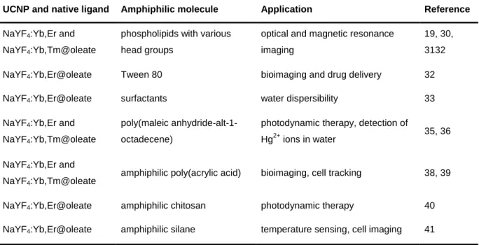
3.3.2 Amphiphilic Coatings ... 25
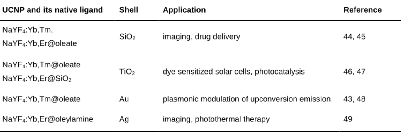
3.3.3 Encapsulation with Inorganic Materials or Noble Metals Forming a Shell ... 30
3.3.4 Replacement of the Native Ligand ... 34
3.4 Conclusions ... 40
References ... 42
4 Particle-Size Dependent Förster Resonance Energy Transfer from Upconversion Nanoparticles to Organic Dyes ... 51
4.1 Abstract ... 51
4.2 Introduction ... 53
4.3 Results and Discussion ... 55
4.3.1 Synthesis of Hydrophobic UCNPs with Controlled Sizes ... 55
4.3.2 Design of the FRET UCNP-Platform ... 56
4.3.3 Size-dependent FRET Efficiency ... 59
4.3.4 Influence of Luminescence Enhancement on FRET Efficiency ... 65
4.4 Conclusions ... 66
4.5 Materials and Methods ... 67
Acknowledgements ... 70
ii Contents
References ... 70
5 Surface Engineering of Upconversion Nanoparticles for Time-Resolved Analysis of ATP-Responsive Energy Transfer... 75
5.1 Abstract ... 75
5.2 Introduction ... 77
5.3 Results and Discussion ... 79
5.3.1 Design, Synthesis and Characterization of UCNPs ... 79
5.3.2 Surface Modification and Attachment of the ATP Aptamer ... 81
5.3.3 Cytotoxicity Studies and Cellular Uptake ... 83
5.3.4 Sensing Properties of Aptamer-modified UCNPs ... 85
5.3.5 Selectivity of the ATP Nanoprobe ... 88
5.4 Conclusions ... 89
5.5 Materials and Methods ... 90
Acknowledgements ... 94
References ... 95
6 Conclusions and Future Perspectives ... 99
6.1 Particle Architectures for FRET-based Applications ... 99
6.2 Future Directions ... 101
6.3 Remaining Challenges ... 104
7 Summary ... 111
8 Zusammenfassung ... 113
Curriculum Vitae ... 115
Publications ... 117
Presentations ... 119
Introduction to Lanthanide-doped Nanomaterials 1
1 I NTRODUCTION TO L ANTHANIDE - DOPED N ANOMATERIALS
1.1 Lanthanide Luminescence
1.1.1 Luminescent Reporters in Bioanalysis
Luminescence has fascinated humans for centuries and is considered one of the most significant and powerful tools in (bio)analytical chemistry today.
1,2Optical biosensors consist of a combination of a bioreceptor as recognition element, e.g. enzymes, nucleic acids, or even whole cells, and an optical reporter system which generates a defined signal directly linked to the concentration of the respective analyte. Examples for the optical transducer are absorption, luminescence and reflectance.
3Fluorescence based detection is often characterized by fast responses and high resolution and sensitivities, which can even reach the single-molecule level,
4,5and both intensity and lifetime of the luminescence emission are available as optical reporter. The application of luminophores as tags and labels for the quantification and imaging of (bio)analytical targets that do not display intrinsic fluorescent properties at all (e.g. most small metabolites, ions) or only to an insufficient degree (e.g.
DNA, proteins) is prevalent in all fields of life science. Central characteristics of ideal luminescent reporters in bioanalysis are (a) high molar absorption coefficients and (b) quantum yields for high brightness, (c) photostability, (d) chemical stability, (e) simple functionalization with receptors (f) solubility in physiological hydrophilic media, such as buffers and cell culture media, and both (g) low cytotoxicity and (h) no excitation/emission wavelengths in the ultra-violet (UV) for applications in living cells combined with minimized photo-damage and light scattering. The importance and impact of luminescence-based biosensing and -imaging in current research is reflected by the selection of Nobel Prize laureates in chemistry during the last decade. Osamu Shimomura, Martin Chalfie and Roger Y. Tsien were awarded the Nobel Prize for the discovery of the green fluorescent protein in 2008 and Eric Betzig, Stefan Hell and William E. Moerner received the Nobel Prize for their outstanding contributions to the development of super-resolution fluorescence microscopy in 2014. These accomplishments have proven that luminescence-based techniques can provide the ability to discover and understand the secrets of life, but the incredible amount of ongoing research demonstrates that there is still much left to be learned in order to cross current boundaries.
The oldest and still most common luminophores are molecular chromophores, i.e. organic
dyes and metal ion complexes.
6The whole visible spectrum of light and the bordering ranges
2 Introduction to Lanthanide-doped Nanomaterials are indeed covered by the huge amount of available luminophores of this type. However, common challenges of molecular luminophores are limited photostability upon prolonged photoexcitation and broad absorption and emission bands exhibiting only small Stokes shifts, which promotes reabsorption of the emitted light and impedes clear separation between excitation light, luminescence emission and background fluorescence of biological materials.
Luminescent nanomaterials, such as quantum dots, carbon dots and gold nanoparticles have been established as alternatives to organic fluorophores to circumvent these issues due to their high photostability and capability of color tuning by modulation of the particle size.
7,81.1.2 Optical Properties of Lanthanide Ions
Lanthanide ions (rare earth ions) represent a class of luminescent materials with exceptional and unusual optical properties. This is represented by their diverse fields of applications, ranging from active materials in solid-state lasers (e.g. Nd
3+- or Er
3+-doped yttrium- aluminium-garnet),
9to Er
3+-doped glass fibers used for telecommunication,
10and lanthanide complexes applied as anti-counterfeiting features,
11or luminescent reporters in theranostics.
12In contrast to many other luminophores, their luminescence shows multiple sharp emission bands and very large Stokes shifts > 100 nm. This means, emitted light can easily be distinguished from excitation radiation, which usually is ultraviolet or visible light.
Their outstanding optical properties arise from the unique electronic structure that all lanthanides have in common. Lanthanides possess the ground state electronic configuration [Xe] 4f
n(n = 0-14). They exist almost exclusively in the trivalent state, Ln(III). The energy levels of their excited states are generally well defined and insensitive to the environment (ligand field) due to the effective shielding of the 4f orbitals by the xenon core. This leads to characteristic, almost line-like emission bands from the respective f-f transitions.
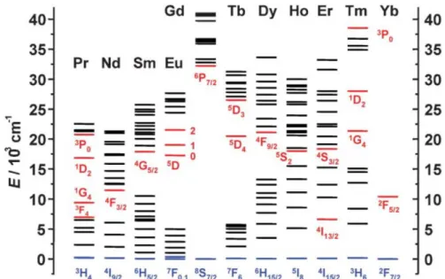
13The energy level diagrams of the Ln(III) aqua-ions are displayed in Figure 1.1.
Lanthanides show ladder-like energy levels with very stable excited states, leading to long
luminescence decay times and efficient population of the higher energy levels. However, the
small energy gaps between the single excited states facilitate non-radiative deactivation
processes and suppress luminescence.
14Samarium, europium, gadolinium, terbium and
dysprosium reveal the largest energy gaps between two neighboring energy levels among
the lanthanide ions and thus exhibit the strongest intrinsic luminescence upon UV excitation
(Stokes emission). The long-lived excited states and luminescence lifetimes of several
hundred microseconds or even milliseconds make these lanthanide-based materials
candidates for time-gated fluorometry in biological imaging.
15Here, background fluorescence
can be almost completely excluded by exploiting the delay between excitation pulse and
Introduction to Lanthanide-doped Nanomaterials 3 acquisition of the luminescence signal, since fluorescence originating from biological matter usually decays within less than 10 ns.
Figure 1.1│ Energy diagrams for the lanthanide aqua-ions. The luminescent levels are shown in red, while the fundamental level is indicated in blue. Adapted from reference 13 with permission of The Royal Society of Chemistry.
The molar absorption coefficient of the lanthanides is generally quite low with often less than 1 M
-1cm
-1, due to the f-f transitions being forbidden according to Laporte's parity rule.
16This limitation can be overcome by the use of high energy excitation sources or the introduction of strongly absorbing light harvesting ligands that collect the excitation energy and subsequently transfer it to the lanthanide ion in the center, which then emits the photon.
17These antenna ligands, however, often are susceptible to photobleaching, just like organic dyes, and limit the solubility of the lanthanide complex. Another possibility is the confinement of ions inside solid host materials that cause a distortion of the orbital symmetry and increase the probability of the f-f transitions.
18Especially during the last two decades a large variety of lanthanide doped nanomaterials has been developed. The emission color of the nanoparticles can be tuned by the amount and type of lanthanide ions doped into the host lattice.
Ln(III) doped nanoparticles display several advantages compared to other nanomaterials that
are often used as alternatives to molecular fluorophores. Unlike II-IV and III-V semiconductor
quantum dots
19and molecular fluorophores,
20Ln(III) based materials show no fluorescence
intermittence (also called blinking).
21Dye doped polymer particles are prone to
4 Introduction to Lanthanide-doped Nanomaterials photobleaching and dye leakage upon swelling in aqueous environments, both of which does not apply for Ln(III) based nanoparticles.
22Due to these exceptional features, lanthanide doped nanoparticles have found their way into bioanalytical and theranostic applications ranging from luminescence imaging and sensing based on Förster resonance energy transfer (FRET)
23to photodynamic therapy.
24We employed Eu
3+-doped GdVO
4nanoparticles for the detection of hydrogen peroxide and designed an enzymatic assay for glucose via measurement of the quantity of H
2O
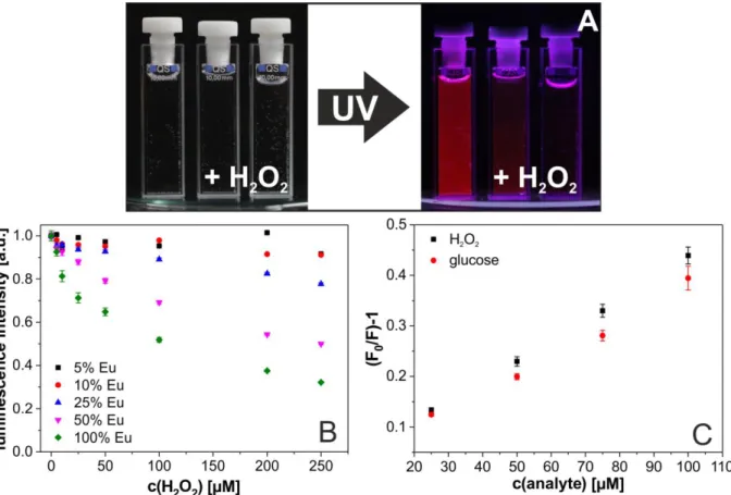
2formed resulting from the catalytic action of glucose oxidase in collaboration with colleagues from the University of Belgrade, Serbia (Figure 1.2).
25Figure 1.2│(A) Digital photograph of a dispersion of GdVO4(50%Eu) nanoparticles in water before and after the addition of two different concentrations of H2O2. The decrease of the luminescence intensity in presence of H2O2
is clearly visible. (B) Quenching of the (normalized) luminescence of Eu3+-doped GdVO4 nanoparticles (λexc 298 nm, λem 618 nm; pH 7.4) by H2O2 as a function of the % fraction of Eu3+ dopand. The total concentration of Eu3+ always was adjusted to 0.4 mM. (C) Stern-Volmer plots of the emission of GdVO4(50%Eu) nanoparticles in the presence of various concentrations of H2O2 and glucose.
Introduction to Lanthanide-doped Nanomaterials 5
1.2 Upconversion Nanoparticles
1.2.1 Mechanism of Upconversion Luminescence
The ladder-like and long-lived metastable excited states of the lanthanide ions enable the occurrence of another optical phenomenon that was first described for bulk materials by Auzel, Ovsyankin and Feofilov in the 1960s: photon upconversion.
26This term describes a non-linear optical process generated by the sequential absorption of at least two low energy photons, which leads to the population of higher energy levels followed by the emission of one photon with higher energy and shorter wavelength than the excitation light. This unique feature makes upconversion luminescent nanoparticles (UCNPs) promising candidates for security printing,
27enhancement of solar cell efficiency,
28implementation as photocatalysts,
29and, most importantly, application as labels, probes and reporters for theranostics, biosensing and bioimaging.
30The excitation wavelength in the near-infrared region (typically at 980 nm) is within the "optical window" of biological tissue, where the absorption of the biological matrix is at a minimum. This allows for deep tissue penetration and simultaneously reduced photo-damage compared to the much higher energy of UV/visible light, which is needed to excite Stokes-emitting molecules and materials. The NIR excitation also triggers virtually no background fluorescence and enables high sensitivity during measurements with extraordinarily high signal-to-noise ratios.
Three main mechanisms for photon upconversion have been described: exited state
absorption, energy transfer upconversion, and photon avalanche.
31Schematic
representations of these mechanisms are shown in Figure 1.3. Energy transfer upconversion
(ETU) is much more efficient than excited state absorption and, unlike photon avalanche,
does not require a critical (high) excitation power and does not result in delayed
luminescence emission caused by the reoccurring relaxation - excitation cycles.
32Therefore,
most existing upconversion materials are based on ETU. The so-called sensitizer ion is
responsible for the resonant photon absorption. The energy is then transferred to a
neighboring activator ion. This leads to the relaxation of the sensitizer ion back to the ground
state and the promotion of the activator ion to a higher energy level. This process is repeated
for one (or several) more times, which leads to the population of the second (or higher)
excited state of the activator. The transition of the activator back to its ground state causes
the emission of a higher energy photon. In such materials upconversion emission can
already be stimulated using low power (power densities starting from 10 W·cm
-2) continuous
wave (CW) diode lasers. The required excitation power is much lower than for simultaneous
6 Introduction to Lanthanide-doped Nanomaterials two-photon absorption processes used for example in two-photon excitation microscopy, in which expensive pulsed lasers operating at power densities up to 10
9W·cm
-2are applied.
33Figure 1.3│ Simplified representations of the three main upconversion processes (A) excited state absorption, (B) energy transfer upconversion, (C) photon avalanche. Dashed arrows symbolize photon absorption, full arrows show photon emission and dotted arrows refer to non-radiative processes. G, E1 and E2 represent ground state, excited level 1, and excited level 2, respectively.
1.2.2 Characteristics and Composition
Inorganic UCNPs usually are composed of an inert host crystal lattice doped with metal ions.
Transition metal ions, e.g. Ti
2+, Re
3+, or Os
4+, have been reported as luminescent dopants,
34but trivalent lanthanide ions show by far the most promising features for efficient photon upconversion. Yb
3+ions represent the most popular sensitizers. They possess a molar absorption coefficient of 10 M
-1cm
-1, which is very high considering that the absorption promotes parity forbidden f-f transitions.
16A typical absorption spectrum of Yb
3+is shown in Figure 1.4. The main absorbance of Yb
3+is in the near infrared (NIR) region at a wavelength of 978 nm. Therefore, the Yb
3+ions acting as sensitizers can be efficiently excited by conventional 980 nm low power (hand held) diode lasers. The energy conserved in the
2F
5/2electronic state of the Yb
3+ions can be transferred to activator ions, which act as the luminescent centers in the nanoparticles. Most common activators are Er
3+and Tm
3+, but upconversion luminescence originating from other lanthanide ions, like Ho
3+, Tb
3+and Eu
3+ions has also been reported.
35,36The upconversion efficiency of UCNPs is strongly dependent on the host material. The
energy transfer dynamics between sensitizer-sensitizer and sensitizer-activator is influenced
by the ion-ion distances within the host crystal lattice. Suitable host lattices in this regard are
metal oxides and halogenides, such as Y
2O
3, YVO
4, LaF
3, and NaYF
4.
31The fluoride host is
superior to oxide materials due to its low phonon energy around 350 cm
-1. This avoids
energy loss caused by multiphonon relaxation between the metastable states inside the
Introduction to Lanthanide-doped Nanomaterials 7 crystal lattice and promotes upconversion efficiency. Fluoride materials with Na
+, Ca
2+, and Y
3+as cations additionally provide chemical stability. Therefore NaYF
4is regarded as the most efficient and most widely used upconversion host material.
Figure 1.4│ Absorption spectrum of oleate-capped β-NaYF4 (20% Yb, 2% Er) UCNPs dispersed in cyclohexane at a particle concentration of 22 mg·mL-1. The optical path length of the cuvette was 5 cm, the slit width was 1 nm.
The ionic radius of Y
3+is very similar to that of trivalent lanthanide ions and can consequently be easily exchanged by the rare earth ions without significantly disturbing the overall crystal structure.
37This offers the option to design a number of various nanomaterials with diverse electromagnetic properties depending on the chosen dopants. Magnetic properties for MRT applications can be introduced, if the host material is doped with Gd
3+ions. Doping with Eu
3+or Tb
3+leads to “downconversion” luminescence upon UV excitation, while doping with Yb
3+, Tm
3+, Er
3+or Ho
3+produces upconversion luminescence.
38The combination of such different doping possibilities enables the design of multimodal nanoparticles. Thermodynamically stable phases of NaYF
4at room temperature are the cubic α-phase and the hexagonal β- phase. However, the upconversion efficiency is about ten times higher in β-NaYF
4compared to α-NaYF
4due to a more favorable spatial arrangement of the dopants within the hexagonal β-crystal phase.
39The probability of resonant energy transfer processes between Yb
3+and Er
3+is increased by optimal ion-ion distances creating efficient photoactive sites in β-NaYF
4.
40900 950 1000 1050
0.1 0.2 0.3 0.4
e x ti n c ti o n [ a .u .]
wavelength [nm]
8 Introduction to Lanthanide-doped Nanomaterials 1.2.3 Photophysical Properties
One of the highest upconversion efficiencies so far has been reported for a β-NaYF
4host lattice doped with either 20 mol% Yb
3+as sensitizer and 2 mol% Er
3+as activator or 25 mol% Yb
3+as sensitizer and 0.3 mol% Tm
3+as activator. Upconversion quantum yields of 0.1% have been reported for these particles (diameter of 22 nm) already at low excitation power densities (around 50 W·cm
-1).
41While higher amounts of Yb
3+would increase the absorption of the material, they also induce increased sensitizer-sensitizer relaxations and activator-sensitizer back transfers that counteract the population of the higher electronic states in the activator ions. The same is true for higher doping concentrations of the activator, which can promote undesired non-radiative activator-activator relaxations.
42The energy level diagrams of the lanthanide ions in NaYF
4:Yb
3+/Er
3+and NaYF
4:Yb
3+/Tm
3+and the mechanisms leading to the population of the excited states are shown in Figure 1.5.
The pair Yb/Er generates two main upconversion emission bands upon excitation at 980 nm,
one in the green region between 500 nm and 550 nm and one in the red region between
640 nm and 680 nm. The green emission originates from the transitions from
2H
11/2and
4S
3/2back to the ground state
4I
15/2in Er
3+. The red emission can be ascribed to the
4F
9/2→
4I
15/2transition. The green emission is more intense than the red one as a result of different
pathways leading to the population of the two distinct emissive states, which imparts a
colloidal dispersion of the nanoparticles with the impression of predominantly green
luminescence. All three excited electronic states in Er
3+are mainly populated by a two-
photonic ETU pathway. Higher order population pathways of energy states occur for the
main upconversion emissions of the pair Yb/Tm. The ultraviolet emission around 360 nm
(
1D
2→
3H
6) and one of two blue (
1D
2→
3F
4) emission bands at 450 nm are populated by the
sequential absorption of four NIR photons. The second blue emission (
1G
4→
3H
6) at 475 nm
is generated by the absorption of three NIR photons. The most intense Tm
3+emission
(
3H
4→
3H
6) is in the NIR around 800 nm and is populated by a two-photon ETU pathway. But
since the NIR and UV emissions are not visible by the human eye, Yb/Tm doped UCNPs
give the impression of blue luminescence. A photograph of colloidal dispersions of
β-NaYF
4:Yb
3+,Er
3+and β-NaYF
4:Yb
3+,Tm
3+UCNPs in cyclohexane indicating the multicolor
main upconversion emission bands is depicted in Figure 1.6.
Introduction to Lanthanide-doped Nanomaterials 9
Figure 1.5│ Energy level diagrams of the lanthanide ions in NaYF4:Yb3+/Er3+ and NaYF4:Yb3+/Tm3+. The solid and dotted black and grey arrows represent photon absorption, the solid colored arrows photon emission, the curly black lines non-radiative relaxations, and the curly blue lines non-radiative relaxations favored by high energy O-H vibrations.
Figure 1.6│ (Middle) Dispersion of hydrophobic NaYF4:Yb3+,Er3+ and β-NaYF4:Yb3+,Tm3+ UCNPs in cyclohexane displaying predominantly blue or green luminescence upon 980 nm CW laser excitation. (Left and right) Corresponding upconversion luminescence spectra upon 980 nm CW laser excitation. The wavelengths of the individual peak maxima are given for each of the two distinct main peaks in either the blue and NIR or the green and red spectral region, respectively.
The small particle dimensions (d < 50 nm) needed for bioanalytical applications, i.e. uptake
into cells and interaction with biomolecules, amplify all surface related effects. The massive
increase of the surface-to-volume ratio by the reduction of the particle dimensions down to
the nm regime leads to increased (non-radiative) surface deactivation caused by quenching
by surface ligands, solvent molecules and crystal defects.
43Consequently, the quantum yield
10 Introduction to Lanthanide-doped Nanomaterials of UCNPs is much lower than that of the bulk material. The upconversion quantum yield in nanoparticles scarcely reaches values above 1% and heavily depends on even slight changes of the particle diameter and excitation power density.
41,44Surface quenching is most pronounced in aqueous solvents as a result of increased deactivation of Ln(III) excited states by O-H vibrations of water molecules.
41Core-shell particle architectures provide a possibility to minimize surface deactivation by protecting the surface of the emissive UCNP core. Inert shells, usually consisting of non-doped host material, efficiently prevent water molecules (or quenchers in general) coming close to the active particle core and lead to enhanced upconversion luminescence quantum yield, emission intensity and lifetime in all solvents.
45A shell thickness of ≥ 10 nm almost completely suppresses surface quenching and no further luminescence enhancement is achieved with even thicker shells.
461.2.4 Synthesis and Surface Modification
Sophisticated and reproducible synthesis protocols are necessary to control the crystal size and composition, ensure monodisperse particle (and shell) growth, avoid crystal defects, and optimize the spatial arrangement of sensitizer and activator ions in the crystal lattice for the design of high quality upconversion nanomaterials.
47Many synthesis methods for the fabrication of efficient UCNPs have been described during the last years. Several recent reviews give a comprehensive overview of the characteristics of different synthetic strategies, including thermal decomposition, co-precipitation, hydro-/solvothermal methods and combinations of these techniques.
48Most prevalent is the high temperature synthesis performed in solvent mixtures of oleic acid and 1-octadecene at 300 °C first reported by Li et al. in 2008.
49This technique enables the fabrication of oleate-capped, pure β-NaYF
4nanocrystals doped with lanthanide ions that display outstanding quality in terms of monodispersity, shape uniformity, and upconversion luminescence efficiency for large batches of several grams of UCNPs.
50The size and shape of the UCNPs can be tuned by altering the ratio of oleic acid and 1-octadecene,
51by varying the Na
+content,
52or by addition of Gd
3+as further dopant.
53An example of β-NaYF
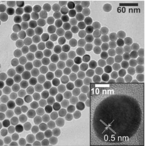
4:Yb,Er UCNPs synthesized by this high temperature procedure is depicted in Figure 1.7. The transmission electron micrograph shows the narrow size distribution structure of the resulting UCNPs.
The surface of the particles is stabilized by oleate molecules, which causes dispersibility only
in hydrophobic organic solvents. Since such particles cannot be dispersed in water due to
the oil-based synthesis, further surface modification is necessary before the UCNPs can be
applied in biosensing and -imaging. Most common surface modification techniques that have
been established for the functionalization of hydrophobic nanoparticles, such as quantum
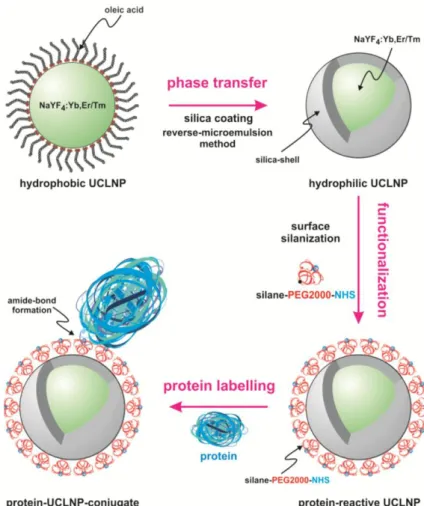
Introduction to Lanthanide-doped Nanomaterials 11 dots, magnetic particles and UCNPs are (a) ligand exchange, (b) additional coating with amphiphilic molecules, and (c) encapsulation with silica. These three strategies greatly differ regarding time consumption and optical and physical properties of the functionalized, hydrophilic UCNPs.
54Amphiphilic coatings and encapsulation with silica are expected to provide better protection against surface deactivation compared to ligand exchange as a result of the additional hydrophobic layer or increased distance between particle surface and solvent molecules, leading to stronger luminescence. They also tend possess higher colloidal stability in physiological media. Colloidal stability depends on an efficient electrostatic and/or steric stabilization, which is challenging to maintain in biological environments with high ionic strengths. Ligand exchange gives the impression of the most versatile strategy resulting in overall smallest particle sizes due to direct attachment of the new ligand to the UCNP surface. Stability in buffered solutions can be improved by the use of polymers as chelating ligands or molecules containing phosphonates as strongly coordinating groups towards the UCNPs. Since all of these factors influence the performance of UCNPs in bioanalytical applications, it of great importance to develop specific surface modification strategies precisely tailored towards the final application.
Figure 1.7│ Transmission electron micrograph (scale bar: 60 nm) of oleate-capped NaYF4:Yb3+,Er3+@NaYF4
core-shell UCNPs with a mean diameter of 28.8 ± 1.0 nm. The core particles used for the synthesis had a diameter of 21.1 ± 1.0 nm, and the thickness of the inert shell is almost 4 nm. The inset shows a higher magnification (scale bar: 10 nm) of one core-shell particle. The difference between core and shell is not visible, since the two very similar materials display the same contrast in the images. The regular orientation of the lattice planes indicates the uniform growth of the NaYF4-shell in all directions. The distance between the visible lattice planes is 0.5 nm.
12 Introduction to Lanthanide-doped Nanomaterials 1.2.5 Current Applications and Challenges
The exceptional optical properties of UCNPs and advances in the field of nanoparticle synthesis leading to the controlled preparation of complex particle architectures meet many criteria that define efficient luminescent reporters in bioanalysis and for theranostic applications
55and have led to UCNPs being the focus of intensive research in these fields over the last decade. They have been applied as luminescent reporters for the detection and quantification of a multitude of different target analytes. A vast number of recent publications report on the development of upconversion nanoprobes for the determination of e.g. heavy metal ions,
56mycotoxins,
57viruses,
58bacteria,
59and hemoglobin.
60Loading of UCNPs with active ingredients enabled imaging guided drug delivery and cancer therapy.
61-63Cellular uptake provided a nanoplatform for photodynamic therapy with UCNPs acting as in situ nanolamps for indirect excitation of photosensitizers with reduced damage to surrounding, healthy tissue.
64,65UCNPs have also been successfully used for super-resolution fluoresence microscopy to achieve nanoscopic resolutions.
66The abundance of ongoing studies demonstrates the demand for new luminescent probes and also the perspectives offered by a complete understanding of upconversion luminescene. However, there are still some unsolved questions limiting the exploitation of the full theranostic potential of UCNPs. One major challenge is the need for efficient surface engineering providing both colloidal stabilization in physiological media and functionalization with selective receptor molecules, and simultaneously enabling (sub)cellular targeting. The combination of all these features while at the same time keeping particle crosslinking and cytotoxicity at a minimum is crucial for the creation of stable, target-responsive nanoprobes.
Another challenge is to increase the understanding of influences on upconversion luminescence emission. The luminescence intensity is dependent on many different parameters, e.g. particle size, architecture, and concentration, solvent, excitation power density, light scattering, and surface coating. All these influences must be considered to design efficient nanoprobes for diverse applications.
67Exploitation of Förster resonance energy transfer (FRET) enables the elimination of many of
these dependencies. This non-radiative energy transfer process takes place between two
(fluorescent) molecules very close to each other, usually within less than 10 nm, and is
extremely distance dependent.
68FRET is the most powerful analytical method for monitoring
bioaffinity reactions, e.g. DNA hybridization
69and receptor interactions,
70due to its ability to
act as a "nanoscopic ruler". The two energy transfer partners must display an overlap of the
luminescence emission spectrum of one molecule (energy donor) with the absorption
spectrum of the second one (energy acceptor). Characteristic for successful FRET is the
Introduction to Lanthanide-doped Nanomaterials 13 reduction of the donor luminescence intensity and lifetime, i.e. the stronger the reduction, the higher the FRET efficiency. In contrast to conventional organic dyes usually used as donors for FRET-based detection schemes, UCNPs possess long lifetimes in the range of 100 µs up to over 1 ms. Their decay times are thus ascertainable with very simple instrumentation, e.g.
a conventional CW laser source and an optical chopper wheel for mechanical intermittence of the excitation light, and changes are easily detectable. Unlike the emission intensity, the lifetime of UCNPs is mostly unaffected by several critical parameters, e.g. (a) its concentration, which can be challenging to determine especially in the case of nanoparticles, (b) radiative absorption processes, i.e. inner filter effects capable of influencing the changes of the emission intensity, (c) fluctuations in the power density of the excitation source, which can cause changing intensity ratios as a result of the nonlinear nature of the absorption process preceding the UC emission, and (d) luminescence light scattering typically induced in all biological tissues. FRET also impacts the acceptor lifetime. When FRET occurs, the luminescence decay time of the acceptor molecule is equal to the donor lifetime. For organic dyes acting as acceptors in combination with UCNP donors this should lead to an elongation of their originally short lifetimes in the lower ns to ps range to > 100 µs. Such an extreme effect is also easily detectable. Despite the considerable advantages of time-resolved FRET studies, most current FRET-based detection schemes rely on changes of the upconversion intensity.
71A more detailed understanding of FRET with UCNPs may promote the development of lifetime-based upconversion nanoprobes and the expansion and improvement within their fields of application.
References
(1) Roda, A.; Mirasoli, M.; Michelini, E.; Di Fusco, M.; Zangheri, M.; Cevenini, L.; Roda, B.;
Simoni, P. Progress in Chemical Luminescence-based Biosensors: A Critical Review.
Biosens. Bioelectron. 2016, 76, 164–179.
(2) Yuan, L.; Lin, W.; Zheng, K.; He, L.; Huang, W. Far-red to Near-infrared Analyte- responsive Fluorescent Probes Based on Organic Fluorophore Platforms for Fluorescence Imaging. Chem. Soc. Rev. 2013, 42, 622–661.
(3) Borisov, S. M.; Wolfbeis, O. S. Optical Biosensors. Chem. Rev. 2008, 108, 423–461.
(4) Deschout, H.; Zanacchi, F. C.; Mlodzianoski, M.; Diaspro, A.; Bewersdorf, J.; Hess, S.
T.; Braeckmans, K. Precisely and Accurately Localizing Single Emitters in Fluorescence Microscopy. Nat. Methods 2014, 11, 253–266.
(5) Ma, F.; Li, Y.; Tang, B.; Zhang, C.-y. Fluorescent Biosensors Based on Single-
Molecule Counting. Acc. Chem. Res. 2016, 49, 1722–1730.
14 Introduction to Lanthanide-doped Nanomaterials (6) Lee, M. H.; Kim, J. S.; Sessler, J. L. Small Molecule-based Ratiometric Fluorescence Probes for Cations, Anions, and Biomolecules. Chem. Soc. Rev. 2015, 44, 4185–4191.
(7) Chen, G.; Roy, I.; Yang, C.; Prasad, P. N. Nanochemistry and Nanomedicine for Nanoparticle-based Diagnostics and Therapy. Chem. Rev. 2016, 116, 2826–2885.
(8) Jaque, D.; Richard, C.; Viana, B.; Soga, K.; Liu, X.; García Solé, J. Inorganic Nanoparticles for Optical Bioimaging. Adv. Opt. Photon. 2016, 8, 1–103.
(9) McCarty, R. J.; Stebbins, J. F. Investigating Lanthanide Dopant Distributions in Yttrium Aluminum Garnet (YAG) Using Solid State Paramagnetic NMR. Solid State Nucl.
Magn. Reson. 2016, 79, 11–22.
(10) Ainslie, B. J. A Review of the Fabrication and Properties of Erbium-doped Fibers for Optical Amplifiers. J. Lightwave Technol. 1991, 9, 220–227.
(11) Andres, J.; Hersch, R. D.; Moser, J.-E.; Chauvin, A.-S. A New Anti-Counterfeiting Feature Relying on Invisible Luminescent Full Color Images Printed with Lanthanide- Based Inks. Adv. Funct. Mater. 2014, 24, 5029–5036.
(12) Teo, R. D.; Termini, J.; Gray, H. B. Lanthanides: Applications in Cancer Diagnosis and Therapy. J. Med. Chem. 2016, 59, 6012–6024.
(13) Bünzli, J.-C. G.; Piguet, C. Taking Advantage of Luminescent Lanthanide Ions. Chem.
Soc. Rev. 2005, 34, 1048–1077.
(14) Gai, S.; Li, C.; Yang, P.; Lin, J. Recent Progress in Rare Earth Micro/Nanocrystals: Soft Chemical Synthesis, Luminescent Properties, and Biomedical Applications. Chem.
Rev. 2014, 114, 2343–2389.
(15) Cardoso Dos Santos, M.; Hildebrandt, N. Recent Developments in Lanthanide-to- Quantum Dot FRET Using Time-gated Fluorescence Detection and Photon Upconversion. Trends Anal. Chem. 2016, 84, 60–71.
(16) Bünzli, J.-C. G. On the Design of Highly Luminescent Lanthanide Complexes. Coord.
Chem. Rev. 2015, 293-294, 19–47.
(17) Moore, E. G.; Samuel, A. P. S.; Raymond, K. N. From Antenna to Assay: Lessons Learned in Lanthanide Luminescence. Acc. Chem. Res. 2009, 42, 542–552.
(18) Han, S.; Deng, R.; Xie, X.; Liu, X. Enhancing Luminescence in Lanthanide-Doped Upconversion Nanoparticles. Angew. Chem. Int. Ed. 2014, 53, 11702–11715.
(19) Galland, C.; Ghosh, Y.; Steinbruck, A.; Sykora, M.; Hollingsworth, J. A.; Klimov, V. I.;
Htoon, H. Two Types of Luminescence Blinking Revealed by Spectroelectrochemistry of Single Quantum Dots. Nature 2011, 479, 203–207.
(20) Dickson, R. M.; Cubitt, A. B.; Tsien, R. Y.; Moerner, W. E. On/Off Blinking and Switching Behaviour of Single Molecules of Green Fluorescent Protein. Nature 1997, 388, 355–358.
(21) Zhou, J.; Xu, S.; Zhang, J.; Qiu, J. Upconversion Luminescence Behavior of Single Nanoparticles. Nanoscale 2015, 7, 15026–15036.
(22) Reisch, A.; Klymchenko, A. S. Fluorescent Polymer Nanoparticles Based on Dyes:
Seeking Brighter Tools for Bioimaging. Small 2016, 12, 1968–1992.
Introduction to Lanthanide-doped Nanomaterials 15 (23) Sy, M.; Nonat, A.; Hildebrandt, N.; Charbonniere, L. J. Lanthanide-based
Luminescence Biolabelling. Chem. Commun. 2016, 52, 5080–5095.
(24) Bulin, A.-L.; Truillet, C.; Chouikrat, R.; Lux, F.; Frochot, C.; Amans, D.; Ledoux, G.;
Tillement, O.; Perriat, P.; Barberi-Heyob, M. et al. X-ray-Induced Singlet Oxygen Activation with Nanoscintillator-Coupled Porphyrins. J. Phys. Chem. C 2013, 117, 21583–21589.
(25) Muhr, V.; Buchner, M.; Hirsch, T.; Jovanović, D. J.; Dolić, S. D.; Dramićanin, M. D.;
Wolfbeis, O. S. Europium-doped GdVO
4Nanocrystals as a Luminescent Probe for Hydrogen Peroxide and for Enzymatic Sensing of Glucose. Sens. Actuators B Chem.
2017, 241, 349–356.
(26) Auzel, F. Upconversion and Anti-Stokes Processes with f and d Ions in Solids. Chem.
Rev. 2004, 104, 139–174.
(27) Meruga, J. M.; Baride, A.; Cross, W.; Kellar, J. J.; May, P. S. Red-Green-Blue Printing Using Luminescence-Upconversion Inks. J. Mater. Chem. C 2014, 2, 2221–2227.
(28) Goldschmidt, J. C.; Fischer, S. Upconversion for Photovoltaics – a Review of Materials, Devices and Concepts for Performance Enhancement. Adv. Opt. Mater. 2015, 3, 510–
535.
(29) Xu, Z.; Quintanilla, M.; Vetrone, F.; Govorov, A. O.; Chaker, M.; Ma, D. Harvesting Lost Photons: Plasmon and Upconversion Enhanced Broadband Photocatalytic Activity in Core@Shell Microspheres Based on Lanthanide-Doped NaYF
4, TiO
2, and Au. Adv.
Funct. Mater. 2015, 25, 2950–2960.
(30) Zhou, J.; Liu, Q.; Feng, W.; Sun, Y.; Li, F. Upconversion Luminescent Materials:
Advances and Applications. Chem. Rev. 2015, 115, 395–465.
(31) Haase, M.; Schäfer, H. Upconverting Nanoparticles. Angew. Chem. Int. Ed. 2011, 50, 5808–5829.
(32) Babu, P.; Martín, I. R.; Venkata Krishnaiah, K.; Seo, H. J.; Venkatramu, V.;
Jayasankar, C. K.; Lavín, V. Photon Avalanche Upconversion in Ho
3+–Yb
3+Co-doped Transparent Oxyfluoride Glass–Ceramics. Chem. Phys. Lett. 2014, 600, 34–37.
(33) Park, Y. I.; Lee, K. T.; Suh, Y. D.; Hyeon, T. Upconverting nanoparticles: A Versatile Platform for Wide-field Two-photon Microscopy and Multi-modal in vivo Imaging. Chem.
Soc. Rev. 2015, 44, 1302–1317.
(34) Gamelin, D. R.; Güdel, H. U. Design of Luminescent Inorganic Materials: New Photophysical Processes Studied by Optical Spectroscopy. Acc. Chem. Res. 2000, 33, 235–242.
(35) Xu, M.; Chen, D.; Huang, P.; Wan, Z.; Zhou, Y.; Ji, Z. A Dual-functional Upconversion Core@Shell Nanostructure for White-Light-Emission and Temperature Sensing. J.
Mater. Chem. C 2016, 4, 6516–6524.
(36) Dong, H.; Sun, L.-D.; Wang, Y.-F.; Xiao, J.-W.; Tu, D.; Chen, X.; Yan, C.-H. Photon Upconversion in Yb
3+-Tb
3+and Yb
3+-Eu
3+Activated Core/Shell Nanoparticles with Dual- band Excitation. J. Mater. Chem. C 2016, 4, 4186–4192.
(37) Naccache, R.; Yu, Q.; Capobianco, J. A. The Fluoride Host: Nucleation, Growth, and
Upconversion of Lanthanide-Doped Nanoparticles. Adv. Opt. Mater. 2015, 3, 482–509.
16 Introduction to Lanthanide-doped Nanomaterials (38) Chen, Q.; Wang, C.; Cheng, L.; He, W.; Cheng, Z.; Liu, Z. Protein Modified Upconversion Nanoparticles for Imaging-guided Combined Photothermal and Photodynamic Therapy. Biomaterials 2014, 35, 2915–2923.
(39) Zeng, J.-H.; Su, J.; Li, Z.-H.; Yan, R.-X.; Li, Y.-D. Synthesis and Upconversion Luminescence of Hexagonal-Phase NaYF
4:Yb, Er
3+Phosphors of Controlled Size and Morphology. Adv. Mater. 2005, 17, 2119–2123.
(40) Aebischer, A.; Hostettler, M.; Hauser, J.; Krämer, K.; Weber, T.; Güdel, H. U.; Bürgi, H.-B. Structural and Spectroscopic Characterization of Active Sites in a Family of Light- Emitting Sodium Lanthanide Tetrafluorides. Angew. Chem. Int. Ed. 2006, 45, 2802–
2806.
(41) Würth, C.; Kaiser, M.; Wilhelm, S.; Grauel, B.; Hirsch, T.; Resch-Genger, U. Excitation Power Dependent Population Pathways and Absolute Quantum Yields of Upconversion Nanoparticles in Different Solvents. Nanoscale 2017, 9, 4283–4294.
(42) Johnson, N. J. J.; He, S.; Diao, S.; Chan, E. M.; Dai, H.; Almutairi, A. Direct Evidence for Coupled Surface and Concentration Quenching Dynamics in Lanthanide-Doped Nanocrystals. J. .Am. Chem. Soc. 2017, 139, 3275–3282.
(43) Wang, F.; Wang, J.; Liu, X. Direct Evidence of a Surface Quenching Effect on Size- Dependent Luminescence of Upconversion Nanoparticles. Angew. Chem. 2010, 122, 7618–7622.
(44) Boyer, J.-C.; van Veggel, F. C. J. M. Absolute Quantum Yield Measurements of Colloidal NaYF
4:Er
3+, Yb
3+Upconverting Nanoparticles. Nanoscale 2010, 2, 1417–
1419.
(45) Wang, F.; Deng, R.; Wang, J.; Wang, Q.; Han, Y.; Zhu, H.; Chen, X.; Liu, X. Tuning Upconversion Through Energy Migration in Core-Shell Nanoparticles. Nat. Mater.
2011, 10, 968–973.
(46) Wang, Y.; Liu, K.; Liu, X.; Dohnalová, K.; Gregorkiewicz, T.; Kong, X.; Aalders, M. C.
G.; Buma, W. J.; Zhang, H. Critical Shell Thickness of Core/Shell Upconversion Luminescence Nanoplatform for FRET Application. J. Phys. Chem. Lett. 2011, 2, 2083–2088.
(47) Ma, C.; Xu, X.; Wang, F.; Zhou, Z.; Wen, S.; Liu, D.; Fang, J.; Lang, C. I.; Jin, D.
Probing the Interior Crystal Quality in the Development of More Efficient and Smaller Upconversion Nanoparticles. J. Phys. Chem. Lett. 2016, 7, 3252–3258.
(48) Yan, C.; Zhao, H.; Perepichka, D. F.; Rosei, F. Lanthanide Ion Doped Upconverting Nanoparticles: Synthesis, Structure and Properties. Small 2016, 12, 3888–3907.
(49) Li, Z.; Zhang, Y.; Jiang, S. Multicolor Core/Shell-Structured Upconversion Fluorescent Nanoparticles. Adv. Mater. 2008, 20, 4765–4769.
(50) Wilhelm, S.; Kaiser, M.; Wurth, C.; Heiland, J.; Carrillo-Carrion, C.; Muhr, V.; Wolfbeis,
O. S.; Parak, W. J.; Resch-Genger, U.; Hirsch, T. Water Dispersible Upconverting
Nanoparticles: Effects of Surface Modification on their Luminescence and Colloidal
stability. Nanoscale 2015, 7, 1403–1410.
Introduction to Lanthanide-doped Nanomaterials 17 (51) Zhang, X.; Blasiak, B.; Marenco, A. J.; Trudel, S.; Tomanek, B.; van Veggel, F. C. J. M.
Design and Regulation of NaHoF
4and NaDyF
4Nanoparticles for High-Field Magnetic Resonance Imaging. Chem. Mater. 2016, 28, 3060–3072.
(52) Dühnen, S.; Rinkel, T.; Haase, M. Size Control of Nearly Monodisperse β-NaGdF
4Particles Prepared from Small α-NaGdF
4Nanocrystals. Chem. Mater. 2015, 27, 4033–
4039.
(53) Wang, F.; Han, Y.; Lim, C. S.; Lu, Y.; Wang, J.; Xu, J.; Chen, H.; Zhang, C.; Hong, M.;
Liu, X. Simultaneous Phase and Size Control of Upconversion Nanocrystals through Lanthanide Doping. Nature 2010, 463, 1061–1065.
(54) Wilhelm, S.; Kaiser, M.; Würth, C.; Heiland, J.; Carrillo-Carrion, C.; Muhr, V.; Wolfbeis, O. S.; Parak, W. J.; Resch-Genger, U.; Hirsch, T. Water Dispersible Upconverting Nanoparticles: Effects of Surface Modification on their Luminescence and Colloidal stability. Nanoscale 2015, 7, 1403–1410.
(55) Chen, G.; Qiu, H.; Prasad, P. N.; Chen, X. Upconversion Nanoparticles: Design, Nanochemistry, and Applications in Theranostics. Chem. Rev. 2013, 114, 5161–5214.
(56) Gu, B.; Zhou, Y.; Zhang, X.; Liu, X.; Zhang, Y.; Marks, R.; Zhang, H.; Liu, X.; Zhang, Q.
Thiazole Derivative-modified Upconversion Nanoparticles for Hg
2+Detection in Living Cells. Nanoscale 2016, 8, 276–282.
(57) Dai, S.; Wu, S.; Duan, N.; Wang, Z. A luminescence resonance energy transfer based aptasensor for the mycotoxin Ochratoxin A using upconversion nanoparticles and gold nanorods. Microchim. Acta 2016, 183, 1909–1916.
(58) Tsang, M.-K.; Ye, W.; Wang, G.; Li, J.; Yang, M.; Hao, J. Ultrasensitive Detection of Ebola Virus Oligonucleotide Based on Upconversion Nanoprobe/Nanoporous Membrane System. ACS Nano 2016, 10, 598–605.
(59) Wu, S.; Duan, N.; Shi, Z.; Fang, C.; Wang, Z. Simultaneous Aptasensor for Multiplex Pathogenic Bacteria Detection Based on Multicolor Upconversion Nanoparticles Labels. Anal. Chem. 2014, 86, 3100–3107.
(60) Jo, E.-J.; Mun, H.; Kim, M.-G. Homogeneous Immunosensor Based on Luminescence Resonance Energy Transfer for Glycated Hemoglobin Detection Using Upconversion Nanoparticles. Anal. Chem. 2016, 88, 2742–2746.
(61) Jalani, G.; Naccache, R.; Rosenzweig, D. H.; Haglund, L.; Vetrone, F.; Cerruti, M.
Photocleavable Hydrogel-Coated Upconverting Nanoparticles: A Multifunctional Theranostic Platform for NIR Imaging and On-Demand Macromolecular Delivery. J.
Am. Chem. Soc. 2016, 138, 1078–1083.
(62) Lin, M.; Gao, Y.; Diefenbach, T. J.; Shen, J. K.; Hornicek, F. J.; Park, Y. I.; Xu, F.; Lu, T. J.; Amiji, M.; Duan, Z. Facial Layer-by-Layer Engineering of Upconversion Nanoparticles for Gene Delivery: Near-Infrared-Initiated Fluorescence Resonance Energy Transfer Tracking and Overcoming Drug Resistance in Ovarian Cancer. ACS Appl. Mater. Infaces 2017, 9, 7941–7949.
(63) Liang, L.; Care, A.; Zhang, R.; Lu, Y.; Packer, N. H.; Sunna, A.; Qian, Y.; Zvyagin, A. V.
Facile Assembly of Functional Upconversion Nanoparticles for Targeted Cancer Imaging and Photodynamic Therapy. ACS Appl. Mater. Interfaces 2016, 8, 11945–
11953.
18 Introduction to Lanthanide-doped Nanomaterials (64) Lu, F.; Yang, L.; Ding, Y.; Zhu, J.-J. Highly Emissive Nd
3+-Sensitized Multilayered Upconversion Nanoparticles for Efficient 795 nm Operated Photodynamic Therapy.
Adv. Funct. Mater. 2016, 26, 4778–4785.
(65) Liu, B.; Li, C.; Xing, B.; Yang, P.; Lin, J. Multifunctional UCNPs@PDA-ICG Nanocomposites for Upconversion Imaging and Combined Photothermal/Photodynamic Therapy with Enhanced Antitumor Efficacy. J. Mater.
Chem. B 2016, 4, 4884–4894.
(66) Liu, Y.; Lu, Y.; Yang, X.; Zheng, X.; Wen, S.; Wang, F.; Vidal, X.; Zhao, J.; Liu, D.;
Zhou, Z. et al. Amplified Stimulated Emission in Upconversion Nanoparticles for Super- resolution Nanoscopy. Nature 2017, 543, 229–233.
(67) Wilhelm, S. Perspectives for Upconverting Nanoparticles. ACS Nano 2017, DOI:
10.1021/acsnano.7b07120.
(68) Hildebrandt, N.; Spillmann, C. M.; Algar, W. R.; Pons, T.; Stewart, M. H.; Oh, E.;
Susumu, K.; Díaz, S. A.; Delehanty, J. B.; Medintz, I. L. Energy Transfer with Semiconductor Quantum Dot Bioconjugates: A Versatile Platform for Biosensing, Energy Harvesting, and Other Developing Applications. Chem. Rev. 2017, 117, 536–
711.
(69) Qiu, X.; Guo, J.; Jin, Z.; Petreto, A.; Medintz, I. L.; Hildebrandt, N. Multiplexed Nucleic Acid Hybridization Assays Using Single-FRET-Pair Distance-Tuning. Small 2017, 13, 1700332.
(70) Messerer, R.; Kauk, M.; Volpato, D.; Alonso Canizal, M. C.; Klöckner, J.; Zabel, U.;
Nuber, S.; Hoffmann, C.; Holzgrabe, U. FRET Studies of Quinolone-Based Bitopic Ligands and Their Structural Analogues at the Muscarinic M1 Receptor. ACS Chem.
Biol. 2017, 12, 833–843.
(71) Su, Q.; Feng, W.; Yang, D.; Li, F. Resonance Energy Transfer in Upconversion
Nanoplatforms for Selective Biodetection. Acc. Chem. Res. 2017, 50, 32–40.
Motivation and Objectives 19
2 M OTIVATION AND O BJECTIVES
The design and synthesis of efficient FRET nanoprobes using UCNPs as energy donors requires detailed understanding of energy transfer cascades occurring within the particles and processes on the interface between particle surface and the solvent. The aim of this work was the complete characterization of FRET processes between UCNPs and organic dyes on the particle surface in order to identify both the ideal particle architecture and surface modification technique to provide maximum FRET efficiency and colloidal stability.
This comprehensive knowledge on the complex interplay between particle size and surface functionalization enables the development of enhanced FRET for an improved performance of UCNPs in bioanalytical and theranostic applications.
First and most importantly, control over the surface chemistry of the innately hydrophobic UCNPs needs to be established in order to create the following essential properties:
(a) colloidal stability in aqueous (physiological) solvents by electrostatic stabilization, (b) preparation of UCNPs exhibiting high brightness in the aqueous environment, (c) incorporation of functional groups for the subsequent attachment of FRET acceptors, selective receptors and targeting elements, (d) generation of high FRET efficiencies by controlling the donor-acceptor distance, (e) maintaining colloidal stability after further functionalization, (f) enabling fast cellular uptake, and (g) low cytotoxicity. Advantages and disadvantages of the variety of available techniques and strategies (such as ligand exchange, additional coating with amphiphilic molecules, and silica shells) with respect to the crucial requirements mentioned before need to be carefully considered and evaluated for the proper choice of surface chemistry.
Second, the particle composition and architecture must be precisely designed and controlled.
One should keep in mind that UCNPs are not molecular emitters, but rather a collection of
single point emitters (the activator ions). Consequently, their behavior as FRET donors in
combination with usually also significantly more than one molecular acceptor on the particle
surface may be entirely different from conventional single donor and single acceptor
systems. Aiming at the design of the ideal particle architecture the effect of UCNP size on the
energy transfer efficiency to organic dyes acting as FRET acceptors needs to be
investigated. “Large” dye decorated UCNPs (> 20 nm) theoretically contain a great number of
emitting lanthanide ions in its centers that are too far away from the surface to be able to
contribute to the energy transfer. But increasing surface to volume ratios may promote
surface deactivation competing with FRET. Thus, the optimum UCNP size for FRET based
20 Motivation and Objectives application should be established by exploring FRET efficiencies determined from lifetime studies of UCNPs with varying sizes and different core-shell architectures for the manipulation of the donor-acceptor distance/ratio and brightness of the UCNPs. The FRET systems should be thoroughly characterized and the most efficient design should be reasonably identified.
The insights gained from the systematical investigation of surface modification techniques
and the impact of particle size on the FRET efficiency with UCNPs acting as donors can be
exploited for the development of an efficient nanoprobe applicable for bioanalysis. Focus
needs to be put on smart particle design by combining the preparation of bright UCNPs
facilitating high FRET efficiencies with proper surface engineering. Protection against water
quenching by inert shells and additional amphiphilic coatings is essential to attain high
upconversion luminescence intensity, but is also linked to increased donor-acceptor
distances compared to ligand exchange strategies. The best compromise between these two
opposing effects must be identified to achieve efficient FRET. Further functionalization with
bioreceptors must not impair the colloidal stability of the UCNPs and needs to facilitate
defined analyte-responsive distance alterations. Structure switching receptors, such as
aptamers and molecular beacons, provide a simple but reproducible way to change the
donor-acceptor distance. This can be exploited to induce FRET by labeling with acceptors or
by introducing specific dyes for the recognition of the structure change in presence of the
analyte. Fast cellular uptake is required in order to allow for subcellular targeting and the
nanoprobe should not exhibit cytotoxicity. The challenge is to combine all these features into
one comprehensive probe design. Detailed characterization of the properties and
performance of such detection systems will reveal insights into refined FRET-based probe
designs for future theranostic applications.
Upconversion Nanoparticles: From Hydrophobic to Hydrophilic Surfaces 21
3 U PCONVERSION N ANOPARTICLES : F ROM H YDROPHOBIC TO H YDROPHILIC S URFACES
3.1 Abstract
Photon upconversion nanoparticles (UCNPs) have emerged as a promising new class of nanomaterials due to their ability to convert near-IR light into visible luminescence.
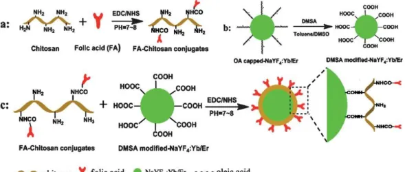
Unfortunately, most efficient methods for preparing UCNPs yield hydrophobic materials, but water-dispersibility is needed in the major fields of applications of UCNPs, that is, in bioimaging, labeling, and bioassays. Numerous methods therefore have been reported in the past years to convert the hydrophobic surface of UCNPs to a more hydrophilic one so to render them dispersible in aqueous systems. We present a classification respective for these strategies and assess the main methods. These include (A) chemical modification of the hydrophobic (typically oleate) ligand on the surface, (B) addition of an extra layer, (C) addition of a thin shell on top of the UCNP, and (D) complete replacement of the original ligand by another one. Chemical modification (A) involves oxidation of the oleate or oleylamine and leads to particles with terminal oxygen functions. This method is less often used because solutions of the resulting UCNPs in water have limited colloidal stability, protocols are time-consuming and often give low yields, and only a limited number of functional groups can be introduced. Methods B and C involve coating of UCNPs with amphiphiles or with shells made from silica oxide, titanium oxide, or metallic gold or silver.
These methods are quite versatile in terms of further modifications, for example, by further
cross-linking or by applying thiol−gold chemistry. Growing an extra shell is, however, often
accompanied by a higher polydispersity. Method D can be divided into subgroups based on
either (i) the direct (single-step) replacement of the native ligand by a new ligand or (ii) two-
step protocols using nitrosyltetrafluoroborate (NOBF
4) or strong acids as reagents to produce
ligand-free UCNPs prior to the attachment of a new ligand. These methods are simple and
versatile, and the distance between the new ligand and the luminescent particle can be well
controlled. However, the particles often have limited stability in buffer systems. The methods
described also are of wider interest because they are likely to be applicable to other kinds of
nanomaterials. We additionally address the need for (a) a better control of particle size and
homogeneity during synthesis, (b) more reproducible methods for surface loading and
modification, (c) synthetic methods giving higher yields of UCNPs, (d) materials displaying
higher quantum yields in water solution without the need for tedious surface modifications,
(e) improved methods for workup (including the suppression of aggregation), (f) new
22 Upconversion Nanoparticles: From Hydrophobic to Hydrophilic Surfaces methods for surface characterization, and (g) more affordable reagents for use in surface modification. It is noted that most synthetic research in the area is of the trial-and-error kind, presumably due to the lack of understanding of the mechanisms causing current limitations.
Finally, all particles are discussed in terms of their biocompatibility (as far as data are available), which is quintessential in terms of imaging, the largest field of application.
Scheme 3.1│ Overview of established surface modification strategies of hydrophobic nanoparticles, including ligand exchange, ligand oxidation, encapsulation with inorganic materials and additional amphiphilic coatings.