Surface Modification and Scan Imaging of Upconverting Nanoparticles
DISSERTATION ZUR ERLANGUNG DES DOKTORGRADES DER NATURWISSENSCHAFTEN (DR. RER. NAT.) DER FAKULTÄT CHEMIE UND PHARMAZIE DER UNIVERSITÄT REGENSBURG
vorgelegt von
Andreas Sedlmeier aus Regensburg
im Jahr 2015
Diese Doktorarbeit entstand in der Zeit von November 2011 bis Dezember 2015 am Institut für Analytische Chemie, Chemo- und Biosensorik an der Universität Regensburg.
Die Arbeit wurde durchgeführt bei Prof. Dr. Otto S. Wolfbeis und Prof. Dr. Antje J. Bäumner unter Anleitung von PD Dr. Hans-Heiner Gorris.
Promotionsgesuch eingereicht: Dezember 2015 Kolloquiumstermin: 29.01.2016
Prüfungsausschuss:
Vorsitzender: Prof. Dr. Frank-Michael Matysik Erstgutachter: Prof. Dr. Antje J. Bäumner Zweitgutachter: PD Dr. Hans-Heiner Gorris Drittprüfer: Prof. Dr. Achim Göpferich
Acknowledgment
First of all, I want to thank Prof. Otto S. Wolfbeis for this interesting topic, the excellent working conditions at his chair and the opportunity to work independently for the first part of my time as a PhD student. Furthermore, I want to express my sincere gratitude to Prof. Dr. Antje J. Bäumner for her willing acceptance of me as a PhD student after Prof. Otto S. Wolfbeis retired, valuable discussions during the second part of my PhD thesis and the continuing possibility to work independently.
I am very grateful to PD Dr. Hans-Heiner Gorris for the direct supervision of my PhD thesis, the abundant discussions and support throughout the whole time as well as the financial support.
I am grateful to Dr. Thomas Hirsch, Dr. Stefan Wilhelm, Verena Muhr and Nadja Leibl for the synthesis of the upconverting nanoparticles (UCNPs), informative discussions and providing the wafers for the plasmon enhancement studies. In addition, I want to thank Dr. Stefan Wilhelm for the transmission electron microscopy imaging of modified UCNPs and Verena Muhr for her guidance during the polyacrylic acid modification.
Additionally, I want to thank the central laboratory of electron microscopy at the department of pathology at the University Hospital of Regensburg and Heiko I. Siegmund in particular for the fast and reliable imaging of the UCNPs samples with informative result discussions and the guidance and performance of the immunogold staining.
Moreover, I am grateful to Prof. Dr. Tero Soukka, Juho Terrijärvi and Ville Haaslahti from Hidex for enabling a research visit at the department of biochemistry & biotechnology at the University of Turku and at the Hidex Company, the provision and explanation of the Chameleon reader device and the support in troubleshooting. I also want to thank Prof. Dr. Paul L. A. M. Corstjens, Hans J. Tanke and Claudia J. de Dood for providing the lateral flow assay series of the Schistosoma circulating anodic antigen (CAA).
I want to thank Dr. Antonín Hlaváček for the productive cooperation in developing the new synthesis route for carboxyl-modified UCNPs and the preparation of electrophoresis gels of UCNPs.
Dr. Mark Schlosser (Institute of Inorganic Chemistry, group of Prof. Dr. Arno Pfitzner) is thanked for the guidance and performance of the infrared spectroscopy measurements.
Furthermore, I want to thank all members of the Institute of Analytical Chemistry, Chemo- and Biosensors for the great support by numerous scientific discussions and the introduction and guidance of or in new techniques and the good atmosphere at the institute. Especially I am grateful
to Gisela Hierlmeier for her great support and motivation both in my PhD work and the lab courses with her assistance. In addition, I want to thank Joachim Rewitzer for the help during the lab courses and in particular his patient troubleshooting for the ICP-OES device.
Christina Liedlbier and Uwe Käfer are thanked for their contribution to the work for this PhD thesis in form of a bachelor thesis and research lab course, respectively. Moreover, I want to thank Matthias Mickert and Lucia Birner for their kind help and assistance and the fruitful discussions.
I am very grateful to Dr. Raphaela Liebherr for her great help and support both in a scientific and private manner. Her kind and reassuring nature helped me during my PhD thesis.
I also want to thank Dr. Heike Mader for the many scientific discussions and sharing her knowledge with me. She is thanked for the critical reading of this thesis and the great help with the English language.
I am grateful to Nicole Guber for her help in all administrative matters and the ongoing encouragement (Danke für alles, Nicole).
Finally and particularly, I want to thank my parents Reinhard and Ingrid Sedlmeier and my sister Carolin for their unremitting support, motivation and help during my whole studies and PhD thesis. I also want to thank my nephew Lukas for being a welcome and happy distraction during the writing of this thesis.
Table of Content
Table of Content
1. Introduction 1
2. Background 4
2.1 Upconversion 4
2.1.1 Mechanisms of Upconversion 4
2.1.2 Design of Upconverting Materials 6
2.1.3 Upconversion in the Nanometer Scale 9
2.1.4 Luminescence Enhancement by Surface Plasmon Resonance 11
2.1.5 Advantages of Upconverting Nanoparticles for Bioanalysis 13
2.2 Surface Modifications of Upconverting Nanoparticles 15
2.2.1 Silica Coating 16
2.2.2 Silanization 17
2.2.3 Ligand Exchange 19
2.3 Click Chemistry 20
2.3.1 Concept of Click Chemistry & Click Reactions 20
2.3.2 Huisgen Cycloaddition 21
2.4 Virus-Like Particles 24
2.4.1 Viruses 24
2.4.2 Definition of Virus-Like Particles 26
2.4.3 Synthesis Routes for Virus-Like Particles 27
2.5 Imaging in Bioanalysis 31
2.5.1 In Vitro Imaging Methods 33
2.5.2 In Vivo Imaging Methods 36
3. Materials & Methods 39
3.1 Chemicals 39
3.2 Instruments 41
3.3 Upconverting Nanoparticles 42
3.4 Surface Modification of Upconverting Nanoparticles 42
3.4.1 Silica Coating 42
3.4.2 Silanization 43
3.4.3 Coating with Polyacrylic Acid 44
3.4.4 Implementation of Click Chemistry 44
3.4.5 Zetasizer & Transmission Electron Microscopy 45
3.5 Synthesis of Virus-Like Particles 46
3.5.1 General Implementation 46
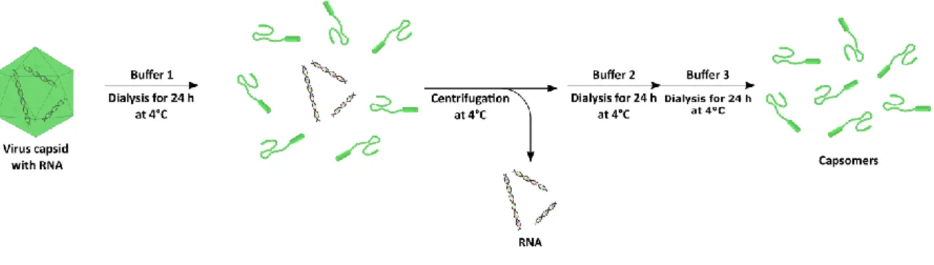
3.5.2 Dialysis 47
3.5.3 Absorption Spectroscopy 47
3.5.4 Immunogold Staining 48
3.6 Chameleon Reader 49
3.6.1 Determination of Limit of Detection 49
3.6.2 Scanning Mode 49
3.7 Gel Electrophoresis 52
3.8 Preparation of Lateral Flow Assays 52
3.9 Wafer Preparation 53
4. Surface Modifications for Biofunctionalization 54
4.1 Demands on Upconverting Nanoparticle for Encapsulation 54
4.2 Silica Shell & Control of Thickness 56
4.3 Amine Functionalization 58
4.4 Carboxylic Acid Functionalization 60
4.5 Coating with Polyacrylic Acid 63
4.6 Phosphonate Functionalization 65
4.7 Alkyne & Azide Functionalization 66
4.8 Carboxylic Acid & Azide Functionalization 70
4.9 Functionalization by Click Chemistry 71
4.10 Summary 74
Table of Content
5. Synthesis of Virus-Like Particles 76
5.1 Disassembly & Reassembly of Virus Capsids 76
5.2 Dialysis during the Synthesis of Virus-Like Particles 79
5.3 Calculations of Capsomer Concentration 80
5.4 Upconverting Nanoparticles 81
5.5 UCNP@Cit as Artificial Cores 81
5.6 UCNP@SiO2 as Artificial Cores 82
5.7 UCNP@SiO2-COOH as Artificial Cores 84
5.8 UCNP@PAA as Artificial Cores 85
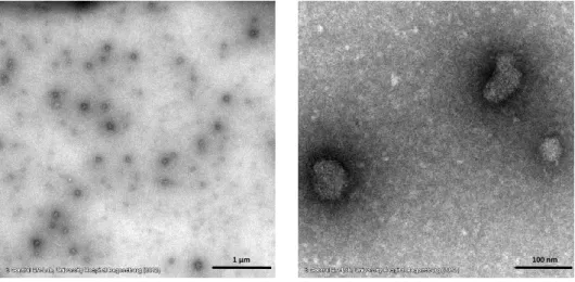
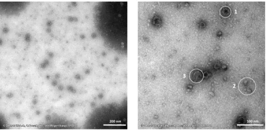
5.9 Immunogold Staining 86
5.10 Summary 91
6. Scan mode & Limit of Detection of the Chameleon Reader 92
6.1 Upconverting Nanoparticles 92
6.2 Limit of Detection in Dispersion 93
6.3 General Optimization Settings for the Scan Mode 95
6.4 Collecting Time 96
6.5 Choice of Filters 98
6.6 Lateral Resolution 102
6.7 Limit of Detection 104
6.8 Scan of Electrophoresis Gels 107
6.9 Scans of Lateral Flow Assays 111
6.10 Luminescence Enhancement by Surface Plasmon Resonance 115
7. Summary 119
7.1 In English 119
7.2 In German 122
8. References 125
9. Appendix 143
9.1 Dynamic light scattering measurements 143
9.2 Zeta potential measurements 150
9.3 Transmission electron microscopy images 155
9.4 Infrared Spectra 156
9.5 Calculations for Synthesis Adjustments of UCNP@SiO2-COOH 157
9.6 Evaluation of Lateral Flow Assays 159
9.7 Wafer Images 160
9.8 Abbreviations 161
10. Curriculum Vitae 163
11. List of Publications and Presentations 164
11.1 Master Thesis 164
11.2 Paper 164
11.3 Abstracts of Posters and Talks 164
Eidesstattliche Erklärung 165
1. INTRODUCTION 1
1. Introduction
The detection of (bio)molecules in in vitro or in vivo is a key issue in various fields of bioanalysis. In addition to the qualitative evidence of these targets, i.e. their presence in the biological system, their quantification is at least equally important. Changes in the (bio)molecule concentration for example can indicate responses of a cell to extracellular stimuli interacting with cell receptors. The detection system for a low concentration of the (bio)molecule has to be highly sensitive to the target but also highly selective to avoid false positive results by chemically or structurally similar compounds.
Both high sensitivity and selectivity can be achieved by using techniques that are based on optical phenomena. Especially fluorescence methods are among the most important detection systems in this regard since they are also versatile, non-invasive, and enable a high spatial resolution.[1]
However, most (bio)molecules that are of high interest in research as well as in clinical studies rarely show intrinsic fluorescence. Therefore, a variety of luminescent labels is available that can be linked to the target with high selectivity, partially even directly in biological samples without the need for sample purification. Organic fluorophores are commonly used for this purpose, since these small molecules can easily be modified by conventional reactions to show the required binding functionality without any changes in their optical properties. A drawback of these labels is their susceptibility to photobleaching which hampers their use in long-term studies. Another class of labels is based on inorganic materials on the nanometer scale. The optical characteristics of nanosized semiconductor materials, so called quantum dots, only rely on the material employed and the nanoparticle size. They show no photobleaching[2, 3], have a higher emission intensity signal, allow an optimization of both the synthesis and the surface modification, and enable the simultaneous introduction of various functionalities and properties into one nanoparticle. Consequently, these fluorescent labels are more suitable for bioanalysis than organic fluorophores. Most quantum dot materials, such as CdS or CdSe, however, display a high (cyto)toxicity due to the heavy metal cadmium rendering them unsuitable for any bioanalytical application[3, 4].
Nanoparticles made of upconverting materials (UCNPs), in contrast, are not only photostable and basically nontoxic, but also enable a shift of the excitation wavelength from the ultraviolet or visible range to the near infrared domain. This change in irradiation light allows studies with drastically reduced photodamage and autofluorescence of biological species. The underlying Anti-Stokes process “upconversion” describes one way to convert low-energy photons (NIR) by sequential absorption into a photon with a shorter wavelength (mostly VIS). This phenomenon, investigated thoroughly by Auzel[5] in the 1960s, occurs with a high efficiency also in nanosized inorganic materials doped with lanthanide ions such as ytterbium, erbium, and/or thulium. The optical characteristics of
the resulting upconversion emission can be divided in two classes according to their dependence on the nanoparticle size. While several parameters such as number, wavelength, and peak width of the emissions are unaffected by the reduction of the upconverting material, other properties like the emission intensity are strongly affected by the altered surface-to-volume ratio owing to enhanced quenching effects.
However, since most syntheses for monodisperse and bright UCNPs rely on hydrophobic size- controlling and surface-capping agents such as oleic acid, the resulting nanoparticles have hydrophobic surfaces unsuitable for any kind of bioanalytical applications. Therefore, surface engineering is indispensable in order to benefit from the advantageous chemical and optical characteristics of UCNPs. Several methods that were originally developed for other nanoparticle types like quantum dots[6] and silica nanoparticles[7, 8] are applicable for these modifications.
However, this surface engineering process has to be adjusted to account for the differences in the surface chemistry of the UCNPs compared to the original targets of these functionalization techniques. Methods based on non-covalent interactions such as ligand exchange or covalent binding such as the growth of a silica shell and a potential silanization creates a hydrophilic UCNP surface and consequently renders these nanoparticles dispersible in aqueous systems. The use of Click chemistry, more precisely the Huisgen cycloaddition of an azide and an alkyne[9, 10], enables a potential direct reaction of the nanoparticle surface with a suitable compound both in vitro and in vivo, since this reaction as well as both functionalities are bioorthogonal[11]. As a result, both functionalities as well as their coupling reaction neither interfere with any biochemical reactions nor are affected by (bio)chemical species. Additionally, both functional groups are exchangeable against each other enabling a flexible optimization of the Click reaction.
Although these modified UCNPs can be used as labels in different bioanalytical applications, they are prone to several detrimental effects. Changes, for example in the composition of the nanoparticle surroundings, can lead to enhanced aggregation tendencies, a loss or alteration of the surface functionalities, or accumulation of the nanoparticles, especially in in vivo studies. Therefore, the creation of a biomimetic surface by using virus capsids is an approach of great interest to obtain stable nanostructures, so-called virus-like nanoparticles (VLPs)[12, 13]. They combine the extremely high long-term stability, biocompatibility[12, 14, 15]
as well as long blood circulation durations of the virus capsids with the unique optical characteristics of UCNPs. The synthesis of these VLPs can be performed according to three different approaches.[12] Firstly, the nucleation and growth of the nanoparticle inside the assembled virus capsid via an introduction of the starting materials; secondly, the attachment of the nanoparticle material to protein parts that later form the inner surface of the
1. INTRODUCTION 3
virus capsid; thirdly, the use of a surface-engineered nanoparticle to act as the new nucleation grain to initiate the capsid self-assembly after the removal of the viral RNA[16, 17].
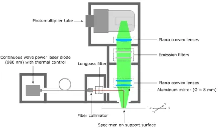
The use of UCNPs as luminescent labels is also hindered by the lack of measurement instruments with a NIR excitation source to detect the upconversion emission in biological systems. Since UCNPs have only recently become commercially available, upconversion devices usually are self-made in most research laboratories. Therefore, the manufacturing and optimization efforts of such devices have to be intensified to enable a more commercial usability of UCNPs. Especially imaging applications with UCNP labels currently rely more on more sophisticated methods such as luminescence microscopy[18-21] or on self-made instruments, especially for whole animal imaging[22-24]. The transfer of scanning measurements of any kind of specimen from these instruments to a commercially available upconversion reader will greatly enhance the rapidity and accuracy of the scan, but foremost the comparability of results from different research groups which is necessary for a standardization of UCNP labels.
The aim of this thesis was on the one hand the creation of virus-like particles (VLPs) with surface- modified UCNPs as the nucleation grains. A variety of modifications, relying either on covalent or non-covalent interactions, were investigated regarding their stability in aqueous systems and thereby their aggregation tendency, any changes in the nanoparticle diameter after the modification, and the strength of the resulting surface charge. As-modified UCNPs with a negative surface charge were decisive for the assembly of virus-like particles (VLP). The concept of Click chemistry, more precisely the copper(I)-catalyzed Huisgen cycloaddition, was also applied on very small UCNPs in order to create a versatile functionalization platform on the nanoparticle surface. This modal functionalization system was suitable for subsequent reactions to generate a negative nanoparticle surface by different functionalities.
On the other hand, this work was focused on adapting and optimizing an upconversion microtiter plate reader for scanning UCNPs in (bio)imaging applications. UCNPs doped with ytterbium and either erbium or thulium were utilized for the instrument optimization as well as for the scanning real sample. The effect of several parameters such as the collecting time, scan point distance and the choice of filters were studied in order to optimize the scanning process. The lateral resolution, the limit of detection, and the signal-to-noise ratio were the most crucial optimization indicators during this process. The optimized scanning mode was applied to real samples by the readout of agarose gel electrophoresis and lateral flow assays. Furthermore, the increase of the UCNP emission by surface plasmon resonance by a gold surface was investigated.
2. Background
2.1 Upconversion
2.1.1 Mechanisms of Upconversion
Upconversion (UC), as described for the first time in the 1960s[5], belongs to the Anti-Stokes processes and describes one way to convert low-energy photons into a photon of higher energy with the others being simultaneous two-photon absorption (STPA) and second harmonic generation (SHG). Similar to other nonlinear optical processes, the upconversion luminescence intensity substantially depends on the power density of the excitation source.[5, 25, 26]
However, there are several photophysical features that clearly differentiate upconversion from other processes such as STPA and SHG and render it much more favorably for bioanalytical applications. Firstly, only discrete, metastable and long-lived energy levels are occupied during the excitation process without the need of virtual levels necessary for STPA and SHG (Figure 2.1). Secondly, the absorption of the two excitation photons has to occur simultaneously with almost no possible delay of the second photon both for the STPA and SHG process owing to these distinction of the energy level, whereas UC relies on the sequential absorption of at least two excitation photons with a longer possible delay between the different absorption processes.
Figure 2.1 Schematic illustration of the two-photon processes “upconversion” (UC), “simultaneous two-photon absorption”
(STPA) and “second harmonic generation” (SHG). Solid lines: ground state (G), discrete energy levels (E1/2); dashed lines:
virtual energy levels; red arrows: absorption; green arrows: emission. The shifted arrangement of the red arrows for UC indicates the sequential photon absorption, while the on-the-line arrangement for STPA and SHG indicates the simultaneous absorption.
Thirdly, the quantum efficiency () of the three processes differs significantly, provided the same excitation density is used. While the efficiency for STPA ( = 10-13) and SHG ( = 10-11) are quite low, UC ( = 10-5 - 10-3) shows an efficiency several magnitudes higher depending on the upconversion mechanism. The high influence of the considered upconverting material on the quantum efficiency has to be taken into account for a conclusive comparison as well.[27]
2. BACKGROUND 5
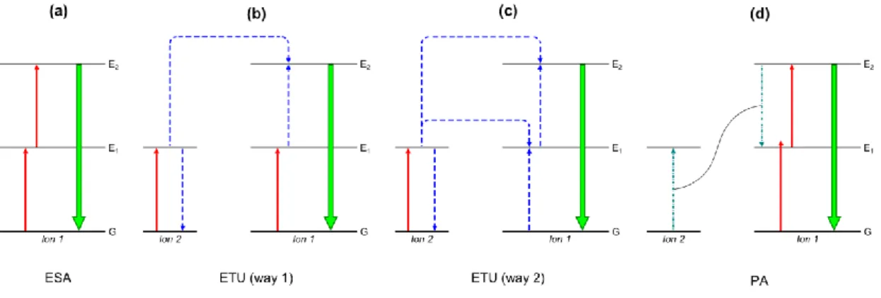
As implied previously, the upconversion process can be ascribed to three mechanisms: excited state absorption (ESA), energy transfer upconversion (ETU), and photon avalanche (PA). After the temporary storage in transition energy levels the accumulated energy of the excitation photons is combined during all three mechanisms to stimulate the emission of higher-energy light, mostly in the visible range.
ESA is the only mechanism taking place in a single ion and thus is independent of the ion concentration (Figure 2.2a). After the absorption of one photon of matching energy to promote the electron from the ground state G to the metastable energy level E1, a second photon excites the electron to the final energy level E2. Finally, the emission of a higher-energy photon occurs by an E2-G transition of the electron.[5, 26, 28]
The ETU mechanism can be derived from the ESA by including a second ion in the excitation mechanism. The precise excitation process can occur according to different excitation sequences and two of them will be described in the following. The first possibility includes the excitation of one electron of each ion to the intermediate energy level E1 (Figure 2.2b). A subsequent energy transfer from ion 1 (energy donor) to ion 2 (energy acceptor) promotes the electron of the energy acceptor to the emitting energy level E2, while the electron of the energy donor returns to the ground state G.
Again, the transition of the electron from E2 to G leads to the emission of a higher-energy photon. In the second excitation sequence one ion completely takes over the role as the energy donor while the other one solely acts as the energy acceptor (Figure 2.2c). In two sequential runs the energy donor (ion 2) absorbs a photon, transfers the energy to the second ion (ion 1) and its electron returns to the ground state and thus promotes the electron of the energy acceptor to energy level E2 resulting in the emission of a higher-energy photon.[5, 26, 28]
Figure 2.2 Schematic illustration of the different mechanisms mainly contributing to the upconversion process: (a) excited state absorption (ESA), (b/c) energy transfer upconversion (ETU, way 1/2), (d) photon avalanche. Solid red arrows:
absorption; dashed blue arrows: energy transfer; dash-dotted teal arrows: cross relaxation; solid green arrows: emission.
The third mechanism PA also employs two ions during the excitation process but a specific threshold of the excitation intensity has to be passed for this mechanism to occur. The first step during the excitation succession is a non-resonant transition of an electron of the ion 1 from G to E1 followed by a resonant excitation to E2 (Figure 2.2d). A cross relaxation between this excited ion and a neighboring ion leads to the transition E2-E1 in ion 1 and the transition G-E1 in ion 2. The absorption of an excitation photon can promote both ions to the E2 level starting the cross relaxation/absorption cycle anew. This “avalanche” process results in an exponential increase of the population of the E2
energy level and thus enables a strong UC emission after the E2-G transition of each ion.[5, 26, 28]
The percentage of the overall upconversion emission of each mechanism differs strongly due to their substantially varying upconversion emission efficiency, i.e. quantum efficiency. The energy transfer upconversion (ETU) results in a nearly simultaneous emission after the excitation process without temporal delay which is independent of the intensity of excitation source. In contrast, the emission takes several seconds to occur after excitation for the photon avalanche (PA) process provided a sufficiently high excitation density is given.[5, 28] Although the ESA process features a lower efficiency by a factor of 100 compared to the ETU mechanism, it is still several magnitudes more efficient than other two-photon processes.[27]
2.1.2 Design of Upconverting Materials
The development of upconverting materials requires a sophisticated and thorough tuning of the inorganic material, the so-called host material, the doped ions, and the dopant concentration to obtain a strong upconversion luminescence. While the host material sets the spatial properties of the dopants and the probability of non-radiative processes, the dopant type and its concentration defines the wavelengths of the upconversion emission and the efficiency of the upconversion process, respectively. Especially the number of different dopants strongly influences the upconversion phenomenon, since for an efficient ETU mechanism[5] two ions are necessary, optimally with differing photophysical properties.
Requirements for the host material with declining priority are a high chemical stability to avoid degradation, low phonon energy to avoid non-radiative relaxation of the excited states of the dopants, and low crystal symmetry to enhance transition probabilities during the upconversion process. Various inorganic crystals including oxides[29-38], oxysulfides[39], oxyfluorides[40-42], phosphates[43], and halides[44-50] are usable to create upconverting materials and meet these characteristics differently. Oxides and phosphates show a high stability in most chemically demanding systems but have high phonon energies of above 500 cm-1 and above 1000 cm-1,
2. BACKGROUND 7 respectively.[28, 29, 51]
Oxysulfides show a strong proneness to acidic conditions. The class of fluoride materials bears a special position compared to the heavier halides. While a very low phonon energy (approx. 300 cm-1)[51] is typical for all halides, only fluorides are chemically stable[28, 29] and are therefore the materials of choice for most research applications. The importance of the last crucial characteristic mentioned above, the extent of crystal symmetry, can significantly be seen when investigating the two modifications of the fluoride NaYF4, one of the most commonly used host materials so far[52-54]. While the cubic modification (-NaYF4) creates a dopant environment with a high symmetry, the crystal structure around the various doped ions differs from each other in a host material with the hexagonal modification (-NaYF4). These differences in the dopant environment strongly influence the transition probability. Its extent increases with rising system asymmetry for specific transitions[26] that are parity forbidden in highly symmetric systems according to the Laporte rule. Consequently, -NaYF4 has a higher upconversion efficiency by one order of magnitude than
-NaYF4.[55-57]
The central role of lanthanide ions as the dopants of choice for upconverting materials can be attributed to several of their unique and intrinsic characteristics. All lanthanides in nature can occur as trivalent ions (Ln3+). The strongly similar ion radius of these ions to other cations like Na+ and Y3+
allows the formation of host materials with less point defects due to doping.[28, 29] Therefore, host materials consisting of these ions with lanthanide doping lead to an enhanced upconversion process compared to other inorganic crystals and thus doped NaYF4 features the highest upconversion efficiency known. Trivalent lanthanide ions with the electronic configuration [Xe] 4fn (n= 0-14) also show only a weak electron-phonon interaction since the electrons in the completely occupied 5s and 5p orbitals (i.e. 5s2 5p6) efficiently shield off the 4f-electrons from the chemical environment.[28, 58]
Although the electron transitions between two f-orbitals, i.e. f-f transitions, are parity forbidden according to the Laporte rule, the use of host lattices with a high asymmetry renders these transitions partially allowed and thus increases their probability, as mentioned above.[26] These two electronic characteristics of lanthanide ions result in narrow and sharp f-f transition bands and a long lifetime of the excited states. A prerequisite for lanthanide ions to show upconversion is the presence of more than one excited energy level given the underlying mechanisms ESA, ETU, and PA (Figure 2.2). Almost all lanthanides meet these conditions more than satisfactory with the only exceptions being lanthanum (La3+), cerium (Ce3+), ytterbium (Yb3+), and lutetium (Lu3+)[28, 59] and thus can act as activators, i.e. as emitting ions during the upconversion process. The lanthanides erbium (Er3+), holmium (Ho3+), and thulium (Tm3+), in particular, are popular activators. Their ladder-like configuration of the 4f energy levels perfectly enables an efficient upconversion process by photon absorption and energy transfers under single wavelength excitation.[59]
Upconverting materials can be created by singly doping the host material with one of these emitting lanthanides. The resulting upconversion process, however, nearly exclusively relies on the quite inefficient ESA mechanism (Figure 2.2a) and these activators also feature low absorption cross sections. Consequently, only a low upconversion luminescence can be observed for these upconverting materials. A raise of the dopant concentration leads to a higher likelihood of occurrence of the ETU mechanism due to a decrease in the ion distance[21, 60] and may enable an envisaged intensity increase of this emission. The impending quenching effect by cross relaxation between two excited ions at higher dopant concentrations[21, 60-62]
yet certainly limits the advantageous effect of this option. Therefore, the level of doping with erbium/holmium or thulium should not exceed 3% and 0.5%, respectively, of the overall lanthanide concentration in the material.[28, 29, 60, 62]
This disadvantageous phenomenon clearly diminishes the usefulness of just one dopant to obtain an efficient and thus bright upconverting material.
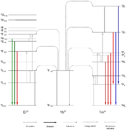
The co-doping with another lanthanide, a so-called sensitizer, can greatly enhance the upconversion efficiency. These additional dopants ideally feature a significantly larger absorption cross section than the activators whose cross section is generally low[28, 63]. The lanthanide ion ytterbium (Yb3+) with only one excited state may not be of use for upconversion as an activator but this lack of any emitting energy levels predestines it as a highly effective sensitizer. Additional to its high absorption cross section[64, 65] the energy gap between its two energy levels 2F7/2 and 2F5/2 perfectly matches the excitation photon energy ( = 980nm) as well as the energy necessary to promote electrons of the activators Er3+ and Tm3+ to their respective emitting energy level.[52, 65, 66]
This condition allows a highly efficient energy transfer to these activators and thus greatly strengthens the probability of the ETU process. In contrast to the activators, the concentration of the sensitizers can be chosen significantly higher, around 15-25% of the overall lanthanide concentration in the material.[28, 60] The resulting upconversion luminescence is by far stronger compared to materials doped with only an activator due to the much higher efficiencies of the ETU and PA process. According to these unique characteristics the doping with both ytterbium (Yb3+) and erbium (Er3+) or thulium (Tm3+) leads to the most efficient upconverting materials known so far. The proposed upconversion process for NaYF4
doped with ytterbium (Yb3+) and erbium (Er3+) or thulium (Tm3+) is given in Figure 2.3.
The upconversion process with erbium as the activator results in emissions in the green and red spectral range. The transitions 2H11/2 - 4I15/2, 4S3/2 - 4I15/2, and 4F9/2 - 4I15/2, are two photon processes and are responsible for the green emissions at 520 nm or 540 nm and the red emission at 655 nm.[67-74]
Thulium-doping leads to an upconversion luminescence located at 450 nm, 475 nm, 646 nm, 696 nm, and 800 nm. The blue emissions corresponding to the 1D2 - 3F4 and the 1G4 - 3H6 transitions are based on a 4- and 3-photon process. The emissions in the red and near infrared range rely almost
2. BACKGROUND 9
exclusively on 2-photon processes and are assigned to the 3F2 - 3H6, 3F3 - 3H6, 3H4 - 3H6 transitions, respectively.[28, 29, 67, 68, 74, 75]
An exception is the 1G4 - 3F4 transition also resulting in an emission at 646 nm but relies on a 3-photon process.[68, 74-76]
The magnitude of luminescence at the different wavelengths strongly differs with the near infrared emission at 800 nm being the strongest and the red emissions at 646 nm and 696 nm being the weakest.[61, 74, 75, 77, 78]
Figure 2.3 Upconversion process with ytterbium as the sensitizer and erbium (left) and thulium (right) as the activators.
2.1.3 Upconversion in the Nanometer Scale
For three decades after the first studies, the practical application of upconversion was limited to the use of the bulk material in form of films, glasses or fibers. Its use was a main focus of research especially in regard of optical devices including solid-state lasers[46, 68], lighting sources[68, 79], and temperature sensors[30, 48]. Thereafter, finally a growing research effort for the development of efficient synthesis routes emerged to obtain upconverting materials in the micro- or nanometer range. Coprecipitation[76, 80, 81]
, thermal decomposition[82], solvothermal synthesis[83-85], and sol-gel processing[86] yield upconverting nanoparticles (UCNPs). The nanoparticle characteristics including size, monodispersity, shape, surface functionality, or phase modification strongly depend on the type of synthesis and its conditions. UCNPs with a hydrophilic surface, a prerequisite for bioanalytical applications, are obtainable directly during synthesis routes performed in aqueous media such as
hydrothermal synthesis[75, 87-90]
or sol-gel processes[38, 86] but these nanoparticles lack a narrow size distribution and a sufficient long term stability in dispersion. Therefore, the synthesis routes such as high-temperature coprecipitation[91-93] and thermal decomposition[82, 94-97]
carried out in high-boiling organic solvents like octadecene are favored, since they yield monodisperse and highly stable UCNPs.
The necessary control of the nanoparticle nucleation and growth during these synthesis routes is preferentially performed by the use of oleic acid which at the same time acts as capping agent on the surface to stabilize the nanoparticles in dispersion. Other potential agents are trioctylphosphine[98], trioctylphosphine oxide[95], N-(2-hydroxyethyl)ethylenediamine[45, 99, 100]
, or oleyl amine[101] but they play only a marginal role. Since all these favorable synthesis techniques result in a hydrophobic UCNP surface, subsequent modification steps to create a hydrophilic surface are indispensable for bioanalysis. A short overview on the most common technique is given in chapter 2.2.
Furthermore, UCNPs are the only choice to use upconversion luminescence as a detection signal in (bio)analysis, since an efficient interaction with biological targets with a high selectivity and sensitivity is only feasible on the nanoscale, the same size dimension as most biomolecules.
However, as the size of the upconverting material is reduced, surface-related effects are amplified due to an increasing surface-to-volume ratio. Most of these effects lead to a decrease of the upconversion emission intensity owing to an enhanced probability of non-radiative relaxation.
Energy migration via lanthanide ions from the nanoparticle center to the surface[56, 71], point defects of the crystal structure of the host material at the surface created by lanthanide doping[102-104], or the high vibrational states of surface-bound molecules matching the phonon states of the host material[103-105] can be considered as reasons or promoters of this detrimental impact on the upconversion luminescence. Especially water with its two O-H bonds show high vibrational states and thus has a stronger quenching effect than organic compounds being either solvent or capping molecules.[106-109]
There are some preventive measures against this quenching including the growth of an pure host material shell, a so called inert shell, on the UCNP surface[105, 106, 110, 111]
or the use of bulky or branched surface ligands[112]. While the first approach increases the distance between the doped lanthanide ions and capping molecules or disrupts the energy transfer to the surface[56, 71, 107, 113]
, the second option entails an improved shielding of the actual UCNP surface from solvent molecules, especially water in aqueous systems. The increase of the UCNP diameter is one of the most apparent options to improve the brightness. However, its benefit for bioanalysis is limited despite the consequent reduced impact of quenching effects on the detected luminescence signal. Especially smaller UCNPs are favorable as labels for bioanalytical applications since they exert only a very low influence on cellular structures. The cellular uptake mechanism such as endocytotic or paracellular
2. BACKGROUND 11
uptake also strongly depends on the particle size.[114-116] Consequently, a compromise between the brightness and the (bio)applicability of UCNPs has to be found.
In the last years the influence of nanomaterials on the environment and especially on the health of living organisms, the so called cytotoxicity, became more and more an object of interest both for researchers as well as for the general public.[117] The prominent example asbestos was a promising material for many different areas of application but showed a high cytotoxicity and carcinogenicity due to the presence of micron-sized fibers in the material that are easily released into the air by different processes.[118-120] Therefore, the determination of the cytotoxicity of UCNPs as a new nanomaterial is crucial for bioanalytical applications. Various investigation methods are applied including viability tests of cells[121-126] or small organisms[127] or in vivo investigation of the nanoparticle biodistribution[22, 123-125, 127, 128]
.
2.1.4 Luminescence Enhancement by Surface Plasmon Resonance
The enhancement of the upconversion luminescence is a key topic of research for the optimization of the synthesis and design of upconverting materials. However, nanosized dimensions and a high hydrophilicity of these materials are required, particularly in bioanalytical fields. These necessary nanoparticle characteristics lead to enhanced surface-related effects like quenching which strongly reduces the achievable brightness, as described earlier. In addition to the coating with a shell made of host material, either undoped or doped[129], other materials proved to be useful to both provide a good shielding of the nanoparticle surface from quenchers and to strengthen the emission intensity.
The plasmonic enhancement of the upconversion luminescence by noble metals is a method whose usage for bioanalysis just recently became the focus of enforced studies. Surface plasmon resonance describes the coherent oscillation of valence electrons of a metal induced by light.[4, 117, 130-132]
This phenomenon has been extensively employed as an analytical measurement signal in the past, especially with the noble metal gold, both in the form of surface layers or nanoparticles.[9, 133-136]
The enhancement of luminescence by this phenomenon is well known for different organic fluorophores[137-141] and quantum dots[142, 143]
if the metal surface is in close proximity to these luminescent species with the right spatial distance. This rise in intensity can be attributed to an increased radiative rate of the emission or a strengthening of the excitation intensity. These effects can also occur simultaneously and thus combine their impact on the upconversion luminescence.[26]
Recent studies showed that this effect also has a beneficial impact on the upconversion luminescence of UCNPs. There are three major structural designs used during these investigations (Figure 2.4) to take advantage of this enhancement[26]: (a) the deposition of UCNPs on a gold or silver
surface[31, 144, 145]
; (b) the attachment of metal nanoparticles on the UCNP surface either by electrostatic interactions[146-152] or by their direct synthesis on the UCNP surface[126, 146, 153, 154]
; (c) the design of a nanoparticle structure with a UCNP/metal core, a metal/UCNP outer shell, and a spacing layer of silica[155-159].
Figure 2.4 Structural designs for plasmon enhancement: (a) deposition of UCNPs on a gold surface with a spacer layer, (b) attachment (top) or direct synthesis (bottom) of gold nanoparticles on the UCNP surface mediated by gold-specific binding sites, (c) coating of an UCNP (left) or gold nanoparticle (right) with a spacer shell on the surface and a second shell made of gold or UCNP material.
All these options lead to an increase of the upconversion luminescence, partially by extremely high factors, but a thorough and efficient optimization of the design is indispensable for such results.
While the enhancement obtainable by (a) and (c) depends first and foremost on the optimal distance between metal and UCNP, a precise tuning of the gold nanoparticle surface or the synthesis parameters for (b) is crucial for a successful attachment. All these factors place high demands on the synthesis reproducibility and accuracy of the different structural designs.
In contrast to these enhancement studies, there is also evidence that the surface plasmon resonance may lead to a decrease of the overall upconversion emission.[26, 146, 155, 160]
This phenomenon can be reduced to either a resonance energy transfer from the upconverting material to the noble metal or absorption of the upconversion emission by the noble metal.[26, 161] The determination of the quantum yield can indicate if an enhancement or a quenching of the upconversion luminescence is prevalent and may be informative of the nature of the associated causes mentioned before. There are well established methods for fluorophores and quantum dots to obtain this information.
However, an unambiguous cause assignment is complicated for UCNPs due to the non-linearity of the upconversion process and thus its dependency on the excitation density.[26, 161] Nevertheless, it is unquestioned that a direct contact or a too close proximity of UCNP and metal strongly promotes luminescence quenching by SPR which again reinforce the necessity of an optimized structure design including a suiting spacer component.
2. BACKGROUND 13
2.1.5 Advantages of Upconverting Nanoparticles for Bioanalysis
Nowadays, organic fluorophores and quantum dots are well established and common luminescent tools for bioanalytical applications. Despite their vast use as probes, labels, or sensor parts they have quite a few intrinsic characteristics which are adverse to their applicability in biological systems. The photophysical and material-related nature of UCNPs addresses several of these drawbacks and thus either eliminates or at least weakens their impact on any potential application. The first advantage of UCNPs in this regard is at the same time one of the most fundamental and representative aspects of the upconversion process: the use of near infrared light as the excitation source. While fluorophores and quantum dots need high-energy UV/VIS irradiation to show luminescence, UCNPs emit visible light upon the absorption of NIR photons, mostly with a wavelength of 980 nm.
The near infrared spectral range between 750 nm and 1000 nm is often referred to as the "optical window"[18, 73, 77, 78, 102, 162-165]
whose limit definition can vary about 100 nm. Photons of these wavelengths show only low interaction with biological systems in contrast to UV photons[166] entailing the following benefits of NIR light for bioanalytical applications. Biological species, above all biomolecules such as proteins or oligonucleotides, show no fluorescence under NIR excitation preventing the occurrence of autofluorescence[5, 167-169]
which is one of the major problems of the use of UV light in bioanalysis. This virtually zero background fluorescence allows studies with higher signal-to-noise ratio and thus improves the sensitivity and/or resolution of the respective application.[22, 95] Also hardly any photodamage against biological samples by NIR excitation is detectable due to its low energy content. Local thermal damaging, however, may occur due to heating effects[170, 171]
. A shift of the excitation wavelength from 980 nm to 800 nm realizable by an additional doping with the sensitizer neodymium (Nd3+) can partly avoid this effect owing to a lower absorption coefficient of water at this wavelength[171, 172]. Furthermore, the penetration depth of NIR light is by far greater than for UV- or visible light ranging from a millimeter to several centimeters[173-176] instead of a few millimeters at most[166, 177, 178]
and thus enables the excitation of labels or probes even in subjacent tissue layers[179].
However, since the emission bands of UCNPs are primarily located in the visible range, i.e. outside the "optical window", absorption and scattering phenomena caused by biological species also start to influence the detectability of the luminescence emission signal.[180] An exception is the emission of thulium-doped materials at 800 nm which falls in the optical window and thus can be detected with a relatively high sensitivity.[49, 106, 162]
Moreover, there is a significant advantage of the upconversion mechanism regarding the NIR excitation source usable compared to other Anti-Stokes processes.
Techniques relying on two-photon absorption, for example, need quite sophisticated and high-cost
excitation sources with very high power density (106-109 W cm-1) that are predominantly utilized in a pulsed mode to avoid sample damages.[28, 87] In contrast, the use of simple and inexpensive continuous-wave laser diodes as the excitation source with a comparatively low power output density (1-103 W cm-1) is both sufficient to induce upconversion and benign for most biological samples.[28, 68, 87, 106, 179]
Luminescent technologies aiming at a long term detection of the target require probes or labels that are compatible with sustained or brief but frequent irradiation intervals and give a continuous signal during excitation. Common organic fluorophores are susceptible to photobleaching, i.e. molecular damages due to intense or prolonged excitation.[20, 181-186]
Although quantum dots hardly photobleach over time[3, 28, 187-191]
, they show an intermittent emission behavior during excitation, so called
"blinking"[4, 133, 192, 193], with a dwell time in the dark state of up to several seconds[2, 3]. In contrast, the upconversion emission remains stable without any indication of "blinking".[168, 194, 195]
It is also unaffected by NIR irradiation over a longer period of time[23, 168, 171, 194-200]
since the luminescence originates from 4f atomic orbitals instead of molecular orbitals[121, 151, 196]
. Therefore, UCNPs surpass both organic fluorophores and quantum dots as luminescent labels or probes in long-term studies.[26, 91, 198, 201]
The emission bands of UCNPs, more precisely their respective wavelength, their width and their number, also result in several features favorable for bioanalytical applications. The emission wavelengths remain unaffected by any changes in UCNP diameter[202], whereas the absorption and emission spectra of quantum dots are strongly size dependent[6, 83, 203-205]
. The emission bands with a width of approximately 10 nm[206, 207]
are extremely sharp and are also well separated from the excitation peak owing to a large Anti-Stokes shift[79, 177]. In contrast, the broad emission bands of organic fluorophores have only a quite small Stokes shift[76]. The most striking characteristic of the upconversion emission spectra, however, is the presence of more than one emission band which enables the use of UCNPs in several important applications. Firstly, the assignment of a specific luminescent code to a sample, also called encoding, allows its direct identification for example in high throughput screening or combinatorial chemistry, especially important in the submicron domain where a purely spatial sample differentiation is impossible.[175, 182, 208, 209]
Secondly, a ratiometric measurement exploits the presence of an unaltered upconversion emission as an intrinsic reference to take drifts and fluctuations of the signal into account and to cancel out their influence on the evaluation.[163, 179, 208, 210, 211]
Thirdly, multiplexing, i.e. the simultaneous detection of several targets in one system[26, 105], can be realized with UCNPs, since the emission wavelengths of nanoparticles with different dopings are spectrally well separated and thus allow an unambiguous detection signal differentiation.[83, 91, 212-219]
2. BACKGROUND 15
As has been mentioned earlier, a low cytotoxicity and thus a high biocompatibility are decisive for the use of luminescent nanomaterials in bioanalysis. Beside its many favorable characteristics, one of the major disadvantages of quantum dots is their harmful effect on biological systems. The most popular material for the quantum dot synthesis, CdSe, is highly susceptible to (photo)oxidation initiated by either air or UV irradiation necessary for excitation.[3, 4, 204, 220, 221]
The heavy metal ion Cd2+
released by this process has been found to be extremely cytotoxic[204, 220-222]. Although this release can be reduced by the growth of a shell made of a material either less susceptible to (photo)oxidation like ZnS or CdS[3, 220, 221, 223]
or inert like silica[224], long-term studies are indispensable to prove a potential improved biocompatibility and thus applicability in bioanalysis[2, 192, 204]
. In contrast, most host materials of UCNPs have a low cytotoxicity as has been shown for ligand-free UCNPs made of Y2O3 [79]
. However, since hardly any UCNPs without any surface ligands or shells are used in bioanalytical applications, the surface composition is most decisive in this regard provided no detachment or degradation of the surface occurs during application. The range of investigated surface functionalities includes among others small molecules such as citrate[123], polymers such as polyacrylic acid (PAA)[57, 125, 225]
, polyethylenimine (PEI)[178], or polyethylene glycol (PEG)[166], biomolecules such as folic acid[199] or oligonucleotides[126], and additional shells such as pure or silanized silica[121, 128, 183, 184, 226]
. All these studies give evidence of a low cytotoxicity independent of the surface rendering UCNPs suitable for bioanalytical applications.
2.2 Surface Modifications of Upconverting Nanoparticles
The hydrophobic character of UCNPs originating from the reaction conditions during most favored syntheses is one of the major challenges that have to be overcome for UCNPs being usable as labels or probes in bioanalysis. Additionally, any surface changes should not affect the desired high monodispersity and long-term dispersion stability obtained after a research-intensive and time- consuming process of synthesis variations and optimizations. Therefore, modification methods have to be found that yield modified nanoparticles void of any aggregation tendencies. The countless publications and the magnitude of excellent reviews[53, 91, 169, 182, 207, 226-228]
about the surface modification and functionalization of UCNPs from different point of views clearly shows the elevated efforts of research groups in this aspect of UCNP studies. Therefore, only a short insight in a few modification options that were used during this thesis is given in this chapter.
2.2.1 Silica Coating
Silica is one of the most popular materials in nanoscience to create nanoparticles[7, 8, 229-237]
or an additional shell on a foreign nanoparticle surface including quantum dots[6, 224, 238]
, metal oxide nanoparticles[181, 239-243]
, noble metal nanoparticles[131, 132, 244, 245]
, or UCNPs[128, 165, 183, 184, 246]
. This favoring of silica in nanosciences, especially in regard to surface coating with an adequate layer thickness, can be attributed to its high chemical inertness[6, 229, 241, 243]
, hydrophilicity[235, 236]
, and the increase of the nanoparticle dispersibility in aqueous systems[8, 78, 80, 128, 165]
. The high transparency[247]
of silica, a premise for its use as a modification material of luminescent compounds, is particularly pronounced in the NIR region enabling an unaltered excitation of silica-coated UCNPs by light at 980 nm or similar wavelengths[51, 248]. Additionally, the upconversion emission in the visible range is only slightly affected by the coating due to scattering[249]. The high biocompatibility[165, 184, 229, 250]
, non-toxicity[231, 239, 250]
, and potential prevention of non-specific binding[78] of biological species justifies the popular use of silica in diverse biological applications as a coating material. Another more application-related advantage of silica is its potential functionalization with a well established and versatile process, the so called "silanization", which will be described in chapter 2.2.2.
There are a great number of methods to cover nanoparticles with a silica shell that are primarily based either on the Stöber process or the microemulsion technique. Although the Stöber process was one of the first synthesis routes for pure silica nanoparticles[7], its applicability can be extended by respective modifications to also grow a silica shell on a hydrophilic nanoparticle surface.[10, 51, 80, 251]
However, since most synthesis routes for UCNPs nowadays yield nanoparticles with a hydrophobic surface, the microemulsion technique (Figure 2.5) clearly is more important in research and application than the Stöber process for the growth of a silica shell on the UCNP surface.
Figure 2.5 Scheme of the microemulsion process in three steps: (1) the formation of reversed micelles in an organic solvent by the detergent encompassing an aqueous ammonia solution, (2) Diffusion of TEOS molecules into the micelles and their hydrolysis initiating the growth of a silica shell, (3) growth of the silica shell by further TEOS hydrolysis until complete consumption.
2. BACKGROUND 17
The central element of this technique is a reversed micelle formed by a detergent in an organic solvent, e.g. cyclohexane. An added tetraalkyl silicate like tetraethyl orthosilicate (TEOS) is hydrolyzed by an aqueous ammonia solution inside of these nanoreactors and forms the silica shell on the UCNP surface by polycondensation. The original hydrophobic surface ligands remain in the organic solvent. The amount of detergent defines the micelle size which should be just slightly larger than the nanoparticle to ensure an inclusion of only a single UCNP in the reversed micelle.[247]
Additionally, the thickness of the resulting silica shell can be adjusted by the amount of the tetraalkyl silicate added[56, 244, 252]
. This parameter is especially important for distance-dependent phenomena and their use in (bio)analytical applications. A relatively thin layer, for example, is required to ensure an efficient luminescence resonance energy transfer (LRET) from a UCNP to surface-bound dyes[78]. Although a coating of UCNPs by a silica shell introduces a variety of advantageous characteristics, it may also be responsible for enhanced aggregation of the modified UCNPs. As mentioned before, the micelle size sets the number of UCNPs encapsulated in its cavity and thus multicore encapsulation by silica may occur if the detergent concentration is inadequately low resulting in larger or non-spherical micelles.[240, 247, 253]
This incorporation of more than one UCNP in a silica shell leads to a higher polydispersity of the nanoparticle dispersion[254] which counteracts the optimization efforts for synthesis routes yielding highly monodisperse UCNPs. Additionally, dispersions of silica-coated UCNPs can show an increased aggregation tendency due to the large surface area and hydrodynamic radius of the modified nanoparticles.[8, 166] The choice of the dispersant composition strongly influences this tendency and an optimization is crucial for the respective silica-coated nanoparticle with a special focus on pH and ionic strength of the dispersant.[69, 255] An easier and more favorable way to reduce aggregation of the silica-coated UCNPs in dispersion is a functionalization of the silica shell by silanization.
2.2.2 Silanization
For many bioanalytical applications, UCNPs have to be equipped with functionalities allowing a directed labeling, also called targeting, or a selective recognition of the respective analyte.
Biomolecules like oligonucleotides, proteins, or antibodies are capable to meet these functions and can be easily attached to a silica surface by physical adsorption. However, a higher control over number and orientation of bound biomolecules is desired for most applications. Covalent binding processes provide a good regulation of the final number of biomolecules by preset binding sites and a high accessibility of the biomolecule's active site. Furthermore, covalently bound functionalities cannot detach from the nanoparticle surface during application. Consequently, a high control over the biomolecule attachment can be exerted by these processes. The silanol groups are not accessible
for the direct binding of biomolecules by common linkage reactions and thus a modification with suitable functional groups by silanization is indispensible for the use of silica-coated UCNPs in bioanalysis. This functionalization can be performed either subsequently or simultaneously to the silica shell growth. During the silanization process the alkoxy groups of a silane with the desired functionality are hydrolyzed and condensate to the silanols on the silica surface.
Amine and carboxylic functionalities are among the most popular binding sites for the attachment of biomolecules due to their great number in biomolecules and the well-established and understood activation with N-hydroxysuccinimide (NHS) and 1-ethyl-3-(3-dimethylaminopropyl) carbodiimide (EDC).[20, 248, 256]
The positive charge of amines present at physiological pH leads to strong aggregation due to the strong electrostatic attraction of amines with the partly negatively charged silanol groups of the silica shell. A conversion into carboxylic acids via anhydrides[218] or the simultaneous silanization with longer-chained or bulkier silanes with negative or neutral head groups[8] are two options to minimize this attraction. In contrast, UCNPs functionalized with carboxylic acids are well stabilized in dispersion by electrostatic repulsion due to the intrinsically negative charge of these groups at physiological pH.
Although the abundant presence of amines and carboxylic acids in biological systems can be favorable for a magnitude of bioanalytical methods, their selective labeling in vitro or in vivo becomes extremely complicated or even impracticable. Functionalities with a high bioorthogonality prove to be convenient substitutes. They enable a binding process which does not interfere or interact with biological processes or species[11] and can still be simply performed with a high selectivity. The bioorthogonal organic azides and terminal alkynes, the components of the copper(I)-catalyzed Huisgen cycloaddition[257-259], can easily be brought onto a surface of silica-coated UCNPs by silanization and are highly selective binding sites for (bio)molecules carrying the complementary functionality.
A way to alter the optical properties of UCNPs takes advantage of the simple and reliable silanization process by binding fluorophores to the silica surface.[183] A radiative or non-radiative energy transfer from the UCNP to the bound fluorophore under NIR irradiation[92] enables the tuning of the UCNP emission and thus the creation of nanolamps with an emission spectra customized for the respective application. In contrast, the additional excitation of the fluorophore with the respective UV/VIS light provides a downconversion emission usable as an additional detection signal for multimodal readout.
Furthermore, this emission can also act as a signal substitute for the upconversion emission. This surrogate detection signal is especially necessary if no instruments with a NIR excitation source for UCNPs are available and applications, for example the imaging electrophoresis gels[260], are thus strongly complicated or even impossible.
![Table 4.2 TEOS and CEST volumes needed for adapted synthesis from literature with a correction factor of 2.06 V TEOS V CEST [µL] [µL] Literature [248] 20 20 Adaption 41 41 Actual used 46 46](https://thumb-eu.123doks.com/thumbv2/1library_info/4131977.1552118/71.892.116.760.714.946/volumes-adapted-synthesis-literature-correction-literature-adaption-actual.webp)