Synthesis and Characterization of Liposomes with Controlled Surface Design
for Analytical Applications
Dissertation zur Erlangung des Doktorgrades der Naturwissenschaften (Dr. rer. nat.)
an der Fakultät Chemie und Pharmazie der Universität Regensburg
Deutschland
vorgelegt von
Carola Hofmann
aus München im Jahr 2019
Die vorliegende Dissertation entstand in der Zeit von März 2015 bis Juli 2019 am Institut für Analytische Chemie, Chemo- und Biosensorik der Universität Regensburg.
Die Arbeit wurde angeleitet von Prof. Dr. Antje J. Bäumner.
Promotionsgesuch eingereicht am: 24. Juli 2019
Kolloquiumstermin: 09. September 2019
Prüfungsausschuss:
Vorsitzender: Prof. Dr. Arno Pfitzner Erstgutachterin: Prof. Dr. Antje J. Bäumner Zweitgutachter: PD Dr. Hans-Heiner Gorris Drittprüfer: Apl. Prof. Dr. Rainer Müller
Für meine Söhne
Danksagung
Zu allererst möchte ich mich bei Prof. Dr. Antje J. Bäumner für die Möglichkeit zur Bearbeitung dieses spannenden Themas, für die ausgezeichnete Betreuung und Unterstützung während der gesamten Arbeit bedanken. Vielen Dank für die zahlreichen und hilfreichen Anregungen während der vielen, wissenschaftlichen Diskussionen.
Vielen Dank an PD Dr. Hans-Heiner Gorris für die Übernahme des Zweitgutachtens, Apl. Prof. Dr. Rainer Müller für die Übernahme der Aufgabe des Drittprüfers und Prof. Dr. Arno Pfitzner für die Übernahme der Funktion des Prüfungsvorsitzenden.
Darüber hinaus geht mein Dank auch an Joachim Rewitzer für die Unterstützung bei den ICP-OES- und ICP-MS Messungen meiner Proben.
Vielen Dank auch an Vanessa Tomanek für die Unterstützung bei den ICP-OES-Messungen meiner Proben, den Zeichnungen für meine Publikationen und für die Wiederholung und Optimierung einiger Messungen für das DNA Preconcentration-Projekt. Besonders bedanken möchte ich mich außerdem für ihre Unterstützung während des gesamten Bakterien-Projekts.
Darüber hinaus gilt mein Dank Susanne Dechantsreiter, Barbara Kaiser, Susanne Märkl und Clemens Spitzenberg für ihre Hilfe während ihrer Bachelorarbeit, Praktika oder WHK-Zeit.
Ebenfalls bedanken möchte ich mich bei meiner Arbeitsgruppe im „4.Stock“ für die tolle Zeit, Unterstützung und auch für die schönen Abende außerhalb der Arbeitszeit. Ich bedanke mich außerdem bei allen Kollegen und Kolleginnen des Instituts für die angenehme Arbeitsatmosphäre.
Außerdem möchte ich mich bei meinen Eltern, Helmut und Claudia, bei meinen zwei kleinen Schwestern, Christina und Iris, und bei meinem Mann, Joachim, dafür bedanken, dass sie zu jedem Zeitpunkt an mich geglaubt haben und mich immer mit unendlicher Geduld und liebevoller Fürsorge unterstützt haben.
Declaration of Collaborations
Most of the theoretical and experimental work presented in this thesis was conducted solely by the author. However, parts of the results were gained in collaboration with other researchers, which are stated in this section in accordance with §8 Abs. 1 Satz 2 Punkt 7 of the “Ordnung zum Erwerb des akademischen Grades eines Doktors der Naturwissenschaften (Dr. rer. nat.) an der Universität Regensburg vom 18. Juni 2009“.
Nanocontainers for Analytical Applications (Chapter 1)
The literature search and writing of the manuscript was done by the author. Axel Duerkop and Antje J.
Baeumner revised the manuscript. AJB is corresponding author.
Tethering Functionality to Lipid Interfaces by a Fast, Simple and Controllable Post Synthesis Method (Chapter 3)
The experimental work was carried out by the author. Guenter Roth provided the lipopeptide-biotin.
Thomas Hirsch, Axel Duerkop and Antje J. Baeumner contributed with strategic discussions. The manuscript was written by the author and revised by GR, TH, AD and AJB. AJB is corresponding author.
Optical Characterization of DNA-tagged Fluorescent Liposomes (Chapter 4)
Most of the experimental work was solely done by the author. The author also wrote this chapter. The experimental work was partly done at Abbott Diagnostics in Lake Forest, IL in cooperation with Qiaoqiao Ruan. Sergey Tetin, Thomas Hirsch, Axel Duerkop and Antje J. Baeumner contributed with strategic discussions. Antje J. Baeumner was the leader of this project.
Electrostatic Interactions of Cationic liposomes for Pathogen Detection (Chapter 5)
The author did most of the experimental work and wrote this chapter. Barbara Kaiser contributed with ζ-potential measurements of the E.coli cells and the fluorescence microtiter plate assay in different buffers as part of a research internship under the supervision of the author. Susanne Märkl contributed with measurements of the interaction of cationic liposomes and magnetic beads. Thomas Hirsch, Axel
Duerkop and Antje J. Baeumner contributed with strategic discussions. Antje J. Baeumner was the leader of this project.
Cationic Liposomes for DNA Preconcentration (Chapter 6)
The author did most of the experimental work and wrote this chapter. Cornelia Hermann contributed with the synthesis and characterization of one of the liposome batches. Vanessa Tomanek and Clemens Spitzenberg repeated and optimized some of the experiments under the supervision of the author.
Antje J. Baeumner contributed with strategic discussions and was the leader of this project.
1
Table of Contents
Summary ... 1
Zusammenfassung... 5
1 Nanocontainers for Analytical Applications ... 9
1.1. Introduction: General Information on Nanocontainers and Possible Applications ... 10
1.2. Synthesis and Surface Functionalization ... 10
1.2.1. Materials for Nanocontainers in (Bio)analysis – Synthesis and Characterization ... 11
1.2.2. Surface Chemistry ... 13
1.3. Relevant Features Supporting Nanocontainer Applications in (Bio)analysis ... 15
1.3.2. Signal Generation Through Controlled Release of Entrapped Molecules... 21
1.3.3. Release-Independent Signal Generation Strategies... 21
1.4. Analytical Applications of Nanocontainers ... 22
1.4.1. Bioassays ... 22
1.4.2. Chemosensors ... 32
1.4.3. Bioimaging and in vivo Applications ... 38
1.5. Conclusion, Future Perspectives and Challenges ... 40
Acknowledgements ... 42
References ... 42
2 Introduction and Structure of the Thesis ... 49
3 Tethering Functionality to Lipid Interfaces by a Fast, Simple and Controllable Post ... Synthesis Method ... 55
3.1. Introduction ... 56
3.2. Experimental ... 59
3.2.1. Materials ... 59
3.2.2. Methods ... 60
3.3. Results and Discussion ... 62
3.3.1. Insertion Method – Preparation and Characterization ... 62
2
3.3.2. Optimization of Insertion Conditions ... 64
3.3.3. Binding Behaviour of Functionalized Liposomes ... 65
3.3.4. Comparison of Lipopeptide-Insertion and Standard Modification ... 68
3.4. Conclusions ... 73
Acknowledgements ... 73
References ... 73
4 Optical Characterization of DNA-tagged Fluorescent Liposomes... 77
4.1. Introduction ... 78
4.2. Experimental ... 79
4.2.1. Materials ... 79
4.2.2. Methods ... 80
4.3. Results and Discussion ... 83
4.3.1. Liposome Preparation and General Characterization ... 83
4.3.2. Theoretical Calculations ... 85
4.3.3. Control Experiments and Imaging ... 86
4.3.4. Fluorescence Correlation Spectroscopy of Liposomes ... 89
4.3.5. Imaging of DNA-Tagged SRB Liposomes ... 91
4.4. Conclusions ... 96
References ... 96
5 Electrostatic Interactions of Cationic Liposomes for Pathogen Detection ... 99
5.1. Introduction ... 100
5.2. Experimental ... 101
5.2.1. Materials ... 101
5.2.2. Methods ... 102
5.3. Results and Discussion ... 105
5.3.1. Liposome Preparation and Characterization ... 105
5.3.2. Electrostatic Interaction with Magnetic Beads ... 106
5.3.3. Bacteria Quantification via Fluorescence Readout ... 108
3
5.3.4. Bacteria Quantification via Chemiluminescence Readout ... 114
5.4. Conclusions ... 117
References ... 117
6 Cationic Liposomes for DNA Preconcentration ... 119
6.1. Introduction ... 120
6.2. Experimental ... 121
6.2.1. Materials ... 121
6.2.2. Methods ... 122
6.3. Results and Discussion ... 125
6.3.1. Liposome Preparation and DNA Extraction... 125
6.3.2. Concepts for DNA Preconcentration with Cationic Liposomes ... 126
6.3.3. Optimization of DNA Preconcentration Using Centrifugation ... 129
6.4. Conclusions ... 132
References ... 133
7 Conclusions and Future Perspectives ... 135
Curriculum Vitae... 143
Publications ... 145
Presentations ... 147
4
1
Summary
Nanocontainers are being applied for many bioanalytical strategies, e.g. in bioassays, in chemosensors or for bioimaging and in vivo applications. They vary with respect to the materials used (such as mesoporous silica, polymers or proteins). These enable different unique characteristics mainly through their cavities or pores for the entrapment of numerous signaling molecules and their large surface area for the attachment of various surface tags. Of special interest is their signal amplification capability where the surface tags enable a specific analyte recognition as well as a controlled pore permeability.
Among all the different types of nanocontainers, liposomes excel by their relatively simple preparation and surface functionalization, their large inner cavity and variety of possible entrapped molecules, short assay response times due to an efficient lysis of the membrane, promising multimodal approaches for clinical diagnosis and therapy as well as by their natural biocompatibility and are thus of major interest in the field of (bio)analysis (Chapter 1).
For all applications in this area a careful control of the vesicle surface is necessary as it not only provides colloidal stability in complex aqueous solutions but also enables a specific binding to surfaces or the recognition of the analyte of interest. Therefore, the surface charge as well as methods for the introduction of specific functionalities were studied in detail in this thesis and the developed liposomes characterized and investigated towards their applicability as signal enhancers for the detection of bacteria or the preconcentration of DNA.
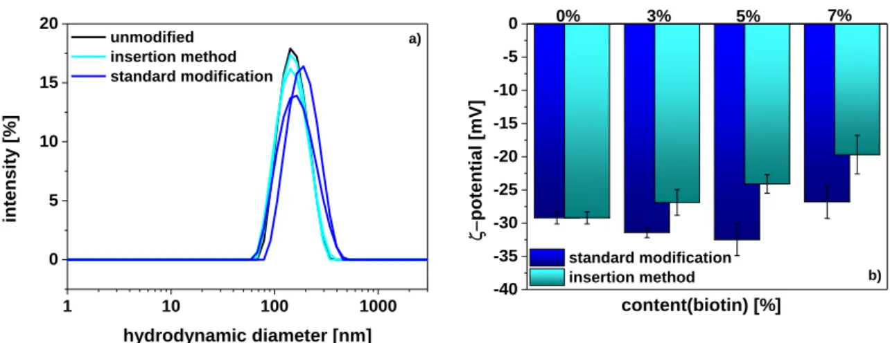
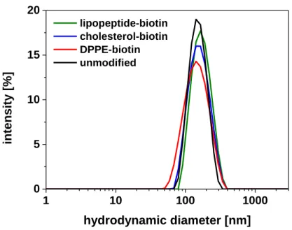
Standard methods for the surface functionalization of liposomes via covalent coupling post synthesis or modification directly during synthesis using functionalized lipids result in the production of numerous functionalized liposomes suitable for many applications. However, due to the need for elevated temperatures and organic solvents, time-consuming preparation and purification steps, low coupling yields, crosslinking or the decoration of the inner and outer leaflet of the bilayer, these methods are accompanied by a comparatively high loss of functionalities and are often unsuitable for fragile moieties. Moreover, large variations in the insertion efficiency, high batch-to-batch differences, and an incorporation limit of 4 mol% in case of direct modification with DPPE-biotin during synthesis were observed. Therefore, an alternative strategy for the insertion of different anchor molecules into dye-loaded liposomes composed of DPPC, DPPG and cholesterol was developed, and the tested molecules studied for their ability to effectively insert into the lipid bilayer and their binding functionality. The best system (lipopeptide-biotin) provided a fast, concentration-controlled functionalization up to 10 mol% in aqueous solution at room temperature and a reliable and quantitative binding to streptavidin with no effect on the analytical properties of the vesicles. Thus,
2 the vesicles only differ by the type or concentration of the functional moiety on the liposome surface which provides a huge potential for applications in the development of bioanalytical assays or for multi-analyte detection but can also be extended to other lipid-based nanomaterials and general analytical or pharmaceutical applications (Chapter 3).
Standard characterization of liposomes includes the determination of the vesicle size, ζ-potential and phospholipid concentration. However, there are several other parameters that can be investigated, such as the number of particles and surface tags or the specific binding of the vesicles to particles or surfaces. Therefore, ssDNA-tagged liposomes were prepared and characterized in detail using the standardly applied methods like DLS or ICP-OES as well as other optical methods. The number of liposomes was e.g. successfully determined via fluorescence correlation spectroscopy. Moreover, a hybridization assay with a fluorophore-tagged complementary oligonucleotide strand was developed to determine the number of ligands on the vesicle surface. In addition, fluorescence microscopy confirmed e.g. a successful purification, the specific binding of the functionalized vesicles to magnetic microparticles and enabled the imaging of the ideal lysis conditions for the applied liposomes (22 mM OG). Also the superior performance of liposomes over simple fluorophore-tagged oligonucleotides with respect to signal amplification was successfully demonstrated (Chapter 4).
The ability of liposomes to strongly enhance signals has already been exploited for many different bioanalytical assays. Here, mainly anionic lipid vesicles are applied as they prevent non-specific binding to most biological molecules or surfaces. The use of cationic liposomes is mainly restricted to pharmaceutical applications, such as in gene delivery. Therefore, a different approach for the use of dye-loaded cationic liposomes has been developed which is based on their electrostatic binding to the negatively charged surface of the model bacterium E.coli. Two different assay concepts were investigated and optimized to exploit this interaction for the detection of E.coli. The first concept is based on centrifugation and the second one on the immobilization of the bacteria to Poly-L-Lysin- coated microtiter plates. Sulforhodamine B-loaded liposomes enabled the analysis via fluorescence and yielded detection limits between 106 and 107 cfu ml-1. This was further improved by the
entrapment of the chemiluminescent marker m-carboxy-luminol. Here, detection limits of
~105 cfu ml-1 were achieved using the centrifugation-based assay. As most bacteria provide a negative surface charge, this method offers the possibility for a simple and universal detection of gram-positive and gram-negative bacteria. However, further optimizations will be necessary to achieve lower limits of detection (Chapter 5).
Besides their use for bacteria detection, preliminary studies also revealed the suitability of cationic liposomes for the preconcentration of genomic DNA. Preconcentration of analytes is a common tool in analytical chemistry and often described for DNA samples. Here, PCR, alcohol precipitation and
3 magnetic beads belong to the most common methods. As in case of bacteria also DNA is negatively charged due to its phosphate backbone. This enables the electrostatic attachment to the cationic vesicle surface. For separation two concepts were investigated. The first concept based on the separation via magnetic beads resulted in only low enrichment factors of ~2. The second concept was based on centrifugation. Here, an incubation time of only 5 min followed by centrifugation for 15 min at 15.000 g resulted in an efficient preconcentration of genomic DNA with enrichment factors up to 75. Thus, these preliminary studies show that cationic liposomes are a promising material in the field of DNA preconcentration and may be able to overcome some of the disadvantages of other methods, such as the need for organic solvents or chaotropic salts (Chapter 6).
4
5
Zusammenfassung
Nanocontainer werden für zahlreiche bioanalytische Strategien eingesetzt, z.B. in Bioassays, in Chemosensoren oder für die Bildgebung und in vivo Anwendungen. Sie variieren in Bezug auf die verwendeten Materialen (wie z.B. mesoporöses Siliziumdioxid, Polymere oder Proteine). Diese bestimmen die spezifischen Eigenschaften vor allem durch ihre Hohlräume und Poren für den Einschluss zahlreicher Signalmoleküle und ihre große Oberfläche für das Anbringen von Oberflächenfunktionalitäten. Ihre Fähigkeit zur Signalverstärkung ist hierbei von besonderem Interesse, wobei funktionelle Gruppen an der Oberfläche sowohl eine spezifische Erkennung des Analyten als auch eine kontrollierte Porendurchlässigkeit ermöglichen. Unter all den verschiedenen Arten von Nanocontainern stechen Liposomen durch ihre relative leichte Herstellung und Oberflächenmodifizierung, ihren großen inneren Hohlraum und die Vielzahl an einschließbaren Molekülen, kurze Assay-Antwortzeiten durch effizientes Lysieren der Membran, vielversprechende multimodale Ansätze für die klinische Diagnostik und Therapie, sowie durch ihre natürliche Biokompatibilität heraus. Sie sind daher von großem Interesse für die (Bio)analytik (Kapitel 1).
Für alle Anwendungen auf diesem Gebiet ist eine sorgfältige Kontrolle der Vesikeloberfläche nötig, da sie nicht nur die kolloidale Stabilität in komplexen, wässrigen Lösungen, sondern auch die spezifische Bindung an Oberflächen oder die Erkennung des Ziel-Analyten ermöglicht. Daher wurde in dieser Arbeit sowohl die Ladung der Liposomoberfläche als auch das Einführen spezifischer Funktionalitäten im Detail untersucht, die entwickelten Liposomen charakterisiert und in Bezug auf ihre Anwendbarkeit als Signalverstärker für die Detektion von Bakterien oder für das Aufkonzentrieren von DNA analysiert.
Standardmethoden für die Oberflächenfunktionalisierung über kovalente Kopplung nach der Synthese oder über direkte Modifizierung während der Synthese mit Hilfe von funktionalisierten Lipiden führt zur Herstellung von zahlreichen, funktionalisierten Liposomen, die für verschiedenste Anwendungen geeignet sind. Allerdings kommt es bei diesen Methoden durch die Notwendigkeit für erhöhte Temperaturen und organische Lösungsmittel, die zeitaufwendige Herstellung und Aufreinigung, niedrige Kopplungsraten, Quervernetzungen oder die Dekoration der äußeren und inneren Membranschicht zu einem verhältnismäßig großen Verlust an Funktionalitäten. Außerdem sind sie dadurch oft nicht für fragile Moleküle geeignet. Darüber hinaus wurden im Fall der direkten Modifizierung mit DPPE-biotin während der Synthese große Variationen in der Einbaueffizienz, hohe Unterschiede zwischen verschiedenen Ansätzen und eine Einbaugrenze von 4 mol% beobachtet. Daher wurde eine alternative Strategie für den Einbau unterschiedlicher Ankermoleküle in Farbstoff- beladene Liposomen aus DPPC, DPPG und Cholesterol entwickelt und die untersuchten Moleküle in
6 Hinblick auf ihre Fähigkeit effektiv in die Lipiddoppelschicht zu insertieren und auf ihre Bindungsfunktionalität hin analysiert. Das beste System (Lipopeptid-biotin) ermöglichte eine schnelle, konzentrations-kontrollierte Funktionalisierung bis zu 10 mol% bei Raumtemperatur in wässriger Lösung und eine zuverlässige, quantitative Bindung an Streptavidin, ohne dabei die analytischen Eigenschaften der Vesikel zu beeinflussen. Daher unterscheiden sich die Vesikel ausschließlich durch die Art oder Konzentration des funktionellen Restes an der Liposomoberfläche, was ein großes Potential für Anwendungen im Bereich der Entwicklung von bioanalytischen Assays oder der Multi- Analyt-Detektion ermöglicht. Darüber hinaus kann diese Methode auch auf andere Lipid-basierte Nanomaterialien oder auf allgemeine analytische oder pharmazeutische Anwendungen ausgeweitet werden (Kapitel 3).
Die standardmäßige Charakterisierung von Liposomen beinhaltet die Bestimmung der Vesikelgröße, ihres ζ-Potentials und ihrer Phospholipidkonzentration. Es gibt allerdings zahlreiche andere Parameter, die ebenfalls untersucht werden können, sowie die Anzahl an Partikeln und Oberflächenfunktionalitäten oder die spezifische Bindung der Vesikel an Partikel oder Oberflächen.
Dazu wurden ssDNA-markierte Liposomen hergestellt und im Detail charakterisiert, wobei sowohl Standardmethoden wie DLS oder ICP-OES zum Einsatz kamen als auch andere optische Methoden. Die Anzahl an Liposomen wurde beispielsweise erfolgreich über Fluoreszenzkorrelationsspektroskopie ermittelt. Darüber hinaus wurde ein Hybridisierungsassay mit einem Fluorophor-markierten, komplementären Oligonukleotidstrang entwickelt, um die Anzahl an Liganden auf der Vesikeloberfläche zu bestimmen. Außerdem bestätigte die Fluoreszenzmikroskopie beispielsweise eine erfolgreiche Aufreinigung, die spezifische Bindung der funktionalisierten Liposomen an magnetische Mikropartikel und ermöglichte die Bildgebung der idealen Lyse-Bedingungen für die angewendeten Liposomen (22 mM OG). Die überlegene Performance der Liposomen im Vergleich zu einfachen Fluorophor-markierten Oligonukleotiden in Bezug auf eine Signalverstärkung wurde ebenfalls erfolgreich gezeigt. (Kapitel 4).
Die Fähigkeit von Liposomen, Signale kräftig zu verstärken, wurde bereits für eine Vielzahl an bioanalytischen Assays genutzt. Dazu wurden hauptsächlich anionische Lipidvesikel verwendet, da sie eine unspezifische Bindung an die meisten biologischen Moleküle und Oberflächen verhindern. Der Einsatz von kationischen Liposomen ist hauptsächlich auf pharmazeutische Anwendungen beschränkt, wie z.B. für den Gentransfer. Daher wurde ein anderer Ansatz für den Einsatz von Farbstoff-beladenen, kationischen Liposomen entwickelt, welcher auf der elektrostatischen Bindung an die negativ geladene Oberfläche des Modell-Bakteriums E.coli beruht. Es wurden zwei verschiedene Assay-Konzepte untersucht und optimiert, um diese Wechselwirkung für den Nachweis von E.coli zu verwenden. Das erste Konzept basiert auf Zentrifugation und das zweite auf der Immobilisierung der Bakterien an Poly-
7 L-Lysin-überzogene Mikrotiterplatten. Sulforhodamin B-beladene Liposomen ermöglichten die Analyse über Fluoreszenz und erzielten Detektionsgrenzen zwischen 106 und 107 cfu ml-1. Das wurde weiter verbessert durch den Einschluss des chemilumineszenten Markers m-Carboxyluminol. Damit wurde mit Hilfe des Zentrifugation-basierten Assays eine Detektionsgrenze von ~105 cfu ml-1 erreicht. Da die meisten Bakterien eine negative geladene Oberfläche besitzen, bietet diese Methode die Möglichkeit für eine einfache und universelle Detektion sowohl von gram-positiven als auch von gram-negativen Bakterien. Allerdings werden weitere Optimierungen nötig sein, um noch niedrigere Detektionsgrenzen zu erreichen (Kapitel 5).
Neben ihrer Verwendung für die Bakteriendetektion, zeigen erste Studien auch die Eignung von kationischen Liposomen für die Aufkonzentrierung von genomischer DNA. Das Aufkonzentrieren von Analyten ist ein weit verbreitetes Mittel in der analytischen Chemie und wird häufig für DNA-Proben beschrieben. Dabei gehören die PCR, die alkoholische Fällung und die Verwendung magnetischer Beads zu den bekanntesten Methoden. Wie im Fall von Bakterien ist auch DNA aufgrund ihres Phosphat-Rückgrats negativ geladen. Dies ermöglicht eine elektrostatische Bindung an die Oberfläche der kationischen Vesikel. Für die Abtrennung ungebundener DNA wurden zwei Konzepte untersucht.
Das erste Konzept, welches auf der Abtrennung über magnetische Beads basiert, erzielte nur geringe Aufkonzentrierungsfaktoren von ~2. Das zweite Konzept basiert auf Zentrifugation. Dabei resultierten eine Inkubationszeit von nur 5 min, gefolgt von Zentrifugation für 15 min bei 15.000 g in einer effizienten Aufkonzentrierung genomischer DNA mit Aufkonzentrierungsfaktoren bis zu 75. Diese ersten Studien zeigen daher, dass kationische Liposomen ein vielversprechendes Material im Bereich der DNA-Aufkonzentrierung sind und möglicherweise einige Nachteile der anderen Methoden, wie z.B.
die Notwendigkeit für organische Lösungsmittel oder chaotrope Salze, beseitigen können (Kapitel 6).
8
9
1 Nanocontainers for Analytical Applications
Abstract
Nanocontainers such as mesoporous silica particles and polymersomes are versatile structures containing holes or pores used for the entrapment of small molecules and the introduction of specific functionalities. They are widely applied in drug delivery, biomedicine, bioreactors and analytical applications. In the latter, nanocontainers usually serve as amplification system. They are hence synthesized to entrap signaling molecules and to bear functional moieties at the outer surface, which in turn enable specific analyte recognition and control of the nanocontainer pore permeability. This review outlines the most important nanocontainer materials, discussing their synthesis, surface chemistry modifications and strategies for molecule entrapment. Their advantages, challenges and limitations in light of (bio)analytical applications are critically discussed in view of other common signal amplification strategies for different assay formats and various detection methods.
__________________________________________________________________________________
This chapter has been published.
C. Hofmann, A. Duerkop, A. J. Baeumner, Angew. Chem. Int. Ed. 2019, 10.1002/anie.201811821.
Author contributions:
The literature search and writing of the manuscript was done by CH. AD and AJB revised the manuscript. AJB is corresponding author.
10
1.1. Introduction: General Information on Nanocontainers and Possible Applications
Nanomaterials consisting of hollow structures, i.e. cavities or pores that are able to entrap small molecules are called nanocontainers. Liposomes are probably the most established type of nanocontainer to find successful application in analytical assays, drug delivery and skin care products.[1–5] Over the last couple of years, new concepts of nanocontainers have been proposed and successfully demonstrated in similar applications. These are made from a variety of materials such as mesoporous silica, polymers,[6] proteins,[7] DNA,[8] gold,[9] metal oxide[10] or carbon.[11] Common to all of these structures is their large surface area and inner volume, and their resulting superior ability to entrap molecules and enable various chemical surface functionalities. The pores and cavities enable the inclusion of molecules such as dyes, drugs, or nanoparticles but can also serve as confined environment for biological and chemical reactions. These unique features drive their application as nanoreactors,[12–15] nanostorage containers in batteries,[16] for environmental remediation[17] or waste water treatment,[18] as biomimetic structures,[19] for drug delivery,[6,20,21] bioimaging,[22,23]
theranostics[9] and in analytical assays[11,24,25] and sensors.[26,27]
Reviews on nanocontainers for sensor applications in the last 5 years are either specialized on one material [1,28] or cover only biomedical or nanoreactor applications, whereas a focus on analytical applications is either missing or kept to a minimum.[7,29] Earlier, in 2012, a more general overview on nanoporous materials for bioanalysis was published by Dai et al..[30] In general, for analytical applications, the cavities and pores are exploited for the entrapment of signaling molecules and the available large surface is functionalized for specific targeting toward the analyte of interest.
This review discusses the applied materials, their syntheses, advantages provided in analytical assays and provides an overview of nanocontainers published between 2013 and 2018 within this topic. The reader is directed to other reviews for specialized overviews on liposomes,[1,4] hollow or mesoporous structures,[29,31] preparation or functionalization of specific nanocontainers[6,14,32] or on applications such as drug delivery,[20,33,34] theranostics,[9] biomimetics,[35] in biomedicine[7,36] and nanoreactors.[12,13,15]
1.2. Synthesis and Surface Functionalization
Analytically relevant nanocontainers and nanocages are discussed in this review and cover mesoporous silica particles, liposomes, and polymer, gold, carbon, protein and metal oxide nanocontainers, respectively. Each material requires specific synthesis, purification and characterization methods. Common to all are strategies for surface chemistry and functionalization to obtain biocompatibility and render the surfaces suitable for analytical applications.
11
1.2.1. Materials for Nanocontainers in (Bio)analysis – Synthesis and Characterization
1.2.1.1. Liposomes
Liposomes are prepared by various methods.[2] The most common techniques are thin film hydration and reverse phase evaporation which produce reliably large amounts of liposomes. Lately, also microfluidic based approaches have been described, however, large-scale production is limited.[4]
Purification of liposomes is usually done by size exclusion chromatography or dialysis. For characterization of the vesicle morphology dynamic light scattering (DLS), zeta-potential measurements, transmission electron microscopy (TEM), scanning electron microscopy (SEM) can be applied. NMR, inductively coupled plasma-optical emission spectroscopy (ICP-OES), Bartlett assay or nanoparticle tracking analysis are used to obtain information on their molecular composition and overall concentration.[25,37–39] The size and lamellarity of liposomes depends on the method used for preparation and can be tuned via extrusion through polycarbonate membranes or sonication. Small lipid vesicles are around 50 nm in size, giant lipid vesicles can be as big as 1 µm.[3] Similarly, the nature of the lipids can tune the overall functionality and for example render liposomes cationic, anionic or non-ionic; can make them highly stable or include trigger molecules for lysis. Liposomes have been intensively studied as cell membrane models, for drug delivery and as signaling means in bioassays.[1]
In all cases, their large hydrophilic cavity, suitable for entrapment of a large number of signaling molecules, the easy surface functionalization, and their inherent biocompatibility makes them highly useful.
1.2.1.2. Mesoporous Silica Nanocontainers
Mesoporous silica nanocontainers are hollow particles with regular, ordered tunable pores. Diameters of 50-180 nm have been reported, the pore sizes are usually in the range of 2.3 to 3.1 nm.[40–42] Larger pores of 25 nm can be found for the entrapment of larger compounds like nanoparticles.[43] Possible synthesis strategies include soft and hard templating routes,[29,31] but a modified Stöber method is the most reported method. Here, a cationic, quaternary ammonium surfactant like cetyltrimethylammoniumbromid (CTAB) is mixed with a silicate like tetraethylorthosilicate (TEOS) in a mixture of water/ ethanol and ammonia. The surfactant forms positively charged micelles and electrostatically forms assemblies with the negatively charged silicate molecules. For functionalization often (3-aminopropyl)triethoxysilane (APTES) is added to the initial mixture.[32] Purification of mesoporous silica is accomplished by simple centrifugation or filtration followed by reflux in a mixture of ethanol and ammonium nitrate or HCl for complete CTAB removal. For characterization of the vesicles’ morphology and composition DLS, zeta-potential measurements, TEM, SEM, atomic force microscopy (AFM), x-ray diffraction (XRD) and Fourier-transform infrared (FT-IR) spectra are used.
12 Furthermore, nitrogen adsorption-desorption isotherms are used to obtain information on the surface area, porosity and pore size.[24,41,42]
1.2.1.3. Polymer Nanocontainers
Polymer nanocontainers are vesicles consisting of an ordered polymer membrane and an aqueous inner cavity. If amphiphilic, non-branched, synthetic block copolymers are applied for the formation the nanocontainers are called polymersomes.[15] They exist in a broad size range of 30-500 nm[12] but for analytical applications they are usually below 400 nm.[44–47] There are several ways to synthesize polymersomes and polymeric capsules.[15] In the bottom up approach the vesicles are built by self- assembly of block copolymers, which consist of a hydrophilic and a hydrophobic block. In the top down approach a thin polymer film is rehydrated to form the polymersomes. Another approach is based on the formation of vesicles during polymerization, which is often applied for polydiacetylene vesicles.
Moreover, phase separation techniques or template-based polymerization have been reported.
Purification of polymersomes is mostly done by dialysis, filtration or size exclusion chromatography.
For characterization of the vesicles’ morphology again DLS, zeta-potential measurements, TEM, SEM or AFM are frequently used.[23,45,47,48]
1.2.1.4. Protein-Based Nanocontainers
The most interesting material for protein-based nanocontainers for analytical applications is ferritin.
Ferritin is a spherically shaped protein with a diameter of 12 nm consisting of 24 subunits, which form an 8 nm big cavity suitable for the entrapment of signaling molecules. Of the two types of ferritin, the iron containing holoferritin and the iron free apoferritin,[20,49] the latter has been used most often for analytical applications.[49,50,51,52] Ferritin is commercially available and can be isolated, e.g. from horse spleen, some human organs, plants, fungi or bacteria.[20] Other protein-based nanocontainers or nanocages like heat-shock proteins, encapsulins or virus capsides have only seldomly been reported for analytical applications.[14] Purification and characterization methods rely heavily on standard biochemical approaches including dialysis, column chromatography, centrifugation, filtration, sodium dodecyl sulfate (SDS) polyacrylamide gel electrophoresis, absorbance measurements, HPLC, Bradford assay or Western blot analysis.[53]
1.2.1.5. Gold Nanocages
Gold nanocages are mostly cubic structures between 20 and 500 nm in size and consist of hollow interiors and porous walls with thicknesses between 2 and 10 nm. They are formed by galvanic replacement of Ag nanocubes with aqueous HAuCl4, and their hollow pores are created by dealloying of the Ag atoms.[9] Purification of gold nanocages is usually done by centrifugation. For characterization of the cage morphology TEM, SEM or energy-dispersive X-ray spectroscopy (EDS), is used as well as UV-vis for investigation of their optical properties.[26,54,55] In addition to using their entrapment
13 capabilities, gold nanocages are analytically also employed due to their localized surface plasmon resonance (LSPR) properties, which can be tuned from 600 to 1200 nm. These optical properties are influenced by the degree of HAuCl4 replacement, and additional incorporation of metals such as Pt or Pd into the walls of the nanocages.[9,20]
1.2.1.6. Carbon-Based Nanocages
Carbon nanocages can be cubic or spherical hollow structures with sizes up to 20 nm, shell thicknesses of 3-5 nm and pore sizes between 3 and ~14 nm.[11,56,57] For their synthesis carbon atom deposition, pyrolysis or hard and soft templating techniques have been described.[20] For example cage type mesoporous silica has been used as hard template and is removed after successful formation.[56,58]
Purification is mostly accomplished using size exclusion chromatography. For characterization of the cage morphology and composition TEM and FT-IR spectra have been reported and absorbance and fluorescence spectra for investigation of the optical properties.[11]
Also here, nitrogen adsorption-desorption isotherms are used to obtain information on the surface area, porosity and pore size.[57,59] Carbon nanocages are usually not applied for the entrapment of signaling molecules, instead, changes in the intrinsic fluorescence of the nanocage are exploited for sensing.[11,58]
1.2.1.7. Metal Oxide Nanocontainers
Metal oxide nanocontainers or nanocages are about 400 nm in size and consist e.g. of CuO, ZnO or Co3O4.[27,60,61] For synthesis the use of metal organic frameworks as template or precursor has been widely applied[61] mostly starting with zeolite imidazolate frameworks (ZIF).[60] After formation of the ZIF-templates core shell structures are formed using an etching and deposition method. By thermal annealing at 450 °C the final structures are formed.[60,62] Purification of metal oxide nanocages is usually done by centrifugation of the template before the final annealing. For characterization of the cage morphology and composition TEM, SEM, XRD, x-ray photoelectron spectroscopy (XPS) or FT-IR spectra are used, and again nitrogen adsorption-desorption isotherms to obtain information on the surface area, porosity and pore size.[10,27,61,63] Also metal oxide nanocontainers are usually not applied for the entrapment of signaling molecules, however, changes in the nanocontainer resistance are exploited for sensing.
1.2.2. Surface Chemistry
Stabilization under physiological conditions or even in simple aqueous buffer environment is a great challenge for nanoparticles and nanovesicles. In addition, the desirable surface chemistry of nanocontainers must be biocompatible and allow for targeted functionalization with tags or probes.[64,65] In some instances, such as with gatekeepers (see Chapter 1.2.2 below), modulating
14 surface tags also have to be added. Table 1.1 provides an overview of common surface functionalization strategies for the different nanocontainer categories.
Table 1.1. Surface functionalization strategies for the different nanocontainer categories.
.
Nanocontainer Biocompatibility/
stability in aqueous environment
Introducing
functionality Common functional groups
Common reaction mechanisms Ref.
Liposomes Naturally Functional lipids -COOH, -NH2, -
OH, -SH Covalent coupling [66], [22], [4]
Functional lipids Biotin, DNA,
folic acid Non-covalent conjugation, e.g.
biotin- streptavidin
[37], [4], [67]
Polymeric Hydrophilic
polymers Functional
polymers -COOH, -NH2, -
SH Covalent coupling [68]
Stimuli responsive polymers
- - [6], [14]
Protein based Naturally Naturally occurring amino acids
-COOH, -NH2, - OH, -SH, -N3, alkine
Covalent coupling [34]
Mesoporous
silica Polycondensation
with silanes -COOH, -NH2, - OH, -SH, -N3
Covalent coupling [14], [68]
Gatekeeper Aptamers;
proteins;
enzymes;
antibodies;
microspheres;
NPs
Electrostatic, covalent; Enzyme cofactor;
Inhibitor(anchor);
[43], [69] [70], [71], [72], [73]; [42]
Carbon based Naturally -COOH, -OH Covalent coupling [68]
Gold based Coating with biocompatible polymers
-SH Covalent coupling [9]
Coating with functional polymers
-COOH, -NH2, -
OH, -SH Covalent coupling [68]
Stimuli- responsive polymers
- - [20]
Gatekeeper Aptamers Electrostatic [55]
Active groups on the nanocontainer surface like carboxyl, hydroxyl, sulfhydryl or amino groups can be used for the attachment of biological receptors. Common covalent coupling techniques include 1- ethyl-3-(3-dimethylaminopropyl)-carbodiimide/N-hydroxysuccinimide (EDC/NHS) chemistry for
15 coupling of carboxyl and amino groups, Michael addition for the conjugation of thiol with maleimide groups, copper catalyzed click chemistry for conjugation of azide and alkyne groups or carbinolamine chemistry for the coupling of aldehyde and amine groups. Also, non-covalent conjugation is possible, e.g. by simple electrostatic interactions or a biotin-streptavidin system.[68]
For the conjugation of gatekeepers, reversible attachment is necessary to enable controlled release of entrapped molecules. Therefore, only weak bonding or non-covalent conjugation is applied, which responds to an external stimulus. This can be a change in pH or temperature, an enzymatic reaction or non-covalent interaction, e.g. with the analyte of interest. As gatekeepers often aptamers,[43]
proteins,[70] enzymes[71] or microspheres[74] are applied.
1.3. Relevant Features Supporting Nanocontainer Applications in (Bio)analysis
Nanocontainers consist of a hollow interior enabling the entrapment of small molecules and have a large surface area for the introduction of various functionalities. Some nanocontainers also possess pores for controlled mass transport in and out of the nanocontainer and in some instances gatekeepers that control this mass-flow. An overview of the features of nanocontainers can be found in Scheme 1.1.
Scheme 1.1. Schematic overview of nanocontainer features, possible entrapment molecules and signalling techniques and principles.
16 These features have been exploited and widely applied for analytical applications ranging from bioassays and biosensors to chemosensors and imaging. In bioassays or sensors, the analyte of interest is recognized by a suitable biological recognition element, e.g. antibodies, antigens, enzymes, receptors, cells, or DNA. This recognition must be convertible into a measurable, concentration- dependent signal.[75] Most sensitive bioassays employ hence a signal amplification strategy to obtain favorable signal-to-noise ratios at low analyte concentrations. Enzymes were the first highly successful signal enhancers used in immunoassays leading to the development of enzyme-linked immunosorbent assays (ELISAs) that are now gold standards in clinical diagnostics.[76] With the advancements in nanotechnology, various nanocontainers have been developed that significantly broadened and improved the possibilities of signal enhancement in bioassays. They have a large surface-to-volume ratio; their cavities or pores can be loaded with a huge amount of signaling molecules, and multiple receptors can be attached to their surface.[1,70] In contrast to enzymes they provide instantaneous signal amplification, can be more robust and can provide higher signal enhancement. An ideal nanocontainer must hence be stable under physiological conditions, not only during the bioassay but also for long-term storage, enable fast and simple surface functionalization and efficient loading with the desired signaling molecules. These can be e.g. small hydrophilic or hydrophobic molecules, proteins, enzymes, DNA probes, or nanoparticles.[1] For signal generation those molecules can either be released in a controlled manner using external stimuli[41] or the signal is generated without release of the entrapped molecules, e.g. in the case of enzymatic reactions inside the container, where the substrate can enter through permeable pores.[44] In literature most examples can be found for liposomes[25,37,66] and mesoporous silica nanocontainers[24,40,41] but also polymersomes,[45] protein,[49]
carbon[11] and gold-based nanocages[55] have been reported.
For bioimaging similar requirements have to be met. Here, the focus lies on the visualization and investigation of tissues, cells, tumor cells, subcellular structures, cellular processes, and interactions of molecules or quantification of ions or metabolites.[77] Therefore, the nanocontainers used for bioimaging usually carry contrast agents, which increase the signal in the areas of interest compared to the background.[78] Those contrast agents like fluorescent dyes, upconverting nanomaterials or magnetic iron oxide, find their applications in simple optical bioimaging, magnetic resonance imaging (MRI), computed chromatography, ultrasound or photoacoustic imaging.[78] Here, nanocontainers must demonstrate in vivo stability, biocompatibility, biodegradability and non-toxic properties,[33]
which has been achieved primarily with liposomes[22] and polymersomes,[23] but also with mesoporous silica[79] and lanthanide-metal organic framework (Ln-MOF) structures.[80]
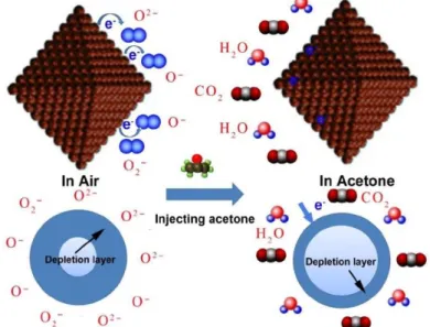
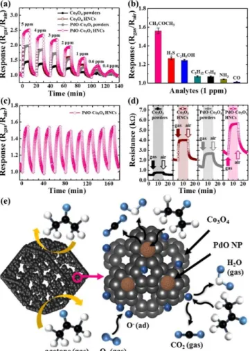
Furthermore, the high surface-to-volume ratio of nanocontainers and hollow pores are exploited for gas sensing, mostly using metal oxides.[27,60,61] Gas diffusion and mass transport to the surfaces are
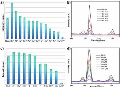
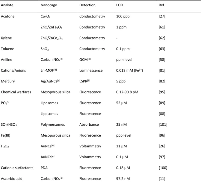
17 enhanced over non-porous macro surfaces[29] hence leading to increased sensitivity. Typically, reducing or oxidizing gases change the resistance of the nanomaterials, which is transduced into a concentration dependent electronic signal. Similarly, nanocontainers can be applied for metal ion sensing. Here the intrinsic properties of the nanocontainer changes upon contact with the analyte.[81] Most examples in literature can be found for hollow Ln-MOF[81] structures or gold nanocages.[82]
1.3.1. Classes of Entrapped Signaling Molecules and Detection Techniques
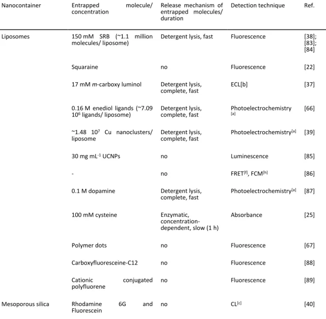
Nanocontainers can entrap several types of molecules. For analytical applications often signaling molecules like dyes, redox molecules or enzyme substrates are used, but also macromolecules such as proteins and DNA, or nanoparticles can be entrapped. Table 1.2 provides an overview of common entrapment molecules and their associated detection strategies.
Table 1.2. Overview on common entrapment molecules, release mechanisms, and associated detection strategies.
Nanocontainer Entrapped molecule/
concentration Release mechanism of entrapped molecules/
duration
Detection technique Ref.
Liposomes 150 mM SRB (~1.1 million
molecules/ liposome) Detergent lysis, fast Fluorescence [38];
[83];
[84]
Squaraine no Fluorescence [22]
17 mM m-carboxy luminol Detergent lysis,
complete, fast ECL[b] [37]
0.16 M enediol ligands (~7.09
106 ligands/ liposome) Detergent lysis,
complete, fast Photoelectrochemistry
[a] [66]
~1.48 107 Cu nanoclusters/
liposome Detergent lysis,
complete, fast Photoelectrochemistry[a] [39]
30 mg mL-1 UCNPs no Luminescence [85]
- no FRET[f], FCM[h] [86]
0.1 M dopamine Detergent lysis,
complete, fast Photoelectrochemistry[a] [87]
100 mM cysteine Enzymatic, concentration- dependent, slow (1 h)
Absorbance [25]
Polymer dots no Fluorescence [67]
Carboxyfluoresceine-C12 no Fluorescence [88]
Cationic conjugated
polyfluorene no Fluorescence [89]
Mesoporous silica Rhodamine 6G and
Fluorescein no CL[c] [40]
18
1.2 mM rhodamine B Gatekeeper, concentration-
dependent, slow (90 min)
Fluorescence [90]
0.2 mg mL-1 rhodamine B Gatekeeper, concentration- dependent, slow (90 min)
Absorbance [70]
1 mM methylene blue;
60 mg mL-1 methylene blue Gatekeeper, complete, slow (120 min);
Gatekeeper, concentration- dependent, slow (80 min)
SWV[d] [41];
[73]
80 µM Cy754 (~256
molecules/ particle) no Fluorescence;
Photoacoustic [91]
Glucose Gatekeeper,
concentration- dependent, slow (80 min)
Fluorescence [24]
1 M glucose; 350 mg mL-1
glucose Gatekeeper,
concentration- dependent, medium (20 min); Gatekeeper, concentration- dependent, medium (25 min)
Glucometer [42];
[74]
150 mM N,N-phenylenebis- (salicylideneimine)dicarboxylic acid
no Fluorescence [79]
50 mM CuSO4 Gatekeeper,
concentration- dependent, medium (30 min)
Photoelectrochemistry[a] [92]
0.08 mM [Ru(bpy)3]2+ Gatekeeper, concentration- dependent, slow (2 h)
ECL[b] [43]
0.16-0.5 mmol rhodamine B Gatekeeper, concentration- dependent, medium (30-35 min)
Fluorescence [93],
[69]
0.32 mmol rhodamine B Gatekeeper, concentration- dependent, slow (60 min)
Fluorescence [94]
BODIPY no Fluorescence [95]
Rhodamine 6G no Fluorescence [96]
AuNCs 100 µM rhodamine B Gatekeeper,
concentration- dependent, slow (1 h)
Fluorescence [55]
[Ru(bpy)3]2+ no ECL[b] [54]
- no LSPR[e] [82]
19
- no Voltammetry [26];
[97]
Polymer e.g. 50 mM CF, 50 mM SRB, 100 mM calcein, 330 mM sodium azide, 3 mM nile red
Enzymatic, complete,
medium (30 min) Fluorescence [45]
30 µM conjugated polymer no Fluorescence [44]
50 µg mL-1 nile red; 40 µg mL-1
nile red no; no FRET[f] [23];
[46]
0.4-1.5 mg mL-1 (~2400
UCNPs/ capsule) no Luminescence [47]
5 mM pyranine Redox-responsive, slow
(2 h) Fluorescence [48]
PDA[g] polymer no Fluorescence [98]
PDA[g] polymer no Absorbance [99]
1,8-naphthalimide-PDA[g]-
conjugate no Fluorescence [100]
52 mg mL-1 Cresol red Competitive
replacement, fast Absorbance [101]
Carbon NCs - no Fluorescence quenching [11]
- no QCM[g] [58]
Apoferritin 10 mM metal phosphates pH-responsive, no
release Square wave anodic
stripping voltammetry [52]
Lead/ cadmium phosphate Acidic, complete, fast
(5 min) Voltammetry [49]
1 mM CuSO4*5 H2O (~1200
CuNPs/ apoferritin) Acidic, complete,
medium (20 min) DPV[h] [51]
Metal oxide - no Conductometric [27]
- no Conductometric [61]
- no Conductometric [62]
- no Conductometric [63]
Ln-MOF - no Luminescence [81]
[a] Photoelectrochemistry: electrochemical current is produced by electron transfer from photoactive materials to semiconductor electrodes via light irradiation. [b] ECL: electrochemiluminescence. [c] CL: chemiluminescence. [d] SWV:
square wave voltammetry. [e] LSPR: localized surface plasmon resonance. [f] FRET: fluorescence resonance energy transfer. [g] QCM: quartz crystal microbalance. [h] DPV: differential pulse voltammetry. [g] PDA: polydiacetylene. [h] FCM:
fluorescence correlation microscopy.
Successful entrapment of the signaling molecules is only possible if properties like size or hydrophilicity match with the nanocontainers’ pores or cavities. The aqueous core of apoferritin can for example only be used for the entrapment of water-soluble compounds. For this the pH dependency of the
20 protein is exploited. At pH 2 it dissociates into its subunits and can be mixed with the entrapment molecules. Upon a rise in pH to 8.5 the protein reassembles into its native structure and encapsulation of the signaling molecules takes place.[1,7,51]
Liposomes provide naturally the possibility to be loaded with both hydrophobic and hydrophilic components due to their bifunctional structure.[38,67,68] Specifically, aqueous marker molecules can be entrapped in high concentrations within the inner cavity, while the membrane layer itself can be intercalated with hydrophobic components. Furthermore, the surface of the lipid bilayer can be functionalized with markers via covalent or adsorptive strategies.[1] The hydrophilicity of the cavity of polymersomes depends on the polymeric membrane. In case of di-block polymeric membranes hydrophobic molecules can be entrapped and hydrophilic molecules in the case of tri-block polymeric membranes.[13] In these cases the entrapment molecules are already added during vesicle formation.
In the case of liposomes and polymersomes, their larger cavities in comparison to other nanocontainers (with pore sizes mostly below 10 nm) enable the entrapment of large numbers of signaling molecules, and even larger molecules (proteins, DNA) or nanoparticles can be loaded. In all instances, entrapment conditions play a crucial role for efficiency, yield and maintaining nanocontainer and entrapped molecule integrities. This is most critical for the entrapment of proteins where denaturation, unfolding and aggregation must be avoided.[102] Therefore, mostly emulsion or rehydration methods under minimized harsh conditions have been applied for efficient protein encapsulation.[102,103] In the case of nanoparticle entrapment into aqueous compartments, surface modification of the particles is necessary to render their usually hydrophobic surface hydrophilic.[85]
Moreover, the hydrodynamic diameter of nanoparticles restrict their overall number to be included in the inner cavity of a nanocontainer so that most nanoparticles used are smaller than 10 nm.[47,85]
In the case of mesoporous silica nanocontainers the pore size and geometry can be tuned and can also be designed to bear either hydrophilic or hydrophobic characteristics.[12] For example, the addition of MTMS to the formulation during synthesis provides an ideal environment for loading hydrophobic dyes such rhodamine 6g and fluorescein.[40] Entrapment is usually conducted after synthesis either non covalently inside the pores or via covalent conjugation on the surface using e.g. dye-APS ((3- aminopropyl)triethoxysilane). The often occurring leakage of non-covalently loaded molecules[91] can be avoided by using gatekeepers.[24,41,42] These physically prevent the encapsulated material from leaking, and interestingly can also serve as target-responsive release systems. Here, the nanocontainer pores need to be big enough for the entrapment of the signaling molecules but at the same time small enough for a successful blocking of the pores.[41] Such analytically highly interesting gatekeepers have also been successfully adapted to gold nanocontainers.[55]
21
1.3.2. Signal Generation Through Controlled Release of Entrapped Molecules
Signal amplification of controlled release systems are based on the large amount of signal generating molecules entrapped inside the cavities or pores of a nanocontainer. Upon release, a signal is either obtained immediately or after reaction with molecules in the vicinity of the nanocontainer. For example, fluorescent dyes entrapped in self-quenching concentrations will emit fluorescence upon dilution in the outer media; electrochemical markers can react on electrode surfaces, enzymes can catalyze reactions etc.
Different mechanisms have been described for controlled release strategies. For example, the nanocontainers can react to external stimuli like temperature, pH or light irradiation.[6] Bushan et al.
demonstrated that by changing the pH from neutral and slightly basic conditions to acidic conditions the disassociation of apoferritin into its subunits takes place, which leads to the release of entrapped molecules.[7] In addition to the same external stimuli,[6] in case of polymeric nanocontainers also enzymes such as hyaluronidase have been used, which degrades specifically hyaluronic acid containing block copolymers.[45] While liposomes also react to external stimuli as described above, detergents are mostly applied in bioassays to lyse the lipid bilayer.[37,84] Somewhat slower release is obtained through enzymatic degradation using sphingomyelinase [25] or phospholipase.[104] For mesoporous silica and gold nanocontainers gatekeepers block the hollow pores. Here, the external stimulus is usually the target analyte, to which the gatekeeper binds more specifically than to the nanocontainer pores resulting in a concentration dependent release of signaling molecules.[42,55] A detailed review on this topic was published by the group of Sancenón et al. in 2016.[105]
1.3.3. Release-Independent Signal Generation Strategies
If no controlled release mechanism is applied, signal amplification is achieved by various strategies. In the case of liposomes, non-quenching concentrations of fluorescent dyes, absorption and refractive index can be detected optically, and size or weight acoustically or via impedance. Also here, the large surfaces and the larger inner cavities provide ultimate advantages. Gao et al. demonstrated that receptors are bound on outer and inner pore surfaces of gold nanocages and hence significantly enhance obtainable signals.[54] Yildiz et al. showed that enzymes bound inside permeable pores of the nanocontainers can react with substrates diffusing into the pores.[44] Similarly, in gas and metal ion sensing analyte molecules penetrate all pores of the metal oxide nanocontainers causing a change in resistance,[27] a change in the luminescence properties of Ln-MOF structures,[81] shifts in the LSPR spectrum of Ag/Au nanocages,[82] or quench the fluorescence of carbon-based nanocontainers.[11]
22
1.4. Analytical Applications of Nanocontainers
1.4.1. Bioassays
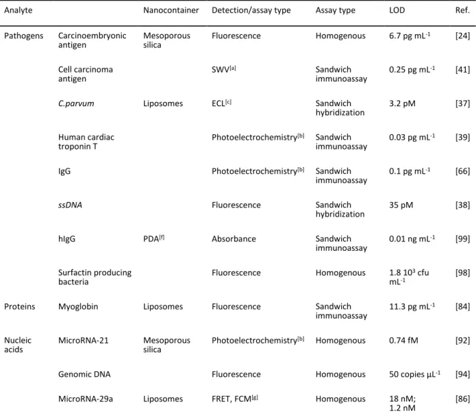
Table 1.3 provides an overview of analytes detected in recent years with the help of nanocontainers.
Assay types, detection methods and corresponding detection limits are provided as well in order to allow a comparison between the analytical performance of nanocontainers described. The sheer number and breadth of analytes studied signify that general bioassay-related challenges have been overcome in the last decade and that nanocontainers can further develop into new standard strategies also applied in commercial products.
1.4.1.1. Heterogenous, Controlled Release Bioassays
In heterogeneous assays nanocontainers are used similar to enzyme labels,[38,51] providing shorter assay times due to instant signal generation and in many instances enable very low limits of detection, as convincingly shown for liposomes, mesoporous silica particles and protein nanocages. For example, 1-methyl-1H-benzimidazole functionalized mesoporous silica nanocontainers were used for the development of a sandwich immunoassay for the detection of squamous cell carcinoma antigen.
Under neutral conditions, β-cyclodextrin functionalized AuNPs serve as gatekeeper for the entrapment of a large amount of methylene blue molecules in the nanocontainer pores. After sandwich formation with the specific antigen on a gold electrode the pH is lowered to acidic conditions, which results in gate opening and release of the signaling molecules. Square wave voltammetry (SWV) was applied for analysis and a detection limit of 0.25 pg mL-1 was achieved. This signal amplification strategy can compete e.g. with immunoassays based on chemiluminescent (CL) labeled antibodies and achieves even lower detection limits than enzymatic amplification.[41] Similarly, magnetic, mannose- functionalized mesoporous silica particles were used for the detection of aflatoxin B1 (AFB1).
rhodamine B was entrapped inside the pores and capped with concanavalin A (ConA) via weak interactions with mannose. The mesoporous silica particles were also used for the attachment of capture antibodies. AuNPs were functionalized with the enzyme invertase and the analyte AFB1. A competitive reaction between free analyte and the functionalized AuNPs took place. For signal generation, sucrose was enzymatically converted to glucose, which can also bind to ConA. This resulted in the displacement of mannose gatekeepers and release of the fluorescent dye. A limit of detection (LOD) of 8 pg mL-1 was achieved, which is up to 30 times lower than with commercially available ELISA kits.[90]
Although low detection limits can be achieved using such setups, heterogenous assays using gatekeepers for signal generation can be very time-consuming as the reported release times range from 90 to 120 minutes to ensure complete release of the guest molecules. Thus, careful evaluation of the perceived improvements over standard materials needs to be considered.