Synthesis, Characterization and Analytical Applications
Dissertation
zur Erlangung des Doktorgrades der Naturwissenschaften (Dr. rer. nat.)
an der Fakultät Chemie und Pharmazie der Universität Regensburg
vorgelegt von Stefan Balk aus Waldmünchen
Juli 2014
under the supervision of Prof. Dr. Burkhard König.
Submission of thesis: 17. July 2014
Date of colloquium: 22. August 2014
Board of Examiners: PD Dr. Richard Weihrich (Chairman) Prof. Dr. Burkhard König (1st Referee) Prof. Dr. David Diaz Diaz (2nd Referee) PD Dr. Rainer Müller (Examiner)
Regensburg, ………...
I would like to express my gratitude to Prof. Dr. Burkhard König for giving me the opportunity to do my Master and PhD theses on this very interesting and versatile topic under his supervision. His helpful advises, the great motivation and the financial support were essential contributions to my work. Furthermore I am very thankful to Prof. Dr. Uday Maitra, who supervised my PhD research internship at IISc. Bangalore and provided a great collaboration. For my defense I am deeply grateful to my second referee Prof. Dr. David Diaz Diaz, my third examiner PD Dr. Rainer Müller and the chairman PD Dr. Richard Weihrich. I want to acknowledge DFG, INDIGO network and DAAD, GDCh and all other founding organizations for financial support and numerous travel grants. A very big
“Thank You!” to Dr. Benjamin Gruber and Dr. Stefan Stadlbauer, who supervised my Master and Bachelor Thesis and contributed in a great part to my work in molecular recognition.
Thanks are also extended to Dr. Rudolf Vasold who helped me with the time-consuming HPLC measurements and the Fraunhofer Institute for the opportunity to use their Zetasizer. Moreover, I would like to mention the permanent staff of our group Viola Rappenegger, Dr. Petra Hilgers, Susanne Schulze, Britta Badziura, Regina Hoheisel, Simone Strauss und Ernst Lautenschlager.
I want to mention my friends and (former) lab mates Dr. Benjamin Gruber, Dr. Florian Schmidt, Tobias Lang, Michal Poznik and Melanie Hacker, they helped with everyday problems in lab and we had a great working atmosphere – Thank you! It was a great time.
Furthermore, I want to thank my colleagues and former members of the König’s group for the wonderful community also besides chemistry, the skiing trips, Chem-Cup, PhD parties and international evenings. Very big thanks also to my friends Sayantan Chatterjee and Stefanie Jäger and all members of Prof. Dr. Uday Maitra’s group for the great time we had together in India. Further thanks go to all the students who contributed to this thesis, namely: Vanessa Haas, Daniel Wutz, Simone Stark, Christian Hundshammer, David Wunderlich und Marleen Häring.
I would particularly like to mention my friends Christian Schäfer, Alen Lovric and Ines Hachani as well as my fellows from the first days at university Dr. Dennis Kühbeck, Tobias Lang, Steffen Pockes, Dr. Christian Wellner, Florian Meier, Wolfram Klosterhuber, Viktor Kais, Dr. Roland Linhardt, Florian Pielnhofer, Dr. Quirin Kainz and Paul Kohls. Thank you for the everlasting time we have had together!
wonderful relationship and her endless support.
Finally, my biggest thanks go to my parents Ingrid and Hubert, my sisters Anja and Bettina and my whole family – Thank you so much! Without their boundless support and never- ending encouragement this thesis would not have been written.
T HANKS
“Live as if you were to die tomorrow. Learn as if you were to live forever.”
Mahatma Gandhi
CHAPTER 1-DYNAMIC INTERFACE IMPRINTING:HIGH AFFINITY PEPTIDE BINDING SITES
ASSEMBLED BY ANALYTE-INDUCED RECRUITING OF MEMBRANE RECEPTORS ... 1
Introduction ... 2
Results and Discussion ... 3
Conclusion ... 8
Experimental Part and Supporting Information ... 9
Notes and References ... 42
CHAPTER 2- THERMALLY INDUCED MOLECULAR IMPRINTING OF LUMINESCENT VESICLES ... 45
Introduction ... 46
Results and Discussion ... 47
Conclusion ... 51
Experimental Part and Supporting Information ... 52
Notes and References ... 63
CHAPTER 3-TERBIUM(III)-CHOLATE FUNCTIONALIZED VESICLES AS LUMINESCENT INDICATORS FOR THE ENZYMATIC CONVERSION OF DIHYDROXYNAPHTHALENE DIESTERS ... 67
Introduction ... 68
Results and Discussion ... 68
Conclusion ... 72
Experimental Part and Supporting Information ... 72
Notes and References ... 84
SUMMARY ... 86
ZUSAMMENFASSUNG ... 87
ABBREVIATIONS ... 88
CURRICULUM VITAE ... 92
1
A
SSEMBLED BYA
NALYTE-
INDUCEDR
ECRUITING OFM
EMBRANER
ECEPTORSDynamic molecular recognition events at biological membrane receptors play a key role in cell signaling. We have prepared artificial membranes with embedded synthetic receptors which dynamically arrange and selectively respond to external stimuli like small peptide ligands.
This Chapter has been published:
B. Gruber, S. Balk, S. Stadlbauer, B. König, Angew. Chem. Int. Ed., 2012, 51, 10060–10063 (Hot Paper).
Author contributions:
BG performed binding studies and wrote the manuscript; SB synthesized compound Cu3 and performed FRET binding studies; SS synthesized compounds Cu4, Zn21, Zn22; BK supervised the project and is corresponding author.
2
Introduction
Molecular recognition between membrane-associated receptors and external ligands as stimuli is important for many biological processes.1-3 The dynamic formation of domains and the clustering of receptors in fluid membranes play a key role e.g. in signal trans- duction leading to highly specific binding of competing multivalent ligands.4-6
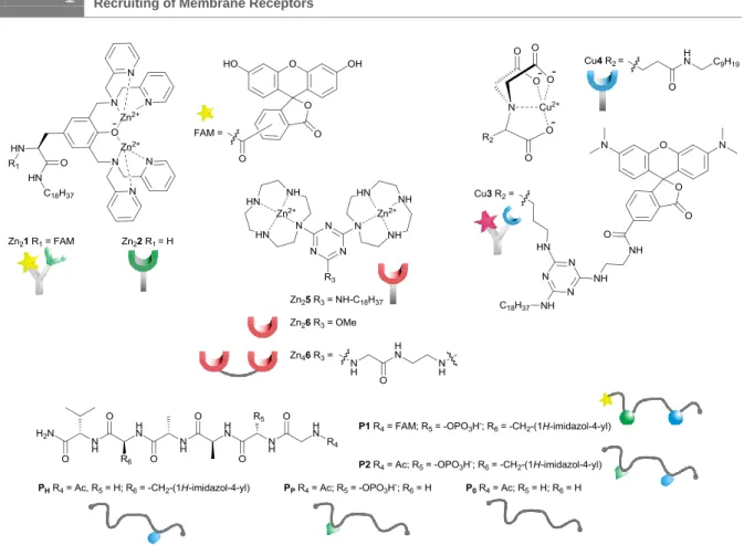
Scheme 1. Structures of synthetic receptors and ligands (counterions omitted).
Various model systems for biological membranes have been developed to understand and mimic multivalent interactions at interfaces7-12 as well as for applications in delivery, sensing and catalysis.13-18 The principle of template-guided assembly has also been extensively exploited in molecular imprinting,19, 20 but the non-covalent arrangement of
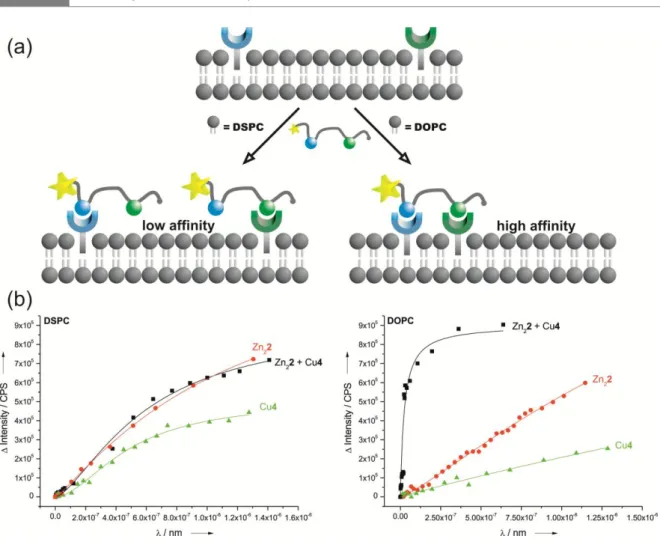
3 orientation triggers a specific response.21-23 Our vesicles with membrane-embedded luminescent receptors produce a characteristic optical response in the presence of small peptides as external ligands. Depending on the functional groups present in the peptide, suitable receptors with complementary binding sites are recruited and arranged in the fluid membrane, which triggers a FRET signal.
Results and Discussion
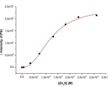
We have previously reported on the recognition of small biomolecules and phosphorylated proteins by multi-site interactions24, 25 at the lipid-water interface using synthetic vesicle membranes with embedded artificial receptors based on transition metal complexes of 1,4,7,10-tetraazacyclododecane (cyclen) and nitrilotriacetic acid (NTA). We now expand this concept to the dynamic recruiting of synthetic receptors allowing the multivalent recognition of phosphorylated peptides. The selective recognition of phospho-serine (pSer) and histidine (His) moieties in model-peptide P1 by metal-complex binding sites was investigated in a previous study in homogeneous solution.26 It was found that in buffered aqueous solution Zn(II)-cyclen receptor Zn26 binds peptide P1 with log K = 4.8, while dimer Zn46, allowing a simultaneous two-prong interaction to pSer and His, shows an affinity of log K = 7.5 (see Table 1, entries 1 and 2 and Supporting Information).
Instead of covalently connecting two receptor sites, as in Zn46, distinct binding sites are now simply embedded in synthetic lipid bilayers. Recruiting and self-organization by the peptide ligand allows the divalent binding of both pSer and His residues of P1 at the membrane interface.
First we embedded Zn25 (1 mol%) in vesicular lipid bilayers from 1,2-distearoyl-sn- glycero-3-phosphocholine (DSPC) using previously reported procedures (see Supporting Information). The emission intensity of peptide P1 increases significantly upon binding to the surface receptors (see SI) and a binding constant of log K = 5.9 (Table 1, entry 3) at ambient temperature was derived. This value is in good agreement with the recognition of pSer by a single Zn25 receptor24 and reasonable, because DSPC’s high phase transition
4
temperature (54 °C)27 restricts diffusion and thus participation of more than a single metal complex binding site in the peptide recognition (cf. Figure 1, Top). Binding affinities could only be enhanced by drastically increasing the vesicle receptor loading to 10 mol%
(Table 1, entry 4) resulting in the formation of tightly packed metal complex patches.16 However, replacing the saturated DSPC lipid by unsaturated 1,2-dioleoyl-sn-glycero-3- phosphocholine (DOPC), which has a much lower transition temperature of -20 °C,27 leads to an increase in peptide P1 binding affinity by more than two orders of magnitude (Table 1, entry 5). The embedded receptor sites can now diffuse in the membrane at room temperature allowing complex formation of one P1 with two Zn25 units by a dynamic assembly process (Figure 1, Top). Unchanged particle sizes determined by dynamic light scattering (Supporting Information, Figure S11) confirm binding of the peptide to receptors of one vesicle (intra-membrane binding mode) and excludes peptide crosslinking of different vesicles (inter-membrane binding).
Entry Lipid Receptor Mol% log K
1 - Zn26 - 4.8[a]
2 - Zn46 - 7.5[a]
3 DSPC Zn25 1 5.9
4 DSPC Zn25 10 8.1
5 DOPC Zn25 1 8.6
6 DSPC Zn22 1 6.2
7 DSPC Cu4 1 5.0
8 DSPC Zn22 + Cu4 1 (each) 6.3
9 DOPC Zn22 1 5.5
10 DOPC Cu4 1 5.8
11 DOPC Zn22 + Cu4 1 (each) 8.8
Table 1. Summary of apparent binding constants for peptide P1 by synthetic receptors in homogeneous solution (entry 1 and 2) and embedded in vesicle membranes (HEPES buffer, pH 7.4, 25 °C).
5 were investigated. The synthesis of amphiphilic Cu(II)-NTA complex Cu4 for the recognition of histidine was reported previously,24 the synthesis of the amphiphilic Zn(II)- 3,5-bis-[(bis-pyridin-2-ylmethyl-amino)-methyl]-4-hydroxy-phenyl (DPA) complex Zn22 for the selective recognition of phosphate28 is described in the Supporting Information. Stable metal complex-doped DOPC and DSPC vesicle membranes were prepared using Zn22 or Cu4 and a mixture of both (1 mol% each). For DSPC membranes with one type of receptor embedded the emission titrations with P1 revealed monovalent binding either to Zn-DPA or to Cu-NTA coordination,29 with log K = 5.0 and 6.2, resp. (Table 1, entries 6 and 7). Job’s plots with P1 as limiting reagent confirm the 1:1 stoichiometry of the binding event (Figure S18). Combining both receptors in gel-phase DSPC-membranes (Figure 1, Bottom) does not increase the binding affinity for peptide P1 (log K = 6.3; Table 1, entries 6, 7, 8). The binding isotherm for the receptor vesicles bearing both complexes resembles the average of the binding properties of the individual receptor vesicles (see SI for data).
This is explained by a random, but fixed position of lipid bilayer embedded metal complex receptors, which prevents the formation of a ternary Zn22-P1-Cu4 complex (see SI for Job´s plots). DOPC membranes, in contrast, show a completely different behaviour:
Whereas vesicles with either Zn22 or Cu4 as embedded receptors give similar binding isotherms and affinity constants (log K = 5.5 and 5.8, resp.; Table 1, entries 9, 10), the liquid-crystalline membrane containing both receptor binding sites shows a significantly higher affinity for peptide P1 in the nanomolar range (log K = 8.8, Table 1, entry 11 and Figure 1, Bottom). We explain this by the divalent binding of P1 by a heterodimeric Zn2- and Cu-receptor assembly in the fluid lipid bilayer (cf. Figure 1, Bottom) resulting from peptide induced self-organization of the membrane receptors. The Job’s plot analysis (Figure S19) supports this conclusion.
6
Figure 1. (Top) Schematic receptor assembly in vesicle membranes: Fixed receptors in gel-phase DSPC-membranes vs. analyte-induced clustering in fluid DOPC-membranes; (Bottom) Emission titrations of P1 vs. DSPC and DOPC- membranes containing Zn21, Cu3 or both receptors.
For bioanalytical applications, the labeling of the peptide analyte must be avoided.
Therefore amphiphilic receptor binding sites that signal the presence of the target analyte by induced FRET were developed: Zn(II)-DPA complex (Zn21) is labeled with carboxyfluorescein (FAM) and Cu(II)-NTA complex Cu3 is rhodamine (TMR) labeled.
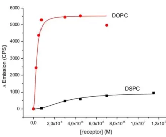
FRET techniques are widely used in molecular biology to measure distances within bio- molecules or to explore membrane structures.30 We use the FRET signal, which is induced by the close distance of the membrane-embedded luminescent binding sites to signal the presence of peptide P2, which in contrast to P1, does not carry a fluorescent label. Divalent binding of P2 to the membrane embedded binding sites recruits Zn21 and Cu3 into close proximity and therefore within the Förster distance of 5.5 nm31 resulting in a FRET signal as optical output (Figure 2, Top).32
7 S22). However, an excess of the target peptide is necessary to reach the maximum energy transfer. Control experiments with monovalent ligands (PP, PH and P0) did not change the observed emission spectra significantly (Figure S23). We exclude inter- vesicular FRET via crosslinking of distinct vesicles as the solution particle size remains unchanged in the presence of P2 (Figures S24, 25).Additionally, vesicles were purified by size exclusion chromatography before binding experiments to remove traces of amphiphilic receptors or micellar aggregates from the solution and the concentrations of the embedded receptors were verified by Zn- and Cu-element analysis using ICP-AES (see SI).33
Figure 2. (Top) Schematic recruiting of labeled membrane receptors by a peptide ligand resulting in a FRET signal; (Bottom) Fluorescence spectra of DOPC vesicles containing labeled receptors Zn21 + Cu3 (1 mol% each) in the absence (red) and presence (green) of peptide P2. Relative FRET emission ratio (I580nm/I520nm) of P2 and monovalent PH and PP as control (inset).
8
As additional control experiments, DOPC and DSPC vesicles with only one of the two binding sites were prepared and used for emission titrations with increasing amounts of corresponding ligands (pSer/PP for Zn21 and His/PH for Cu3). As expected, no significant change in the optical properties of the fluorescent receptor-vesicles was observed (Figure S21). DSPC vesicles with both co-embedded receptors Zn21 and Cu3 (0.1 mol%
each) show a small energy transfer between the FAM- and TMR-labels in the absence of the peptide ligand (Figure S20), which might be explained by the partial formation of Zn21/Cu3 heterodynes. Upon addition of any mono- or divalent ligand no change in emission was detected, as the gel-phase state of the DSPC membranes at room temperature limits the diffusion of the embedded metal complexes (Figure S22).
Conclusion
In conclusion, we demonstrated the specific recognition of peptides by dynamic interface imprinting of synthetic binding sites embedded in vesicle membranes. The amphiphilic metal complex binding sites are recruited in the membrane by the target peptide resulting in multivalent interactions and nanomolar binding affinity. The use of amphiphilic binding sites with FRET pair chromophores, leads to self-organization of a specific analyte epitope at the lipid-water interface and a FRET signal indicating the presence of the target analyte.
The experimental data proof that dynamic binding site recruitment in fluid membranes by analytes (dynamic interface imprinting, DII) leads to the formation of high affinity epitopes.
The process allows the specific detection of functional groups in peptides with nanomolar affinity. To further improve the sensitivity and selectivity of functionalized luminescent vesicles in chemical bioanalysis the number and nature of simultaneously embedded binding sites may be increased, including peptides or nucleotides as recognition moieties.
The combined use of several chromophores in the membrane gives a specific spectroscopic output depending on their spatial arrangement. Such functionalized luminescent vesicles may in some applications replace antibody based assays.
9 Fluorescence measurements were performed with UV-grade solvents (Baker or Merck) in 1 cm quartz cuvettes (Hellma) and recorded on a Varian ‘Cary Eclipse’ fluorescence spectrophotometer with temperature control. Absorption was recorded on a Varian Cary BIO 50 UV/VIS/NIR Spectrometer with temperature control by use of a 1 cm quartz cuvettes (Hellma) and Uvasol solvents (Merck or Baker). PCS measurements were performed on a Malvern Zetasizer Nano at 25 °C using 1 cm disposable polystyrene cuvettes (VWR). NMR spectra were recorded on Bruker Avance 600 (1H: 600.1 MHz, 13C: 150.1 MHz, T = 300 K), Bruker Avance 400 (1H: 400.1 MHz, 13C: 100.6 MHz, T = 300 K), Bruker Avance 300 (1H: 300.1 MHz, 13C: 75.5 MHz, T = 300 K). The chemical shifts are reported in [ppm] relative to external standards (solvent residual peak). The spectra were analyzed by first order, the coupling constants are given in Hertz [Hz].
Characterization of the signals: s = singlet, d = doublet, t = triplet, q = quartet, m = multiplet, bs = broad singlet, psq = pseudo quintet, dd = double doublet, dt = double triplet, ddd = double double doublet. Integration is determined as the relative number of atoms. Assignment of signals in 13C-spectra was determined with DEPT-technique (pulse angle: 135 °) and given as (+) for CH3 or CH, (–) for CH2 and (Cq) for quaternary C. Error of reported values: chemical shift: 0.01 ppm for 1H-NMR, 0.1 ppm for 13C-NMR and 0.1 Hz for coupling constants. The solvent used is reported for each spectrum. Mass spectra were measured on Varian CH-5 (EI), Finnigan MAT 95 (CI; FAB and FD), Finnigan MAT TSQ 7000 (ESI) with Xenon as ionization gas for FAB. IR spectra were recorded on Bio- Rad FTS 2000 MX FT-IR and Bio-Rad FT-IR FTS 155. Melting Points were determined on Büchi SMP or a Lambda Photometrics OptiMelt MPA 100. Thin layer chromatography (TLC) analyses were performed on silica gel 60 F-254 with a 0.2 mm layer thickness.
Detection via UV light at 254 nm / 326 nm or through staining with ninhydrin in EtOH.
Column chromatography was performed on silica gel (70–230 mesh) from Merck.
Commercially available solvents of standard quality were used. Starting materials were used without any further purification. Phospholipids were purchased from Avanti Polar Lipids Inc. Commercially available solvents of standard quality were used. If not otherwise stated, purification and drying was done according to accepted general procedures.34
10
Elemental analyses were carried out by the Centre for Chemical Analysis of the Faculty of Chemistry and Pharmacy of the University of Regensburg.
Synthesis of amphiphilic bis-Zn(II)-DPA complexes Zn21 and Zn22
Receptor Zn21 and Zn22 were prepared from a tyrosine based bis-DPA metal chelating lipid which was synthesized according to a modified protocol of the procedure for the binuclear tyrosine based bis-DPA reported by Hamachi et al.35 Derivatization of the tyrosine scaffold on large scale was possible by standard peptide solution chemistry.
Scheme S1a. Synthesis of amphiphilic tyrosine based bis-dpa zinc complexes. (a) Boc2O, DCM, RT, 20 h; (b) EDC, HOBt, DIPEA, DMF, toluene, 60 °C, 22h; (c) 2,2`-dipicoloylamine, paraformaldehyde, H2O, iPrOH, 80 °C, 30 min, reflux, 17 h; (d) HCl / ether, DCM, RT, 16 h.
11 Scheme S1b. Synthesis of amphiphilic tyrosine based bis-dpa zinc complexes. (e) ZnCl2, MeOH, RT, 2 - 3 h; (f) 5/6-carboxyfluorescein, DIPEA, TBTU, HOBt, DMF, 40 °C, 22 h.
Amide formation of 2536 with octadecylamine gave amphiphilic tyrosine derivative 26 in good yields. Mannich type reaction using 2,2`-dipycolylamine and paraformaldehyde according to literature known procedure gave the protected metal chelating lipid 27.35 Removal of the Boc group in acidic conditions (HCl/ether) yielded compound 28 which was finally treated with two equivalents of ZnCl2 to give Zn22.
A fluorescein label was introduced by amide formation of 28 with an isomeric mixture of 5/6-carboxy fluorescein. Subsequent treatment with ZnCl2 gave the fluorescent amphiphilic binuclear tyrosine based metal complex Zn21.
12
[2-(4-Hydroxy-phenyl)-1-octadecylcarbamoyl-ethyl]-carbamic acid tert-butyl ester (26) Boc-Tyr-OH 25 (2.50 g, 8.9 mmol), DIPEA (5.0 mL, 29.3 mmol), EDC (1.73 mL, 9.8 mmol), and HOBt (1.32 g, 9.8 mmol) were dissolved in DMF (4 mL) under ice cooling and stirred for 45 min. Subsequently a solution of octadecylamine (2.89 g, 9.8 mmol) in 25 mL DMF was added slowly. The reaction was allowed to warm to room temp. and was stirred over night (22 h) at 60 °C. The reaction progress was monitored by TLC (CHCl3 /MeOH 9:1). After completion of the reaction the solvent was removed and the crude product was loaded on flash silica gel and purified by flash column chromatography (CHCl3 / MeOH 95:5, Rf = 0.40) yielding 26 (3.82 g, 7.2 mmol, 81 %) as a colourless solid.
MP: 115 °C. – 1H-NMR (600 MHz; CDCl3): (ppm) = 0.87 (t, 3J = 7.0 Hz, 3 H, C28H3), 1.25 (s, 30 H, C13H2 – C27H2), 1.36 (bs, 2 H, COSY: C12H2), 1.42 (s, 9 H, COSY: C1H3), 2.83- 3.02 (m, 2 H, COSY: C5H2), 3.07-3.24 (m, 2 H, COSY: C11H2), 4.21 (m, 1 H, HMBC, COSY: C4H), 5.17 (bs, 1 H, COSY: NHa), 5.91 (bs, 1 H, COSY: NHb), 6.74 (d, 2J = 8.5 Hz, 2 H, HMBC: C8H), 7.02 (d, 2J = 7.7 Hz, 2 H, HMBC: C7H). – 13C-NMR (150 MHz; CDCl3): (ppm) = 14.1 (+, 1 C, C28H3), 22.7 (–, 1 C, alkyl-CH2), 26.8 (–, 1 C, alkyl-CH2), 28.3 (+, 3 C, C1H3), 29.2 (–, 1 C, alkyl-CH2), 29.3 (–, 1 C, alkyl-CH2), 29.5 (–, 1 C, C12H2), 29.60, 29.65, 29.69 (–, 10 C, alkyl-CH2), 31.9 (–, 1 C, alkyl-CH2), 37.9 (–, 1 C, C5H2), 39.6 (–, 1 C, C11H2), 56.2 (+, 1 C, HMBC: C4H3), 80.3 (Cq, 1 C, C2), 115.6 (+, 2 C, HMBC: C7H), 130.4 (+, 2 C, HMBC: C8H), 128.1 (Cq, 1 C, HMBC: C6), 155.2 (Cq, 1 C, HMBC: C9), 155.6 (Cq, 1 C, HMBC: C3), 171.3 (Cq, 1 C, HMBC: C10). – IR (ATR) [cm-1]:
~
[cm] = 3335, 3306, 2959, 2917, 1682, 1655, 1523, 1468, 1367, 1295, 1236, 1168, 1046, 896, 799. – MS (ESI(+), DCM/MeOH + 10 mmol/L NH4Ac): m/z (%) = 533.6 (100) [MH+], 477.4 (19) [MH+ - C4H8]+, 555.5 (36) [MNa+]. – Elemental analysis calcd. (%) for C32H56N2O4: C 72.14, H 10.59, N 5.26; found C 72.13, H 10.63, N 5.00. – MF: C32H56N2O4 – FW: 532.81 g/mol.13 (2-{3,5-Bis-[(bis-pyridin-2-ylmethyl-amino)-methyl]-4-hydroxy-phenyl}-1-octadecyl carbamoyl-ethyl)- carbamic acid tert-butyl ester (27) 2,2’-Dipicolylamine (2.35 g, 11.8 mmol) and paraformaldehyde (0.56 g, 18.9 mmol) were dissolved in 30 mL water / isopropanol (5:3) and the pH was adjusted to 8 by adding 1 M NaOH. After stirring at 80 °C for 35 min, compound 26 (2.51 g, 4.7 mmol) was added, and the reaction mixture was refluxed for 17 h. After cooling to room temperature the solvent was evaporated and the residue was dissolved in ethyl acetate. The solution subsequently was washed with saturated NaHCO3 (3x) and brine (3x) followed by drying over MgSO4. After removal of the solvent in vacuo the crude product was purified by flash column chromatography on flash silica gel (ethyl acetate / MeOH 2:1, Rf = 0.1) obtaining 27 (1.1 g, 1.15 mmol, 24 %) as a colourless oil. 1H-NMR (400 MHz; acetone-d6): (ppm)
= 0.86 (t, 3J = 6.9 Hz, 3 H, C37H3), 1.14-1.28 (m, 30 H, C22H2 – C36H2), 1.29 (s, 11 H, HSQC: C1H3, C21H2), 2.87 (dd, 3J = 13.5 Hz, 2J = 6.5 Hz, 1 H, HSQC, COSY: C6H2), 2.94 (dd, 3J = 13.5 Hz, 2J = 7.4 Hz, 1 H, HSQC, COSY: C6H2), 3.02 (dd, 3J = 12.9 Hz,
2J = 6.6 Hz, 2 H, HSQC: C20H2), 3.74 (d, 2J = 13.7 Hz, 2 H, HMBC, COSY: C10H2), 3.80 (d,
2J = 13.7 Hz, 2 H, HMBC, COSY: C10H2), 3.85 (s, 8 H, COSY: C12H2), 3.34 (dd,
3J = 14.4 Hz, 2J = 7.1Hz, 1 H, COSY: C20H2), 5.98 (d, 3J = 8.2 Hz, 1 H, HSQC, COSY:
N4H), 7.10 (s, 2 H, HMBC, COSY: C8H), 7.20 (ddd, 3J = 7.4 Hz, 4.9 Hz, 1.1 Hz, 4 H, HMBC: C16H), 7.29 (bs, 1 H, HSQC, COSY: N19H), 7.55 (d, 3J = 7.8 Hz, 4 H, HMBC:
C14H), 7.69 (dt, 3J = 7.7 Hz, 2J = 1.9 Hz, 4 H, HMBC: C15H), 8.52 (m, 4 H, HMBC: C17H), 10.96 (bs, 1 H, HSQC: OH). – 13C-NMR (100 MHz; acetone-d6): (ppm) = 14.3 (+, 1 C, C37H3), 23.3 (–, 1 C, alkyl-CH2), 27.5 (–, 1 C, alkyl-CH2), 28.5 (+, 3 C, C1H3), 29.9 (–, 1 C, alkyl-CH2), 30.22 (–, 1 C, alkyl-CH2), 30.27 (–, 1 C, alkyl-CH2), 30.4 (–, 9 C, alkyl-CH2,
14
solvent peak), 32.6 (–, 1 C, alkyl-CH2), 38.8 (–, 1 C, COSY: C6H2), 39.7 (–, 1 C, HSQC:
C20H2), 55.3 (–, 2 C, HMBC, COSY: C10H2), 60.1 (–, 4 C, HMBC, COSY: C12H2), 56.9 (+, 1 C, HMBC: C5H3), 79.0 (Cq, 1 C, HMBC, HSQC, C2), 122.8 (+, 4 C, HMBC: C16H), 123.8 (+, 4 C, HMBC: C14H), 124.6 (Cq, 2 C, HMBC: C9), 127.7 (Cq, 1 C, HMBC: C7), 131.3 (+, 2 C, HMBC, COSY: C8H), 137.3 (+, 4 C, HMBC, COSY: C15H), 149.7 (+, 4 C, HMBC, COSY: C17H), 155.8 (Cq, 1 C, HMBC: C11), 155.8 (Cq, 1 C, HMBC: C11), 159.9 (Cq, 1 C, HMBC: C3), 160.2 (Cq, 4 C, HMBC: C13), 171.9 (Cq, 1 C, HMBC: C18). – IR (ATR) [cm-1]:
~
[cm] = 3383, 2922, 2852, 1706, 1656, 1590, 1569, 1433, 1364, 1288, 1245, 1167, 1048, 995, 791. – MS (ESI(+), DCM/MeOH + 10 mmol/L NH4Ac): m/z (%) = 478.5 (100) [M + 2 H+]2+, 955.8 (85) [MH+]. – Elemental analysis calcd. (%) for C58H82N8O4: C 72.92, H 8.65, N 11.73; found C 72.38, H 8.34, N 11.63 – MF: C58H82N8O4 – FW: 955.35 g/mol.
2-Amino-3-{3,5-bis-[(bis-pyridin-2-ylmethyl-amino)-methyl]-4-hydroxy-phenyl}-N-octadecyl- propionamide (28) Boc protected 27 (700 mg, 0.73 mmol) was dissolved in DCM under ice cooling and mixed with HCl/ether (3.7 mL; 1 mL / 0.2 mmol Boc group). The reaction was allowed to warm to room temp. and was stirred for additional 16 hours. After completion the reaction mixture was evaporated to dryness, the residue was suspended in saturated NaHCO3 and extracted with ethyl acetate (4x). Subsequently the organic phase was dried over MgSO4 and the solvent was removed in vacuo yielding 28·(497 mg, 0.58 mmol, 79 %) as a pale yellow oil. 1H-NMR (300 MHz; DMSO-d6): (ppm) = 0.82 (t, 3J = 6.9 Hz, 3 H, CH3), 1.04 (bs, 4 H, alkyl-CH2), 1.19 (s, 28 H, alkyl-CH2), 2.57 (dd, 3J = 13.5 Hz, 2J = 7.1 Hz, 1 H, Tyr- CH2), 2.73 (dd, 3J = 13.5 Hz, 2J = 7.1 Hz, 1 H, Tyr-CH2), 2.91 (m, 2 H, CH2), 3.29 (t,
3J = 6.4 Hz, 1 H, C H), 3.65 (s, 4 H, Ar-CH2-pyr), 3.74 (s, 8 H, N-CH2-pyr), 6.98 (s, 2 H, Ar-CH), 7.21 (m, 4 H, Pyr-CH), 7.46 (d, 3J = 8.0 Hz, 4 H, Pyr-CH), 7.70 (dt, 3J = 7.6 Hz,
15 CH2), 31.4 (–, 1 C, alkyl-CH2), 38.0 (–, 1 C, CH2), 40.2 (–, 1 C, CH2), 53.9 (–, 2 C, CH2), 55.9 (+, 1 C, CH), 58.8 (–, 4 C, CH2), 122.1 (+, 4 C, CH), 122.5 (+, 4 C, CH), 123.1 (Cq, 1 C), 127.4 (Cq, 1 C), 129.8 (+, 2 C, CH), 136.5 (+, 4 C, CH), 148.5 (+, 4 C, CH), 153.7 (Cq, 1 C), 158.5 (Cq, 4 C), 173.5 (Cq, 1 C). – IR (ATR) [cm-1]:
~
[cm] = 2921, 2852, 1658, 1590, 1522, 1474, 1433, 1372, 1233, 1149, 1048, 996, 754. – MS (ESI(+), DCM/MeCN/TFA): m/z (%) = 428.4 (100) [M + 2 H+]2+, 299.6 (16) [M + 3 H+ + MeCN]3+, 855.8 (14) [MH+], 286 (12) [M + 3 H+]. – HRMS Calcd for C53H75N8O2 855.6013; Found:855.6000. – MF: C53H74N8O2 – FW: 855.23 g/mol.
Amphiphilic binuclear Zn(II)-Dpa complex (Zn22) To a solution of compound 28 (36.5 mg, 43 µmol) in MeOH (2 mL) was added dropwise a methanolic solution of ZnCl2 (100 mM, 858 µL, 86 µmol). After stirring the reaction mixture for 2 h at room temperature, the methanol was removed in vacuo. The residue was dissolved in water and was lyophilized yielding complex 12 as a colourless solid in quantitative yield. MP: 147 °C. – IR (ATR) [cm-1]:
~
= 2924, 2852, 1676, 1608, 1573, 1477, 1440, 1303, 1270, 1156, 1100, 1053, 1023, 763. – UV (MeOH): max (log = 261 (4.076), 296 (3.436). – MS (ESI(+), H2O/MeOH + 10 mmol/L NH4Ac): m/z (%) = 1099.7 (100) [M3+ + 2 CH3COO-]+, 520.4 (69) [M3+ + CH3COO-]2+. – MF: C53H74N8O2Zn2Cl4 – FW: 1127.79 g/mol.16
Isomeric mixture of amphiphilic, fluorescein labeled Dpa ligand (29) An isomeric mixture of 5/6-carboxyfluorescein (246 mg, 0.65 mmol), DIPEA (323 µL, 1.87 mmol), TBTU (315 mg, 0.98 mmol), and HOBt (133 mg, 0.98 mmol) were dissolved under nitrogen atmosphere in dry DMF (4 mL) under ice cooling and stirred for 1 h.
Subsequently 28 (400 mg, 0.47 mmol) in 4 mL DMF was added slowly. The reaction was allowed to warm to room temperature and was stirred 22 h at 40 °C. The reaction progress was monitored by TLC (CHCl3 / MeOH 9:1). After completion of the reaction the solvent was removed and the crude product was purified by preparative HPLC yielding the isomers 29 as an orange-yellow solid. MP: 97 – 99 °C. – UV (MeOH): max (log ) = 224 (4.629), 260 (4.158), 454 (3.500), 481 (3.481). – IR (ATR) [cm-1]:
~
[cm] = 2924, 2853, 1757, 1670, 1609, 1438, 1377, 1246, 1178, 1126, 955, 837, 798, 755, 719. – MS (ESI(+), DCM/MeCN/TFA): m/z (%) = 607.6 (100) [M + 2 H+]2+, 405.3 (65) [M + 3 H+]2+, 1213.9 (11) [MH+]. – LC-MS (+ p ESI Q1MS ; RT 40 min): m/z (%) = 607.6 (100) [M + 2 H+]2+, 1213.9 (65) [MH+]+, 405.3 (38) [M + 3 H+]2+. – MF: C74H84N8O8 – FW: 1213.53 g/mol.Purification by preparative HPLC:
Column: LabID75 Phenomenex Luna 250 x 21.2 mm 10 um / Ser.Nr. 453159-3
Flow: 21 mL / min
17 Detection wavelength: 220 nm
Gradient: 0 min – 5 % MeCN / H2O (+ 0.0059 % TFA) 43 min – 72 % MeCN / H2O (+ 0.0059 % TFA)
45 min – 98 % MeCN / H2O (+ 0.0059 % TFA) 55 min – 98 % MeCN / H2O (+ 0.0059 % TFA) Retention time: ca. 42 min
Fluorescent binuclear Zn(II)-Dpa complex (Zn21) To a solution of compound 29 (10.5 mg, 8.7 µmol) in MeOH (2 mL) was added dropwise a methanolic solution of MnCl2 (100 mM, 174 µL, 17.4 µmol). After stirring the reaction mixture for 2.5 h at room temperature, the methanol was removed in vacuo. The residue was dissolved in water and was lyophilized yielding complex 14 as an orange solid in quantitative yield (13 mg, 8.7 µmol). MS (ESI(+), H2O/MeOH + 10 mmol/L NH4Ac):
18
m/z (%) = 701.7 (100) [M3+ + CH3COO-]+, 1398.0 (8) [M3+ - H+ + CH3COO-]+, 1458.0 (7) [M3+ + 2 CH3COO-]+. – MF: C74H84N8O8Zn2Cl4 – FW: 1486.10g/mol.
Synthesis of amphiphilic Cu(II)-NTA complex Cu3
Receptor Cu3 was prepared by connecting a carboxytetramethylrhodamine label, an ornithine based NTA binding site and a hydrophobic anchor via a central cyanuric chloride linker and complexation of the amphiphilic ligand with Cu(II).
Scheme S2. Synthesis of an amphiphilic, fluorescent copper complex: (a) Cbz chloride, DCM, 0°C, 3 h; (b) tert butyl acetate, HClO4, RT, 72 h; (c) bromo tert butyl acetate, DMF, 55°C, 20 h; (d) Pd/C.H2, EtOH, RT, 72 h.
19 Scheme S3. Synthesis of an amphiphilic, fluorescent copper complex: (e) octadecylamine, K2CO3, acetone, RT, 20 h; (f) Cbz-diamine 31, K2CO3, acetone, 50°C, 48 h; (g) NTA-precursor 35, collidine, dioxane, 140°C (MW), 1.5 h; (h) Pd/C, H2, EtOH, RT, 48 h;
(i) carboxytetramethylrhodamine, TBTU, HOBt, DIPEA, DMF, 0°C, 1 h / 40°C, 24 h; (j) TFA, RT, 24 h; (k) Cu2(OH)2CO3, MeOH, 60°C, 4 h.
20
Ethylenediamine 30 is Cbz-protected to give mono-amine 31. Cbz-protected L-ornithine 32 is converted into the butyl acetate ester 33. The amine group of 33 reacts in a nucleophilic substitution with bromo-tert-butyl-acetate to give 34 and yields after deprotection the free amine 35. To introduce the hydrophobic anchor octadodecanylamine 13 is tethered by nucleophilic substitution to cyanuric chloride 36 yielding compound 37.
Subsequently Cbz-protected ethylenediamine 31 is attached to 37 yielding 38 to connect carboxytetramethylrhodamine as fluorescent label. Next, the NTA-precursor 35 is coupled to the unprotected functional group of the linker 38 via nucleophilic substitution yielding 39. Hydrogenolytic Cbz-deprotection gives compound 40, which is tethered to an in situ formed active ester of carboxytetramethylrhodamine. Treatment of 41 with TFA leads to the free acid 42. In a last step copper is chelated by the acid groups of the NTA-complex 42 yielding the target receptor Cu3.
Synthesis of benzyl (2-aminoethyl)carbamate (31) Ethylenediamine 30 (12.1 mL, 180 mmol) was dissolved in DCM (240 mL) and a solution of benzyl chloroformate (2.5 mL, 18 mmol) in DCM (60 mL) was added dropwise over 1.5 h at 0 °C. The reaction mixture was stirred at this temperature for further 1.5 h and at room temperature overnight. The formed precipitate was filtered off, the filtrate was washed three times with brine (150 mL) and the organic phase was dried over MgSO4. Evaporation of the solvent leads to a pale-yellow, crystalline raw product which was purified via column chromatography (silica gel, CHCl3/MeOH/Et3N 70:29:1; Rf = 0.09). The product 31 (2.07 g, 10.7 mmol, 59 %) was obtained as pale-yellow oil which crystallizes at 0 °C. 1H-NMR (400 MHz, CDCl3), [ppm]: 1.70 (2 H, bs, NH2-CH2), 2.73 (2 H, pt, H2N- CH2-CH2), 3.17 (2 H, dd, CH2-CH2-NH), 5.05 (2 H, s, O-CH2-C), 5.65 (1 H, bs, CH2-NH-C), 7.29 (5 H, m, CHarom). – 13C-NMR (100 MHz, CDCl3), [ppm]: 156.84 (NH-CO2), 136.64 (CH2-C-C5H5), 128.51, 128.48, 128.02v (C-C5H5), 66.60 (O-CH2-C), 43.71 (CH2-CH2-NH), 41.62 (H2N-CH2-CH2). – MS (ESI(+), H2O + 0.1% FA/MeCN + 0.1% FA): m/z (%) = 195.1 (100) [MH+].
21 Synthesis of tertbutyl protected 2-amino-5-(((benzyloxy)carbonyl)amino) pentanoic acid (33) Benzyloxycarbonyl ornithine 32 (10.00 g, 37.55 mmol) was dissolved in tert butyl acetate (471 mL, 3.47 mol), perchloric acid (70 %, 5.64 mL, 67.59 mmol) was added and the solution was stirred at RT for 72 h. The reaction mixture was cooled to 0°C and four times extracted with 0.5 M hydrochloric acid (300 mL). 2 M sodium hydroxide solution was added to the aqueous phase and the target molecule was extracted from the basic solution with diethyl ether (three times, 200 mL), dried over Na2SO4 and the solvent was removed in vacuo. 33 was obtained as yellow oil (8.61 g, 26.71 mmol, 71 %). – 1H-NMR (400 MHz, CDCl3): [ppm] = 1.43 (s, 9H, CH3), 1.55 – 1.70 (m, 4H, 2x CH2), 3.18 (m, 2H, CH2-NH), 3.30 (m, 1H, CH), 5.05 (s, 2H, CH2-Cbz), 5.25 (s, 1H, NH), 7.25 – 7.36 (m, 5H, CHaromat.). – 13C-NMR (100 MHz, CDCl3): [ppm] = 26.0 (CH3), 28.2 (CH2), 40.9 (CH2), 54.4 (CH), 66.6 (CH2), 81.3 (CHquart.), 128.6 (CHarom.), 137.0 (Cquart,arom.), 156,5 (Ccarbonyl), 175,0 (Ccarbonyl). – MS (ESI(+), H2O + 0.1% FA/MeCN + 0.1% FA):
m/z (%) = 323.0 (100) [MH+], 645.4 (36) [2MH+].
Synthesis of di-tert-butyl 2,2'-((5-(((benzyloxy)carbonyl)amino)-1-(tert-butoxy)-1- oxopentan-2-yl)azanediyl)diacetate (34) Two-fold protected ornithine 33 (8.61 g, 26.71 mmol), tert butyl bromoacetate (15.50 mL, 106.82 mmol) and DIPEA (22.71 mL, 133.53 mmol) were dissolved in dry DMF (100 mL) under nitrogen atmosphere and the mixture was stirred at 55 °C for 20 h. Subsequently
22
the solvent was removed under vacuo yielding a dark brown solid which was suspended in ethyl acetate (60 mL) and filtered off. The residue was washed three times with hexane/ethyl acetate (3:1, 120 mL). After evaporation a brown oil (17.54 g) was obtained.
The crude product was purified via column chromatography (flash silical gel, hexanes/EA 3:1; Rf = 0.27) to give 34 as pale yellow solid (12.74 g, 23.mmol, 86 %). – 1H-NMR (400 MHz, CDCl3): [ppm] = 1.38 (s, 27H, CH3), 1.55 – 1.70 (m, 4H, 2x CH2), 3.16 – 3.25 (m, 3H, CH and CH2-NH), 3.30 – 3.44 (m, 4H, 2x CH2-Ntert.), 5.00 (s, 2H, CH2-Cbz), 5.22 (s, 1H, NH), 7.21 – 7.27 (m, 5H, CHaromat.). – 13C-NMR (100 MHz, CDCl3):
[ppm] = 26.0 (CH3), 28.2 (CH2), 40.6 (CH2), 54.0 (CH), 65.1 (CH2), 66.4 (CH2), 81.2 (CHquart.), 128.0 (CHarom.), 136.8 (Cquart,arom.), 156.5 (CHcarbonyl), 175.1 (CHcarbonyl). – MS (ESI(+), H2O + 0.1% FA/MeCN + 0.1% FA): m/z (%) = 551.2 (100) [MH+].
Synthesis of di-tert-butyl 2,2'-((5-amino-1-(tert-butoxy)-1-oxopentan-2- yl)azanediyl)diacetate (35) Cbz-protected NTA-precursor 34 (484 mg, 0.51 mmol) was dissolved in ethanol (15 mL) and Pd/C (500 mg) was added. The reaction mixture was stirred at RT and under hydrogen atmosphere (30 bar) for 72 h. Subsequently Pd/C was filtered off via celite and the solvent was evaporated yielding the yellow oil 35 (372 mg, 0.45 mmol, 89%). 1H-NMR (300 MHz, CDCl3) [ppm] = 5.00 (bs, 1 H, NH), 3.41 (s, 2 H, CH2-N), 3.38 (s, 2 H, CH2-N), 3.31 (m, 1 H, CH-N), 2.85 (m, 2H, CH2-NH2), 1.52- 1.87 (m, 4 H, 2x CH2alkyl), 1.41 - 1.38 (m, 27 H, CH3). – 13C-NMR (100 MHz, CDCl3) [ppm] = 171.82 (COO), 170.68 (COO), 81.40 (COquart.), 81.03 (COquart.), 65.21 (CH-N), 57.79 (CH2-N), 40.36 (CH2-NH2), 28.13 (CH2alkyl), 28.04 (CH3), 27.04 (CH2-CH), 18.30 (CH3). – MS (ESI(+), H2O + 0.1% FA/MeCN + 0.1% FA): m/z (%) = 417.3 (100) [MH+].
23 Synthesis of 4,6-dichloro-N-propyl-1,3,5 triazin-2-amine (37) Octadecylamine (1.02 g, 5.50 mmol) was dissolved in acetone (40 mL) and a solution of 2,4,6-trichloro-1,3,5-triazine 36 (1.47 g, 5.44 mmol) in acetone (25 mL) and water (5 mL) was added at 0 °C, followed by the addition of K2CO3 (2.25 g, 16.28 mmol) in water (10 mL). The reaction mixture was stirred at 0 °C for further 30 min and at RT for 20 h.
After the solvent was removed in vacuo the residue was washed with brine (5 mL) and extracted three times with CHCl3 (25 mL). The organic phases were dried over MgSO4 and the solvent was evaporated to give a white powder (2.20 g). The crude product was purified via silica column chromatography (hexanes/EA 10:1; Rf = 0.37) yielding the white solid 37 (1.30 g, 3.11 mmol, 57 %). – 1H-NMR (300 MHz, CDCl3) [ppm] = 3.47 (q, 2H, CH2-NH), 1.59 (m, 2H, CH2alkyl), 1.25 -1.32 (m, 30H, 15 x CH2alkyl), 0.88 (t, 3H, CH3). –
13C-NMR (100 MHz, CDCl3) [ppm] = 171.01 (Carom.-NH), 165.83 (Carom.-Cl), 41.58 (CH2alkyl), 31.94 (CH2alkyl), 29.48 (CH2alkyl), 26.64 (CH2alkyl), 22.71 (CH2alkyl), 14.14 (CH3). – MS (ESI(+), H2O + 0.1% FA/MeCN + 0.1% FA): m/z (%) = 417.2 (100) [MH+].
Synthesis of benzyl (2-((4-chloro-6-(propylamino)-1,3,5-triazin-2-yl) amino)ethyl)carbamate (38) Amphiphilic triazine 37 (4.50 g, 10.80 mmol) was dissolved in acetone (50 mL) followed by the addition of Cbz-protected ethylendiamine 31 (2.10 g, 10.80 mmol) and K2CO3 (7.46 g, 54.00 mmol). The mixture was stirred at 50 °C for 40 h before the solvent was removed in vacuo. The residue was redissolved in CHCl3 (100 mL) and washed three times with a
24
water/brine solution (2:1, 100 mL). The organic phases were dried over MgSO4 and the solvent was evaporated. The obtained crude product was recrystallized from EA yielding 38 as a light brown solid (3.46 g, 6.01 mmol, 56 %). 1H-NMR (300 MHz, CDCl3)
[ppm] = 7.29 (m, 5 H, CHarom), 5.08 (s, 2 H, CH2-Cbz.), 3.56 (m, 2 H, CH2alkyl), 3.27- 3.45 (m, 4 H, CH2-CH2), 1.53 (t, 2 H, CH2alkyl), 1.25 (bs, 30 H, 15 x CH2alkyl), 0.88 (t, 3 H, CH3). –
13C-NMR (100 MHz, CDCl3) [ppm] = 136.53 (Carom), 128.07- 128.54 (CHarom.), 66.72 (CH2-CH2), 41.05 (CH2alkyl), 31.94 (CH2alkyl), 29.38 - 29.72 (CH2alkyl), 22.71 (CH2alkyl), 14.14 (CH3). – MS (ESI(+), H2O + 0.1% FA/MeCN + 0.1% FA): m/z (%) = 574.4 (100) [MH+].
Synthesis of di-tert-butyl 2,2'-((5-((4-((2-(((benzyloxy)carbonyl)amino) ethyl)amino)-6- (octadecylamino)-1,3,5-triazin-2-yl)amino)-1-(tert-butoxy)-1-oxopentan-2- yl)azanediyl)diacetate (39) Amine 35 (1.00 g, 2.47 mmol) was suspended in dioxane (10 mL) followed by the addition of the two-fold substituted triazine 38 (1.42 g, 2.47 mmol) and collidine (1.63 µl, 12.37 mmol). The reaction was carried out in a microwave (150 W, 1.7 bar) at 140 °C for 1.5 h. After evaporation a brown oil was obtained. The crude product was purified via column chromatography (flash silica gel, EA/hexanes/Et3N 75:20:5; Rf = 0.43) to give product 39 as a dark yellow oil (0.48 g, 0.51 mmol, 21 %). 1H-NMR (300 MHz, CDCl3)
[ppm] = 7.29 - 7.33 (m, 5 H, CHarom), 5.07 (s, 2 H, CH2-Cbz), 3.26 - 3,50 (m, 13 H, CH and 6 x CH2alkyl), 1.70 (m, 2 H, CH2alkyl), 1.64 (m, 2 H, CH2alkyl), 1.50 (t, 2 H, CH2alkyl), 1.43 (bs, 30 H, CH2alkyl), 1.24 (bs, 27 H, CH3), 0.88 (t, 3 H, CH3). – 13C-NMR (100 MHz, CDCl3)
[ppm] =172.22 (COO), 170.66 (COO), 136.73 (CHarom)), 128.45 (CHarom)), 127.96 (CHarom), 81.11 (COquart), 80.67 (COquart), 66.49 (CHCOO), 65.1, (CH2O), 53.82 (CH2-N), 40.68 (CH2-NH), 40.39 (CH2-NH), 31.92 - 26.22 (CH2alkyl), 26.97 (CH3), 22.69 (CH2-CH3), 14.12 (CH3). – MS (ESI(+), H2O + 0.1% FA/MeCN + 0.1% FA): m/z (%) = 478.4 (100) [M + 2 H+]2+, 955.8 (64) [MH+].
25 Synthesis of di-tert-butyl 2,2’-(5-((4-((2-aminoethyl)amino)-6-(octodecylamino)-1,3,5-
triazin-2-yl)amino)1-(tert-butoxy)-1-oxopentan-2-yl)azanediyl)diacetate (40) Cbz-protected spacer moiety 39 (190 mg, 0.20 mmol) was dissolved in ethanol (15 mL) and Pd/C (500 mg) was added. The reaction mixture was stirred at RT and under hydrogen atmosphere (30 bar) for 48 h. Subsequently Pd/C was filtered off via celite and the solvent was evaporated yielding 40 as light-yellow oil (156 mg, 0.19 mmol, 95%).
1H-NMR (300 MHz, CDCl3) [ppm] = 3.26 – 3.58 (m, 13 H, CH and 6 x CH2alkyl), 1.71 (m, 2 H, CH2alkyl), 1.64 (m, 2 H, CH2alkyl), 1.55 (t, 2 H, CH2alkyl), 1.42 (bs, 30 H, CH2alkyl), 1.24 (bs, 27 H, CH3), 0.88 (t, 3 H, CH3). – 13C-NMR (100 MHz, CDCl3) [ppm] =172.22 (COO), 170.66 (COO), 136.73 (CHarom)), 128.45 (CHarom)), 127.96 (CHarom), 81.11 (COquart), 80.67 (COquart), 66.49 (CHCOO), 65.1. (CH2O), 53.82 (CH2-N), 40.68 (CH2-NH), 40.39 (CH2-NH), 31.93 - 26.22 (CH2alkyl), 26.97 (CH3), 22.69 (CH2-CH3), 14.13 (CH3). – MS (ESI(+), H2O + 0.1% FA/MeCN + 0.1% FA): m/z (%) = 411.2 (70) [M + 2 H+]2+, 431.8 (100) [M + 2 H+ + MeCN]2+, 821.7 (30) [MH+].
26
Synthesis of 4-((2-((4-((4-(bis(2-(tert-butoxy)-2-oxoethyl)amino)-5-(tert-butoxy)-5- oxopentyl)amino)-6-(octadecylamino)-1,3,5-triazin-2-yl)amino) ethyl)carbamoyl)-2-(6- (dimethylamino)-3-(dimethyliminio)-3H-xanthen-9-yl)benzoate (41) Carboxytetramethylrhodamine (180 mg, 0.42 mmol), TBTU (148 mg, 0.46 mmol), HOBt (62 mg, 0.46 mmol) and DIPEA (284 µL, 1.67 mmol) were dissolved in dry DMF (4.5 mL) under nitrogen atmosphere at 0°C and the reaction mixture was stirred at RT. After 1 h a solution of the receptor-precursor 40 (343 mg, 0.42 mmol) in dry DMF (3 mL) was added dropwise over 35 min. The reaction mixture was warmed to 45 °C and stirred for another 40 h. Subsequently the solvent was removed and the pink residue was purified via column chromatography (flash silica gel, EA/MeOH/Et3N 75:20:5) to give 41 as magenta oil.
(76 mg, 0.06 mmol, 15 %). 1H-NMR (300 MHz, CDCl3) [ppm] = 0.80 (t, 3 H, CH3), 1.11 (m, 2 H, CH2 alkyl), 1.17 (m, 28 H, CH2 alkyl ), 1.27 (m, 2 H, CH2 alkyl), 1.35 (s, 27 H, CH3 Boc), 1.45 – 1.73 (m, 4 H, CH2 alkyl), 3,00 (m, 12 H, CH3-N), 3.16-3.55 (m,13 H, CH2 and CH), 7.22 (s, 1H, arom.), 7.34 – 7.49 (m, 4 H, arom), 7.64 – 7.67 (m, 4H, arom.). – 13C-NMR (100 MHz, CDCl3) [ppm] = 8.7, 14.1, 22.7, 28.0, 28.3, 29.7, 31.9, 45.7, 53.4, 110.7, 110.9, 118.5, 124.0, 125.1, 127.8. – MS (ESI(+), H2O + 0.1% FA/MeCN + 0.1% FA):
m/z (%) = 1234.0 (100) [MH+].
27 Synthesis of 4-((2-((4-((4-(bis(carboxymethyl)ammonio)-4-carboxybutyl) amino)-6- (octadecylamino)-1,3,5-triazin-2-yl)amino)ethyl)carbamoyl)-2-(6-(dimethylamino)-3- (dimethyliminio)-3H-xanthen-9-yl)benzoate (42) 41 (92 mg, 0.08 mmol) was dissolved in TFA (1.6 mL, 20.77 mmol) and stirred at RT for 30 h. TFA was evaporated in vacuo, the residue was suspended in water (3 mL) and lyophilized giving product 42 (83 mg, 0.08 mmol, 98 %) as a magenta solid. 1H-NMR (600 MHz, CDCl3) [ppm] = 0.86 (t, 3 H, CH3), 1.15 – 1.28 (m, 28 H, CH2 alkyl), 1.36-1.38 (m, 4 H, CH2 alkyl ), 1.39 – 1.42 (m, 12 H, CH3), 3.09 – 3.15 (m, 5 H, CH-(CH2)2), 3.16 – 3.20 (m, 8 H, CH2-NH), 3.69 – 3.71 (m, 4 H, CH2-COOH), 6.61 (bs,1 H, NH), 6.74 (bs,1 H, NH), 6.98 (bs,1 H, NH), 7.39 (t, 1H, CHarom.), 7.41 – 7.53 (m, 4 H, CHarom.), 7.56 (d, 1H, CHarom.), 7.75 – 7.83 (m, 2H, CHarom.), 8.02 (d, 1H, CHarom.), 8.61 (bs,1 H, NH), 9.12 (bs,1 H, NH). – 13C-NMR (150 MHz, CDCl3) [ppm] = 14.1 (+, 1 C, CH3), 17.3 (+, 2 C, CH3), 18.5 (+, 2 C, CH3), 22.6 (-, 1 C, CH2 alkyl), 28.8 – 29.7 (-, 16 C, CH2 alkyl), 31.9 (-,1 C, CH2 alkyl), 40.6 (+, 1 C, CH), 41.0 (-, 1 C, CH-(CH2)2), 42.5 (-, 1 C, CH-(CH2)2), 46.3 (-, 4C, CH2-NH), 54.4 (-, 2 C, CH2-COOH), 109.8 (+, 1 C, CHarom.), 112.6 (+, 1 C, CHarom.), 114.1 (+, 1 C, CHarom.), 114.7 (-, 1 C, Cq), 114.8 (-, 1 C, Cq), 116.7 (-, 1 C, Cq), 118.6 (-, 1 C, Cq), 119.7 (+, 1 C, CHarom.), 124.8 (+, 1 C, CHarom.), 127.0 (+, 1 C, CHarom.), 128.1 (+, 1 C, CHarom.), 128.6 (+, 1 C, CHarom.), 128.9 (+, 1 C, CHarom.), 130.4 (-, 1 C, Cq), 130.9 (-, 1 C, Cq), 136.6 (-, 1 C, Cq), 142.5 (-, 1 C, Cq), 157.0 (-, 1 C, Cq), 157.3 (-, 1 C, Cq), 160.6 (-, 1 C, Cq), 160.9 (-, 1 C, Cq), 161.1 (-, 1 C, Cq), 161.4 (-, 1 C, Cq). – MS (ESI(+), DCM/MeOH + 0.1% FA): m/z (%) = 533.5 (100) [M + 2 H+]2+, 1065.7 (26) [MH+].
28
Synthesis of the amphiphilic, labeled Cu(II)-NTA-complex (Cu3) Deprotected 42 (83.0 mg, 0.08 mmol) was dissolved in MeOH (1.5 mL) and a suspension of Cu(OH)2CO3 and a very small amount of MeOH was added. The reaction was completed by stirring the mixture at RT for 20 h and at 60°C for 3 h. Subsequently the warm solution was filtered immediately and the solvent was evaporated. The residue was suspended in water (2 mL) and lyophilized yielding Cu3 (89.0 mg, 0.08 mmol, 99 %) as a dark magenta solid. MS (ESI(-), H2O/MeOH + 10 mmol/L NH4Ac): m/z (%) = 1124.4 (100) [M - H - 2 H2O]-.
29 Scheme S4. Structures and symbols of (non-)amphiphilic receptors and ligands (counterions omitted).
Receptor-ligand binding in homogeneous (Zn26, Zn46) and vesicular (Zn25) solution
Figure S1. (a) Schematic recognition of P1 in homogeneous aqueous solution by previously developed receptors26 Zn26 and Zn46; (b) Schematic recognition of P1 by membrane-embedded receptor Zn25 in DSPC and DOPC vesicles.
30 Vesicles
Preparation
In small glass reaction vessels 1 - 5 µmol of DOPC or DSPC were dissolved in chloroform and optionally 0.1-10 mol% of dissolved metal complexes were added and mixed. The solvent was completely removed under reduced pressure at 25 °C (DOPC) or 75˚C (DSPC) and an appropriate amount of buffer (HEPES 10 mM, pH 7.4) was added to obtain lipid concentrations of 1 - 2 mM. Vigorous shaking at 25 °C (DOPC) or 75 °C (DSPC) for 5 - 10 minutes yielded a turbid multi-lamellar vesicle suspension. Small uni- lamellar dispersions were obtained by extrusion through 100 nm-pore size polycarbonate membranes with a LiposoFast liposome extruder from Avestin.37
Metal complex concentration
For all vesicles the receptor concentration refers to the outer surface exposed binding sites, as only these should be reactive, with the assumption that embedded compounds distribute equally in both layers of the vesicle membrane and the bilayer thickness for the prepared vesicles amounts to 5 nm.11, 38-40
Dynamic light scattering (DLS)
Vesicle size distributions were determined using Dynamic light scattering (DLS). The standard size distribution of species obtained after extrusion is the following:
31 Figure S2. Typical size distribution of 100 nm-sized vesicles.
In order to remove low molecular weight solutes vesicles can be purified by size exclusion chromatography via small spin columns.41 For this Sephadex LH-20 SEC medium was swollen in buffer solution prior to use. 5 mL bed volume per mL vesicle suspension was transferred into a small plastic syringe with filter support and removed plunger and the solvent removed by centrifugation (Eppendorf bench top centrifuge, 15 sec @ 4400 rpm).
Vesicle solutions were added onto the column and recovered by subsequent centrifugation.
Cu- and Zn-quantification via atomic emission spectroscopy (ICP-AES)
In order to verify the receptor concentration of prepared vesicles their total metal content was measured via ICP-AES on a Spectro Flame-EOP spectrometer. Standard curves were recorded in Millipore water to quantify Cu and Zn in stock solutions of Cu3 and Zn21.
Metal contents were again determined after receptor-embedding in vesicle membranes and purification of vesicular solutions via SEC.