In vitro and in vivo characterization of
histone deacetylase inhibitors as potential therapeutics for autosomal recessive proximal
spinal muscular atrophy (SMA)
Inaugural-Dissertation zur
Erlangung des Doktorgrades
der Mathematisch-Naturwissenschaftlichen Fakultät der Universität zu Köln
vorgelegt von Markus Rießland
aus Köln
Köln 2009
The Doctoral Thesis "In vitro and in vivo characterization of histone deacetylase inhibitors as potential therapeutics for autosomal recessive proximal spinal muscular atrophy (SMA)" was performed at the Institute of Human Genetics, Institute of Genetics and Centre for Molecular Medicine Cologne (CMMC) of the University of Cologne from July 2005 to 2009.
Berichterstatter/in Prof. Dr. rer. nat. Brunhilde Wirth Prof. Dr. rer. nat. Manolis Pasparakis
Tag der letzten mündlichen Prüfung: 20.11.2009
Für meine Eltern
ACKNOWLEDGEMENTS
First, I wish to thank my supervisor Professor Dr. Brunhilde Wirth, for giving me the opportunity to work on various very interesting projects, for helpful discussions and encouragement, and for generous support to attend scientific congresses and further education. I appreciate that I could perform my thesis in Brunhilde Wirth´s laboratory.
I thank my examiners, Prof. Dr. Manolis Pasparakis and Prof. Dr. Ansgar Büschges.
A very big “Thank You!” to all past and present members of the Institute for Human Genetics and especially of the “SMA group” for ever being nice colleagues. I would like to thank Anja Förster for her excellent technical support and for always being motivated and interested. I thank Lutz Garbes for very helpful discussions about virtually everything and for carefully reading this manuscript. I thank Irmgard Hölker for staying by my left side since a quite long time and for her helpfulness. I thank Bastian Ackermann and Sandra Kröber, for always being nice and helpful.
With you it was never boring in the lab! Thank You! A big “Thank You!” to Ylva Mende and Miriam Jakubik, for their nice and patient way to introduce me into “mouse-work”. Moreover, I thank Lars Brichta for all that he taught me at the beginning of my scientific carreer.
I am very grateful to Dr. Eric Hahnen for various helpful discussions, his help with the “Erlangen- project” and supplying me with new HDACis. I thank Jan Hauke and Sebastian Seufert for being always helpful in the lab.
I thank Karin Boß for carefully reading the manuscript.
I thank Darius Zlotos for synthesizing SAHA. I thank Prof. Sendtner for supplying us with SMA- like mice. A big thank to J.J. Buggy and Gloucester pharmaceuticals for supplying us with PCI- 34051 and FK228. I thank Christian Tränkle for performing the in vitro HDAC activity test.
I thank Christoph Patsch for motivating discussions, being a perfect fellow student and being a good friend.
I wish to thank Sandra Stelter for her great support in everything and for just being there.
I thank parents and my grandmother. Without their everlasting support, it would never have been possible to study biology or to perform this work. Thank you! I thank Sonja Antweiler for being the best big sister one can imagine.
Table of contents
List of abbreviations ...V
1 Introduction... 1
1.1 The clinical picture and diagnosis of proximal spinal muscular atrophy... 1
1.1.1 Classification of proximal SMA ... 6
1.1.1.1 SMA type I (Werdnig-Hoffmann disease, MIM #253300)... 6
1.1.1.2 SMA type II (intermediate form, MIM #253550)... 6
1.1.1.3 SMA type III (Kugelberg-Welander, MIM #253400) ... 7
1.1.1.4 SMA type IV (adult SMA, MIM #271150)... 7
1.2 The molecular basis of proximal spinal muscular atrophy (SMA) ... 8
1.2.1 The SMN gene ... 9
1.2.2 The SMN transcript ... 10
1.2.3 The SMN protein ... 13
1.2.3.1 Housekeeping functions of SMN... 13
1.2.3.2 Neuron specific functions of SMN ... 15
1.2.3.3 Muscle specific functions of SMN... 16
1.3 SMA animal models...17
1.3.1 SMA mouse models ... 17
1.3.1.1 Classic knock-out of murine Smn... 17
1.3.1.2 SMN2 transgenic mice modeling an SMA phenotype ... 18
1.3.1.3 Conditional Smn knock-out mice... 19
1.3.2 Further SMA animal models ... 19
1.4 SMA therapy...20
1.5 Epigenetic chromatin modifications ...22
1.5.1 Class I histone deacetylases... 24
1.5.2 Class IIA histone deacetylases... 25
1.5.3 Class IIB histone deacetylases... 25
1.5.4 Class IV histone deacetylase... 25
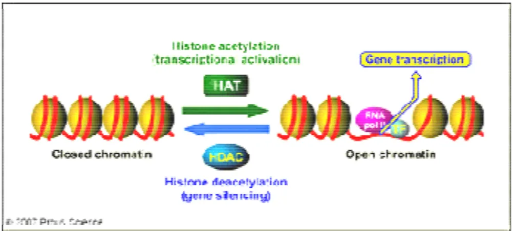
1.5.5 Histone acetyltransferases (HATs)... 25
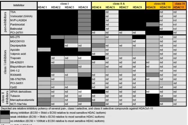
1.5.6 Histone deacetylase inhibitors ... 26
2 Aims... 30
3 Materials and Methods... 31
3.1 Fibroblasts derived from SMA patients...31
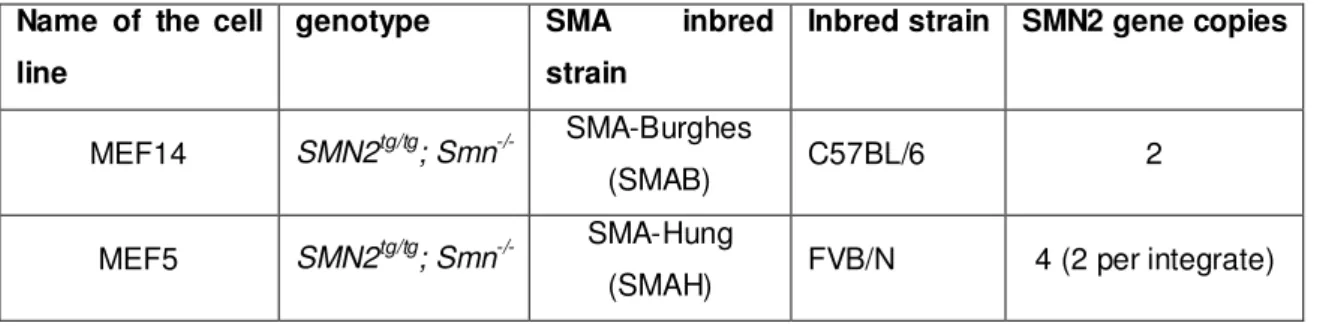
3.2 Mouse inbred strains ...31
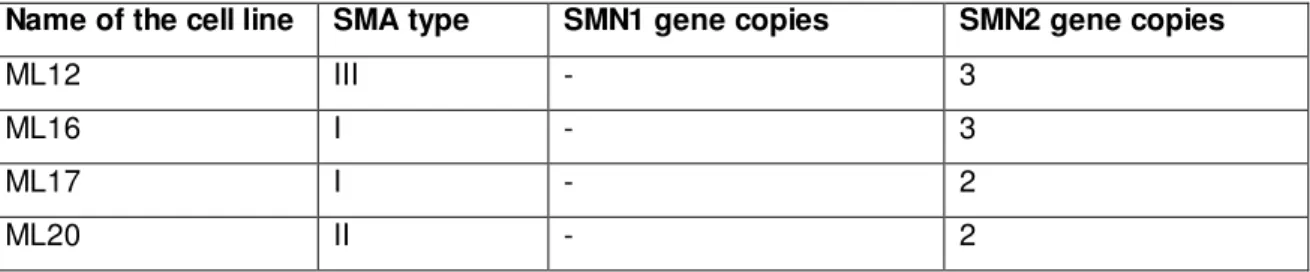
3.3 Cell lines derived from SMA patients...32
3.4 Equipment and Chemicals...32
3.4.1 Chemicals ...34
3.4.2 Kits ...34
3.4.3 Reagents, enzymes and additional supplies for cell culture procedures...35
3.4.3.1 Reagents ...35
3.4.3.2 Enzymes...36
3.4.3.3 Additional materials for cell culture procedures...36
3.4.3.4 Additional materials for laboratory mouse in vivo procedures...37
3.4.3.5 Purchased human cDNA clones...37
3.5 Antibodies...37
3.5.1 Primary antibodies ...37
3.5.2 Secondary antibodies ...37
3.6 Solutions and Media...38
3.6.1 Frequently used buffers and solutions ...38
3.6.2 Media for eukaryotic cell and tissue culture procedures ...42
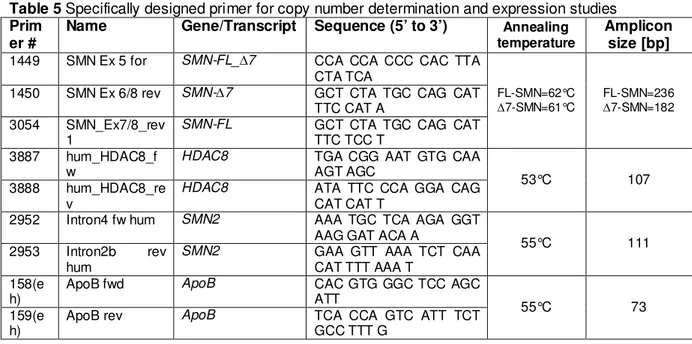
3.7 Primers and siRNAs...43
3.8 Software, internet programs, and databases...45
3.9 Cell culture procedures...46
3.9.1 Cell culture of primary fibroblasts derived from SMA patients...46
3.9.2 Stimulation of primary fibroblast cell lines with chemical substances ...47
3.9.3 MTT assay...47
3.9.4 Transient transfection of primary human fibroblasts...48
3.10 Animal breeding and characterization of SMA-like mice ...50
3.10.1 Animal breeding ...50
3.10.2 Motor ability test and determination of weight progression of mice ...50
3.10.3 Treatment of mice with test substances ...51
3.10.4 Generation of murine embryonic fibroblasts ...52
3.10.5 Preparation of mouse organs ...52
3.11 Molecular biology methods...53
3.11.1 Isolation of genomic DNA ...53
3.11.2 Photometric determination of the DNA concentration ...53
3.11.3 Fluorimetric determination of the DNA concentration...53
3.11.4 Isolation of total RNA from mouse organs...54
3.11.5 Isolation of total RNA from primary fibroblast cell cultures...54
3.11.6 Determination of the RNA concentration ...54
3.11.7 Photometric RNA concentration analysis ...54
3.11.8 Fluorimetric RNA concentration analysis with RiboGreen® dye ...55
3.11.9 Reverse transcription (cDNA synthesis)...55
3.11.10Polymerase chain reaction (PCR)...55
3.11.11Analysis of the number of transgenic SMN2 copies by quantitative real-time PCR ...56
3.11.12Analysis of gene expression by quantitative real-time PCR... 56
3.11.13Analysis of gene expression by one-step reverse transcription - quantitative real-time PCR ... 57
3.11.14Agarose gel electrophoresis for separation of DNA fragments... 57
3.12 Proteinbiochemical and immunological methods...58
3.12.1 Extraction of proteins from mouse organs... 58
3.12.2 Extraction of proteins from primary fibroblast cell cultures... 59
3.12.3 Protein contents determined according to the Bradford method... 59
3.12.4 Discontinuous denaturing polyacrylamide gel electrophoresis (SDS-PAGE)... 59
3.12.5 Transfer of proteins to nitrocellulose membrane by wet blotting (Western blot)... 60
3.12.6 Ponceau staining of proteins on nitrocellulose membranes ... 60
3.12.7 Immunostaining of membranes with antibodies and detection of signals with chemiluminescence reagent... 61
3.12.8 Chromatin Immunoprecipitation ... 62
3.13 Histochemistry ...64
3.13.1 Nissl staining... 64
3.13.2 Hematoxylin and eosin staining ... 64
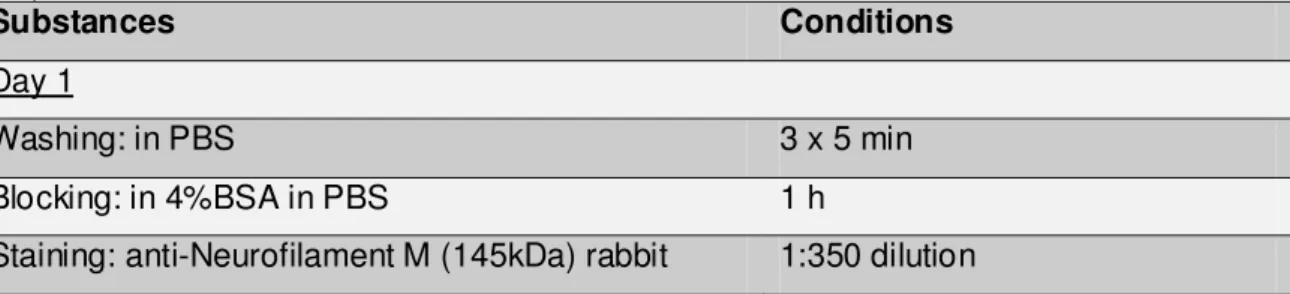
3.13.3 Immunohistochemistry... 65
3.13.4 Analysis of the motor neuron, NMJ and muscle stainings ... 66
3.14 Statistical methods...67
4 Results... 68
4.1 In vitro experiments with histone deacetylase (HDAC) inhibitors in fibroblast cell lines derived from SMA patients and SMA mouse models....69
4.1.1 Treatment of SMA fibroblast cultures with M344... 69
4.1.1.1 Impact on SMN2 RNA levels... 69
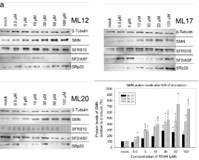
4.1.1.2 SMN and splicing factor protein level change under M344 ... 71
4.1.1.3 Change of the number of gems... 74
4.1.1.4 Cytotoxicity of M344 in SMA fibroblasts ... 76
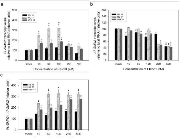
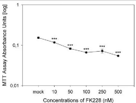
4.1.2 Treatment of SMA fibroblast cultures with FK228 (Depsipeptide)... 77
4.1.2.1 Impact on SMN2 RNA levels... 78
4.1.2.2 SMN and SFRS10 protein level change under FK228... 80
4.1.2.3 Change of the number of gems... 82
4.1.2.4 Cytotoxicity of FK228 in SMA fibroblasts... 84
4.2 Identification of the specific histone deacetylase (HDAC) regulating the expression of SMN2...85
4.2.1 Effects of histone deacetylase (HDAC) knock-down experiments on SMN2 expression... 85
4.2.2 Chromatin immunoprecipitation of HDAC8 in the promoter region of SMN2... 87
4.2.3 HDAC overexpression experiments in SMA fibroblasts and the effect on
SMN2 expression ...89
4.2.4 Treatment of SMA fibroblasts with the HDAC8 specific inhibitor PCI-34051...90
4.2.4.1 Impact on SMN2 RNA levels ...90
4.2.4.2 SMN and SFRS10 protein level change under PCI-34051...92
4.2.4.3 Cytotoxicity of PCI-34051 in SMA fibroblasts...94
4.3 In vivo treatment of SMA mouse models with the histone deacetylase (HDAC) inhibitor suberoylanilide hydroxamic acid (SAHA)...95
4.3.1 SAHA treatment of the SMAB mouse model...95
4.3.1.1 Determination of the transgene copy-number in the SMAB mouse model...96
4.3.1.2 Effects of SAHA treatment in the SMAB mouse model ...100
4.3.2 SAHA treatment of the SMAH mouse model...103
4.3.2.1 Establishment of the treatment concentration and application method ...103
4.3.2.2 Effects of SAHA treatment on survival of the SMAH mice...107
4.3.2.3 Effects of SAHA treatment on body weight of SMAH mice...109
4.3.2.4 Effects of SAHA treatment on motor ability of SMAH mice...111
4.3.2.5 Effects of SAHA treatment in the SMAH mouse model on SMN2tg expression...113
4.3.2.6 Histological changes in the SMAH mouse model after SAHA treatment...123
5 Discussion ... 129
5.1 In vitro experiments with the pan-HDACis M344 and FK228... 130
5.2 Identification of HDAC8 to be specifically involved in SMN2 expression regulation... 133
5.2.1 In vitro findings with regard to the current state of research...136
5.3 In vivo characterization of SAHA treatment in mouse models for SMA... 139
5.4 Future prospects... 150
5.4.1 Future prospects for potential SMA therapy...151
6 Summary... 153
7 Zusammenfassung ... 155
8 Publications, lectures, poster contributions and awards... 158
9 References... 160
10 Appendix...XI
List of abbreviations
A adenine
APS ammonium persulfate bp base pair
BSA bovine serum albumin
C cytosine
CD cluster of differentiation cDNA complementary DNA cen centromeric
CK creatine kinase cM centimorgan cm centimeter
CNS central nervous system DEPC diethylpyrocarbonate
D-MEM Dulbecco’s modified Eagle medium DMSO dimethylsulfoxide
DNA deoxyribonucleic acid EBV Epstein-Barr virus
EDTA ethylenediaminetetraacetic acid e.g. exempli gratia
EMG electromyography ESE exonic splicing enhancer ESS exonic splicing silencer et al. et alii
FCS fetal calf serum
FDA Food and Drug Administration FITC fluorescein isothiocyanate FL full length
G guanine
G acceleration due to gravity
h hours
HAT histone acetyltransferase HDAC histone deacetylase HMT histone methyltransferase HP-β-CD hydroxypropyl-β-cyclodextrin
i.e. id est
ISS intronic splicing silencer kb kilobases
kDa kilodalton
l liter
LAT lysine acetyltransferase LLN lower limit of normal
M molar
m milli-
Mb megabases MDa megadalton min minutes ml milliliter mm millimeter mM millimolar
mRNA messenger RNA
NCV nerve conduction velocity n.d. not determined
ng nanogram
nm nanometer nM nanomolar nmol nanomol n.s. not significant
OHSC organotypic hippocampal slice culture OMIM Online Mendelian Inheritance in Man P postnatal day
p probability (statistical significance) PAA polyacrylamide
PAGE polyacrylamide gel electrophoresis PBS phosphate-buffered saline
PCR polymerase chain reaction pH power of hydrogen
pmol picomol
RNA ribonucleic acid RNAi RNA interference rpm revolutions per minute RT reverse transcription
SAHA suberoylanilide hydroxamide acid s.c. subcutaneous
SD standard deviation SDS sodium dodecyl sulfate SEM standard error of the mean siRNA small interfering RNA
SMA autosomal recessive spinal muscular atrophy SMN survival motor neuron
T thymidine
TEMED N,N,N’,N’-tetramethylethylenediamine ter telomeric
UV ultraviolet VPA valproic acid
µ micro-
µg microgram
µl microliter µM micromolar µm micrometer
# number
1 Introduction
1.1 The clinical picture and diagnosis of proximal spinal muscular atrophy
Spinal muscular atrophies (SMA) present as a broad spectrum of diverse clinical pictures. All spinal muscular atrophies are caused by the loss of lower motor neurons in the spinal cord, resulting in denervation of especially voluntary muscles leading to muscle weakness and atrophy as main symptoms of the disease. The occurrence of the atrophy may differ between proximal or distal, symmetrical or asymmetrical muscle atrophy. Also a wide mode of inheritance is found, ranging from sporadic occurrence over X-chromosome linked to recessive or dominant heredity. Possible forms of spinal muscular atrophies include X-linked disease (XL-SMA), spinal muscular atrophy with respiratory distress (SMARD), spinal and bulbar muscular atrophy (Kennedy’s disease), and distal spinal muscular atrophy.Autosomal recessive proximal spinal muscular atrophy is the most common form of spinal muscular atrophies and, therefore, is simply referred to as “SMA”. It is the subject of this thesis and, unless mentioned otherwise, the term “SMA” used herein refers to the autosomal recessive proximal from of spinal muscular atrophies.
With an occurring incidence in the western European population of 1 in 6,000 to 10,000 life-births and a carrier frequency of 1:35 (Czeizel and Hamula 1989; Feldkotter et al. 2002; Pearn et al. 1978), SMA is the second most frequent autosomal recessive disorder in humans and the leading genetic cause of infant death today (Montes et al.
2009).
The major characteristic hallmark of SMA is the degeneration of α-motor neurons in the anterior horns of the spinal cord, leading to hypotonia and muscle weakness finally resulting in muscle atrophy. Other neurological systems (including brain and sensory nerves) usually display no dysfunction and affected individuals show normal intelligence.
Muscular weakness starts at the voluntary muscles of the proximal limbs, such as upper arms and legs, rather than in the distal ones, like hands or feet. The atrophy is more pronounced and begins earlier in the legs than in the arms. SMA is a progressive disease starting from muscle weakness and resulting in atrophy of the muscles. SMA patients show further symptoms, like tremor of the hands and an explicit decrease of deep reflexes. Furthermore, fasciculations of the tongue are observable in SMA patients.
Muscle groups in the face and in the eyes are typically not involved.
The serum level of the creatine kinase (CK) activity is a common and very sensitive marker of increased muscle membrane permeability. Elevated CK activity points towards a muscle damage (Schlattner et al. 2006). CK activity is a common diagnostic tool for
myocardial infarction or muscular dystrophy. While in these cases the CK activity level is highly increased (10-fold and more above the normal level), in SMA patients only a mild elevation can be detected. Therefore, a highly augmented CK amount is a criterion of exclusion for SMA diagnosis.
Nowadays, one of the main diagnostic tools for SMA is electromyography (EMG).
Electromyography is a technique for evaluating and recording the activation signal of muscles. With the help of an apparatus called “electromyograph” EMG is performed and the results are recorded in an electromyogram. By the use of this apparatus it is possible to detect the electrical potential generated by myoblasts in an active and in an inactive state. Electromyographic investigations in affected SMA patients show a typical pattern of denervation, normally without sensory involvement. Additionally, SMA patients reveal specific EMG characteristics, such as spontaneous muscle activity with fibrillations and fasciculations of single muscle fibers and motor units. The nerve conduction velocity (NCV) in SMA patients is normal or mildly reduced, but not lower than 70% compared to healthy individuals.
As a guideline for the diagnosis of SMA, in 1992 the SMA Consortium defined the following list of major exclusion criteria for SMA (Munsat and Davies 1992):
• CNS dysfunction
• arthrogryposis
• involvement of other neurologic systems or other organs (e.g. hearing, cardiac or vision)
• sensory loss
• eye muscle weakness
• marked facial weakness
• marked increase in creatine kinase (CK) activity levels.
Still, in very rare SMA cases it has been observed that the disease may sometimes associate with arthrogryposis, CNS involvement, increased CK values or heart defects (Rudnik-Schoneborn et al. 1996). Nevertheless, it remains elusive, whether these observations are true causal associations or just random findings.
To screen out the misdiagnosis of muscular dystrophy or other neuromuscular disorders and to verify the diagnosis for SMA, muscle biopsy of patients is an established means. These histological samples are evaluated histochemically, and may allow distinguishing SMA from other denervating diseases. There are two types of muscle fibers. Muscle fibers of the type I have a red appearance, because they contain
the oxygen binding protein myoglobin and they show slow fatigue since they generate ATP by oxidative metabolism, and therefore are suited for endurance. Due to the absence of myoglobin, type II fibers appear white. Besides the oxidative metabolism type II fibers make use of the anaerobic metabolism, hence they are faster to fatigue, but they are efficient for short bursts of speed and power. In a muscle biopsy from a typical chronic SMA patient groups of atrophic and hypertrophic fibers or fiber-type grouping are most often found (see Figure 1). Moreover, SMA skeletal muscle usually shows atrophic fibers with islands of group hypertrophy (Buchthal and Olsen 1970). Furthermore, evidence of skeletal muscle denervation can be observed.
Figure 1Histopathology of spinal muscular atrophy
Motor commands generated in the cerebral cortex are transmitted through spinal cord α-motor neurons (red cell in spinal cord anterior horn and green arrow in (A)). The spinal cord anterior horn region shows an absence of motor neurons in a patient (B) compared with those in the healthy control (A). Skeletal muscle of a patient (D) shows hypertrophic fibers (hollow arrowhead) surrounded by group atrophy (green arrowhead) compared with healthy fibers with uniform morphology in normal infantile muscle (C). Despite the atrophy of muscle fibers in spinal muscular atrophy, muscle spindles (black asterisk) are not affected and become more conspicuous (D). All slides are stained with haematoxylin and eosin. (Taken from (Lunn and Wang 2008))
Besides the atrophy of both muscle fiber types, an additional distinctive feature is the presence of a small number of scattered hypertrophic type I fibers presumably resulting
from physiologic hypertrophy. Nevertheless, normal-appearing fibers may be present.
Although the muscle biopsies of SMA patients show neither, significant necrosis, degeneration, regeneration, lipid accumulation, nor connective tissue proliferation, some of these features may sometimes occur in older chronic SMA patients, suggesting a secondary myopathic process.
In the year 1995 Lefebvre and colleagues identified the SMA disease determining gene, called survival of motor neuron 1 gene (Lefebvre et al. 1995). Since then it has been possible to confirm the clinical diagnosis of SMA by molecular genetic testing. To verify the clinical diagnosis of SMA, the SMN1 gene on chromosome 5q is screened for specific mutations (deletions/gene conversions of exon 7 or exon 7 and 8). Furthermore, this highly important diagnostic tool may also be applied in prenatal diagnosis and carrier testing. The gain of knowledge regarding SMA led to the modification of the primary diagnostic criteria proposed by the International SMA Consortium (Munsat and Davies 1992) by Zerres and Davies in 1999 (Zerres and Davies 1999). In 2008, Lunn and Wang proposed another guideline for proper SMA diagnostics shown in Figure 2.
Figure 2 Diagnostic algorithm for spinal muscular atrophyEvery patient presenting with clinical symptoms resembling spinal muscular atrophy (SMA) should be tested for homozygous deletion of SMN1, which would confirm the diagnosis of SMN-associated SMA (5q SMA). A negativeSMN1test should be followed by a repeat clinical examination for atypical clinical features (e.g., contractures, eventration of hemidiaphragms, congenital absence of muscles) and laboratory testing for creatine kinase (CK) and electrophysiological studies such as EMG and a nerve conduction study (NCS). If lower motor neuron disease is suggested by EMG, SMN1copynumber will establish ifSMN1sequencing is indicated to identify intragenic mutations in patients with a singleSMN1copy. If twoSMN1copies are detected, theninvestigation should be directed towards other motor neuron diseases by further diagnostic work-up such as muscle or nerve biopsy, imaging studies, metabolic screens, and genetic testing. ALS=amyotrophic lateral sclerosis. NMJ=neuromuscular junction. SMARD=SMA with respiratory distress. Red boxes=diagnostic assessments. Blue boxes=physical and laboratory findings. Green boxes=final diagnoses. Taken from (Lunn and Wang 2008).
1.1.1 Classification of proximal SMA
Based on different motor functions and on the age of onset, spinal muscular atrophy has been divided into four clinical types: severe type I; intermediate type II; mild type III and adult-onset type IV as a very mild form (Munsat and Davies 1992; Zerres and Rudnik- Schoneborn 1995).
1.1.1.1 SMA type I (Werdnig-Hoffmann disease, MIM #253300)
This type of SMA is the most severe form and is also called acute SMA. The clinical picture of SMA type I was first described by Werdnig in 1891 (Werdnig 1891) and independently by Hoffmann in 1893 (Hoffmann 1893).
Usually type I SMA is diagnosed within the first 6 months of life. The onset of the disease is before the age of 6 months and death occurs within the first two years of life.
Recent data suggest a mean age of survival of ~7 months for an SMA type I patient (Rudnik-Schoneborn et al. 2009, in press). In rare cases, the symptoms may begin prenatally (in the third trimester). Werdnig-Hoffmann disease is the most common form of SMA and accounts for about 50% of patients diagnosed with spinal muscular atrophy (Markowitz et al. 2004). Besides a generalized muscle weakness (“floppy infants”) the affected children suffer from hypotonia and are never able to sit unaided. The patients show symmetrical flaccid paralysis and no control of head movement. Typical for SMA type I patients is the paradoxical breathing (inward bony thorax movement with outward abdominal movement during inspiration) and a bell-shaped upper torso, which is due to their spared diaphragm, combined with weakened intercostal muscles. The bulbar denervation results in characteristic tongue fasciculation and weakness with poor suck and problems in swallowing. The increased risk of aspiration pneumonia is an important cause of morbidity and mortality. In 1995, it was postulated that 8% of affected children reach the tenth year of life (Zerres and Rudnik-Schoneborn 1995). However, that study might have included non SMN1-linked patients, since at that time SMN1 had not yet been identified.
1.1.1.2 SMA type II (intermediate form, MIM #253550)
Affected children with SMA type II show first disease symptoms after six, but before 18 months of life. SMA type II is of intermediate severity and patients are able to sit independently but never learn to walk and need support to stand. Life expectancy is reduced, but usually children do not die before the age of two years and more than 70%
grow older than 20 years. Surgical or orthotic intervention is required to correct the often
developing kyphoscoliosis. Frequent symptoms are also fine tremors with digit extension or hand grips. Comparable to patients with type I disease, clearing of tracheal secretions and coughing might become difficult because of poor bulbar function and weak intercostal muscles. A very frequent cause of death during adolescence is respiratory insufficiency.
1.1.1.3 SMA type III (Kugelberg-Welander, MIM #253400)
A less severe form of SMA was for the first time reported in the year 1956 by Kugelberg and Welander (Kugelberg and Welander 1956).
The so called juvenile SMA is quite variable in its age of onset, but generally first symptoms of SMA type III occur after the 18th month of life. Depending on the exact age of onset, SMA type III is divided into two subtypes: if first symptoms are diagnosed before the age of three years, the disease is called type IIIa, if the patient is older than three years he or she SMA bears type IIIb (Zerres and Rudnik-Schoneborn 1995). This juvenile form of SMA is very heterogeneous as some affected individuals may need wheelchair assistance in childhood, whereas others may continue to walk throughout their whole lives. A quiet common orthopedic feature of this progressive form is the development of scoliosis.
1.1.1.4 SMA type IV (adult SMA, MIM #271150)
The first symptoms of this adult form of SMA typically occur in the second or third decade of life (Wirth 2002). The patients are comparatively mildly affected and have a normal life expectancy. No respiratory problems are observed and affected individuals are able to walk in adult years.
1.2 The molecular basis of proximal spinal muscular atrophy (SMA)
The identification of the SMA disease causing gene started in the year 1990, when scientists succeeded in mapping SMA patients of type I, II, and III by linkage analysis to a region of about 10 cM on chromosome 5q11.2 - q13.3 (Brzustowicz et al. 1990; Gilliam et al. 1990; Melki et al. 1990). A few years later the development of many new polymorphic markers enabled the researchers to narrow down the initial quite large interval to a critical disease region of less than 1 Mb (DiDonato et al. 1994; Melki et al.
1993; Melki et al. 1994; Soares et al. 1993; Wirth et al. 1995; Wirth et al. 1994). This newly identified region contained a highly complex unstable sub-region of about 500 kb, which was subjected to intrachromosomal rearrangements, including gene duplications, gene conversions, and de novo deletions (Lefebvre et al. 1995). This complex genomic organization considerably hindered the researches to construct a uniform physical map (Lefebvre et al. 1995; Melki et al. 1994; Roy et al. 1995a; Roy et al. 1995b; Thompson et al. 1993).
Finally in the year 1995, the survival motor neuron gene (SMN) was identified as the disease-causing gene by the use of a human fetal brain cDNA library (Lefebvre et al.
1995).
Today it is well-known that the genomic SMA region is prone to de novo rearrangements, including unequal crossovers, intrachromosomal rearrangements, and gene conversions (Melki et al. 1994; Wirth et al. 1997). In this critical 500 kb SMA segment five genes have been identified: the survival of motor neuron gene (SMN); the BIRC1 (baculoviral IAP repeat-containing 1) gene, also known as NAIP (neuronal apoptosis inhibitory protein); the SERF1 (small EDRK-rich factor 1) gene, also known as H4F5, the GTF2H2 (general transcription factor IIH) gene or p44; and the OCLN (occludin) gene (Lefebvre et al. 1995; Schmutz et al. 2004) (see Figure 3). This dynamic 500 kb genomic SMA region can be present in 0 to 4 copies per chromosome 5 and is flanked by the single-copy genes RAD17 (proximal) and BDP1 (also known as TFNR) (distal) (Kelter et al. 2000; von Deimling et al. 1999).
Figure 3 Schematic overview of the genomic SMA region on chromosome 5q13. Many genes in the vicinity of the SMN genes are duplicated and inverted.
1.2.1 The SMN gene
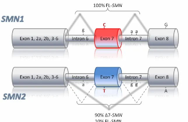
Lefebvre and colleagues identified the SMA disease-determining gene called survival motor neuron gene (SMN) in the year 1995 (Lefebvre et al. 1995). Two copies of the SMN gene are located in the genomic SMA candidate region: the telomeric (SMN1) and the centromeric (SMN2) duplicate (Lefebvre et al. 1995). These gene copies are almost identical except for five silent nucleotide differences not affecting the amino acid sequence: one each in exon 7 (840C→T, codon 280, nt position 27141), exon 8 (nt position 27869) and intron 6 (nt position 27092) and another two in intron 7 (nt positions 27289 and 27404), respectively (see Figure 4) (Burglen et al. 1996; Lefebvre et al.
1995). To date there have several other gene variants been described, but discrimination between SMN1 and SMN2 has not been possible, since these variants have been found in both gene copies (Brahe et al. 1996; Hahnen and Wirth 1996; Monani et al. 1999).
The SMN gene has a size of 28 kb on genomic level and consists of 9 exons (1, 2a, 2b-8) and 8 introns with an open reading frame of 882 bp (294 codons) (Chen et al.
1998; Lefebvre et al. 1995). The ~1.5 kb SMN transcripts are expressed in all somatic tissues and encode a 294 amino acid protein of 38 kDa (Lefebvre et al. 1995). The expression level is ~50-fold to 100-fold higher in spinal cord as compared to other tissues (Coovert et al. 1997; Lefebvre et al. 1997). Interestingly, the SMN protein is very highly conserved from yeast to man (Miguel-Aliaga et al. 1999; Paushkin et al. 2000;
Schrank et al. 1997). On an evolutionary background, the SMN gene duplication occurred for the first time in primates and therefore took place after the split of primates from rodents, since mice and rats have only one copy namely Smn. Nevertheless, the gene copy SMN2 is unique to humans, since chimpanzees never carry this converted gene despite having multiple SMN gene copies in their genome (Rochette et al. 2001).
Over 98% of patients with spinal muscular atrophy have a homozygous SMN1 deletion, rearrangement, or mutation (Hahnen et al. 1995; Lefebvre et al. 1995). All these patients, however, retain at least one copy of SMN2, which undergoes alternative splicing and produces mainly a truncated mRNA isoform, which lacks exon 7 (major product) or exon 5, or both (see 1.2.2). In these individuals the loss of the SMN1 gene causes SMA, while the severity of the SMA phenotype inversely correlates with the number of SMN2 copies (Lefebvre et al. 1995; Wirth 2000). In very rare cases some individuals are protected from developing SMA, although they lack SMN1. These persons express the only known fully protective modifier of SMA, namely Plastin 3 (Oprea et al. 2008).
Figure 4 Schematic representation of the genomic structure, nucleotide and splicing differences between SMN1 and SMN2. The SMN gene copies can be distinguished by 5 nucleotide exchanges of which only the C T transition in exon 7 (marked in red) lies within the coding region. The nucleotide exchange in exon 7 is a translationally silent mutation. Therefore full-length SMN1 and SMN2 mRNA encode identical proteins of 294 amino acids. However, the CT transition affects an exonic splicing enhancer and/or silencer causing alternative splicing of SMN2 pre-mRNA. SMN2∆7 transcripts produce a truncated and unstable protein.
1.2.2 The SMN transcript
Although the SMN1 and the SMN2 gene encode for an identical protein, they differ in 5 nucleotide exchanges, all of which are translationally silent. SMN1 produces almost exclusively full length transcripts, whereas because of the C to T transition at position +6 in exon 7 of SMN2, 90% of the SMN2 transcripts are alternatively spliced lacking exon 7 (∆7-SMN2). Only 10% of SMN2 pre-mRNA produce full length transcripts (FL-SMN2).
The first stop codon of the FL-transcripts is located at the end of exon 7; hence these transcripts are translated into a FL-SMN protein consisting of 294 amino acids. The ∆7- SMN2 transcripts lack exon 7 and therefore encode a truncated SMN protein of only 282 amino acids. The skipping of exon 7 leads to the use of an alternative stop codon located in exon 8, therefore a truncated protein is generated, which C-terminally lacks 16 amino acids encoded by exon 7 but instead contains four amino acids encoded by exon 8. This shortening of the protein reduces its oligomerization capacity and decreases its stability.
Thus, the appropriate protein function of SMN is severely reduced (Lorson and Androphy 2000; Lorson et al. 1998).
Furthermore, it has been shown that both SMN genes are able to produce three more transcripts. One alternative transcript is the ∆3-SMN, which lacks exon 3, another one is
∆5-SMN lacking exon 5 and, additionally, there are SMN mRNA occurring which lack both, exon 5 and exon 7 (∆5,7-SMN) (Chang et al. 2001; Gennarelli et al. 1995; Singh et al. 2006).
At a closer look on the splicing function of the SMN protein, the ∆3-SMN protein reveals an interesting feature of the Tudor domain, which is located in exon 3 of SMN.
Researchers have proven that the disruption of this respective domain reduces the ability of SMN to interact with the Sm (Smith antigen) proteins, which is essential for the splicing involvement of the SMN protein (Buhler et al. 1999; Mohaghegh et al. 1999; Sun et al. 2005) (see also protein function of SMN chapter 1.2.3.1).
Focusing on the SMN pre-mRNA splicing, it is necessary to mention the C to T transition in SMN2 exon 7 again. This particular nucleotide exchange lies directly within an exonic splicing enhancer (ESE) (Lorson and Androphy 2000; Lorson et al. 1999). It destroys a heptamer motive, which is normally (in the case of SMN1) recognized by the splicing factor SF2/ASF (splicing factor 2 (SF2), a positive splicing factor that is also known as the alternative splicing factor (ASF)) (Cartegni and Krainer 2002). The hence resulting alternative splicing produces a transcript lacking exon 7. A concurring hypothesis instead postulates that the C to T transition forms a new exonic splicing silencer (ESS), which recruits the splicing factor hnRNP A1, and that this leads to the exclusion of exon 7 (Kashima and Manley 2003).
The splicing pattern of the SMN2-pre-mRNA is subjected to the influence of different splicing factors. It has been shown in in vivo splicing experiments that the SR- like splicing factor SFRS10 (Serine/Arginine rich splicing factor 10; formally known as hTra2-β1, the human homolog of transformer-2 from Drosophila melanogaster) specifically binds to a GA-rich region, a second ESE in the central part of SMN exon 7.
Overexpression of SFRS10 restores the alternative splicing pattern of SMN2, so that 80% of full length transcripts are produced (Hofmann et al. 2000).
Both splicing factors SF2/ASF and SFRS10 interact specifically and directly with exon 7 of the SMN pre-mRNA (Cartegni and Krainer 2002; Hofmann et al. 2000).
Splicing factors like SRp30 and hnRNP-G act on the exon 7 inclusion via binding to SFRS10 (Bose et al. 2008; Helmken and Wirth 2000; Hofmann and Wirth 2002; Young et al. 2002). The splicing factor SF2/ASF is interacting with the U2 class of small nuclear ribonuclear protein (U2 snRNP) and its auxiliary factor (U2AF) at the branch point located in intron six to support removal of the intron and successful pre-mRNA splicing during SMN1 transcription (Cartegni and Krainer 2002). A detailed description of the splicing process of SMN pre-mRNA is given in Figure 5.
Furthermore, several chemical compounds were found to influence the splicing pattern of SMN2. Previously it was shown in a lymphoblastoid cell line for sodium butyrate to increase the FL-SMN2 level 4-fold by forcing the inclusion of exon 7 (Chang et al. 2001). In the following years the capability to boost full-length levels of transcripts was proven in fibroblast cultures for Aclarubicin (Andreassi et al. 2001) and in our laboratory for valproate (Brichta et al. 2003).
Figure 5 Pre-mRNA splicing of SMN1 and SMN2
In SMN1, an exonic splicing enhancer (ESE), which contains the nucleotide cytosine (C) at position six in exon seven (Ex7+6), is recognised by splicing factor 2 or alternative splicing factor (SF2/ASF), which interacts (thick black arrow) with the U2 class of small nuclear ribonuclear protein (U2 snRNP) to remove intron six. Other splicing factors (e.g. SFRS10) determine splicing through interactions (thin black arrow) with ESE elements found centrally within exon seven.
Serine and arginine (SR)-rich proteins might also exert a positive splicing effect. In SMN2, the ribonucleotide uridine (transcribed from thymidine) at Ex7+6 favours exon seven exclusion by binding to heterogeneous nuclear ribonuclear protein (hnRNP) A1, a negative splicing factor.
Moreover, SF2/ASF no longer recognises this sequence motif. Binding of hnRNP A1 is also believed to prohibit U2 snRNP binding to the branch point, which results in about 90% of SMN2 final mRNA transcripts with no exon seven. The positive splicing factors downstream (thin black arrow) are functioning and could account for exon 7 inclusion in about 10% of SMN2 transcripts.
(taken from (Lunn and Wang 2008))
1.2.3 The SMN protein
The SMN protein, 38 kDa in size, consists of 294 amino acid and is found in virtually all cells of the human body (Carvalho et al. 1999; Lefebvre et al. 1995; Liu and Dreyfuss 1996; Young et al. 2001; Young et al. 2000). The full-length SMN protein localizes both in the nucleus and the cytoplasm. Immunostaining of various cell types revealed a diffuse cytoplasmic distribution of SMN, while it is found in bright dot-like structures in the nucleus (Burlet et al. 1998; Coovert et al. 1997; Liu and Dreyfuss 1996). These SMN- containing structures are often observed in close proximity to or completely overlapping with the coiled bodies [also Cajal bodies; nuclear domains that are enriched in spliceosomal U snRNPs (Fakan et al. 1984)], and therefore are termed gemini of coiled bodies (gems) (Liu and Dreyfuss 1996; Liu et al. 1997; Young et al. 2000).
Another isoform of SMN consisting only of exons 1-3 and a part of intron 3 is only observed in axons (aSMN) (Setola et al. 2007).
Since the SMN protein possesses diverse functions in many different environments, its functions are subsequently subdivided into a) housekeeping functions, b) neuron specific functions and c) muscle specific functions:
1.2.3.1 Housekeeping functions of SMN
o Biogenesis of snRNPs (pre-mRNA splicing)
The so called small nuclear ribonucleoproteins (snRNPs) play an important role in every cell. The snRNPs are active in recognizing and removing introns from pre-mRNAs in the nucleus. Every snRNP is comprised of a small nuclear RNA (snRNA) and a protein part.
The protein part is made of a number of specific proteins that are unique for each snRNP and the so called Sm (Smith antigen) proteins. The Sm proteins are essential for the splicing process. SMN plays here an important role in assembling Sm proteins onto the snRNAs to produce an active snRNP particle (Eggert et al. 2006; Gubitz et al. 2004;
Pellizzoni 2007; Raker et al. 1999). Therefore, the SMN protein acts in an active complex in the cytoplasm of every cell. This so called “SMN complex” is composed of SMN, GEMIN2–8 and UNR-interacting protein (UNRIP). SMN protein itself is acting as an oligomer as it has been found to self-associate, and it has been suggested that oligomerization is crucial for SMN function (Lorson et al. 1998; Pellizzoni et al. 1999).
The exact numbers of SMN monomers in an SMN complex is unknown, it has been shown to form higher-order complexes ranging from 20S to 80S and has been proven to
be impaired in SMN mutants of SMA patients (Lorson et al. 1998; Paushkin et al. 2002;
Pellizzoni 2007; Pellizzoni et al. 1999).
o Transcription
The SMN protein was shown to interact with its exon 6 with mSin3a (Zou et al. 2004).
mSin3a is a well known transcription corepressor that is involved in histone deacetylation and chromatin remodelling. SMN and the mSin3a protein are involved in the formation of a protein supra-complex exceeding 40,000 kDa in size (Zou et al. 2004). Since mSin3a associates with histone deacetylases and methytransferases and other proteins known to regulate chromatin remodelling (Yang et al. 2003), it has been postulated that SMN might be involved in the transcriptional repression of critical genes in motor neurons, via mSin3 associated histone modification enzymes (Zou et al. 2004).
o Stress response
If a cell is exposed to environmental stress such as heat shock, exposure to relevant chemical compounds or to UV irradiation, it starts a protective cellular process called
“stress response“. While specific stress-induced genes are activated in response to stress most other genes are silenced. It has been shown that ~50% of total mRNA transcripts are actively recruited and dynamically sorted into cytosolic compartments forming the so-called stress granules (SGs) (Kedersha et al. 1999). This process has been shown to be dependent on the eukaryotic initiation factor (eIF) 2α and its phosphorylation status (Clemens 2001a, b). The SGs have been proven to serve as transient local storage compartment for mRNAs under stress conditions (Nover et al.
1989). Interestingly, it has been observed that the SMN protein co-localizes and interacts with T-cell internal antigen-1 (TIA-1) and TIAR (TIA-1-related protein), which are known RNA-binding proteins that accumulate in SGs (Hua and Zhou 2004). It has been stated that SMN granules can be seen as stress granules (Hua and Zhou 2004). This suggests that SMN contributes to formation and function of SGs.
o Translation regulation
Since SMN is known to be involved in mRNA localization and transport along the axons, SMN is widely discussed to be involved in translational regulation. It has been shown that the fragile X mental retardation syndrome protein (FMRP), which plays an important role in transport of mRNPs and in their translation, interacts with SMN. Therefore, it has been discussed that SMN may have a specific function in regulating translation in neurons (Piazzon et al. 2008).
1.2.3.2 Neuron specific functions of SMN
o Axonal mRNA transport
For the C-terminal exon 7 of SMN was shown that it contains a sequence motif, which is responsible for the cytoplasmic localization of the SMN protein. The overexpression of an exon-7 deletion mutant was characterized by nuclear accumulation of SMN and reduced neurite outgrowth. An active transport of SMN granules to developing neurites and growth cones was observed, indicating an involvement of SMN in axonal transport of mRNA and mRNA binding proteins (Zhang et al. 2003). Recently, a novel isoform of SMN was identified, called a-SMN (axonal SMN). This isoform consists of exons 1-3 and a part of intron 3 of SMN and is exclusively found in axons of motor neurons (Setola et al. 2007). a-SMN, preferentially encoded by SMN1, stimulates motor neuron axonogenesis and induces axonal-like growth in non-neuronal cells (Setola et al. 2007).
o Neurite outgrowth
Not only the axonal isoform of SMN (a-SMN) plays an important role in neurite outgrowth (see chapter above), but also the selective overexpression of the SMN C-terminal domain has been shown to induce neurite outgrowth similar to full length protein and could rescue SMN knock-down effects. Furthermore, it has been stated that knock-down of endogenous SMN led to a significant change in the G-/F-actin ratio, indicating a role for SMN in actin dynamics. In regard to these observations, SMN might be involved in microfilament metabolism in axons of motor neurons (van Bergeijk et al. 2007).
Moreover, the knock-down of Smn in zebrafish embryos has been shown to lead to an earlier branching of axons, underlining the important role of Smn in motor axon–
specific pathfinding and in motor axon development (McWhorter et al. 2003).
Overexpression of the recently identified protective modifier of SMA, Plastin-3, has been observed to rescue this axonal phenotype by restoring the axonal length of motor axons (Oprea et al. 2008).
o Neuromuscular junction formation
Neuromuscular junctions (NMJ) are the synapses of the axon terminal of a motor neuron with the motor endplate of a muscle. The neurotransmitter acetylcholine passes the signal through the neuromuscular junction to the motor endplate. In an SMA mouse model in which Smn protein levels are severely reduced, the maturation of the NMJs was shown to be seriously impaired. Reduced Smn level has been shown to lead to an impairment of the maturation of acetylcholine receptor (AChR) clusters into pretzel-like
structures. Furthermore, pre-synaptic defects, such as poor terminal arborization have been observed. The described NMJ malformations have been postulated to lead to functional deficits at the NMJ characterized by intermittent neurotransmission failures (Kariya et al. 2008). Moreover, a recent publication indicated a severely disturbed synaptic vesicle release at NMJs in an SMA-like mouse model (Kong et al. 2009).
1.2.3.3 Muscle specific functions of SMN
o Myoblast fusion, sarcomeric Z-disc localization
Although SMA is a neuromuscular disease affecting primarily the motor neurons and, as a secondary effect, the muscles leading to atrophy, features like scattered hypertrophic type I fibers, significant necrosis, lipid accumulation or connective tissue proliferation, are rarely found in older SMA patients, suggesting a secondary myopathic process.
Therefore, several studies searched for a muscle specific role of SMN. Indeed, some functions may be specific for this tissue, but these findings are still subject to discussions. One work has proven that the knock-down of Smn in myoblasts led to severe defects in myoblast fusion and an abnormal myotube morphology (Shafey et al.
2005). Another publication revealed that the whole Smn protein complex is localized to the sarcomeric Z-disc and is a direct target of calpain-3-mediated proteolytic cleavage (Walker et al. 2008).
Noteworthily, the hypothesis that SMA might be caused by a muscle specific reduction of SMN was rebutted by the finding that neuronal specific expression of SMN in a Smn knock-out mouse rescues the phenotype, whereas the muscle specific expression does not (Gavrilina et al. 2008a).
1.3 SMA animal models
Since humans are the only known species carrying an SMN2 gene copy, all animals used to model SMA have only one endogenous Smn gene that is equivalent to SMN1.
Supporting the importance of SMN, in all organisms, loss of Smn leads to lethality.
Nevertheless, the levels of maternal Smn (Smn derived from the mother’s Smn gene as opposed to the embryo’s) defines the time point of lethality. In Smn knock-out mice for example, there is only a minimal amount of maternal Smn leading to an early embryonic lethality (Schrank et al. 1997). In contrast to mice, in the egg-laying animal D.
melanogaster death occurs later, when the maternal Smn level drops below a crucial point (Chan et al. 2003). The resulting hypothesis that loss of SMN in a specific tissue in conditional Smn mutants results in the death of that tissue was proven by different approaches (see chapter 1.3.1.3) (Cifuentes-Diaz et al. 2001; Frugier et al. 2000; Vitte et al. 2004).
1.3.1 SMA mouse models
To analyze the pathomechanism of SMA, many different SMA mouse models have been generated over the past years. Mice carry only one Smn gene, which is equivalent to the human SMN1, and lack another copy comparable to the human SMN2 (DiDonato et al.
1999; DiDonato et al. 1997; Viollet et al. 1997). In the following chapters, different SMA mouse models will be highlighted.
1.3.1.1 Classic knock-out of murine Smn
Already in 1997, Schrank and co-workers described the first classical knock-out of the murine Smn gene. The researches disrupted exons 2-4 of the Smn gene and observed early embryonic lethality before implantation between day E2.5 and E3.5. The analyzed blastocysts and morulae have revealed a disorganized, multicystic structure with extensive cellular degeneration (Schrank et al. 1997).
In the year 2000, the group of Li genetically replaced a 1.6 kb fragment of the Smn gene including exon 7 with a HPRT (hypoxanthine phosphoribosyltransferase) cassette and observed normal embryos at E3.5 but not at E6.5, indicating an essential role of Smn in the embryonic development (Hsieh-Li et al. 2000).
Heterozygous Smn+/--mice showed no symptomatic muscle atrophy, but a ~50%
reduction of the Smn protein level and a resulting degeneration of the α-motor neurons after 6 months of life (Jablonka et al. 2000).
1.3.1.2 SMN2 transgenic mice modeling an SMA phenotype
Since the homozygous ubiquitous knock-out of the endogenous Smn gene leads to early embryonic lethality, researchers tried to mimic SMA in mice by additionally integrating copies of the human SMN2 gene. In the year 2000, two independent groups managed to generate an SMA-like phenotype in mice. Monani and co-workers integrated a 34 kb large BamHI fragment of human SMN2, isolated from a cosmid clone into the mouse genome. Mice with an Smn-/-;SMN2tg/tg genotype represent an SMA-like phenotype, with degeneration of α-motor neurons and resulting muscle atrophy. Comparable to SMA patients, the severity of the disease correlates inversely with the SMN2 copy number.
While Smn-/-;SMN2tg/tg-mice, carrying one SMN2 copy per integrate, die between 1-8 days, Smn-/-;SMN2tg/tg-mice with 8-16 SMN2 copies show a complete rescue of the phenotype (Monani et al. 2000). Hsieh-Li and co-workers integrated a BAC clone containing the human SMN2 gene into the murine DNA. The resulting Smn-/-;SMN2tg/tg- mice carry 4 SMN2 copies (two per integrate), are fertile and live for about one year (Hsieh-Li et al. 2000). Smn-/-;SMN2tg/wt-mice show a severe form of SMA and survive for about 9.9 days (own observation).
Another mouse model resembling a milder form of SMA (SMA type III mice) is based on the already mentioned Monani-SMA mouse model. In a study by Arthur Burghes´ laboratory, researchers have introduced a transgene carrying a missense (A2G) mutation in exon 1 of the SMN gene into the severe SMA genetic background.
Smn-/-;SMN2tg/tg;SMNA2Gtg/wt-mice exhibited many of the clinical and pathological characteristics of type III (mild) SMA, suggesting a stabilization-capacity of the truncated SMN protein for the low expressed full-length SMN protein amount. These mice suffer from motor neuron degeneration and its associated effects. They include evidence of motor axon degeneration, loss and sprouting, muscle atrophy, and abnormal EMG patterns (Monani et al. 2003). Moreover, researchers could recently show that a previously human SMN1 mutation (SMN(A111G)) (Sun et al. 2005), an allele capable of snRNP assembly, can rescue mice that lack Smn and contain either one or two copies of SMN2 (SMA mice). However, the SMNA111G transgene has been proven not to be able to rescue the SMA phenotype alone. Moreover, the normal survival of SMNA111G-high- expressing Smn-/-;SMN2tg/tg;SMNA111Gtg/wt-mice has directly been correlated with snRNP assembly activity in spinal cord and the correction of snRNA levels (Workman et al.
2009).
Furthermore, the transgene expression of the truncated SMN2∆7 cDNA on a Smn-/-
;SMN2tg/tg background was observed to increase the lifespan of SMA-like animals from 5 to 14 days. This was an in vivo proof that the SMN∆7-protein has the capability to
associate with the SMN-protein and to stabilize the whole SMN-complex and that SMN∆7 has no detrimental effect on the SMA animals (Le et al. 2005).
1.3.1.3 Conditional Smn knock-out mice
To investigate tissue specific functions of Smn, several Cre/LoxP mediated conditional knock-out strains were generated.
With the help of a neuron-specific enolase promoter driven Cre-recombinase (NSE-Cre) Frugier and colleagues disrupted the endogenous Smn in neurons of mice.
Profound motor axon but no cell body loss and muscle paralysis of neurologic origin was detected, the mean age of these SMA-like animals was 25 days (Frugier et al. 2000).
The important role of Smn in the skeletal musculature was proven by the finding that an α-skeletal actin promoter (HSA-Cre) driven Cre, causing a muscle specific deletion of Smn exon 7, which leads to muscle paralysis after 3 weeks, resulting in a progressive myopathy and finally death after a mean age of 33 days (Cifuentes-Diaz et al. 2001).
A liver specific deletion of Smn exon 7 led to massive iron overload in the liver of E16 mice, dramatic liver atrophy, dysfunction and lack of regeneration, culminating in late embryonic lethality (Vitte et al. 2004).
Neuron specific overexpression driven by a prion promoter could rescue the SMA phenotype in a severe SMA mouse model (genotype: Smn-/-;SMN2tg/tg , mean survival 5 days), whereas the muscle specific expression (HAS promoter) could not (Gavrilina et al.
2008a). This reflects the finding that SMA results from SMN depletion-mediated degeneration of motor neurons and is not caused by a primary muscular defect.
1.3.2 Further SMA animal models
The essential housekeeping function of Smn was highlighted by the finding that loss of Smn by gene disruption in a relatively simple organism like Schizosaccharomyces pombe is lethal (Owen et al. 2000; Paushkin et al. 2000).
RNA interference (RNAi) and knock-out experiments in the nematode (roundworm) Caenorhabditis elegans led to embryonic lethality and developmental defects or to movement defects with later knock-down of Smn in adult animals (Burt et al. 2006;
Miguel-Aliaga et al. 1999).
In the genetic model organism Drosophila melanogaster it was shown that mutations of Smn which disrupt its self-organization and which therefore can be considered equivalent to null alleles, lead to larval lethality or embryonic lethality if maternal Smn was removed (Chan et al. 2003). A mutation that causes loss of Smn in the adult thorax of the fly led to loss of ability to fly and jump (Rajendra et al. 2007),
whereas different small interfering RNA knock-down approaches of Smn resulted in lethality at different stages, depending on different knockdown attempts (Chang et al.
2008).
Antisense morpholino knock-down of Smn in Zebrafish (Danio rerio) resulted in abnormal axon patterning and death (McWhorter et al. 2003).
1.4 SMA therapy
To date, there is no cure for SMA available. Symptomatic treatment like physical therapy to maintain joint mobility and to decrease the incidence of contractures and respiratory drainage are very important. Due to the gain of knowledge about the molecular basis of SMA, its underlying pathomechanism is well understood. Thus, the development of therapeutic approaches underwent a boost in the past years. A major aim is to prevent potential SMA patients from developing the disease. Besides so-called non-targeted strategies, which aim at the development of neuroprotective or neurotrophic agents that are able to protect α-motor neurons from degeneration, the unique feature of SMA, that every patient possesses at least one SMN2 copy-gene, capable of ameliorating the phenotype, is a further target of a prospective therapy.
An overview of the therapeutic approaches for SMA, which have been established until now is given in Figure 6.
Figure 6 Strategies for potential spinal muscular atrophy therapy