Characterization of the (pro)renin receptor in vitro and in vivo
Dissertation
zur Erlangung des akademischen Grades
doctor rerum naturalium (Dr. rer. nat.) im Fach Biologie
eingereicht an der
Mathematisch-Naturwissenschaftlichen Fakultät I der Humboldt-Universität zu Berlin
von
Diplom-Biologin Ulrike Maschke
Präsident der Humboldt-Universität zu Berlin Prof. Dr. Jan-Hendrik Olbertz
Dekan der Mathematisch-Naturwissenschaftlichen Fakultät I Prof. Dr. Andreas Herrmann
Gutachter: 1. Prof. Andreas Herrmann 2. Prof. Dominik N. Müller
3. Prof. Oliver Daumke
Tag der mündlichen Prüfung: 17.04.2012
Content Content
Content ... 2
1. Abstract ... 7
2. Introduction ... 8
2.1. The (pro)renin receptor ... 8
2.2. RAS-related functions of PRR ... 10
2.2.1. The renin-angiotensin system (RAS) ... 10
2.2.2. Prorenin und renin... 11
2.2.3. PRR and RAS... 13
2.3. RAS-independent functions of the PRR... 16
2.3.1. vacuolar H+-ATPase (vATPase) ... 16
2.3.1.1. Structure of the vATPase ... 16
2.3.1.2. Proton transport of the vATPase ... 18
2.3.1.3. Regulation of vATPase activity ... 18
2.3.2. PRR and vATPase... 19
2.3.3. Physiological and pathophysiological function of the vATPase... 20
2.3.4. WNT/β-catenin pathway ... 22
2.3.5. PRR and WNT signalling... 23
2.3.6. WNT/β-catenin signalling in development and disease... 24
2.4. T cells ... 25
2.4.1. T cell function ... 25
2.4.2. T cell development ... 27
2.4.3. T cells and PRR... 28
2.5. Objectives of the work ... 30
3. Materials and methods ... 31
3.1. Materials... 31
3.1.1. Chemicals ... 31
3.1.2. Enzymes ... 31
3.1.3. Clones... 31
3.1.4. Kits and standards ... 31
3.1.5. Bacterial Strains ... 32
3.1.6. Media and antibiotics ... 32
3.1.7. Buffers... 33
Content
3.1.9. Constructs and mutants ... 34
3.1.10. Antibodies and staining reagents for flow cytometry ... 38
3.1.11. Recombinant proteins... 38
3.1.12. Animals ... 38
3.1.13. Primers and oligonucleotides ... 38
3.1.14. Hardware ... 39
3.1.15. Software ... 39
3.1.16. Statistics ... 40
3.2. Molecular biology methods... 40
3.2.1. Construct design... 40
3.2.2. Polymerase chain reaction (PCR) ... 40
3.2.3. Restriction digest... 40
3.2.4. Agarose gel electrophoresis ... 40
3.2.5. DNA gel extraction ... 40
3.2.6. Ligation ... 41
3.2.7. Transformation ... 41
3.2.8. Preparation of heat competent E.coli cells ... 41
3.2.9. Plasmid purification ... 41
3.2.10. Site directed mutagenesis ... 41
3.2.11. Bacterial storage... 41
3.2.12. RNA isolation... 42
3.2.13. cDNA transcription ... 42
3.2.14. Real-Time PCR ... 42
3.2.15. Mouse genotyping ... 42
3.2.16. Isolation of genomic DNA ... 42
3.3. Biochemical methods ... 43
3.3.1. SDS-polyacrylamide gel electrophoresis (SDS-PAGE) ... 43
3.3.2. Coomassie staining... 43
3.3.3. Determination of protein concentration ... 43
3.3.4. Small-scale over-expression and solubility test ... 43
3.3.5. Large-scale over-expression... 44
3.3.6. Large-scale purification... 44
3.3.6.1. Cell lysis and sample preparation... 44
3.3.6.2. Affinity chromatography... 44 3
Content
3.3.6.3. Tag cleavage... 45
3.3.6.4. Removal of Prescission Protease... 45
3.3.6.5. Size-exclusion chromatography ... 45
3.3.7. Protein storage... 46
3.3.8. GST pull-down assay ... 46
3.3.9. Limited proteolyis assay... 46
3.3.10. Chemical cross-linking... 46
3.4. Biophysical methods ... 47
3.4.1. Right-angle light-scattering (RALS)... 47
3.4.2. Circular dichroism (CD)... 47
3.4.3. Analytical Ultracentrifugation (AUC) ... 48
3.4.3.1. Sedimentation velocity... 48
3.4.3.2. Sedimentation equilibrium ... 48
3.4.4. Protein crystallization trials... 48
3.4.5. Nuclear magnetic resonance (NMR) spectroscopy... 49
3.5. Cell biological methods... 50
3.5.1. Isolation of lymphocytes from blood ... 50
3.5.2. Isolation of single cell suspensions from spleen and thymus... 50
3.5.3. CD4+ cell isolation ... 50
3.5.4. Isolation of CD4-/CD8- double negative lymphocytes... 50
3.5.5. Flow cytometry ... 51
3.5.6. Apoptosis staining ... 51
4. Results ... 52
4.1. Expression and design of recombinant PRR proteins ... 52
4.1.1. Protein test-expression and solubility ... 52
4.1.2. Expression of hsPRR (170-303)... 54
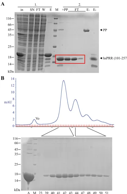
4.1.3. Expression of hsPRR (101-257)... 55
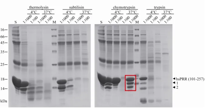
4.1.4. Limited proteolysis of hsPRR (101-257) ... 57
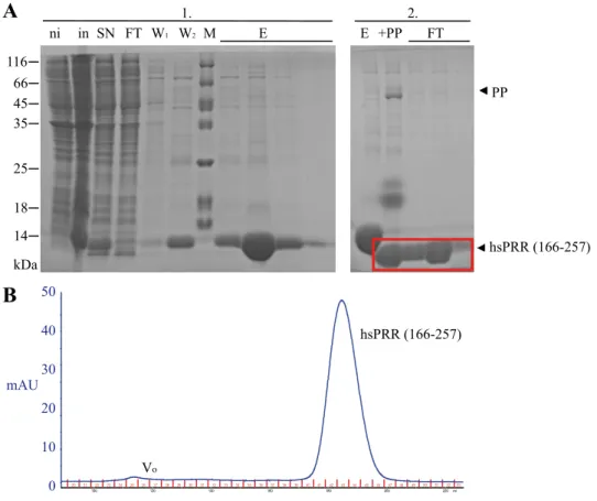
4.1.5. Expression of hsPRR (166-257)... 58
4.1.6. Expression of PRR proteins from different species ... 59
4.2. Characterization of purified hsPRR proteins ... 59
4.2.1. Characterization of hsPRR (170-303) ... 59
4.2.1.1. Secondary structure determination of hsPRR (170-303) ... 59
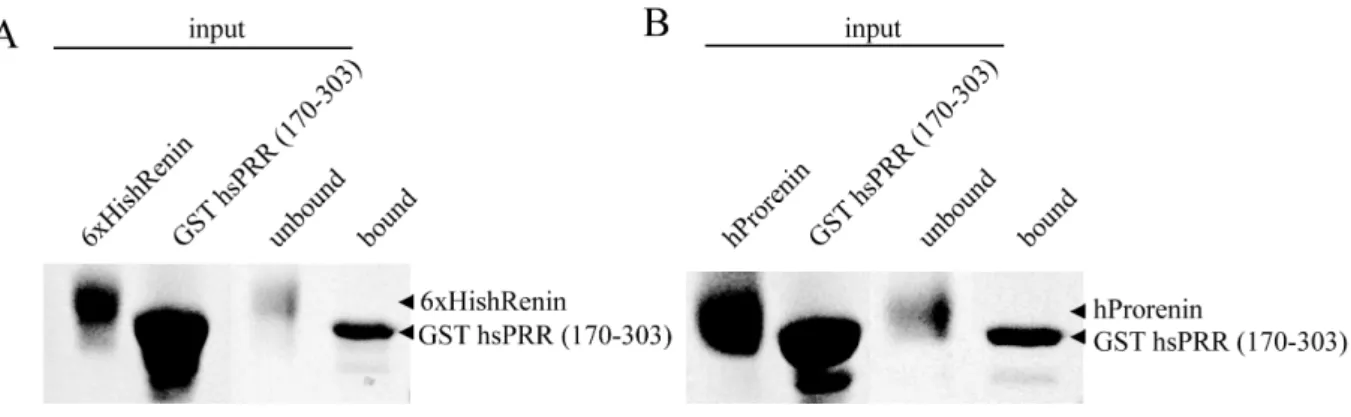
4.2.1.2. Interaction of hsPRR (170-303) with (pro)renin... 60
Content
4.2.2. Characterization of hsPRR (101-257) ... 61
4.2.2.1. Secondary structure determination of hsPRR (101-257) ... 61
4.2.2.2. Interaction study of hsPRR (101-257) with (pro)renin ... 62
4.2.2.3. Analysis of the oligomerization of hsPRR (101-257)... 63
4.2.3. Characterization of hsPRR (166-257) ... 68
4.2.3.1. Interaction study of hsPRR (166-257) with renin ... 75
4.2.4. Characterization of homologous PRR from different species... 76
4.3. Structural studies ... 78
4.3.1. Crystallization trials ... 78
4.3.2. Nuclear magnetic resonance (NMR) spectroscopy... 79
4.4. Function of PRR in T cells... 81
4.4.1. Conditional PRR knock-out model ... 81
4.4.2. Characterization of lymphocytes in the cKO model ... 83
4.4.3. Characterization of T cell proliferation and apoptosis ... 85
4.4.4. Characterization of thymocyte maturation... 86
5. Discussion ... 89
5.1. Characterization of PRR constructs ... 89
5.1.1. Purification of PRR constructs... 89
5.1.2. Binding to renin and prorenin ... 90
5.1.3. Oligomerization of PRR proteins... 92
5.1.4. Structural investigations of the PRR proteins ... 94
5.2. Conditional deletion of PRR in T cells ... 96
5.2.1. PRR cKO causes a decrease in T cell numbers due to a block in ... 96
development ... 96
5.2.2. Block in the transition from DN3-DN4 might be due to PRR affecting pre- TCR signalling ... 97
5.2.3. Role of the vATPase in T cell development ... 98
5.2.4. PRR deletion decreases but does not completely reduce T cells ... 99
5.3. Outlook and perspectives ... 100
6. Zusammenfassung... 102
7. Appendix ... 103
8. Abbreviations ... 105
9. Acknowledgement... 107
10. Publications ... 109 5
Content
10.1. Peer-reviewed journal articles... 109
10.2. Active Congress participation ... 109
10.2.1. Talks ... 109
10.2.2. Poster... 109
11. Curriculum vitae... 110
12. Eigenständigkeitserklärung ... 111
13. References ... 112
1. Abstract 1. Abstract
The (pro)renin receptor (PRR) is an evolutionary conserved transmembrane receptor that was first discovered to bind renin and prorenin. Upon binding, PRR was shown to influence the activity of the renin-angiotensin-system (RAS) and to induce MAP kinase signalling. It was previously shown that a truncated, transmembrane part of PRR was associated to vacuolar H+- ATPase (vATPase), a proton pump which is important for acidification. Recently, a new function of PRR in the WNT/β-catenin signalling pathway was described. Here, the PRR was shown to be an adaptor between WNT receptors and the vATPase. The precise mechanisms by which PRR functions, are still elucidative but the PRR is supposed to regulate various cellular processes.
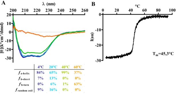
Currently, no biochemical characterization or structural analysis is available for PRR. In order to gain understanding of the function of the PRR, structural studies were performed with several truncated proteins of the extracellular part of the PRR. All PRR proteins (hsPRR170- 303, hsPRR 101-257 or hsPRR 166-257) showed an overall alpha helical folding and did not bind renin or prorenin. The oligomeric assembly of the proteins was investigated. The hsPRR (101-257) was shown to be in a concentration and pH dependent monomer/oligomer equilibrium, whereas hsPRR (166-257) is only present in a monomer/dimer equililibrium.
These data are the basics for further structural and functional studies.
Additionally, conditional KO animals are an excellent tool to investigate the physiological role of the PRR in vivo. As the major mediator of the Wnt/β-catenin signaling pathway, β- catenin, is crucial for T cell maturation, a conditionel deletion of PRR in T cells was analyzed.
PRR deletion resulted in a loss of mature T cells. Moreover, a defect in T cell maturation in the thymus was determined. Our data showed that PRR is critical for proper T cell development and support the hypothesis that PRR contributes to Wnt/β-catenin signaling in T cells.
7
2. Introduction 2. Introduction
The (pro)renin receptor (PRR) was first described in 2002 by Nguyen et al as a receptor that binds renin or prorenin 1. On the basis of this it was supposed that PRR was a new member of the renin angiotensin system (RAS); an important cascade involved in maintaining blood pressure homeostasis 2. However, during the last few years it has become clear that the PRR is not only involved in blood pressure regulation but also plays a role in WNT signalling, vacuolar H+-ATPase mediated acidification and consequently influences early cellular development and neuronal function. The precise function of this receptor and how it is involved in cellular processes is still unclear.
2.1. The (pro)renin receptor
PRR is ubiquitously expressed in all cell types. It consists of 350 amino acids with a molecular weight of 39 kDa. The amino terminus contains a short 16 residue signal peptide, and a long extracellular domain (amino acids 17 to 303). The intracellular C-terminus covers amino acids 327 to 350 with the amino and C-terminus flanking the single transmembrane region from amino acids 306-326 (Figure 1).
full-length PRR soluble PRR
1 SP
19 275RKTR278 303 326 304 350
ID
ED TM
327
associated to vATPase
18
Furin cleavage site
Figure 1. Schematic representation of the domains of PRR.
The PRR consists of a short intracellular (ID), transmembrane (TM), extracellular domain (ED) and signalpeptide (SP). A furin cleavage site (275RKTR278) is located in the extracellular domain.
The PRR shows no homology to any known proteins 1. The extracellular domain possesses a furin cleavage site (275RKTR278). Furin is a cellular endoprotease that proteolytically activates large numbers of proprotein substrates in secretory pathway compartments 3. Upon cleavage with furin, a 28 kDa soluble form of the PRR is detectable in the conditioned media of cultured cells 4, 5. This soluble PRR fraction is generated intracellularly and is hypothesized to be secreted into the blood. Yoshikawa et al have recently described the presence of a
2. Introduction ADAM19 is the protease responsible for cleaving the extracellular region of PRR, instead of furin 6. These studies both illustrate the existence of a soluble PRR, but its function remains unknown.
It was subsequently realised that PRR was not described for the first time by Nguyen et al 1. In 1998, Ludwig et al co-purified a shorter fragment of PRR (8.9 kDa) with the V0 subunit of the vacuolar H+-ATPase (vATPase) from chromaffin granule membranes 7. This fragment of PRR was later renamed ATP6AP2 (vATPase associated protein 2). Approximately 70 amino acids were characterized, consisting of the C-terminal intracellular region, the transmembrane region and some additional amino acids from the extracellular domain. A link between PRR and vATPase was assumed, and therefore PRR is also known as ATP6AP2 7. Interestingly, only a minor amount of the PRR is localized at the plasma membrane, whereas the majority is located in intracellular vesicles 8.
The intracellular domain of PRR contains only 24 amino acids and a crystal structure of part of this domain was published 9. Fusion of 18 amino acids of this domain to maltose binding protein forms a flexible loop. The authors state that these short fragments form a dimer, but the dimerization interface remains unknown. Currently, no other structural information is available about the PRR.
The PRR is found in a wide variety of species from invertebrates, such as Drosophila, to vertebrates, such as Xenopus and up to mammals including human, mouse and rat.
Interestingly, not all of these species have a functional RAS, which, so far, is found almost exclusively in mammals. A gene for renin 10 as well as all other components of the RAS (http://www.uniprot.org/) is found in zebrafish, but whether these animals have a functional RAS is unclear. In Xenopus, no RAS components except for the angiotensin II type I receptor are present, and in Drosophila no RAS components exist. Despite this, PRR demonstrates a high evolutionary conservation. Bader and Burckle described that the N-terminal extracellular region of PRR is exclusively conserved in vertebrates, whereas the intracellular and transmembrane domain display a high amino acid sequence identity among all species investigated 11. These authors raise the hypothesis that the PRR might have diverse functions due to the different regions of the protein having different evolutionary backgrounds. They propose that the renin binding capacity of the extracellular domain is evolutionary younger,
9
2. Introduction whilst the highly conserved C-terminus has a different function. Additionally, they suggest that the PRR might be involved in processes that are independent of renin and prorenin 11.
The only known human disease associated mutation of PRR is described in patients suffering from mental retardation and epilepsy. An X-linked mutation in an exonic splice enhancer results in the production of a shorter PRR fragment with a deletion of exon 4 and thereby reducing full-length PRR levels by around 50% 12. The blood pressure of these patients was unchanged. Mutagenesis of the PRR in zebrafish also displays early developmental abnormalities. These mutants demonstrate eye and body hypopigmentation, neuronal cell death and have early lethality 13. Conventional complete knock-out approaches in mice were not successful as no chimeras could be obtained 11, 14. These results all suggest an important function for the PRR in early cellular development and also neurogenesis, perhaps in a renin and prorenin independent manner.
Details are already known about the function of the PRR in vivo; in particular its role in the cardiovascular system 15, its implication on WNT/β-catenin signalling 16 and its link to vATPase 7, 17. These described roles of the PRR cover a wide area, including RAS-related and RAS-independent functions, and will be explored in the following sections.
2.2. RAS-related functions of PRR
2.2.1. The renin-angiotensin system (RAS)
The renin-angiotensin system (RAS) is a hormone cascade controlling and regulating cardiovascular, renal and adrenal functions. Blood pressure is increased by the secretion of specific hormones, which regulate water and sodium resorption thereby influencing blood volume and also inducing the constriction of blood vessels. The proteolytic enzyme renin is produced in juxtaglomerular cells of the kidney. The only known substrate of this protease is angiotensinogen. Angiotensinogen is synthesized predominatly in the liver, with minor amounts also detectable in the heart, kidney and adipose tissue. Cleavage of angiotensinogen by renin is the first step in the RAS cascade. The decapeptide angiotensin I is then cleaved at its N-terminus by the angiotensin converting enzyme (ACE), producing the octapeptide angiotensin II. Angiotensin II is the intrinsic effector of the RAS and binds to the angiotensin II type 1 receptor (AT1R). Angiotensin II induces both directly (via binding to the AT1R) and indirectly (via a stimulation of the aldosterone production) increased sodium and water
18
2. Introduction blood vessels, an increased thirst and salt appetite. All of these actions results in an increase in blood pressure (Figure 2). Angiotensin II directly acts on juxtaglomerular cells and is thereby able to regulate the secretion of renin by a negative feedback loop 19. The availability and activity of renin is the rate limiting step in the RAS. Therefore, this is the most obvious target for blood pressure regulation.
Figure 2. The renin-angiotensin system (RAS).
Angiotensinogen is cleaved by renin to produce angiotensin I (AngI), which is further converted to angiotensin II (AngII) by the angiotensin converting enzyme (ACE). AngII binds to the angiotensin II type 1 receptor (AT1) and increases blood pressure.
The classical circulating RAS is complemented by additional local RAS in diverse tissues. A prerequesite for the existence and functionality of a local RAS is the production of the individual RAS components in the tissue or alternatively, an uptake of the soluble proteins such as renin or angiotensinogen from the circulation. Since the site of expression of renin is exclusively in the kidney, a mechanism for the uptake of renin from the circulation into tissue is still under investigation. A local RAS is described for the heart, blood vessels, retina and kidney 20, 21.
2.2.2. Prorenin und renin
Renin is a specific aspartyl protease which exclusivly uses angiotensinogen as a substrate.
Renin folds in predominatly β-sheet conformation, typical for the aspartic proteinase family (Figure 3B). The active cleft in the renin molecule contains two catalytically active aspartyl residues, aspartic acid 38 and 226 22.
11
2. Introduction
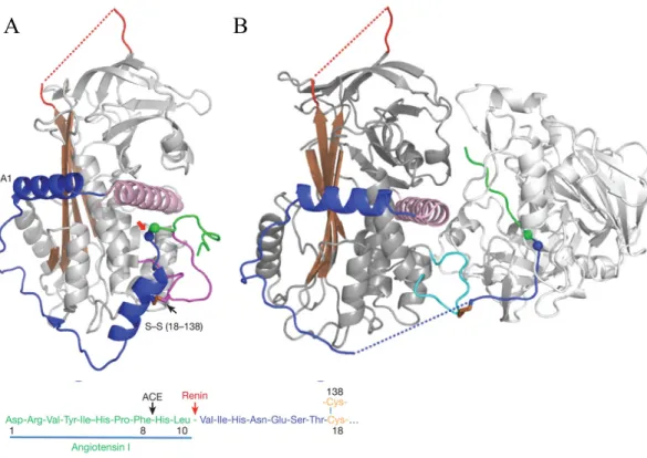
Recently, the crystal structure of human angiotensinogen was solved alone (Figure 3 A) and in complex with renin (Figure 3 B). It was shown that the processing of angiotensinogen by renin is modulated by a redox switch in angiotensinogen protein. Reduced angiotensinogen does not interact with renin that is bound to the PRR, whereas oxidized angiotensinogen is converted with a 4-fold increase after binding to renin 23.
A B
Figure 3. Crystal structure of human angiotensinogen and renin.
A) Crystal structure of human angiotensinogen alone. The N-terminus contains two unique helices (blue), the angiotensin peptide is shown (green) and the renin cleavage site (blue and green balls). B) Human angiotensinogen (grey) with renin (light grey) showing the displacement of the angiotensin peptide. The figures are taken from Zhou et al. 23.
Prorenin contains an additional 43 N-terminal amino acids. This propeptide is hypothesised to cover renins enzymatic cleft and prevent the binding and cleavage of angiotensinogen. No structural information for prorenin is available.
Prorenin can be activated proteolytically or non-proteolytically 24. In vivo, proconvertase I 25 and cathepsin B 26 proteolytically remove the prosegement. This happens exclusively in the juxtaglomerular cells. In vitro, activation of prorenin can occur by addition of trypsin or
2. Introduction physiological conditions such as low pH (~ pH 3.3) and reduced temperature (~4ºC) 27, 28. Under these conditions, the propeptide is thought to no longer cover the active cleft of prorenin.
For a long time, it was assumed that prorenin was only the inactive precursor of renin 2, 29. However, recent findings suggest that prorenin could have important physiological or pathophysiological functions. Under physiological conditions, less than 2% of cirulating prorenin is in the active, uncleaved conformation 29. In healthy patients, the plasma prorenin:renin ratio is approximately 10 to 1. Prorenin is constitutively secreted into the plasma, whereas renin is stored in secretory vesicles and released only after certain stimuli.
The site of renin expression is only in the juxtaglomerular cells of the kidney, whereas prorenin is also produced in the eye, adrenal gland, placenta, testis and ovaries 30. Under certain circumstances, the plasma prorenin concentration can rise. During pregnancy, prorenin levels are increased and they are used as a marker for the onset of microalbuminuria in diabetic patients 31, 32. Microalbuminuria describes the onset of kidney failure, because the kidney leaks small amounts of albumin into the urin. Its defined by elevated albumin concentrations between 30-300 mg/l urin 33.
For years, it was supposed that a binding partner for renin or prorenin exists. An intracellular renin binding protein (Rbp) was found to bind renin and inactivate it 34, but deletion of this gene did not result in any change in blood pressure or the RAS 35. In 2001, Saris et al discovered that renin and prorenin bind with high affinity to the mannose-6- phosphate/insulin-like growth factor II receptor 36, 37. After binding, the ligand is internalised and degraded. Therefore, this receptor is thought to be solely responsible for renin and prorenin clearance, and has no influence on angiotensin I formation 38. As mentioned above, in 2002 Nguyen et al described the binding of renin and prorenin with the PRR 1.
2.2.3. PRR and RAS
Nguyen showed in 1996 that renin binds specifically to human mesangial cells 39. A mesangial cell line without an expression contruct of PRR DNA shows a specific binding to renin or prorenin. Binding to PRR increases the activity of renin five-fold and unexpectedly, prorenin is also non-proteolytically activated thereby resulting in the production of angiotensin I (Figure 4) 1. Batenburg et al showed that renin binds PRR with a kD of 20 nM and prorenin with a kD of 7 nM, concluding that prorenin is the endogenous binding partner
13
2. Introduction of PRR 40. PRR is expressed ubiquitously in all cell types. Especially high levels are found in the heart, brain, placenta and hematopoetic cells and lower levels in the kidney and liver 1.
extracellular intracellular
AOG
prorenin Ang I
PRR
cell membrane
ERK pERK
Ang II
Figure 4. RAS-dependent function of the PRR.
The PRR binds renin and prorenin. Upon binding, prorenin is activated non-proteolytically and processes angiotensiongen (AOG) to angiotensin I (Ang I). Also, the ERKinase becomes phosphorylated and activates the MAP kinase pathway.
Several signal transduction pathways and effector mechanisms are triggered in an angiotensin II independent manner upon binding of renin or prorenin to the PRR. To analyze PRR mediated signal transduction, cells were pretreated with AT1R blockers to exclude angiotensin II induced effects, and then stimulated with renin or prorenin. These experiments show that the mitogen activated protein kinase (MAPK), Erk1/2, is activated in vitro in monocytes 41, mesangial cells 1, 42, vascular smooth muscle cells 43-45 and endothelial cells 46 after stimulation with renin or prorenin. This stimulation induced an increase in the transforming growth factor β 42, 47, 48, which in turn stimulated the expression of the profibrotic genes plasminogen activator inhibitor 1 42, 44, 47, fibronectin 42, collagen I 42 and inflammatory associated proteins such as cyclooxygenase 2 49, interleukin 1 50, 51 and tumor necrosis factor α 51. Interaction with renin or prorenin also induced cell proliferation in mesangial cells 1. Another study shows in cardiomyoctes, prorenin induced angiotensin II independent activation of the p38 MAP kinase. Thus, a role for the PRR in this signal transduction pathway is likely 52. A yeast two-hybrid screen identified the transcription factor promyelotic zincfinger (PLZF) as a new binding partner for the PRR. After activation of the PRR by renin, PRR was shown to interact with PLZF which then translocates into the nucleus and inhibits the expression of PRR 53, 54.
2. Introduction It is hypothesized that PRR is directly involved in pathology because of its involvement in several signalling pathways resulting in the expression of inflammatory and fibrotic genes.
Additionally, a polymorphism in the PRR gene was shown to be associated with blood pressure in Japanese men 55. This was also confirmed by another group studying a Caucasian cohort, who has an identical phenotype 55, 56. Also a transgenic rat overexpressing the PRR develops elevated blood pressure levels after 6 month of age 57. Another group reported glomerulosclerosis in a similar animal model 58. Both animal models had a moderately mild phenotype with a late onset arguing for a rather marginal role of the PRR in cardiovascular pathology.
Since its discovery, discussion has centered on the development of PRR inhibitors which would block the angiotensin II mediated effects of PRR and therefore be beneficial antihypertensive therapies. Ichihara et al designed a decapeptide inhibitor (handle region peptide, HRP) which mimicked some of the amino acids of the prosegment of prorenin called the “handle” region. This region was shown to be important for the non-proteolytic activation of prorenin 59. Since prorenin becomes activated by binding to the PRR, this peptide is proposed to competitivly inhibit the binding of prorenin to the PRR. However, several conflicting studies arose with this putative PRR inhibitor. In diabetic animals, HRP prevents the formation of glomerulosclerosis 51. HRP treatment in spontanous hypertensive rats results in reduced cardiac fibrosis, proteinuria and glomerulosclerosis 59, 60. Also in diabetic AT1R KO mice, HRP prevents the formation of glomerulosclerosis 61. Based on these results, it is proposed that PRR has pathologic actions via an angiotensin II independent pathways.
Beneficial effects of HRP treatment are also shown in pathologies such as retinal neovascularization and ocular inflammation 62. For all of these studies, only prorenin is considered as a binding partner for the PRR. In contrast to these findings, several other publications could not confirm the beneficial effects of HRP. Susic et al found in their model of spontanous hypertensive rats that HRP does not ameloriate cardiac hypertrophy 63. Several other animal models also do not confirm the observed beneficial effects of HRP. In double transgenic animals overexpressing renin and angiotensinogen 43, as well as in Goldblatt hypertensive animals 64, no ameliorated cardiac or renal factors are observed upon treatment with HRP. These studies suggest that the efficiency of HRP as a therapeutic compound for renovascular damage needs further examination. So far no other inhibitor for the PRR has been described. Without further knowledge about the detailed cellular function of PRR it remains controversial as to whether PRR should be seen as a drug target. The angiotensin II
15
2. Introduction independent functions of the PRR are still not completely understood, and the consequences of blocking them are not clear. Another aspect is the direct inhibition of renin, as this will result in higher renin levels. Renin might not longer be able to process angiotensinogen, but an interaction with the receptor is likely and the induction of its signal transduction 65.
2.3. RAS-independent functions of the PRR 2.3.1. vacuolar H+-ATPase (vATPase)
As previously mentioned, when PRR was first discovered it was found to be associated with the vATPase 7. The vATPase is an ATP-dependent proton pump. Its main function is the regulation of pH in intracellular compartments and extracellular fluids, which is essential for a variety of cellular processes 66.
2.3.1.1. Structure of the vATPase
vATPase is structurally and mechanistically related to F-ATPase, and even more closely to archaebacterial ATPase. vATPase is a rotary proton pump which hydrolyses ATP. In contrast to F-ATPase, vATPase is not able to synthesise ATP 67. vATPase is divided into two domains. It consists of a cytoplasmic 650 kDa V1 domain where ATP hydrolysis occurs and a 260 kDa membrane V0 domain responsible for proton translocation (see Figure 5) 66, 68. All together, vATPase is composed of 14 protein subunits. Additionally, different isoforms of these subunits are described for several tissues and cellular locations, further increasing the complexity of this pump. The V1 complex contains 8 subunits: A, B, C, D, E, F, G, H. Three copies of each of the A and B subunits are present, which form an alternating hexamer with three catalytic sites in the A subunit for ATP hydrolysis, and three nucleotide binding sites at the AB interface. Surrounding the AB ring are two copies of the G and E subunits, which form heterodimers. Further present in the complex are 1 or 2 copies of the H subunit, and a single copy of all other subunits. Functionally, a peripheral stalk, composed of the C, E, G, H subunits, and a central stalk, composed of the D and F, subunits are defined. The central stalk provides the energy to the rotation necessary for proton transport, whereas the peripheral stalk is hypothesized to prevent rotation of the AB hexamer. The peripheral stalk, the AB complex, and subunits a and e of the V0 domain, form the stationary part of vATPase called the “stator”.
The V0 domain consists of the subunits a, c, c´, c´´, d and e. Subunit a has eight or nine transmembrane regions with one buried arginine residue that connects the V1 and V0 complex
2. Introduction and is absolutely necessary for proton translocation. The c, c´ and c´´ subunits form the so- called proteolipid ring. These highly hydrophobic subunits all possess a single buried glutamate residue, which is also important for proton transport. The d subunit sits atop of the proteolipid ring and connects the V0 domain with the central stalk of the V1 domain. The function of the highly hydrophobic e subunit has so far not been described 68.
Figure 5. Schematic representation of the structure of vATPase.
vATPase is composed of the V1 (orange and green) and the V0 domain (blue) and its subunits (A-H (V1) and a-e (V0)). The V1 domain is responsible for ATP hydrolysis, while the V0 domain translocates the protons across the membrane. For details see text. The figure is taken from Forgac 68.
The correct assembly and targeting of vATPase is critical for its function in certain tissues and cellular compartments. The V1 domain is able to self-assemble, whereas the V0 domain requires the support of several endoplasmatic reticulum assembly factors such as VMA (vacuolar membrane ATPase activity) 21, VMA 12, VMA 22 and Pkr169-71. The targeting of vATPase to its site of activity is mediated by several isoforms. Four isoforms of the a subunit have been described, which all differ in the cell type in which they are expressed and the cellular location in which they function. Multiple isoforms of the same subunit are also possible in one cell 68, 72. For example, the a1 subunit is found in presynaptic nerves, in synaptic vesicles and on the plasma membrane 73. The a2 subunit is expressed in renal proximal tubule cells in the apical endosomes 74. In contrast, the a3 subunit is localised on the plasma membrane of osteoclasts 75, whereas a4 is located in the apical membrane of renal intercalated cells 76. Most of the other V0 subunits have two isoforms, where one isoform is
17
2. Introduction ubiquitously expressed and the second isoform is only expressed in a specific cell type or tissue 68. PRR is not thought to be directly involved in the formation of vATPase complex, but is associated to the V0 domain. Therefore, PRR might play role in assembly of the V0
domain.
2.3.1.2. Proton transport of the vATPase
Protons are translocated after ATP hydrolysis via an active transport mechanism, through the rotation of the proteolipid ring, subunits D, F and d 77. Protons enter from the cytoplasmic side of the membrane through a hemi-channel of subunit a, and protonate the buried glutamate in the c, c´ or c´´ subunit. Following ATP hydrolysis, the proteolipid and accessory subunits rotate, thereby displacing the protons. The protonated glutamate then interacts with the buried arginine in the a subunit 66. This arginine becomes protonated and releases the proton into the lumen of the vesicular compartment or the extracellular fluid 78. The stoichiometry for this process is between 1:2 and 1:3. Three, since there are three ATP binding sites in the AB hexamer and 6-10 protonable sites in the proteolipid, depending on the amount of proteolipids in the ring 68.
Bafilomycin and concanamycin are inhibitors for vATPase and are proposed to block proton transport by circumventing the helical rotation around their own axes of the proteolipids. This helical swivelling is important for the rotation and the proton transport of the proteolipid ring
79-82.
2.3.1.3. Regulation of vATPase activity
Since pH is crucial for many cellular processes, vATPase activity is highly regulated. Several mechanisms are described for the regulation of vATPase activity. One mechanism is the reversible dissociation of the domains V1 and V0. This mechanism is best understood in yeast and insect cells, but also occurs in mammalian cells 68. In yeast, it was shown that after depletion of glucose, which consequently resulted in a reduction of ATP consumption, the two domains of vATPase dissociated 83. This process is reversible, but dissociation and assembly are regulated differently. Assembly in yeast requires a protein complex called RAVE 84, whereas dissociation is not dependent on this complex. The mechanism for the reversible dissociation is not fully understood, but it is hypothesized that the non-homologous region of the A subunit plays a role in dissociation 85. Also, the RAS/cAMP/protein kinase A pathway has been described to be involved in the dissociation of vATPase 86. A role for PRR
2. Introduction in assembly or dissociation of vATPase is possible. Because of its transmembrane localization, an interaction with the V0 domain appears feasible. Thus, regulation of vATPase activity could be mediated via an interaction between the V0 domain and the PRR. The first evidence for an involvement of PRR in vATPase assembly and function came from Kinouchi et al. They showed that deletion of the PRR results in a downregulation of the V0 subunits and affect the stability and assembly of the V0 subunits, thereby compromising vesicular acidification 87.
Besides reversible dissociation, another regulatory mechanism of vATPase activity is the endocytosis or exocytosis of vATPase at the plasma membrane. In this case, vATPase is stored in vesicles close to the plasma membrane in renal epithelial cells and is transported to the membrane upon induction by a glucose stimulus. The complex is internalised and trafficked to intracellular storage compartments if proton transport is reduced 88.
A third hypothesized mechanism for the regulation of vATPase activity is the increase/decrease of the coupling efficiency of ATP hydrolysis and proton translocation. This mechanism is controlled by the different a subunit isoforms. Different yeast homologs for the a subunit showed altered coupling efficiency suggesting a tighter coupling of proton transport and ATP hydrolysis 72. A special mechanism to regulate the activity of vATPase is the formation of a reversible disulfide bond in the subunit a, causing a disruption of the catalytic function of the ATPase 89.
A tight regulation of vATPase activity is important for cellular function and is achieved by these various processes. A role for the PRR in these mechanisms is possible, but needs further investigation.
2.3.2. PRR and vATPase
It has been shown that PRR is associated to vATPase 7. Another link between PRR and vATPase was shown by Advani et al, where PRR was demonstrated to co-localize with the B subunit of vATPase in the kidney 90. Additionally, Cruciat et al showed that PRR co- immunoprecipitated with transmembrane subunits of vATPase 16. All of these effects were first shown in vitro, until recently where support for a link between PRR and vATPase came from in vivo studies. The first conditional knock-out of PRR in cardiomyocytes resulted in lethal heart failure accompanied with impaired autophagic degradation 87. The authors explain
19
2. Introduction these phenomena by the defective acidification of intracellular compartments, which is necessary for protein degradation. They showed that malfunction of this acidification was due to a disordered assembly of vATPase; evidenced by deletion of PRR reducing protein levels of subunits of transmembrane vATPase 87. Furthermore, conditional KO of PRR in podocytes also caused a defect in protein degradation and autophagy 91, 92. Podocytes form the main filtration barrier in the kidney and deletion of PRR in this cell type resulted in kidney failure and early lethality. Additionally, similar to the phenotype observed in cardiomyocytes, the authors observed a block in podocyte lysosome processing, which they propose could be due to defective vATPase function and impaired acidification 91. Another group described the same podocyte model with a similar phenotype. They confirmed again that deletion of PRR suppressed expression of the c subunit of the V0 domain of vATPase, resulting in deacidification of intracellular vesicles 92. All of these studies point towards PRR having an important role in the function and regulation of vATPase in vivo.
2.3.3. Physiological and pathophysiological function of the vATPase
vATPase is involved in a variety of cellular processes where acidification is necessary.
Depending on its cellular localization it is involved in different functions. For example, vATPase is found in cellular compartments such as endosomes, lysosomes, Golgi-derived vesicles, clathrin-coated vesicles, secretory vesicles and the central vacuoles of plants and fungi. In these compartments, vATPase is engaged with protein trafficking including processes such as clathrin-mediated endocytosis, receptor recycling, lysosomal degradation and secretion. In clathrin-mediated endocytosis, ligand-receptor complexes are clustered in clathrin-coated pits which then bud off from the plasma membrane as clathrin-coated vesicles and form early- or sorting-endosomes. Sorting endosomes are then acidified by vATPase and the internalised ligand-receptor complex dissociates. The receptor is then shuttled back to the cell surface and the ligand is further trafficked to the lysosome. In lysosomes, proteases are only active at low pH and thus acidification by vATPase is essential for the degradation of proteins 68. Not only for protein degradation, but also for the processing of proteins vATPase is necessary. In secretory vesicles, vATPase is essential for the activation of proteases at low pH cleaving prohormones, such as in pancreatic beta cells, where proinsulin is converted to insulin 93. Additionally, vATPases provide the membrane potential or pH gradient required to drive the transport of neurotransmitters in secretory vesicles 94. Previously, it was shown that PRR is expressed in the brain and plays a role in the neural control of cardiovascular
2. Introduction functions 95. Besides the acidification in intracellular compartments, the vATPase is also involved in development in Xenopus embryos 96.
vATPase is also found on the plasma membrane of a variety of cells. Mutations in subunits of vATPase are involved in several pathophysiologies. In particular, cell surface vATPase in the kidney is important for several processes. Alpha-intercalated cells in the kidney have a large amount of vATPases in their apical membrane, which acts to secrete protons into the lumen of the distal tubule and collecting duct. This mechanism is important for the acid/base homeostasis of the urine 97, and a mutation in the B subunit of the vATPase results in an inherited form of distal renal tubule acidosis 98. Osteoclasts are another cell type in which vATPase has an important role. At the plasma membrane, vATPase creates an acidic pH necessary for the activity of digestive enzymes that degrade the bone. A mutation in vATPase subunit a3 in osteoclasts results in osteopetrosis 99. This disease is characterized by thickening of the bone, due to malfunctioning of the bone degradation machinery. Interestingly, this same mutation in the a3 subunit additionally results in a decreased insulin secretion in beta cells in the pancreas 75. vATPase is also critical for sperm maturation in the vas deferens.
Here, the epididymal clear cells secrete protons into the lumen of the epididymus to generate an acidic pH required for the development and maintenance of sperm 100. Additionally, vATPase plays a crucial role for maintaining the homeostasis of neutrophils and macrophages when they are recruited to sites of inflammation. At these sites there is often an acidic pH, and thus, these immune cells require a functional vATPase at the plasma membrane in order to maintain their neutral intracellular pH 101. vATPase also influences angiogenesis of endothelial cells, especially in migration, invasion and tumor metastasis. In the case of tumor cells, vATPase acidifies the extracellular surrounding, thereby promoting invasion by providing the low pH required for cathepsins to digest the extracellular matrix 102. These studies highlight that vATPase is important for several pathophysiological processes.
Besides the above described diseases associated with vATPase, a role for vATPase in bacterial and viral infections is also known. Several viruses and toxins enter the cell with the help of vATPase. One example is the influenza virus, which is able to infect the cell due to the low pH in the endocytosed viral vesicles which facilitates fusion of the viral and endosomal membranes, resulting in the release of viral RNA into the host cell 103. The low pH in intracellular vesicles additionally aids the entry of bacterial toxins such as anthrax or diphtheria into the cell 104. vATPases are also hypothesized to play a role in infection by HIV.
21
2. Introduction In this case, the virus enters the cell via the CD4 receptor on T cells, within endosomes.
Acidification by vATPase is crucial for the intracellular release of the virus. Additionally, the H subunit of vATPase has been shown to interact with the virus protein NEF and regulate CD4 internalization so as to prevent multiple infections 105.
Taken together, these facts show that vATPase is involved in several diseases and is also crucial for physiological processes. This makes vATPase an important complex for the maintenance of cellular function. Therefore, the function of the PRR and its importance for vATPase, in vitro and in vivo, should be elucidated further.
2.3.4. WNT/β-catenin pathway
A recent publication described PRR as an important protein involved in WNT/β-catenin signalling 17. The WNT/β-catenin canonical signalling pathway has an essential role in cell proliferation, stem cell maintenance, cell fate decision, organized cell movements and the establishment of tissue polarity. It is frequently deregulated in human cancer and degenerative disease 106. Besides the canonical WNT pathway, non-canonical WNT pathways exist, among them the planar cell polarity pathway 107.
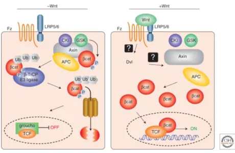
The canonical WNT pathway depends on the presence of WNT (see Figure 6). WNT is a secreted signalling molecule and binds to its cell surface receptor Frizzled. Upon binding to Frizzled, the complex interacts with the membrane-bound co-receptor LRP6 (low-density lipoprotein receptor-related protein 6). In the next step, Dishevelled is recruited to the receptor complex and becomes activated. Dishevelled is another key component of the canonical WNT pathway and inhibits the destruction complex. This destruction complex is composed of the proteins axin, glycogen synthase kinase 3 (GSK3), adenomatous polyposis coli (APC) and casein kinase 1 (CK1). The destruction complex promotes the proteolytic degradation of β- catenin. Dishevelled destroys this complex, but the mechanism for this is not clear. In turn, β- catenin accumulates in the cytoplasm and translocates into the nucleus where it interacts with the transcription factor family TCF/LEF to induce gene expression. After interaction of β- catenin with TCF, groucho dissociates and target gene transcription starts. In the absence of WNT, β-catenin is complexed with the destruction complex, where it is phosphorylated by GSK3 which then targets it to the β-TrCP E3 ligase. This complex promotes the ubiquitinylation of β-catenin, subsequently resulting in the proteolytic degradation of β- catenin by the proteasome 107, 108.
2. Introduction
Figure 6. Schematic representation of the canonical WNT/β-catenin pathway.
In the absence of Wnt signal, β-catenin is recruited into the APC/Axin/GSK3/CKI complex, and is phosphorylated by GSK3. Phosporylated β-catenin binds to β–Trcp E3 ligase of the proteosome machinery and is targeted for degradation. Wnt binds to its frizzled Fz receptor and LRP5/6 co-receptor and activates dishevelled (Dvl), leading to the inhibition of APC/Axin/GSK3 mediated β-catenin degradation. Stabilized β- catenin forms a transcriptional complex with LEF/TCF and activates downstream targets. The figure is taken from Cardigan and Pfeiffer 108.
There are also several non-canonical pathways, such as the planar cell polarity (PCP) pathway. The biggest difference between the non-canonical and the canonical-pathways is the independence of β-catenin. The PCP pathway is best understood in Drosophila and only a little is know about these pathways in mammalian cells 106. Frizzled and dishevelled form a complex that binds and activates small Rho GTPases and JNK (c-Jun N-terminale kinase) 106. In this thesis, I will focus on the canonical-WNT pathway.
2.3.5. PRR and WNT signalling
A new function of PRR was proposed by Cruciat et al in 2010, where they describe PRR as a component of the WNT receptor complex 17. They show that PRR binds to WNT receptors Frizzled and LRP6 and hypothesize that PRR in combination with vATPase, influences LRP6 phosphorylation and activation. This phosphorylation process may only occur under acidic conditions mediated by vATPase. Additionally, they describe a PRR knock down in Xenopus that results in body morphogenesis and pigmentation defects displaying a similar phenotype as already described for zebrafish. All these functions are renin-independent 17.
23
2. Introduction In the same year, two other publications confirmed the importance of PRR for non-canonical WNT signalling. They both show in Drosophila that PRR interacts with Frizzled 109, 110. The authors hypothesize that the complex of vATPase and PRR, contributes to the function and signalling of Frizzled and thereby mediates non-canonical WNT (PCP) signalling. They also report that a knock down of PRR in Xenopus results in a defective PCP signalling phenotype, similar to previously published ones 109. Thus, PRR is not only involved in the canonical WNT pathway described by Cruciat et al 17, but also plays a role in the non-canonical WNT pathway. It has been hypothesized that PRR is a conserved modulator of both non-canonical and canonical WNT pathways bridging the WNT receptors with vATPase 17, 109.
2.3.6. WNT/β-catenin signalling in development and disease
The canonical WNT pathway is highly conserved in evolution and is a fundamental signalling pathway in all species 111. The impact of WNT signalling on development and disease is described in the following.
The use of gain-of-function and loss-of-function mutations of WNT components in animal models have helped to obtain insight into the diverse functions of the WNT pathway in development 112. The WNT pathway has an enormous role in stem cell fate and proliferation
108. It has also been shown that WNT signalling is critical for the proliferation of intestinal epithelial cells 113 and hair follicle stem cells 114. WNT signalling also controls differentiation of mesenchymal stromal stem cells and regulates the self-renewal of hematopoietic stem cells
112. For example a gain-of-function mutation that results in constitutive active β-catenin in hematopoetic precursors results in impaired self renewal of these cells 115, 116.
In terms of disease, the WNT pathway is implicated in cancer progession. A large number of mutations in WNT pathway related genes are associated with leukaemia, colon cancer and a variety of other cancers 111. APC is a tumour suppressor gene and allelic mutations, resulting in loss of both functional alleles, is present in almost 80 % of all sporadic colorectal cancers.
Mutation of APC results in an over-activation of the canonical WNT pathway, resulting in β- catenin being no longer bound to the degradation complex and thereby inducing the uncontrolled proliferation of intestinal epithelial cells 117. Some colon cancers are linked to a loss of function mutation of Axin, which in turn are correlated to hepatocellular carcinomas
111.
2. Introduction A deregulated WNT pathway is involved in several additional diseases. Skeletal disorders and bone formation are linked to deregulated WNT components. Expression of gain-of-function LRP5 in mice results in high bone density and elevated numbers of active osteoblasts, while loss-of-function of LRP5 leads to reduced bone mass in animal models 111. Interestingly, vATPase is also involved in bone formation; mutations in vATPase subunit a3 in osteoclasts results in osteopetrosis 99. Disturbed WNT/β-catenin signalling is as well involved in neuronal disorders, like Alzheimers disease and, moreover, in wound healing and cardiovascular disease 107. Mice models with defective WNT signalling are more prone to infarct rupture after induction of myocardial infarction and neovascularisation of the infarct area is influenced 118. The contribution of PRR to these diseases that are associated with a deregulated WNT/ β-catenin pathway is yet to be determined.
2.4. T cells
T cells are a specialised type of immune cell which play a central role in cell mediated immunity. Their main function is the recognition of intracellular pathogens and activation of B cells 119. To become a functional T cell, they undergo several steps of maturation in the thymus for which the canonical WNT signalling pathway is essential 120. PRR is ubiquitously expressed and shows a high level of gene expression in hematopoietic tissue (personal communication, M. Andrade, MDC, Berlin, Germany). As described above, PRR is proposed to be an adaptor between the WNT receptors and vATPase, and thus, might be important in T cell maturation 17.
2.4.1. T cell function
The immune system is composed of several cell types originating from pluripotent hematopoetic stem cells from the bone marrow. These hematopoetic stem cells separate into common myeloid progenitors that give rise to granulocytes, macrophages, dendritic cells and erythrocytes and into common lymphoid progenitors. The common lymphoid progenitors give rise to B and T cells, which are both components of the adaptive immunity, and to natural killer cells 119.
T cells are defined by their maturation in the thymus and their expression of a T cell receptor (TCR). The TCR is a cell surface receptor responsible for the recognition of antigens and the activation of T cells. The TCR is a heterodimer consisting of a α- and β-chain or a γ- and δ- chain. The γδ T cells are engaged in early and late immune responses, and function as effector
25
2. Introduction cells and immune regulators. Because γδ T cells are the first T lymphocytes to arise in ontogeny, it seems likely that they play an important role in immune protection early in development 121.
The majority of T cells express the CD3 TCR that is composed of α- and β-chains, which play a major role in cell-mediated immunity. These T lymphocytes are further separated into T helper cells, cytotoxic T cells and regulatory T cells. T helper cells express the CD4 protein on their cell surface, whereas cytotoxic T cells express CD8 on their surface and therefore they are known as CD4+ and CD8+ T cells, respectively. CD4 and CD8 are co-receptors for the TCR that are required to bind to the major histocompatibility complex (MHC) present on all antigen presenting cells, such as dendritic cells or macrophages. CD4 binds to MHC class II, whilst CD8 interacts with MHC class I. This interaction is important for antigen recognition and T cell response 119. For activation of T cells, signalling via the TCR MHC complex is essential, but also co-stimulatory signals are required. The best known signal is the CD28-mediated co-stimulus. In response to this, T cells clonally expand and become activated 122. CD8+ T cells are responsible for the killing of pathogen infected cells. The CD8+ cells induce formation of a pore and activate apoptotic pathways. CD4+ T cells are not directly involved in cell apoptosis, instead they assist to activate CD8+ T cells or macrophages, and effect the maturation of B cells 119. Upon activation, the CD4+ T cells differentiate into several subtypes expressing diverse cytokines, thereby modulating the immune response. For example, Interleukin 12 induces the development of Th1 cells that secrete interferon γ and support the cellular immune response. In contrast interleukin 4 stimulates the expansion of Th2 cells affecting the humoral immune response 123. Th17 cell development is triggered by the presence of TGF β, interleukin 6 and 21. These cells are characterized by the expression of interleukin 17. Th17 cells are important in clearing pathogens during host defence reactions and inducing tissue inflammation in autoimmune disease 124, 125. The last T cell subtype is regulatory T cells. These are induced by TGF β and are supposed to have suppressor and regulatory functions for the immune system 126. Most of these cells die within 1 or 2 weeks after activation, and only a small subset survives as effector memory T cells or central memory T cells 123.
2. Introduction 2.4.2. T cell development
For the existence and function of T cells, their maturation process is crucial. The WNT/β- catenin pathway plays a key role in their development and thus the role of the PRR in this process should be analyzed closer.
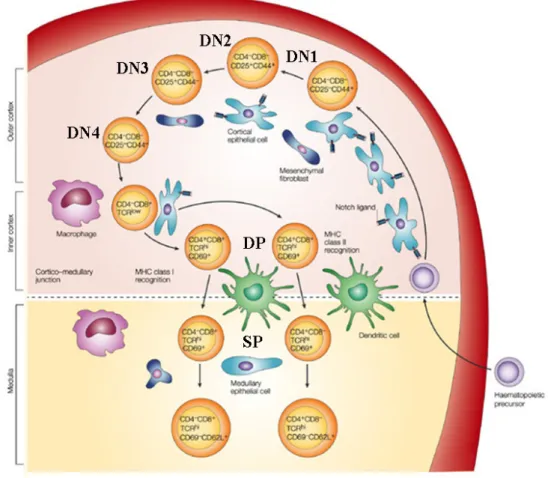
T cell maturation is a highly regulated and complex procedure. Initially, common lymphoid progenitors migrate to the thymus. They do not express the TCR or the co-receptor CD4 and CD8 and are therefore called double negative (DN; CD4-/CD8-) cells. These DN cells are further characterized by the expression of the two surface markers CD44 and CD25, corresponding to the α-chain of the interleukin-2 receptor. In the first DN stage, the cells are negative for CD25 and positive for CD44 and are also referred to as DN1 (CD25-/CD44+) (Figure 7). During the next maturation step (DN2; CD25+/CD44+) the cells downregulate the hematopoetic stem cell marker cKIT and upregulate CD25. With this, they lose their potential to form other cells, such as B cells and natural killer cells and are committed to the T cell lineage 127. Until this point the cells are moderately proliferating, whilst during the DN3 (CD25+/CD44-) stage they stop proliferation and downregulate CD44. During the following rearrangement of the β-chain, the TCR gene is genetically recombined to recognize different antigens. Cells which successfully rearranged their β-chain, present the pre-TCR on their surface; cells that do not rearrange successfully undergo apoptosis. The pre-TCR is composed of a β-chain and a non-rearranged α-chain. This point is the first important stage for T cell maturation called the “β-selection checkpoint” 128, 129. β-catenin deletion results in a developmental block at the DN3 stage to DN4, pointing towards an important role of WNT/
β-catenin signalling in the β-selection checkpoint 120. When cells further proceed with their next maturation process (DN4; CD25-/CD44-), they downregulate CD25 and start to express the CD4 and CD8 co-receptors, therefore becoming double positive cells (DP; CD4+/CD8+).
More then 90 % of the cells die at this stage during a positive selection process. Only cells that moderately bind to MHC class I or II complexes are chosen, whereas non-functional T cells are eliminated by “death by neglect”, because they do not obtain important signals for their further survival. The surviving cells start to express high levels of the TCR with a rearranged TCR α-chain, whilst the pre-TCR is downregulated. Between 5-10 % of the remaining cells are negatively selected because they bind to self antigens and undergo apoptosis. Only 2 % of T cells survive these selection rounds and transit from a DP cell, to a single positive (SP) mature CD4+ (helper) or CD8+ (cytotoxic) cells. These cells then move from the thymus to the blood stream. 128. The T cell repertoire is established during early
27
2. Introduction years, as the thymus shrinks during adolescence. However, T cell production remains continuous 127
.
Figure 7. Overall scheme of T cell development in the thymus.
The hematopoetic precursors migrate from the bone marrow to the thymus. The double negative (DN) cells are characterized by the absence of CD4-/CD8-. These cells can be further subdivided into DN1-DN4 cells characterized by the expression of CD25 and CD44. Thereafter DN cells start to express the CD4+/CD8+ antigen (DP) and finally develop into single positive CD4+ or CD8+ cells. For details see text. The figure is adapted from Zúñiga-Pflücker 130
2.4.3. T cells and PRR
A role for PRR in T cells has not been described before, but as WNT/β-catenin signalling and vATPase are involved in T cell function and development a role for PRR is likely.
β-catenin is the central mediator of WNT signalling and a conditional deletion in T cells results in an impaired T cell development 120. The authors showed that the amount of T cells was drastically reduced in lymphoid tissues and the circulation, while B cells were not affected. The lack of β-catenin did not result in an increased apoptosis or in a dramatic
2. Introduction block in development was shown by analyzing the thymocytes. The pre-TCR signalling seems to be dependent on functional WNT signalling 120.
Another publication implicates that vATPase also plays an important role for T cell development. Pua et al published that autophagy is critical for lymphocyte development and function and suggest an essential role for survival and proliferation. Autophagy is an intracellular degradation process depending on acidification. Mice that are deficient for ATG5 had reduced numbers of T cells with a dramatically increased number of dead cells and a proliferation defect 131. vATPase was shown to play a role in the infection of HIV. It helps the virus to enter the CD4+ T cell and prevents multiple infections of the same cell 105, 132 Acidification of the lytic granules of the cytotoxic T cell is dependent on vATPase and therefore necessary for their function 133. All these observations point to an important role of vATPase and WNT/β-catenin signalling for T cell function and maturation and therefore imply a fundamental role for PRR for T cells. Additionally T cell subtypes are important to cardiovascular function and target-organ damage 134 .
A better understanding of the function of the PRR for these cells would give insight into T cell regulation. Additionally, this cKO model would provide knowledge about the in vivo implication of PRR for vATPase and WNT/β-catenin pathway.
29
2. Introduction 2.5. Objectives of the work
The aim of this thesis was to characterize the PRR. This characterization might help in the future to obtain a better understanding of the role of the PRR for the RAS, WNT/β-catenin pathway and the vATPase, both in vitro and in vivo.
No structural data is available for the soluble region of PRR. Therefore, the first aim of my thesis was to obtain structural information by protein X-ray crystallography of the soluble region of PRR. With the help of structural data, additional information about possible binding sites to renin or prorenin or other interaction partners could be analyzed and would give insights into the function of the soluble region of PRR.
Specific aims therefore were to find soluble, truncated proteins of PRR that could be purified in high amounts and quality in an E.coli expression system. These constructs were then analyzed for their secondary structure to obtain information about their folding. Next, binding to renin or prorenin of these constructs was analyzed. In addition these PRR proteins were analyzed for their oligomerization behaviour. All constructs were used for crystallization trials.
The second aim of my thesis was to analyse the role of the PRR in an in vivo system, using a conditional knockout approach. The hypothesis tested that the deletion of PRR in T cells would result in phenotype similar to that of a β-catenin knockout, displaying a maturation defect at the DN3 to DN4 cell stage. With this, the possible link of PRR to WNT/β-catenin pathway should be analyzed to understand the function of the PRR.
In this project it was analyzed if PRR deletion results in a decrease of T cells in peripheral lymphoid tissue. Furthermore it was determined what kind of T cells, CD4+ or CD8+ cells, are reduced in this cKO model. To explain the reduction of T cell it was further analyzed if this is due to a developmental defect. For that reason, T cells were examined during their maturation process in the thymus. The proportion of typical cellular stages of thymus maturation, double negative, double positive and single positive CD4+ or CD8+ T cells, was determined. The double negative cell subset was further analyzed for the proportion of DN3 and DN4 cells to draw conclusion for possible blocks in the distinct developmental steps.
3. Materials and Methods 3. Materials and methods
3.1. Materials
3.1.1. Chemicals
Chemicals were obtained from the following companies: Karl Roth GmbG & Co. KG (Karlsruhe, Germany), Jena Bioscience (Jena, Germany), Merck KGaA (Darmstadt, Germany), Sigma-Aldrich GmbH (Hannover, Germany), Amersham-Pharmacia (Freiburg, Germany), Qiagen (Hilden, Germany), GE Healthcare (Munich, Germany), BD Bioscience (Heidelberg, Germany) and Miltenyi Biotec GmbH (Bergisch Gladbach, Germany).
3.1.2. Enzymes
KOD DNA Polymerase Novagen (Darmstadt, Germany) PFU DNA Polymerase Roboklon (Berlin, Germany)
T4 DNA Polymerase New England Biolabs (Frankfurt a.M., Germany)
DNase I Roche (Mannheim, Germany)
Restriction Nucleases
(BamHI, XhoI, EcoRI, NotI, DpnI)
New England Biolabs (Frankfurt a.M., Germany)
Prescission Protease GE Healthcare (Munich, Germany) Trypsin, Chymotrypsin, Thermolysin, Subtilisin Sigma Aldrich (Hannover, Germany) Superscript III Reverse Transkriptase Invitrogen (Darmstadt, Germany)
Proteinase K Roth GmbG & Co. KG (Karlsruhe, Germany)
3.1.3. Clones
cDNA clones for the human (Homo sapiens), mouse (Mus musculus), zebrafish (Danio rerio) and frog (Xenopus laevis) PRR were purchased from imaGenes (Berlin, Germany).
3.1.4. Kits and standards Kits
QIAgen Plasmid Purification (Mini) Kit Qiagen (Hilden, Germany) QIAquick Gel Extraction Kit Qiagen (Hilden, Germany)
Rneasy Kit Qiagen (Hilden, Germany)
DNAse Digest Qiagen (Hilden, Germany) Superscript III cDNA Synthesis Kit Invitrogen (Darmstadt, Germany) NuPageNovex Bis Tris precast gel system Invitrogen (Darmstadt, Germany)